
Intracoronary Thrombogenicity in Patients with Vasospastic Angina: An Observation Using Coronary Angioscopy


Abstract
:1. Introduction
2. Material and Methods
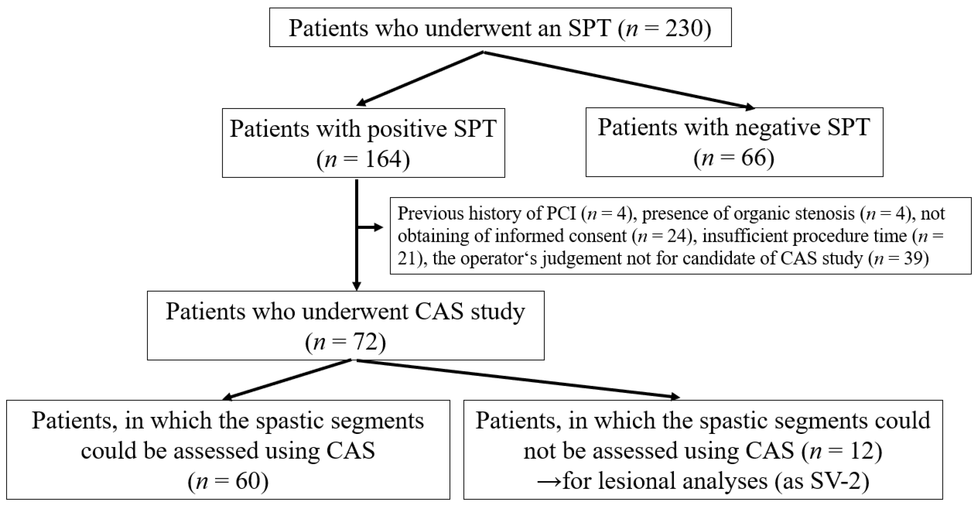
2.1. Patient Selection
2.2. Coronary Angiography, SPT
2.3. Assessment of CAS
2.4. Clinical Factors Assessed in the Present Study
2.5. Statistical Analyses
3. Results
3.1. Patients’ Characteristics in VSA Patients with Intracoronary Thrombi
3.2. Lesion Characteristics with Intracoronary Thrombi
4. Discussion
5. Conclusions
Author Contributions
Funding
Institutional Review Board Statement
Informed Consent Statement
Data Availability Statement
Acknowledgments
Conflicts of Interest
References
- Yasue, H.; Kugiyama, K. Coronary spasm: Clinical features and pathogenesis. Intern. Med. 1997, 36, 760–765. [Google Scholar] [CrossRef] [Green Version]
- Yasue, H.; Nakagawa, H.; Itoh, T.; Harada, E.; Mizuno, Y. Coronary artery spasm—Clinical features, diagnosis, pathogenesis, and treatment. J. Cardiol. 2008, 51, 2–17. [Google Scholar] [CrossRef] [Green Version]
- JCS Joint Working Group. Guidelines for diagnosis and treatment of patients with vasospastic angina (Coronary Spastic Angina) (JCS 2013). Circ. J. 2014, 78, 2779–2801. [Google Scholar] [CrossRef] [PubMed] [Green Version]
- Kunadian, V.; Chieffo, A.; Camici, P.G.; Berry, C.; Escaned, J.; Maas, A.; Prescott, E.; Karam, N.; Appelman, Y.; Fraccaro, C.; et al. An EAPCI Expert Consensus Document on Ischaemia with Non-Obstructive Coronary Arteries in Collaboration with European Society of Cardiology Working Group on Coronary Pathophysiology & Microcirculation Endorsed by Coronary Vasomotor Disorders International Study Group. Eur. Heart J. 2020, 41, 3504–3520. [Google Scholar] [CrossRef] [PubMed]
- Ford, T.J.; Stanley, B.; Good, R.; Rocchiccioli, P.; McEntegart, M.; Watkins, S.; Eteiba, H.; Shaukat, A.; Lindsay, M.; Robertson, K.; et al. Stratified Medical Therapy Using Invasive Coronary Function Testing in Angina: The CorMicA Trial. J. Am. Coll. Cardiol. 2018, 72, 2841–2855. [Google Scholar] [CrossRef]
- Scalone, G.; Niccoli, G.; Crea, F. Pathophysiology, diagnosis and management of MINOCA: An update. Eur. Heart J. Acute Cardiovasc. Care 2018, 8, 54–62. [Google Scholar] [CrossRef]
- Montone, R.A.; Niccoli, G.; Fracassi, F.; Russo, M.; Gurgoglione, F.; Camma, G.; Lanza, G.A.; Crea, F. Patients with acute myocardial infarction and non-obstructive coronary arteries: Safety and prognostic relevance of invasive coronary provocative tests. Eur. Heart J. 2018, 39, 91–98. [Google Scholar] [CrossRef]
- Reynolds, H.R.; Maehara, A.; Kwong, R.Y.; Sedlak, T.; Saw, J.; Smilowitz, N.R.; Mahmud, E.; Wei, J.; Marzo, K.; Matsumura, M.; et al. Coronary Optical Coherence Tomography and Cardiac Magnetic Resonance Imaging to Determine Underlying Causes of MINOCA in Women. Circulation 2021, 143, 624–640. [Google Scholar] [CrossRef]
- Pelliccia, F.; Pepine, C.J.; Berry, C.; Camici, P.G. The role of a comprehensive two-step diagnostic evaluation to unravel the pathophysiology of MINOCA: A review. Int. J. Cardiol. 2021, 336, 1–7. [Google Scholar] [CrossRef]
- Etsuda, H.; Mizuno, K.; Arakawa, K.; Satomura, K.; Shibuya, T.; Isojima, K. Angioscopy in variant angina: Coronary artery spasm and intimal injury. Lancet 1993, 342, 1322–1324. [Google Scholar] [CrossRef]
- Suzuki, H.; Kawai, S.; Aizawa, T.; Kato, K.; Sunayama, S.; Okada, R.; Yamaguchi, H. Histological evaluation of coronary plaque in patients with variant angina: Relationship between vasospasm and neointimal hyperplasia in primary coronary lesions. J. Am. Coll. Cardiol. 1999, 33, 198–205. [Google Scholar] [CrossRef] [Green Version]
- Shin, E.S.; Ann, S.H.; Singh, G.B.; Lim, K.H.; Yoon, H.J.; Hur, S.H.; Her, A.Y.; Koo, B.K.; Akasaka, T. OCT-Defined Morphological Characteristics of Coronary Artery Spasm Sites in Vasospastic Angina. JACC Cardiovasc. Imaging 2015, 8, 1059–1067. [Google Scholar] [CrossRef] [Green Version]
- Shin, E.S.; Her, A.Y.; Ann, S.H.; Balbir Singh, G.; Cho, H.; Jung, E.C.; Shim, E.B.; Koo, B.K.; Akasaka, T. Thrombus and Plaque Erosion Characterized by Optical Coherence Tomography in Patients With Vasospastic Angina. Rev. Esp. Cardiol. (Engl. Ed.) 2017, 70, 459–466. [Google Scholar] [CrossRef]
- Kitano, D.; Takayama, T.; Sudo, M.; Kogo, T.; Kojima, K.; Akutsu, N.; Nishida, T.; Haruta, H.; Fukamachi, D.; Kawano, T.; et al. Angioscopic differences of coronary intima between diffuse and focal coronary vasospasm: Comparison of optical coherence tomography findings. J. Cardiol. 2018, 72, 200–207. [Google Scholar] [CrossRef] [Green Version]
- Teragawa, H.; Oshita, C.; Ueda, T. History of gastroesophageal reflux disease in patients with suspected coronary artery disease. Heart Vessel. 2019, 34, 1631–1638. [Google Scholar] [CrossRef]
- Sato, K.; Kaikita, K.; Nakayama, N.; Horio, E.; Yoshimura, H.; Ono, T.; Ohba, K.; Tsujita, K.; Kojima, S.; Tayama, S.; et al. Coronary vasomotor response to intracoronary acetylcholine injection, clinical features, and long-term prognosis in 873 consecutive patients with coronary spasm: Analysis of a single-center study over 20 years. J. Am. Heart Assoc. 2013, 2, e000227. [Google Scholar] [CrossRef] [Green Version]
- Komatsu, S.; Ohara, T.; Takahashi, S.; Takewa, M.; Minamiguchi, H.; Imai, A.; Kobayashi, Y.; Iwa, N.; Yutani, C.; Hirayama, A.; et al. Early detection of vulnerable atherosclerotic plaque for risk reduction of acute aortic rupture and thromboemboli and atheroemboli using non-obstructive angioscopy. Circ. J. 2015, 79, 742–750. [Google Scholar] [CrossRef] [Green Version]
- den Heijer, P.; Foley, D.P.; Hillege, H.L.; Lablanche, J.M.; van Dijk, R.B.; Franzen, D.; Morice, M.C.; Serra, A.; de Scheerder, I.K.; Serruys, P.W.; et al. The ‘Ermenonville’ classification of observations at coronary angioscopy—Evaluation of intra- and inter-observer agreement. European Working Group on Coronary Angioscopy. Eur. Heart J. 1994, 15, 815–822. [Google Scholar] [CrossRef] [PubMed]
- Ueda, Y.; Asakura, M.; Yamaguchi, O.; Hirayama, A.; Hori, M.; Kodama, K. The healing process of infarct-related plaques. Insights from 18 months of serial angioscopic follow-up. J. Am. Coll. Cardiol. 2001, 38, 1916–1922. [Google Scholar] [CrossRef] [Green Version]
- Dai, K.; Matsuoka, H.; Kawakami, H.; Sato, T.; Watanabe, K.; Nakama, Y.; Ishihara, M. Comparison of Chronic Angioscopic Findings of Bare Metal Stents, 1st-Generation Drug-Eluting Stents and 2nd-Generation Drug-Eluting Stents- Multicenter Study of Intra-Coronary Angioscopy After Stent (MICASA). Circ. J. 2016, 80, 1916–1921. [Google Scholar] [CrossRef] [PubMed] [Green Version]
- Miyoshi, T.; Higaki, A.; Kawakami, H.; Yamaguchi, O. Automated interpretation of the coronary angioscopy with deep convolutional neural networks. Open Heart 2020, 7, e001177. [Google Scholar] [CrossRef]
- Teragawa, H.; Oshita, C.; Orita, Y. Clinical significance of prolonged chest pain in vasospastic angina. World J. Cardiol. 2020, 12, 450–459. [Google Scholar] [CrossRef]
- Ong, P.; Athanasiadis, A.; Hill, S.; Vogelsberg, H.; Voehringer, M.; Sechtem, U. Coronary artery spasm as a frequent cause of acute coronary syndrome: The CASPAR (Coronary Artery Spasm in Patients With Acute Coronary Syndrome) Study. J. Am. Coll. Cardiol. 2008, 52, 523–527. [Google Scholar] [CrossRef] [PubMed] [Green Version]
- Kanwar, S.S.; Stone, G.W.; Singh, M.; Virmani, R.; Olin, J.; Akasaka, T.; Narula, J. Acute coronary syndromes without coronary plaque rupture. Nat. Rev. Cardiol. 2016, 13, 257–265. [Google Scholar] [CrossRef]
- Tsujita, K.; Yamanaga, K.; Komura, N.; Sakamoto, K.; Miyazaki, T.; Oimatsu, Y.; Ishii, M.; Tabata, N.; Akasaka, T.; Sueta, D.; et al. Clinical and morphological presentations of acute coronary syndrome without coronary plaque rupture—An intravascular ultrasound study. Int. J. Cardiol. 2016, 220, 112–115. [Google Scholar] [CrossRef]
- Morimoto, S.; Shiga, Y.; Hiramitsu, S.; Yamada, K.; Nomura, S.; Miyagi, Y.; Nomura, M.; Mizuno, Y. Plaque rupture possibly induced by coronary spasm--an autopsy case of acute myocardial infarction. Jpn. Circ. J. 1988, 52, 1286–1292. [Google Scholar] [CrossRef] [PubMed]
- Lin, C.S.; Penha, P.D.; Zak, F.G.; Lin, J.C. Morphodynamic interpretation of acute coronary thrombosis, with special reference to volcano-like eruption of atheromatous plaque caused by coronary artery spasm. Angiology 1988, 39, 535–547. [Google Scholar] [CrossRef]
- Tanaka, A.; Shimada, K.; Tearney, G.J.; Kitabata, H.; Taguchi, H.; Fukuda, S.; Kashiwagi, M.; Kubo, T.; Takarada, S.; Hirata, K.; et al. Conformational change in coronary artery structure assessed by optical coherence tomography in patients with vasospastic angina. J. Am. Coll. Cardiol. 2011, 58, 1608–1613. [Google Scholar] [CrossRef] [PubMed] [Green Version]
- Miyamoto, S.; Ogawa, H.; Soejima, H.; Takazoe, K.; Kajiwara, I.; Shimomura, H.; Sakamoto, T.; Yoshimura, M.; Kugiyama, K.; Yasue, H.; et al. Enhanced platelet aggregation in the coronary circulation after coronary spasm. Thromb. Res. 2001, 103, 377–386. [Google Scholar] [CrossRef]
- Oshima, S.; Yasue, H.; Ogawa, H.; Okumura, K.; Matsuyama, K. Fibrinopeptide A is released into the coronary circulation after coronary spasm. Circulation 1990, 82, 2222–2225. [Google Scholar] [CrossRef] [PubMed] [Green Version]
- Morikawa, Y.; Uemura, S.; Ishigami, K.; Soeda, T.; Okayama, S.; Takemoto, Y.; Onoue, K.; Somekawa, S.; Nishida, T.; Takeda, Y.; et al. Morphological features of coronary arteries in patients with coronary spastic angina: Assessment with intracoronary optical coherence tomography. Int. J. Cardiol. 2011, 146, 334–340. [Google Scholar] [CrossRef] [PubMed]
- Takagi, Y.; Takahashi, J.; Yasuda, S.; Miyata, S.; Tsunoda, R.; Ogata, Y.; Seki, A.; Sumiyoshi, T.; Matsui, M.; Goto, T.; et al. Prognostic stratification of patients with vasospastic angina: A comprehensive clinical risk score developed by the Japanese Coronary Spasm Association. J. Am. Coll. Cardiol. 2013, 62, 1144–1153. [Google Scholar] [CrossRef] [Green Version]
- Sueda, S.; Miyoshi, T.; Sasaki, Y.; Sakaue, T.; Habara, H.; Kohno, H. Gender differences in sensitivity of acetylcholine and ergonovine to coronary spasm provocation test. Heart Vessel. 2016, 31, 322–329. [Google Scholar] [CrossRef]
- Yamagishi, M.; Miyatake, K.; Tamai, J.; Nakatani, S.; Koyama, J.; Nissen, S.E. Intravascular ultrasound detection of atherosclerosis at the site of focal vasospasm in angiographically normal or minimally narrowed coronary segments. J. Am. Coll. Cardiol. 1994, 23, 352–357. [Google Scholar] [CrossRef] [Green Version]
- Saito, S.; Yamagishi, M.; Takayama, T.; Chiku, M.; Koyama, J.; Ito, K.; Higashikata, T.; Seguchi, O.; Honye, J.; Kanmatsuse, K. Plaque morphology at coronary sites with focal spasm in variant angina: Study using intravascular ultrasound. Circ. J. 2003, 67, 1041–1045. [Google Scholar] [CrossRef] [Green Version]
- Koizumi, T.; Yokoyama, M.; Namikawa, S.; Kuriyama, N.; Nameki, M.; Nakayama, T.; Kaneda, H.; Sudhir, K.; Yock, P.G.; Komiyama, N.; et al. Location of focal vasospasm provoked by ergonovine maleate within coronary arteries in patients with vasospastic angina pectoris. Am. J. Cardiol. 2006, 97, 1322–1325. [Google Scholar] [CrossRef]
- Ishii, M.; Kaikita, K.; Sato, K.; Yamanaga, K.; Miyazaki, T.; Akasaka, T.; Tabata, N.; Arima, Y.; Sueta, D.; Sakamoto, K.; et al. Impact of aspirin on the prognosis in patients with coronary spasm without significant atherosclerotic stenosis. Int. J. Cardiol. 2016, 220, 328–332. [Google Scholar] [CrossRef] [PubMed]
- Lim, A.Y.; Park, T.K.; Cho, S.W.; Oh, M.S.; Lee da, H.; Seong, C.S.; Gwag, H.B.; Yang, J.H.; Song, Y.B.; Hahn, J.Y.; et al. Clinical implications of low-dose aspirin on vasospastic angina patients without significant coronary artery stenosis; A propensity score-matched analysis. Int. J. Cardiol. 2016, 221, 161–166. [Google Scholar] [CrossRef]


| Group I | Group II | ||
|---|---|---|---|
| Intracoronary Thrombus (−) | Intracoronary Thrombus (+) | p-Value | |
| No. (%) | 42 (70) | 18 (30) | |
| Age (years) | 65 ± 12 | 63 ± 10 | 0.6 |
| Male/Female | 16/26 | 14/4 | <0.01 |
| Body mass index | 24.4 ± 4.4 | 24.8 ± 3.8 | 0.75 |
| Coronary risk factors (%) | |||
| Current smoker (%) | 15 (36) | 13 (72) | <0.01 |
| Hypertension (%) | 17 (40) | 5 (28) | 0.35 |
| Dyslipidemia (%) | 20 (48) | 7 (39) | 0.53 |
| Diabetes mellitus (%) | 6 (14) | 14 (78) | 0.45 |
| Family history of CAD (%) | 13 (31) | 5 (28) | 0.81 |
| Chronic kidney disease (%) | 9 (21) | 4 (22) | 0.95 |
| LVEF (%) on echocardiogram | 65 ± 8 | 65 ± 7 | 0.98 |
| Group I | Group II | p-Value | |
|---|---|---|---|
| Total cholesterol (mg/dL) | 210 ± 34 | 198 ± 39 | 0.24 |
| Triglyceride (mg/dL) | 120 ± 50 | 145 ± 79 | 0.14 |
| High-density lipoprotein cholesterol (mg/dL) | 64 ± 14 | 58 ± 19 | 0.24 |
| Low-density lipoprotein cholesterol (mg/dL) | 120 ± 34 | 107 ± 40 | 0.2 |
| Fasting blood sugar (mg/dL) | 105 ± 22 | 119 ± 45 | 0.1 |
| Haemoglobin A1C (%) | 5.9 ± 0.6 | 6.2 ± 1.0 | 0.23 |
| eGFR (mL/min/1.73 m2) | 70.7 ± 13.6 | 73.3 ± 19.9 | 0.56 |
| D-dimer (µg/mL) | 0.5 (0.5, 0.6) | 0.5 (0.5, 0.5) | 0.1 |
| C-reactive protein (mg/dL) | 0.07 (0.04, 0.20) | 0.12 (0.03, 0.20) | 0.5 |
| Brain natriuretic peptide (pg/mL) | 20 (10, 33) | 17 (13, 40) | 0.84 |
| Group I | Group II | p-Value | |
|---|---|---|---|
| Medications before admission | |||
| Coronary vasodilators (%) | 16 (38) | 4 (22) | 0.23 |
| Statins (%) | 15 (30) | 7 (39) | 0.82 |
| Aspirin (%) | 6 (14) | 3 (17) | 0.81 |
| Frequency of chest symptoms (/month) | 4 (1, 2) | 9 (2, 17) | 0.36 |
| Duration from onset to admission (month) | 18 (4, 39) | 15 (2, 84) | 0.36 |
| Variant angina (%) | 0 (0) | 1 (6) | 0.12 |
| Severely concomitant symptoms (%) | 5 (12) | 9 (50) | <0.01 |
| Presence of multi-vessel spasm (n, %) | 23 (39, 59) | 9 (12, 75) | 0.32 |
| SV-1 | N-SV | p-Value | SV-2 | p-Value | |
|---|---|---|---|---|---|
| (SV-1 vs. N-SV) | (SV-1 vs. SV-2 vs. N-SV) | ||||
| No. | 70 | 20 | 22 | ||
| LAD/LCX/RCA | 51/5/14 | 4/3/13 | <0.01 | 12/0/10 | <0.01 |
| Atherosclerotic change on coronary angiogram (%) | 20 (29) | 6 (32) | 0.8 | 4 (18) | 0.56 |
| Yellow plaque (%) | 56 (80) | 14 (70) | 0.34 | 14 (64) | 0.26 |
| Degree of yellow plaque | 1 (1, 2) | 1 (0, 1) | 0.2 | 1 (1, 1) | 0.81 |
| Intracoronary thrombus (%) | 18 (26) | 0 (0) | <0.01 | 0 (0) | <0.01 |
| Intracoronary Thrombus (−) | Intracoronary Thrombus (+) | p-Value | |
|---|---|---|---|
| No. | 52 | 18 | |
| LAD/LCX/RCA | 38/3/ 11 | 13/2/3 | 0.72 |
| Atherosclerotic change on coronary angiogram (%) | 9 (17) | 11 (61) | <0.01 |
| Yellow plaque (%) | 41 (71) | 15 (83) | 0.68 |
| Degree of yellow plaque | 1 (1, 2) | 1 (1, 2) | 0.4 |
| Focal spasm (%) | 14 (27) | 11 (71) | <0.01 |
| Segmental spasm (%) | 23 (44) | 16 (90) | <0.01 |
| Total or subtotal spasm (%) | 13 (25) | 9 (50) | 0.04 |
| Spasm-induced dose of ACh-L, M, H | 6/32/14 | 6/10/ 2 | 0.07 |
| ST-T elevation on ECG (%) | 7 (13) | 6 (33) | 0.08 |
Publisher’s Note: MDPI stays neutral with regard to jurisdictional claims in published maps and institutional affiliations. |
© 2021 by the authors. Licensee MDPI, Basel, Switzerland. This article is an open access article distributed under the terms and conditions of the Creative Commons Attribution (CC BY) license (https://creativecommons.org/licenses/by/4.0/).
Share and Cite
Teragawa, H.; Orita, Y.; Oshita, C.; Uchimura, Y. Intracoronary Thrombogenicity in Patients with Vasospastic Angina: An Observation Using Coronary Angioscopy. Diagnostics 2021, 11, 1632. https://doi.org/10.3390/diagnostics11091632
Teragawa H, Orita Y, Oshita C, Uchimura Y. Intracoronary Thrombogenicity in Patients with Vasospastic Angina: An Observation Using Coronary Angioscopy. Diagnostics. 2021; 11(9):1632. https://doi.org/10.3390/diagnostics11091632
Chicago/Turabian StyleTeragawa, Hiroki, Yuichi Orita, Chikage Oshita, and Yuko Uchimura. 2021. "Intracoronary Thrombogenicity in Patients with Vasospastic Angina: An Observation Using Coronary Angioscopy" Diagnostics 11, no. 9: 1632. https://doi.org/10.3390/diagnostics11091632