

Prone versus Supine FDG PET/CT in the Staging of Breast Cancer

 , , , ,
Abstract
:1. Introduction
2. Materials and Methods
2.1. Patient Selection
2.2. Imaging Technique
2.3. Image Interpretation
2.4. Statistical Analysis
3. Results
3.1. Primary Breast Lesions
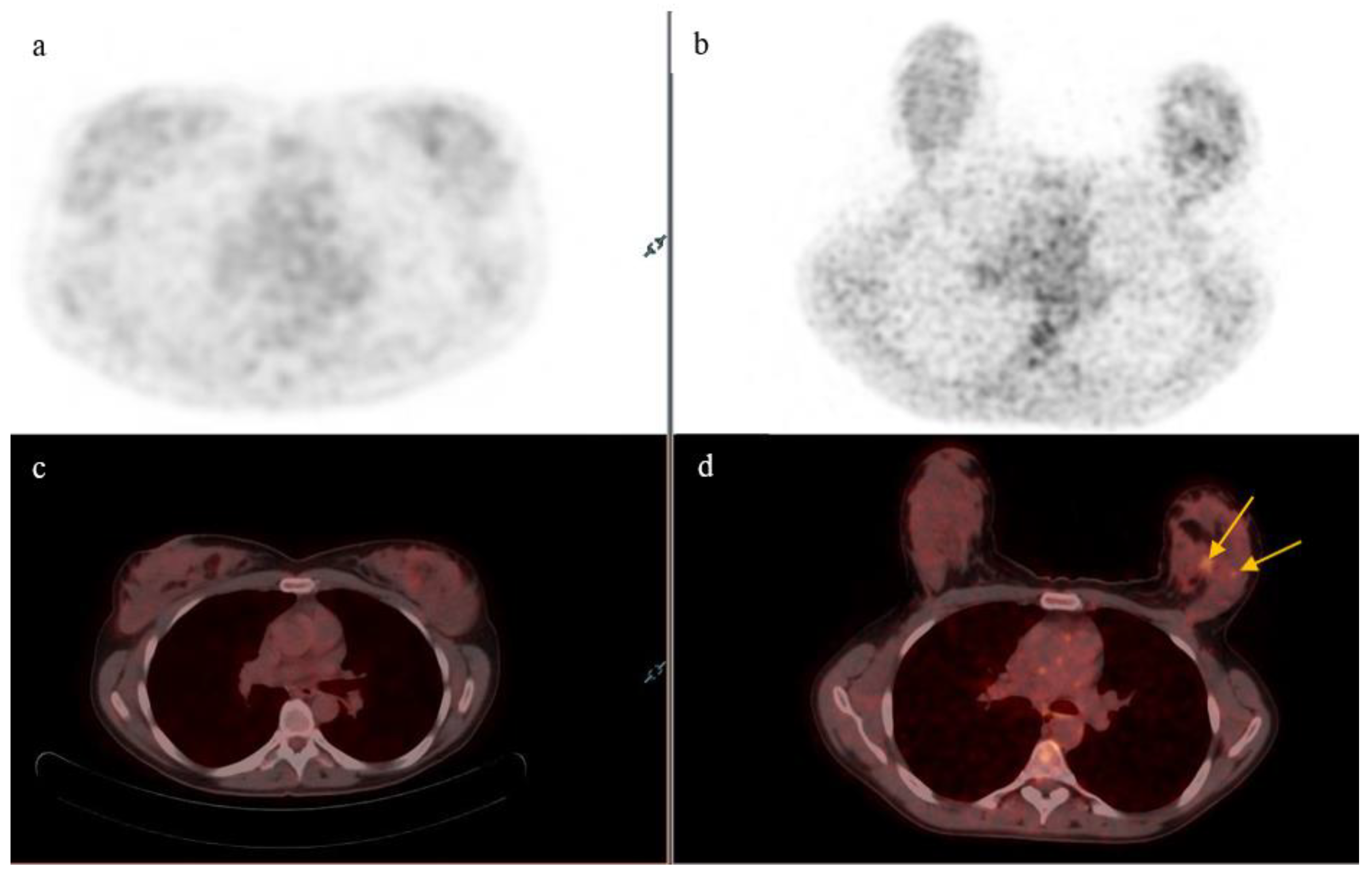
- On supine position acquisition, primary breast lesions were missed in three patients, all of which were detected on prone position. One such example is seen in Figure 2,
- A single lesion was detected on supine position in two patients who, on prone position, each had two lesions detected (Table 1),
- One lesion was detected on supine position in a patient who was found to have three lesions on prone acquisition (Table 1).
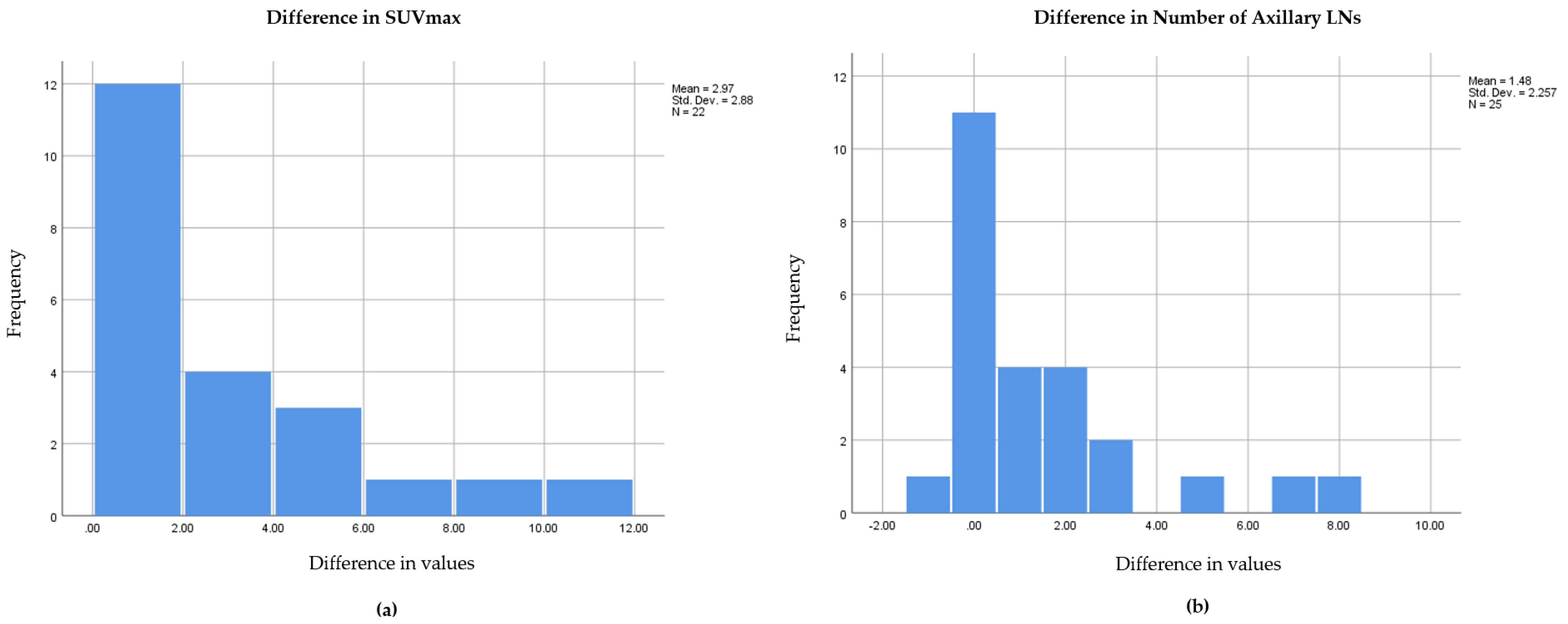
3.2. Lymph Nodes
3.3. Bone Metastasis
3.4. Pathology
3.5. Staging and Prognosis
4. Discussion
5. Conclusions
Author Contributions
Funding
Institutional Review Board Statement
Informed Consent Statement
Data Availability Statement
Conflicts of Interest
References
- Arnold, M.; Morgan, E.; Rumgay, H.; Mafra, A.; Singh, D.; Laversanne, M.; Vignat, J.; Gralow, J.R.; Cardoso, F.; Siesling, S.; et al. Current and future burden of breast cancer: Global statistics for 2020 and 2040. Breast 2022, 66, 15–23. [Google Scholar] [CrossRef]
- Giaquinto, A.N.; Sung, H.; Miller, K.D.; Kramer, J.L.; Newman, L.A.; Minihan, A.; Jemal, A.; Siegel, R.L. Breast Cancer Statistics, 2022. CA Cancer J. Clin. 2022, 72, 524–541. [Google Scholar] [CrossRef] [PubMed]
- Gradishar, W.J.; Moran, M.S.; Abraham, J.; Aft, R.; Agnese, D.; Allison, K.H.; Anderson, B.; Burstein, H.J.; Chew, H.; Dang, C.; et al. Breast Cancer, Version 3.2022, NCCN Clinical Practice Guidelines in Oncology. J. Natl. Compr. Canc. Netw. 2022, 20, 691–722. [Google Scholar] [CrossRef] [PubMed]
- Han, S.; Choi, J.Y. Impact of 18F-FDG PET, PET/CT, and PET/MRI on Staging and Management as an Initial Staging Modality in Breast Cancer: A Systematic Review and Meta-analysis. Clin. Nucl. Med. 2021, 46, 271–282. [Google Scholar] [CrossRef] [PubMed]
- Groheux, D.; Mankoff, D.; Espié, M.; Hindié, E. 18F-FDG PET/CT in the early prediction of pathological response in aggressive subtypes of breast cancer: Review of the literature and recommendations for use in clinical trials. Eur. J. Nucl. Med. Mol. Imaging 2016, 43, 983–993. [Google Scholar] [CrossRef]
- Kasem, J.; Wazir, U.; Mokbel, K. Sensitivity, Specificity and the Diagnostic Accuracy of PET/CT for Axillary Staging in Patients with Stage I-III Cancer: A Systematic Review of the Literature. In Vivo 2021, 35, 23–30. [Google Scholar] [CrossRef]
- Vogsen, M.; Jensen, J.D.; Gerke, O.; Jyling, A.M.B.; Asussen, J.T.; Christensen, I.C.; Braad, P.-E.; Thye-Rønn, P.; Søe, K.L.; Ewertz, M.; et al. Benefits and harms of implementing [(18)F]FDG-PET/CT for diagnosing recurrent breast cancer: A prospective clinical study. EJNMMI Res. 2021, 11, 93. [Google Scholar] [CrossRef]
- Wahl, R.L.; Siegel, B.A.; Coleman, R.E.; Gatsonis, C.G. Prospective multicenter study of axillary nodal staging by positron emission tomography in breast cancer: A report of the staging breast cancer with PET Study Group. J. Clin. Oncol. 2004, 22, 277–285. [Google Scholar] [CrossRef]
- Kikano, E.G.; Avril, S.; Marshall, H.; Jones, R.S.; Montero, A.J.; Avril, N. PET/CT Variants and Pitfalls in Breast Cancers. Semin. Nucl. Med. 2021, 51, 474–484. [Google Scholar] [CrossRef]
- Khalkhali, I.; Mena, I.; Diggles, L. Review of imaging techniques for the diagnosis of breast cancer: A new role of prone scintimammography using technetium-99m sestamibi. Eur. J. Nucl. Med. 1994, 21, 357–362. [Google Scholar] [CrossRef]
- Kalli, S.; Semine, A.; Cohen, S.; Naber, S.P.; Makim, S.S.; Bahl, M. American Joint Committee on Cancer’s Staging System for Breast Cancer, Eighth Edition: What the Radiologist Needs to Know. Radiographics 2018, 38, 1921–1933. [Google Scholar] [CrossRef] [PubMed] [Green Version]
- Abramson, R.G.; Lambert, K.F.; Jones-Jackson, L.B.; Arlinghaus, L.R.; Willam, J.; Abramson, V.G.; Chakravarthy, A.B.; Yankeelov, T.E. Prone Versus Supine Breast FDG-PET/CT for Assessing Locoregional Disease Distribution in Locally Advanced Breast Cancer. Acad. Radiol. 2015, 22, 853–859. [Google Scholar] [CrossRef] [PubMed] [Green Version]
- Hayato, K.; Masatoshi, I.; Teruhiko, F.; Teruhiko, F.; Seiji, K.; Masafumi, U.; Kenkichi, B.; Teruo, M.; Kawamura, H.; Etsuyo, O.; et al. Improved breast cancer detection of prone breast fluorodeoxyglucose-PET in 118 patients. Nucl. Med. Commun. 2008, 29, 885–893. [Google Scholar]
- Teixeira, S.C.; Koolen, B.B.; Vogel, W.V.; Wesseling, J.; Stokkel, M.P.M.; Vrancken, P.; Marie-Jeanne, T.F.; van der Noort, V.; Rutgers, E.J.; Olmos, V.; et al. Additional Prone 18F-FDG PET/CT Acquisition to Improve the Visualization of the Primary Tumor and Regional Lymph Node Metastases in Stage II/III Breast Cancer. Clin. Nucl. Med. 2016, 41, e181–e186. [Google Scholar] [CrossRef]
- Yutani, K.; Tatsumi, M.; Uhera, T.; Nishimura, T. Effect of patients’ being prone during FDG PET for the diagnosis of breast cancer. AJR Am. J. Roentgenol. 1999, 173, 1337–1339. [Google Scholar] [CrossRef] [Green Version]
- Stokkeland, P.J.; Andersen, E.; Bjørndal, M.M.; Mikalsen, A.M.; Aslaksen, S.; Hyldmo, P.K. Maintaining immobilisation devices on trauma patients during CT: A feasibility study. Scand. J. Trauma Resusc. Emerg. Med. 2017, 25, 84. [Google Scholar] [CrossRef] [Green Version]
- Li, X.; Abramson, R.G.; Arlinghaus, L.R.; Chakravarthy, A.B.; Abramson, V.; Mayer, I.; Farley, J.; Delbeke, D.; Yankeelov, T.E. An algorithm for longitudinal registration of PET/CT images acquired during neoadjuvant chemotherapy in breast cancer: Preliminary results. EJNMMI Res. 2012, 2, 62. [Google Scholar] [CrossRef] [Green Version]
- Boellaard, R.; Delgado-Bolton, R.; Oyen, W.J.G.; Giammarile, F.; Tatsch, K.; Eschner, W.; Verzijibergen, F.J.; Barrington, S.F.; Pike, L.C.; Weber, W.A.; et al. FDG PET/CT: EANM procedure guidelines for tumour imaging: Version 2.0. Eur. J. Nucl. Med. Mol. Imaging 2015, 42, 328–354. [Google Scholar] [CrossRef]
- Wangerin, K.A.; Muzi, M.; Peterson, L.M.; Linden, H.M.; Novakova, A.; O’Sullivan, F.; Kurland, B.F.; Mankoff, D.A.; Kinahan, P.E. Effect of (18)F-FDG uptake time on lesion detectability in PET imaging of early stage breast cancer. Tomography 2015, 1, 53–60. [Google Scholar] [CrossRef]
- Kaida, H.; Ishibashi, M.; Fujii, T.; Kurata, S.; Ogo, E.; Tanaka, M.; Hayabuchi, N. Improved detection of breast cancer on FDG-PET cancer screening using breast positioning device. Ann. Nucl. Med. 2008, 22, 95–101. [Google Scholar] [CrossRef]
- Erdogan, E.B.; Aydin, M. Investigation of Added Value of Imaging Performed in a Prone Position to Standard (18)F-Fluorodeoxyglucose Positron Emission Tomography/Computed Tomography Imaging for Staging in Patients with Breast Cancer. Mol. Imaging Radionucl. Ther. 2022, 31, 23–32. [Google Scholar] [CrossRef] [PubMed]
- Hashem, T.; Abdelmoez, A.; Rozeka, A.M.; Abdelazeem, H. Intra-mammary lymph nodes, an overlooked breast cancer prognostic tool? World J. Surg. Oncol. 2021, 19, 114. [Google Scholar] [CrossRef] [PubMed]
- Nassar, A.; Cohen, C.; Cotsonis, G.; Carlson, G. Significance of intramammary lymph nodes in the staging of breast cancer: Correlation with tumor characteristics and outcome. Breast J. 2008, 14, 147–152. [Google Scholar] [CrossRef] [PubMed]
- Vidal-Sicart, S.; Perades, P.; Zanón, G.; Pahisa, J.; Martinez-Román, S.; Caparrós, X.; Vilalta, A.; Rull, R.; Pons, F. Added value of intraoperative real-time imaging in searches for difficult-to-locate sentinel nodes. J. Nucl. Med. 2010, 51, 1219–1225. [Google Scholar] [CrossRef] [Green Version]
- Heusner, T.A.; Freudenberg, L.S.; Kuehl, H.; Hauth, E.A.M.; Veit-Haibach, P.; Forsting, M.; Bockissh, A.; Antoch, G. Whole-body PET/CT-mammography for staging breast cancer: Initial results. Br. J. Radiol. 2008, 81, 743–748. [Google Scholar] [CrossRef]
- van Loevezijn, A.A.; Stokkel, M.P.M.; Donswijk, M.L.; van Wekhoven, E.D.; van der Noordaa, M.E.M.; van Duijnhoven, F.H.; Peeters, M.-J.T.F.D. [(18)F]FDG-PET/CT in prone compared to supine position for optimal axillary staging and treatment in clinically node-positive breast cancer patients with neoadjuvant systemic therapy. EJNMMI Res. 2021, 11, 78. [Google Scholar] [CrossRef]
- Faast, A.; Ikeda, D.M.; Pittman, S.; Demartini, W.; Kozlov, A. FDG Avid Abnormalities in the Breast: Breast Cancer Mimics. Curr. Radiol. Rep. 2021, 9, 8. [Google Scholar] [CrossRef]
- Skawran, S.; Gennari, A.G.; Dittli, M.; Treyer, V.; Muehlematter, U.J.; Maurer, A.; Burger, I.A.; Mader, C.; Messerli, O.; Grünig, H.; et al. [(18)F]FDG uptake of axillary lymph nodes after COVID-19 vaccination in oncological PET/CT: Frequency, intensity, and potential clinical impact. Eur. Radiol. 2022, 32, 508–516. [Google Scholar] [CrossRef]
- Minamimoto, R.; Kiyomatsu, T. Effects of COVID-19 vaccination on FDG-PET/CT imaging: A literature review. Glob. Health Med. 2021, 3, 129–133. [Google Scholar] [CrossRef]
- McIntosh, L.J.; Bankier, A.A.; Vijayaraghaven, G.R.; Licho, R.; Rosen, M.P. COVID-19 Vaccination-Related Uptake on FDG PET/CT: An Emerging Dilemma and Suggestions for Management. AJR Am. J. Roentgenol. 2021, 217, 975–983. [Google Scholar] [CrossRef]


| Patient Number | Number of Primary Breast Lesions Detected on Prone | Highest SUVmax on Prone | Number of Primary Breast Lesions Detected on Supine | Highest SUVmax on Supine |
|---|---|---|---|---|
| 1 | 2 | 21.11 | 2 | 15.40 |
| 2 | 2 | 5.97 | 1 | 4.96 |
| 3 | 1 | 3.30 | 0 | - |
| 4 | 1 | 2.30 | 0 | - |
| 5 | 1 | 4.20 | 1 | 3.30 |
| 6 | 1 | 3.96 | 0 | - |
| 7 | 1 | 7.15 | 1 | 5.20 |
| 8 | 1 | 5.90 | 1 | 3.80 |
| 9 | 1 | 12.30 | 1 | 7.30 |
| 10 | 1 | 5.50 | 1 | 5.50 |
| 11 | 3 | 4.11 | 1 | 3.90 |
| 12 | 1 | 19.90 | 1 | 18.30 |
| 13 | 2 | 4.70 | 1 | 4.40 |
| 14 | Multiple | 5.90 | Multiple | 4.20 |
| 15 | 1 | 8.40 | 1 | 7.70 |
| 16 | 1 | 6.00 | 1 | 4.70 |
| 17 | 1 | 32.90 | 1 | 30.20 |
| 18 | 1 | 7.10 | 1 | 5.50 |
| 19 | 1 | 7.50 | 1 | 5.90 |
| 20 | 1 | 13.80 | 1 | 4.30 |
| 21 | 1 | 9.56 | 1 | 6.80 |
| 22 | 1 | 7.40 | 1 | 3.00 |
| 23 | 1 | 20.30 | 1 | 9.90 |
| 24 | 1 | 11.50 | 1 | 4.80 |
| 25 | 1 | 5.30 | 1 | 2.10 |
| Patient Number | Intramammary Nodes (Prone; Supine) | Axillary Nodes (Prone; Supine) | Internal Mammary Nodes (Prone; Supine) | Supraclavicular Nodes (Prone; Supine) |
|---|---|---|---|---|
| 1 | 0; 0 | 12; 4 | 0; 0 | 0; 0 |
| 2 | 0; 0 | 1; 1 | 0; 0 | 0; 0 |
| 3 | 0; 0 | 3; 1 | 0; 0 | 1; 0 |
| 4 | 0; 0 | 3; 0 | 2; 2 | 0; 0 |
| 5 | 0; 0 | 2; 0 | 0; 0 | 0; 0 |
| 6 | 1; 0 | 0; 0 | 0; 0 | 0; 0 |
| 7 | 0; 0 | 0; 0 | 0; 0 | 0; 0 |
| 8 | 0; 0 | 0; 0 | 0; 0 | 0; 0 |
| 9 | 0; 0 | 2; 0 | 0; 0 | 0; 0 |
| 10 | 0; 0 | 8; 3 | 1; 1 | 0; 0 |
| 11 | 0; 0 | 3; 2 | 0; 0 | 0; 0 |
| 12 | 0; 0 | 2; 1 | 0; 0 | 0; 0 |
| 13 | 0; 0 | 3; 4 | 0; 0 | 0; 0 |
| 14 | 1; 0 | 10; 3 | 0; 0 | 0; 0 |
| 15 | 0; 0 | 3; 0 | 0; 0 | 0; 0 |
| 16 | 0; 0 | 0; 0 | 0; 0 | 0; 0 |
| 17 | 0; 0 | 0; 0 | 0; 0 | 0; 0 |
| 18 | 0; 0 | 0; 0 | 0; 0 | 1; 1 |
| 19 | 0; 0 | 1; 0 | 0; 0 | 0; 0 |
| 20 | 0; 0 | 1; 0 | 0; 0 | 0; 0 |
| 21 | 0; 0 | 0; 0 | 0; 0 | 0; 0 |
| 22 | 0; 0 | 0; 0 | 0; 0 | 0; 0 |
| 23 | 0; 0 | 0; 0 | 0; 0 | 0; 0 |
| 24 | 0; 0 | 8; 6 | 0; 0 | 0; 0 |
| 25 | 0; 0 | 0; 0 | 0; 0 | 0; 0 |
Disclaimer/Publisher’s Note: The statements, opinions and data contained in all publications are solely those of the individual author(s) and contributor(s) and not of MDPI and/or the editor(s). MDPI and/or the editor(s) disclaim responsibility for any injury to people or property resulting from any ideas, methods, instructions or products referred to in the content. |
© 2023 by the authors. Licensee MDPI, Basel, Switzerland. This article is an open access article distributed under the terms and conditions of the Creative Commons Attribution (CC BY) license (https://creativecommons.org/licenses/by/4.0/).
Share and Cite
Nassar, L.; Kassas, M.; Abi-Ghanem, A.S.; El-Jebai, M.; Al-Zakleet, S.; Baassiri, A.S.; Naccoul, R.A.; Barakat, A.; Tfayli, A.; Assi, H.; et al. Prone versus Supine FDG PET/CT in the Staging of Breast Cancer. Diagnostics 2023, 13, 367. https://doi.org/10.3390/diagnostics13030367
Nassar L, Kassas M, Abi-Ghanem AS, El-Jebai M, Al-Zakleet S, Baassiri AS, Naccoul RA, Barakat A, Tfayli A, Assi H, et al. Prone versus Supine FDG PET/CT in the Staging of Breast Cancer. Diagnostics. 2023; 13(3):367. https://doi.org/10.3390/diagnostics13030367
Chicago/Turabian StyleNassar, Lara, Mutaz Kassas, Alain S. Abi-Ghanem, Malak El-Jebai, Safaa Al-Zakleet, Amro S. Baassiri, Rami Abou Naccoul, Andrew Barakat, Arafat Tfayli, Hazem Assi, and et al. 2023. "Prone versus Supine FDG PET/CT in the Staging of Breast Cancer" Diagnostics 13, no. 3: 367. https://doi.org/10.3390/diagnostics13030367