Article

Double Anterior Interventricular Arteries: Prevalence and Morphological Types—A Dissection Study

by
Ecaterina Daescu
1,2,
Alexandra Enache
2,3,4,
Emanuela Stan
2,3,4,
Sorin Lucian Bolintineanu
1,
Laura Andreea Ghenciu
5,
Alexandra Corina Faur
1,
Agneta Maria Pusztai
1,* and
Delia Elena Zahoi
1
1
Department I of Anatomy and Embryology, “Victor Babes” University of Medicine and Pharmacy, 300041 Timisoara, Romania
2
Institute of Legal Medicine Timisoara, 300610 Timisoara, Romania
3
Department of Neuroscience, Discipline of Forensic Medicine, Bioethics, Deontology and Medical Law, “Victor Babes” University of Medicine and Pharmacy, 300041 Timisoara, Romania
4
Ethics and Human Identification Research Center, Department of Neurosciences, “Victor Babes” University of Medicine and Pharmacy, 300041 Timisoara, Romania
5
Department of Functional Sciences, “Victor Babes” University of Medicine and Pharmacy Timisoara, 300041 Timisoara, Romania
*
Author to whom correspondence should be addressed.
J. Pers. Med. 2024, 14(9), 1007; https://doi.org/10.3390/jpm14091007
Submission received: 21 August 2024 / Revised: 12 September 2024 / Accepted: 20 September 2024 / Published: 22 September 2024
(This article belongs to the Section Clinical Medicine, Cell, and Organism Physiology)

Abstract

:
Background: This study aimed to evaluate the prevalence of double anterior interventricular artery using the dissection method. Metode: A retrospective study was conducted between 2010 and 2024 at the Anatomy and Embryology Laboratory of the Victor Babes University of Medicine and Pharmacy in Timisoara. Eighty cases were analyzed for morphological variants of the coronary arteries, especially the anterior interventricular artery. Results: Two cases of double anterior interventricular arteries were identified. In the first case, the two anterior interventricular arteries originated from the anterior interventricular branch of the left coronary artery. In the second case, an additional anterior interventricular artery with an aortic origin was found running along the lower third of the two interventricular grooves. This shape has not been described before in the specialized literature. Conclusions: Knowing the potential variations of the double left anterior descending artery is critical for interpreting cardiac imaging and choosing and planning percutaneous and surgical reperfusion strategies.

1. Introduction

The arterial supply of the heart is provided by the two (right and left) coronary arteries. The right coronary artery arises from the right coronary aortic sinus, with the origin typically located superior to the right semilunar valve (in 10 percent of cases, the ostium is below the cusps margin). It initially passes anterior and to the right, between the right auricle and the pulmonary trunk. Further, it reaches the atrioventricular sulcus and descends almost vertically along it to the right cardiac border, curving around it into the posterior part, where it approaches its junction with both the interatrial and interventricular sulci. The right coronary artery supplies the right ventricle, the right atrium, the sinoatrial node, the atrioventricular node, and the posterior third of the interventricular septum through its branches, with the left coronary artery supplying the apex. The left coronary artery is larger in caliber than the right and supplies a greater volume of myocardium. It originates from the left coronary aortic sinus; the ostium is below the cusp margin, leading into two major initial branches—the circumflex and anterior interventricular branches [1]. Through its branches, it supplies the left atrium, the left ventricle, and the anterior two thirds of the interventricular septum.
The coronary arteries have numerous anatomical variations, including the site of origin, number, course, dimensions (caliber or length), branching pattern, and vascular territory, among others. Most anatomical variants have no repercussions and are discovered accidentally. However, some can lead to severe arrhythmias, the interruption of myocardial perfusion (either intermittent or chronic), or even sudden cardiac death [2]. The double anterior interventricular artery is a very rare coronary anomaly that occurs, according to various studies, with a frequency ranging from 0.13 percent to 5.96 percent of the population [3,4].
This study aimed to evaluate the prevalence of double anterior interventricular artery using the dissection method.

2. Materials and Methods

This study was conducted at the Department of Anatomy and Embryology of the ‘Victor Babes’ University of Medicine and Pharmacy in Timisoara, between 2010 and 2024, on a set of 80 hearts from formalin-fixed adult human cadavers, collected and dissected in the laboratory, in accordance with current legislation. Gross dissection was performed using the standard technique. We removed the pericardium and carefully examined the vascular structures: aorta, pulmonary artery, pulmonary veins, superior vena cava, and inferior vena cava were then sectioned and the heart was removed. In particular, the coronary arteries were carefully dissected. The study classified any deviations from the typical structure into distinct morphological types and analyzed numerous aspects of the coronary arteries, including their origin, number, course, and branching pattern.

3. Results

The analysis of the morphological variations of the anterior interventricular artery on the study material revealed the presence of double anterior interventricular arteries in two cases (2.5 percent).
In the first case (Figure 1), the anterior interventricular branch (anterior descending artery) originated from the left coronary artery. After a short path along the anterior interventricular groove, it branched into two long anterior interventricular arteries that descended on either side of the anterior interventricular groove, almost to the apex of the heart. Aside from the right branch’s initial intramural course, this morphological type resembles type XIII, as described by Pellegrini, J.R., et al. [5].
In the second case (Figure 2 and Figure 3), an initial analysis of the origin of the coronary arteries from the aortic sinus revealed the presence of an additional artery. Starting from their aortic origins, the three arteries were dissected, and their course was traced to identify them. The right coronary artery, which arose from the right coronary sinus, was located in the coronary sulcus between the right atrium and the right auricle, extending in a posterior direction. After the right marginal branch stemmed from it, the right coronary artery continued in a posterior direction, splitting into two branches of similar size. The first branch descended diagonally down the diaphragmatic surface of the right ventricle, terminating in two branches intended for this specific region. The second branch crossed the posterior portion of the coronary sulcus to the junction with the posterior interventricular sulcus, after which it descended into the posterior interventricular sulcus, extending approximately to the middle third of this sulcus.
The left coronary artery also exhibited morphological peculiarities. It divided into three branches of approximately equal caliber immediately after its origin: the circumflex branch, the left marginal branch, and the anterior interventricular branch. The circumflex branch traversed the posterior portion of the coronary sulcus, approached the posterior interventricular sulcus, and ended by giving off two branches destined for the diaphragmatic surface of the left ventricle. The left marginal branch followed a descending trajectory, branching out at the level of the left pulmonary surface. From its origin, the left anterior interventricular branch (anterior descending artery) positioned itself between the pulmonary artery and the left auricle, crossing the anterior portion of the coronary sulcus to reach the sternocostal surface of the heart. Shorter than usual, it traversed the upper two-thirds of the anterior interventricular sulcus, after which it exited and ended on the sternocostal surface of the left ventricle. In the middle third (between the origin and entry into the anterior interventricular sulcus), it had an intramural course (muscular bridge). The aortic sinus, anterior to the right coronary artery, was the origin of the third artery, later named the supernumerary anterior interventricular artery (right) due to its trajectory. From its origin, the artery had a short path between the right auricle (which partially covered it) and the pulmonary artery, positioning itself anterior to the latter to reach the sternocostal surface at the base of the right ventricle. From this level, the artery’s course obliquely descended toward the left. In the middle third of the sternocostal surface, the right anterior interventricular artery had a trajectory almost parallel to the anterior interventricular branch of the left coronary artery. Further, it continued its oblique descending path toward the anterior interventricular sulcus, traversing its lower third and reaching the level of the cardiac apex. After crossing the apical notch of the heart, it continued its course on the diaphragmatic surface, traversing the lower third of the posterior interventricular sulcus.
In this case, both the anterior interventricular branch (originating from the left coronary artery) and the posterior interventricular branch (originating from the right coronary artery) are present. A particular feature of this case is the presence of a long supernumerary anterior interventricular artery (right), which originates directly from the right coronary sinus and extends beyond the apical notch of the heart, entering the lower third of the posterior interventricular sulcus. The short left anterior interventricular branch (anterior descending artery) also passes through muscle in its middle third before it enters the anterior interventricular sulcus.
As a similarity, both the anterior and posterior interventricular sulci each contain two arteries. The posterior interventricular branches originating from the left coronary artery and the posterior interventricular branches originating from the right coronary artery have shorter paths; each traverses the upper two-thirds of their respective interventricular sulcus. Simultaneously, the supernumerary/right anterior interventricular artery of aortic origin traverses the lower third of both interventricular sulci.
The presented case is quite complex as it includes multiple anomalies in the number, origin, course, and termination of the coronary arteries.

4. Discussion

Over time, multiple studies have been conducted regarding the morphological variability of the coronary arteries, and the frequency of cases reported in the specialized literature varies depending on the size of the study sample, the method (dissection, angiography, angio-CT, or MRI), sex, geographical area, etc. Given their very low frequency, most are case presentations.
Coronary artery anomalies correspond to patterns of heart vascularization that are generally encountered very rarely; characterized by abnormal origin, course or termination of the coronary arteries; and most of them are asymptomatic and discovered incidentally [6,7]. In the general population, they appear with a frequency ranging from 0.3 percent to 5.8 percent [2,8].
Andishmand, A., et al., reported an incidence of coronary artery anomalies of 1.26 percent [9]. Al-Umairi. R.S., et al. in a cohort of 4445 patients who underwent coronary-computed tomography angiography, identified 59 patients (1.3 percent) with coronary artery anomalies [10]. Kashyap, J.R., et al. reported an incidence of coronary artery anomalies of 2.06 percent [11]. Gräni, C., et al. reported an incidence of coronary artery anomalies of 2.6 percent [12]. Şahin, T., and Ilgar, M., in a study of 5200 multidetector computed tomography coronary angiography, reported a 2.61 percent incidence of coronary artery anomalies [13]. Sidhu et al. reported an incidence of coronary artery anomalies of 3.06 percent [14].
The anterior interventricular branch (anterior descending artery) usually originates from the left coronary artery and traverses the anterior interventricular sulcus toward the cardiac apex. Along its trajectory, it gives off several branches: the anterior conal artery, diagonal branches, and septal branches.
The double anterior interventricular artery is a rare congenital anomaly defined as the presence of two distinct arteries that approach the anterior interventricular sulcus. This category also includes cases where there are two branches, typically one short and one long, that traverse and supply different parts of the anterior interventricular sulcus; these may have different origins and courses.
Waterson et al. made the first mention of this congenital anomaly in 1939 [15].
Double anterior interventricular arteries are very rare coronary anomalies; their reported incidence varies between 0.11 percent [10], 0.13 percent [3], 0.56 percent [11], 0.68 percent [14], 0.77 percent [13], 1.3 percent [16], and 4 percent [17].
A double left anterior descending artery arising from the left and right coronary arteries is an extremely rare congenital coronary anomaly. The specialized literature has identified several case presentations with double anterior interventricular arteries originating from different sources: the right coronary artery and the left coronary artery [3,17,18]; double anterior interventricular arteries originating from the right coronary sinus or the left coronary sinus [19]; double anterior interventricular arteries originating from the right coronary sinus or the left coronary artery [20]; and double anterior interventricular arteries originating from the right coronary sinus [21].
Spindola-Franco et al. conducted the first study that analyzed and classified double anterior interventricular arteries in 1983. They categorized these cases into four morphological types, considering the origin, course, and length of the arteries that approach the anterior interventricular sulcus [3]. Over time, researchers included additional morphological types to this primary classification (Table 1).
This study’s first case of double AIA/LAD is similar to Type XIII described by Pellegrini, J.R., et al. [5]. However, in its initial phase, the bifurcating branch that descends to the right of the AIS has an intramural course. We propose that this morphological type of double anterior interventricular artery is considered subtype XIIIA.
The second identified case has a right AIA (the long AIA/LAD), which emerges from the right coronary sinus and has a distinct ostium. It has a pre-pulmonary trajectory, moving towards the AIS and traversing its lower third to reach the cardiac apex. It then crosses the heart’s apical notch and continues its course on the diaphragmatic surface, extending through the lower third of the posterior interventricular sulcus. The left anterior interventricular artery (the short AIA/LAD) traverses the upper two-thirds of the AIS, after which it exits and terminates on the sternocostal surface of the left ventricle. In its middle third (between its origin and entry into the AIS), it exhibits an intramural course (muscular bridge).
There are no clinical data regarding any potential cardiac conditions in the individuals involved.
Therefore, we suggest classifying this case as Type XIV of the double anterior interventricular artery due to its unique characteristics.
A comprehensive understanding of these morphological variants is crucial to avoid the misinterpretation of CT coronary angiography images, as well as for accurate diagnosis, planning, and therapeutic management [23].
The occurrence of various coronary anomalies can be understood or explained from the perspective of the embryological development of the coronary system of the heart.
Recent studies on embryos have clarified many aspects, but controversies still exist regarding the origin of various precursors and the mechanisms involved in the formation of the coronary system. Proepicardial cells, which cover the transverse septum and the venous sinus at the beginning of embryonic heart development, represent a major source of precursors. These cells later migrate to form the epicardium, which surrounds the heart [24]. The primordial coronary vessels appear in the 28-day-old embryo as small vascular islands formed from epicardial cells. They are located in the coronary and interventricular sulci, at the level of the cardiac apex, and in other regions of the ventricular wall [25]. At 33 days, these islands start to grow and spread out, creating multiple vascular networks in different subepicardial areas. From these structures branches penetrate deeper into the myocardium, forming small vessels and capillaries [26]. A peritruncal ring forms from the subepicardial vascular structure surrounding the initial portion of the major arteries [27,28]. Recent studies confirm that the proximal portion of the coronary arteries forms through the aortic wall penetration by the endothelial cells of the peritruncal ring, thus forming the coronary ostia [26,29].
Aortic wall penetration is a process of localized apoptosis in areas rich in VEGF-C receptors. The epicardium is an important place to find cells that are high in VEGF receptors, which help the coronary plexus grow [30]. Researchers have also demonstrated that the absence of VEGF inhibits the formation of ostia, alters the connection of coronary arteries with the aorta, and can thereby lead to coronary anomalies [29,31].
The final morphology of the coronary system depends on the branching of coronary arteries from the aorta. Any anomalies in the development or connection of the two can lead to different variants in the number or location of the coronary ostia.

5. Conclusions

Our study highlighted two cases of double AIA/LAD out of the eighty analyzed (2.5%), and we propose their inclusion in types XIIIA and XIV
Knowing the potential variations of the double left anterior descending artery is critical in interpreting cardiac imaging as well as in choosing and planning percutaneous and surgical reperfusion strategies.

Author Contributions

Conceptualization, E.D. and D.E.Z.; methodology, E.D., S.L.B. and A.M.P.; validation, D.E.Z., A.E. and S.L.B.; formal analysis, E.S. and A.E.; investigation, E.D., A.M.P. and A.C.F.; resources, E.D., S.L.B. and D.E.Z.; data curation, E.S., L.A.G. and A.C.F.; writing—original draft preparation, E.S. and L.A.G.; writing—review and editing, S.L.B. and A.M.P.; visualization, A.E. and E.D.; supervision, E.D. and D.E.Z. All authors have read and agreed to the published version of the manuscript.

Funding

This research received no external funding.

Institutional Review Board Statement

The study was conducted in accordance with the Declaration of Helsinki and approved by the Committee on Research Ethics of the Victor Babes University of Medicine and Pharmacy Timisoara (approval No. 42/30 July 2024 rev).

Informed Consent Statement

Not applicable.

Data Availability Statement

The raw data supporting the conclusions of this article will be made available by the authors on request.

Acknowledgments

We would like to acknowledge the Victor Babes University of Medicine and Pharmacy Timisoara for their support in covering the costs of publication for this research paper.

Conflicts of Interest

The authors declare no conflicts of interest.

Abbreviations

Left Coronary ArteryLCA
Anterior Interventricular ArteryAIA
Left Anterior Descending ArteryLAD
Anterior Interventricular SulcusAIS
Left VentricleLV
Right VentricleRV
Right Coronary ArteryRCA
Anterior Interventricular BranchAIB
Right Ventricular Outflow TractRVOT
Left Marginal BranchLMB
Circumflex ArteryCX

References

  1. Loukas, M.; Standring, S. (Eds.) Gray’s Anatomy. The Anatomical Basis of Clinical Practice, 42nd ed.; Elsevier: Amsterdam, The Netherlands, 2021; pp. 1068–1097. [Google Scholar]
  2. Agarwal, P.P.; Dennie, C.; Pena, E.; Nguyen, E.; LaBounty, T.; Yang, B.; Patel, S. Anomalous Coronary Arteries That Need Intervention: Review of Pre- and Postoperative Imaging Appearances. Radiographics 2017, 37, 740–757. [Google Scholar] [CrossRef] [PubMed]
  3. Spindola-Franco, H.; Grose, R.; Solomon, N. Dual left anterior descending coronary artery: Angiographic description of important variants and surgical implications. Am. Heart J. 1983, 105, 445–455. [Google Scholar] [CrossRef] [PubMed]
  4. Maggialetti, N.; Greco, S.; Lorusso, G.; Mileti, C.; Sfregola, G.; Brunese, M.C.; Zappia, M.; Belfiore, M.P.; Sullo, P.; Reginelli, A.; et al. The Role of Coronary CT Angiography in the Evaluation of Dual Left Anterior Descending Artery Prevalence and Subtypes: A Retrospective Multicenter Study. J. Pers. Med. 2023, 13, 1127. [Google Scholar] [CrossRef] [PubMed]
  5. Pellegrini, J.R.; Munshi, R.; Alvarez Betancourt, A.; Tokhi, B.; Makaryus, A.N. “Two for One”, Novel Dual Left Anterior Descending Artery (LAD) Variant: Type XIII. Cureus 2021, 13, e14717. [Google Scholar] [CrossRef] [PubMed]
  6. Gentile, F.; Castiglione, V.; De Caterina, R. Coronary Artery Anomalies. Circulation 2021, 144, 983–996. [Google Scholar] [CrossRef]
  7. Villa, A.D.; Sammut, E.; Nair, A.; Rajani, R.; Bonamini, R.; Chiribiri, A. Coronary artery anomalies overview: The normal and the abnormal. World J. Radiol. 2016, 8, 537–555. [Google Scholar] [CrossRef]
  8. Alexander, R.W.; Griffith, G.C. Anomalies of the coronary arteries and their clinical significance. Circulation 1956, 14, 800–805. [Google Scholar] [CrossRef]
  9. Andishmand, A.; Montazerghaem, H.; Pedarzadeh, A.; Varastehravan, H.R.; Mohammadi, H.; Moghadam, R.N.; Azimizadeh, M.; Ahrar, M.H.; Khezri, A.; Andishmand, M. Prevalence and characteristics of coronary artery anomalies (CAAS) in 3016 symptomatic adult participants undergoing coronary computed tomography angiography (CCTA): A single-center retrospective study in Iran. J. Cardiovasc. Thorac. Res. 2023, 15, 218–222. [Google Scholar] [CrossRef]
  10. Al-Umairi, R.S.; Al-Kindi, F.; Al-Tai, S. Prevalence and Spectrum of Coronary Anomalies Detected on Coronary Computed Tomography Angiography. Sultan Qaboos Univ. Med. J. 2019, 19, e108–e113. [Google Scholar] [CrossRef]
  11. Kashyap, J.R.; Kumar, S.; Reddy, S.; Rao k, R.; Sehrawat, O.; Kashyap, R.; Kansal, M.; Reddy, H.; Kadiyala, V.; Uppa, L. Prevalence and Pattern of Congenital Coronary Artery Anomalies in Patients Undergoing Coronary Angiography at a Tertiary Care Hospital of Northern India. Cureus 2021, 13, e14399. [Google Scholar] [CrossRef]
  12. Gräni, C.; Benz, D.C.; Schmied, C.; Vontobel, J.; Possner, M.; Clerc, O.F.; Mikulicic, F.; Stehli, J.; Fuchs, T.A.; Pazhenkottil, A.P.; et al. Prevalence and characteristics of coronary artery anomalies detected by coronary computed tomography angiography in 5634 consecutive patients in a single centre in Switzerland. Swiss Med. Wkly. 2016, 146, w14294. [Google Scholar] [PubMed]
  13. Şahin, T.; Ilgar, M. Investigation of the Frequency of Coronary Artery Anomalies in MDCT Coronary Angiography and Comparison of Atherosclerotic Involvement between Anomaly Types. Tomography 2022, 8, 1631–1641. [Google Scholar] [CrossRef] [PubMed]
  14. Sidhu, N.S.; Wander, G.S.; Monga, A.; Kaur, A. Incidence, Characteristics and Atherosclerotic Involvement of Coronary Artery Anomalies in Adult Population Undergoing Catheter Coronary Angiography. Cardiol. Res. 2019, 10, 358–368. [Google Scholar] [CrossRef] [PubMed]
  15. Waterston, D.; Orr, J.; Cappell, D.F. Sir James Mackenzie’s Heart. Br. Heart J. 1939, 1, 237–248. [Google Scholar] [CrossRef]
  16. Şeker, M. Prevalence and morphologic features of dual left anterior descending artery subtypes in coronary CT angiography. Radiol. Med. 2020, 125, 247–256. [Google Scholar] [CrossRef]
  17. Bozlar, U.; Uǧurel, M.Ş.; Sarı, S.; Akgün, V.; Örs, F.; Taşar, M. Prevalence of dual left anterior descending artery variations in CT angiography. Diagn. Interv. Radiol. 2015, 21, 34–41. [Google Scholar] [CrossRef]
  18. Maroney, J.; Klein, L.W. Report of a new anomaly of the left anterior descending artery: Type VI dual LAD. Catheter. Cardiovasc. Interv. 2012, 80, 626–629. [Google Scholar] [CrossRef]
  19. Manchanda, A.; Qureshi, A.; Brofferio, A.; Go, D.; Shirani, J. Novel variant of dual left anterior descending coronary artery. J. Cardiovasc. Comput. Tomogr. 2010, 4, 139–141. [Google Scholar] [CrossRef]
  20. Celik, T.; Bozlar, U.; Ozturk, C.; Balta, S.; Verim, S.; Demir, M.; Demirkol, S.; Iyisoy, A. A new anomaly of the left anterior descending artery: Type X dual LAD. Indian Heart J. 2015, 67, S14–S17. [Google Scholar] [CrossRef]
  21. Al-Umairi, R.S.; Al-Kindi, F.A.; Al-Tai, S.A. A new variant of dual left anterior descending artery anomaly: Type XI. Sultan Qaboos Univ. Med. J. 2018, 18, e386–e388. [Google Scholar] [CrossRef]
  22. Pandey, N.N.; Shaw, M.; Sharma, A.; Ganga, K.P.; Gulati, G.S. Yet another novel variant of dual left anterior descending artery: Type XII. Heart. Lung. Circ. 2020, 29, 33–35. [Google Scholar] [CrossRef] [PubMed]
  23. Baz, R.O.; Refi, D.; Scheau, C.; Savulescu-Fiedler, I.; Baz, R.A.; Niscoveanu, C. Coronary Artery Anomalies: A Computed Tomography Angiography Pictorial Review. J. Clin. Med. 2024, 13, 3920. [Google Scholar] [CrossRef] [PubMed]
  24. Ishii, Y.; Langberg, J.; Rosborough, K.; Mikawa, T. Endothelial cell lineages of the heart. Cell. Tissue Res. 2009, 335, 67–73. [Google Scholar] [CrossRef] [PubMed]
  25. Conte, G.; Pellegrini, A. On the development of the coronary arteries in human embryos, stages 14–19. Anat. Embryol. 1984, 169, 209–218. [Google Scholar] [CrossRef] [PubMed]
  26. Thiene, G.; Frescura, C.; Padalino, M.; Basso, C.; Rizzo, S. Coronary Arteries: Normal Anatomy With Historical Notes and Embryology of Main Stems. Front. Cardiovasc. Med. 2021, 8, 649855. [Google Scholar] [CrossRef]
  27. Bogers, A.J.; Gittenberg-de Groot, A.C.; Dubbeldam, J.A.; Huysmans, H.A. The inadequacy of existing theories on development of the proximal coronary arteries and their connexions with the arterial trunks. Int. J. Cardiol. 1988, 20, 117–123. [Google Scholar] [CrossRef]
  28. Bogers, A.J.; Gittenberger-de Groot, A.C.; Poelmann, R.E.; Péault, B.M.; Huysmans, H.A. Development of the origin of the coronary arteries, a matter of ingrowth or outgrowth? Anat. Embryol. 1989, 180, 437–441. [Google Scholar] [CrossRef]
  29. Tomanek, R.; Angelini, P. Embryology of coronary arteries and anatomy/pathophysiology of coronary anomalies. A comprehensive update. Int. J. Cardiol. 2019, 281, 28–34. [Google Scholar] [CrossRef]
  30. Sharma, B.; Chang, A.; Red-Horse, K. Coronary Artery Development: Progenitor Cells and Differentiation Pathways. Annu. Rev. Physiol. 2017, 10, 1–19. [Google Scholar] [CrossRef]
  31. Gittenberger-de Groot, A.C.; Vrancken Peeters, M.P.; Bergwerff, M.; Mentink, M.M.; Poelmann, R.E. Epicardial outgrowth inhibition leads to compensatory mesothelial outflow tract collar and abnormal cardiac septation and coronary formation. Circ. Res. 2000, 87, 969–971. [Google Scholar] [CrossRef]
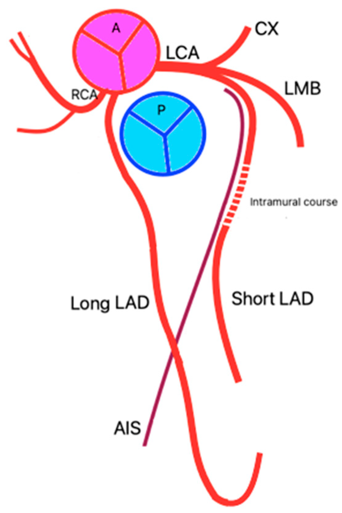
Figure 1. (a) The double anterior interventricular artery (descending anterior artery) originating from the left coronary artery, and after a short path, it branches into two long anterior interventricular arteries that descend on one side and the other of the anterior interventricular sulcus until close to the top of the heart; the right bifurcation branch presents, in its first portion, an intramuscular route. (b) Schematic representation.
Jpm 14 01007 g001
Figure 2. Type XIV of double anterior interventricular artery (descending anterior artery). The right anterior interventricular artery/long anterior descending artery, with a separate origin, in the right coronary sinus (c,f), which exceeds the apex of the heart approaching the distal portion of the posterior interventricular sulcus (a,d,e). The left anterior interventricular artery/short anterior descending artery, with its origin in the left coronary artery, presents a muscular bridge in its middle portion (a,b).
Jpm 14 01007 g002
Figure 3. Type XIV—schematic representation.
Jpm 14 01007 g003
Table 1. Classification of dual AIA/LAD.
TypeOrigin
Short AIA/LAD
Origin
Long AIA/LAD
Course
Short AIA/LAD
Course
Long AIA/LAD
I [3]AIB/LADAIB/LADProximal AISCourses along the left ventricular side of the proximal AIS and re-enters the distal AIS.
II [3]AIB/LADAIB/LADProximal AISDescends along the right ventricular side of the proximal AIS and re-enters the distal AIS.
III [3]AIB/LADAIB/LADProximal AISIt had an intramyocardial course in the proximal portion of the septum and appears distally at the level of the AIS or terminates intramyocardially.
IV [3]LCARCAProximal AISFollowing an anomalous pre-pulmonic trajectory anterior to the RVOT and entering the distal AIS.
V [19]LCSRCS Proximal AISIt had an intramyocardial course before reaching the distal portion of the AIS.
VI [18]LCARCAProximal AISFollows a course between the RVOT and the aortic root and then enters the distal portion of the AIS.
VII [17]AIB/LADAIB/LADProximal AISCourses on the left ventricular side of the proximal AIS and then enters the distal AIS.
VIII [17]LCAThe middle of the RCAProximal AIS Traverses the diaphragmatic surface of the right ventricle and reaches the distal portion of the AIS at the apex.
IX [17]AIB/LADAIB/LADProximal AISPassing on the LV side of the mid AIS, re-entering distally into the AIS, and ending before reaching the cardiac apex.
X [20]LCARCSProximal AISCourses along an anomalous pre-pulmonic course anterior to RVOT and re-enters the distal AIS.
XI [21]RCSRCSFollowed an intramyocardial course, through the anterior part of the interventricular septum and terminates in the proximal AIS.Courses along an anomalous pre-pulmonic course anterior to RVOT and re-enters the distal AIS.
XII [22]LCA (that originates from RCS)RCSProximal AISCourses anterior to the main pulmonary artery and terminates in the distal AIS.
XIII [5]-AIB/LAD-Two long AIA/LAD which descend to the right and left sides of the AIS, extending toward the cardiac apex.
XIIIA-AIB/LAD-Two long AIA/LAD which descend to the right and left sides of the AIS extending toward the cardiac apex; the bifurcating branch that descends to the right of the AIS has an intramural course.
XIVLCARCSTraverses the upper two-thirds of the AIS and terminates on the sternocostal surface of the left ventricle. In its middle third (between its origin and entry into the AIS), it exhibits an intramural course.It had a pre-pulmonary course, moving towards the AIS and traversing its lower third to reach the cardiac apex. It then crosses the heart’s apical notch and continues its course on the diaphragmatic surface, extending through the lower third of the posterior interventricular sulcus.
Type XIIIA and Type XIV are described in this study.
Disclaimer/Publisher’s Note: The statements, opinions and data contained in all publications are solely those of the individual author(s) and contributor(s) and not of MDPI and/or the editor(s). MDPI and/or the editor(s) disclaim responsibility for any injury to people or property resulting from any ideas, methods, instructions or products referred to in the content.

