Article

Acinetobacter baumannii from Samples of Commercially Reared Turkeys: Genomic Relationships, Antimicrobial and Biocide Susceptibility

1
Institute of Poultry Diseases, School of Veterinary Medicine, Freie Universität Berlin, 14163 Berlin, Germany
2
Veterinary Centre for Resistance Research (TZR), Freie Universität Berlin, 14163 Berlin, Germany
3
Institute of Microbiology and Epizootics, Centre for Infection Medicine, School of Veterinary Medicine, Freie Universität Berlin, 14163 Berlin, Germany
4
Institute for Medical Microbiology, Immunology and Hygiene, Faculty of Medicine and University Hospital Cologne, University of Cologne, 50935 Cologne, Germany
5
German Center for Infection Research (DZIF), Partner Site Bonn-Cologne, 50935 Cologne, Germany
6
Center for Molecular Medicine Cologne, Faculty of Medicine and University Hospital Cologne, University of Cologne, 50935 Cologne, Germany
*
Author to whom correspondence should be addressed.
Microorganisms 2023, 11(3), 759; https://doi.org/10.3390/microorganisms11030759
Submission received: 3 February 2023 / Revised: 5 March 2023 / Accepted: 7 March 2023 / Published: 16 March 2023
(This article belongs to the Section Veterinary Microbiology)

Abstract

:
Acinetobacter baumannii is especially known as a cause of nosocomial infections worldwide. It shows intrinsic and acquired resistances to numerous antimicrobial agents, which can render the treatment difficult. In contrast to the situation in human medicine, there are only few studies focusing on A. baumannii among livestock. In this study, we have examined 643 samples from turkeys reared for meat production, including 250 environmental and 393 diagnostic samples, for the presence of A. baumannii. In total, 99 isolates were identified, confirmed to species level via MALDI-TOF-MS and characterised with pulsed-field gel electrophoresis. Antimicrobial and biocide susceptibility was tested by broth microdilution methods. Based on the results, 26 representative isolates were selected and subjected to whole-genome sequencing (WGS). In general, A. baumannii was detected at a very low prevalence, except for a high prevalence of 79.7% in chick-box-papers (n = 118) of one-day-old turkey chicks. The distributions of the minimal inhibitory concentration values were unimodal for the four biocides and for most of the antimicrobial agents tested. WGS revealed 16 Pasteur and 18 Oxford sequence types, including new ones. Core genome MLST highlighted the diversity of most isolates. In conclusion, the isolates detected were highly diverse and still susceptible to many antimicrobial agents.

1. Introduction

Acinetobacter baumannii are nonmotile, oxidase-negative, aerobic, Gram-negative coccobacilli [1]. These bacteria are associated with nosocomial infections worldwide [2]. Although A. baumannii is an opportunistic pathogen, it has led to many outbreaks in hospitals and care-facilities with high morbidity and mortality rates [3]. These infections are mainly caused by outbreak strains, which can spread rapidly between patients [4]. Many disease conditions, including ventilator-associated pneumonia, bloodstream infection, urinary tract infection, wound infection and meningitis have been described [3], and A. baumannii has been shown to be a common co-infecting agent in COVID-19 patients in intensive care units [5,6]. A. baumannii can rapidly develop antimicrobial resistance [7,8] due to various resistance mechanisms, such as β-lactamase production, efflux pump overexpression, alterations at the target sites of the antimicrobial agents, and decreased membrane permeability [9]. A. baumanni is intrinsically resistant to a number of antimicrobial agents, such as penicillin, ampicillin, amoxicillin, amoxicillin-clavulanic acid, aztreonam, first generation cephalosporins (cephalothin, cefazolin), second generation cephalosporins (cefuroxime), cephamycines (cefoxitin, cefotetan), clindamycin, daptomycin, fusidic acid, glycopeptides (vancomycin), linezolid, macrolides (erythromycin, azithromycin, clarithromycin), quinupristin-dalfopristin, rifampin, ertapenem, trimethoprim, chloramphenicol, and fosfomycin [10]. Moreover, multi-drug resistance properties include resistance not only to the most commonly used antimicrobial agents, but also to last-resort antimicrobial agents in human medicine [11,12]. Due to its outstanding ability to escape antimicrobial therapy, A. baumannii is listed among the ESKAPE pathogens, which also include Enterococcus faecium, Staphylococcus aureus, Klebsiella pneumoniae, Pseudomonas aeruginosa, and Enterobacter spp. [13,14].
Concerning farm animal populations and their environment, there is little information about the distribution of A. baumannii, with data on antimicrobial resistance especially lacking [15]. A recent study on isolates from cattle has shown that they harbour a highly diverse population of A. baumannii, which are susceptible to most antimicrobial agents [16].
Concerning poultry, in 2011 a case was described in which a highly virulent strain of A. baumannii led to an outbreak in a commercial chicken farm in China, during which more than 3000 six-day-old chicks died [17]. Otherwise, the isolation of A. baumannii has only occasionally been described from chickens and was not related to outbreaks or diseases [18,19]. Among other bird species, in Poland, 25% of 661 white stork (Ciconia ciconia) nestlings were tested positive for A. baumannii [18]. There are also reports of single A. baumannii isolates found in geese [18], falcons [20], and other birds of which the species was not published [21,22,23]. However, in general, birds are not considered as a primary host for A. baumannii [24].
In environmental samples associated with poultry, A. baumannii isolates have been obtained from sewage water of a poultry slaughterhouse [25]. It has also been detected in the air of a duck hatchery. The authors considered that these bacteria might be a possible trigger for respiratory diseases in hatchery workers [26,27]. Liu et al. also point out that cross-infections between humans and chicks through handling may be possible [17]. Therefore, the dissemination of A. baumannii in poultry livestock may have far-reaching consequences for public health [18]. In addition, poultry meat might potentially be a threat to public health, as A. baumannii has been isolated from raw turkey and chicken meat [28,29,30,31,32].
In our pilot study, we focused on the occurrence of A. baumannii in samples from commercially reared turkeys for meat production, as information concerning these farm animals and especially their antimicrobial resistance profiles are missing [15]. Collected isolates were characterised by pulsed-field gel electrophoresis and whole-genome sequencing, and tested for antimicrobial and biocide susceptibility.

2. Materials and Methods

2.1. Sample Collection and Isolation

In total, 250 samples from 95 different farms were collected from allegedly healthy commercial fattening turkey flocks distributed all over Germany (n = 94) and the Czech Republic (n = 1) as part of a Salmonella surveillance in 2019. This included 118 chick-box-papers (paper with wood shavings on which the turkey chicks were transported from the hatchery to the production house containing meconium) from one-day-old turkey chicks taken on arrival at the production house from 81 farms (with 24 farms providing more than one sample). Six unused chick-box-papers were also examined as negative controls. In addition, 50 boot swab samples (containing one pair of boot swabs each) taken during the rearing period and 82 boot swab samples from turkeys leaving for the slaughterhouse were investigated. Data and subsequent results were compiled, assessed, and evaluated using Microsoft Excel (Microsoft Office 2019). After pre-enrichment in buffered peptone water (Thermo Scientific, Wesel, Germany) at 37 °C for 16 to 18 h, approximately 10 μL enrichment broth was streaked on chromogenic media Brilliance UTI Clarity agar (Thermo Scientific, Wesel, Germany) and incubated at 37 °C for 24 h. Buffered peptone water without any supplements was analysed as sterility control. In addition, 393 diagnostic samples sent to the Institute of Poultry Diseases, Freie Universität Berlin, Berlin, Germany between 2018–2020 were examined. These included liver and yolk sac samples from 88 one-to six-day-old commercial turkey chicks, as well as 217 lung- and heart-swabs from commercial turkeys. Cultivation was performed on Columbia agar with 5% sheep blood (Thermo Scientific, Wesel, Germany) and Brilliance UTI Clarity agar at 37 °C for 24 h.
Presumptive colonies were selected, sub-cultured, and confirmed to species level by matrix-assisted laser desorption/ionisation time of flight mass spectrometry (MALDI-TOF MS) (Bruker Daltonic GmbH, Bremen, Germany). All isolates were stored at −20 °C in brain heart infusion (BHI) medium (Roth, Karlsruhe, Germany) until further use.

2.2. Antimicrobial Susceptibility Testing

Antimicrobial susceptibility testing was performed by broth microdilution according to the instructions of the Clinical and Laboratory Standards Institute (CLSI, 2022) [10]. The Acinetobacter isolates were tested with custom-made microtiter plates (MCS Diagnostics, Swalmen, The Netherlands) for their susceptibility to 18 antimicrobial agents or combinations: colistin, streptomycin, neomycin, trimethoprim/sulfamethoxazole, gentamicin, nalidixic acid, ciprofloxacin, enrofloxacin, marbofloxacin, tetracycline, doxycycline, florfenicol, imipenem, ceftiofur, cefquinome, cefotaxime, cefoperazone, and tiamulin. This test panel was the same as used in the GERM-Vet programme, the German national resistance monitoring programme of veterinary pathogens, for Gram-negative bacteria. The reference strain Escherichia coli ATCC® 25922 served as quality control. The minimal inhibitory concentration (MIC) values were interpreted as susceptible, intermediate, or resistant using the human-specific clinical breakpoints from CLSI [10], as veterinary-specific clinical breakpoints are not available for Acinetobacter spp.

2.3. Biocide Susceptibility Testing

Biocide susceptibility testing was performed for four different biocides—benzalkonium chloride (a quaternary ammonium compound), octenidine (a bispyridine) as well as chlorhexidine and polyhexanide (two biguanides)—using commercial microtitre plates (sifin diagnostics GmbH, Berlin, Germany) and the protocol from Schug et al. [33], with some adaptations [34]. The use of commercial microtitre plates led to an adaptation of the protocol by adding only 30 µL bacterial suspension of a density of 0.5 McFarland to 12 mL single-concentrated tryptic soy broth (TSB) and the microtitre plates were inoculated with 100 µL per well according to the manufacturer’s recommendation. These plates contained the biocides in 11 or 12 two-fold dilution steps: benzalkonium chloride (0.000008–0.016%), octenidine (0.000016–0.016%), chlorhexidine (0.000008–0.008%), and polyhexanide (0.000016–0.032%). The reference strain Pseudomonas aeruginosa ATCC® 15442 served as quality control [34].

2.4. Macrorestricton Analysis with Subsequent Pulsed-Field Gel Electrophoresis

Macrorestricton analysis using the enzyme ApaI (New England Biolabs, Frankfurt, Germany) and subsequent pulsed-field gel electrophoresis (PFGE) were performed for a preliminary characterisation of the 99 A. baumannii isolates as previously published [35], with a minor modification: for restriction analysis with ApaI (30U), the plug slices were incubated overnight at 25 °C. A Lambda PFGE ladder (New England Biolabs, Frankfurt, Germany) with a size range from 48.5 to 1018 kb served as size marker. Electrophoresis was performed using the CHEF-DR III system (Bio-Rad Laboratories, Düsseldorf, Germany). Gels were stained with GelRed (Biotium, San Francisco, CA, USA) and scanned with the laboratory’s imaging system (BIO RAD Molecular Imager GelDocTM XR+ with Image LabTM Software, Düsseldorf, Germany). An isolate from diagnostics (141_Diagnostik) served as internal control on each gel. Cluster analysis concerning the percentage similarity was performed with BioNumerics software, version 7.6.3 (Applied Maths, bioMérieux). Similarities were calculated with the dice coefficient (optimization 1.5%, tolerance 1.5%) and the unweighted pair group method with arithmetic mean (UPGMA) [35]. Pulsotypes were defined at a threshold value of ≥80% (named alphabetically) and at a threshold value of ≥87% (additional numeric marking) [35,36].

2.5. Whole-Genome Sequencing

For whole-genome sequencing (WGS), 26 isolates were selected, including at least one isolate per PFGE pulsotype (cut off level of ≥80%). DNA was isolated using the Master Pure DNA Purification Kit for Blood Version II (Epicentre Biotechnologies) as published by the manufacturer. The libraries were prepared using the Nextera XT DNA Library Preparation Kit (Illumina Inc., San Diego, CA, USA) according to the manufacturer’s instructions. The 2 × 300 bp paired-end sequencing in 40-fold multiplexes was performed on the Illumina MiSeq platform (Illumina) with MiSeq Reagent Kit v3 (600-cycle) (Illumina). For sequence assembly, the Illumina reads were trimmed by Trim Galore v0.6.6 (RRID:SCR_011847) and quality checked by FastQC [37]. De novo assembling was carried out using Unicycler v0.4.9. [38]. Antimicrobial resistance genes were detected using ABRicate [39] with NCBI AMRFinderPlus [40], ResFinder [41], and CARD [42] databases. Plasmid replicons were searched for using ABRicate [39] applied to the PlasmidFinder 2.1 database (https://cge.food.dtu.dk/services/PlasmidFinder/ accessed on 21 February 2023). The databank PubMLST (https://pubmlst.org/ accessed on 21 February 2023 [43]) was used to confirm the species with ribosomal multilocus sequence typing (rMLST) [44] and to compare and identify sequence types (ST) using both the Pasteur [45] and the Oxford [46,47] scheme. New STs and new alleles were submitted to PubMLST [43]. The generated genomes were used for core genome multilocus sequence typing (cgMLST) with SeqSphere+ v7.5.5 (Ridom GmbH, Münster, Germany) [48]. This typing scheme is based on a core genome of 2390 alleles. However, the calculations for the minimum spanning tree presented here were done on the basis of only 1943 alleles as all missing values were excluded. Detected β-lactamases were compared with those listed in the Beta-Lactamase DataBase (www.bldb.eu accessed on 21 February 2023) [49]. Accession numbers and bioproject number are presented in the Data Availability section.

3. Results

3.1. Isolation

Ninety-nine A. baumannii isolates were collected during the study period. A. baumannii was detected in 79.7% (n = 94) of the 118 chick-box-papers. In two chick-box-papers, two morphologically different A. baumannii isolates were recovered. Two further A. baumannii isolates (2.4%) were found among the 82 boot swab samples tested from turkeys before slaughter. None of the 50 boot swab samples taken during the rearing period were positive for A. baumannii (Table 1). Taken together, 1.5% of the boot swab samples contained A. baumannii. The six unused chick-box-papers tested negative. A. baumannii was detected in one of the 217 swabs (0.5%) sent in for bacteriological diagnostics. The single positive pooled heart-lung-swab originated from a seven-week old turkey (isolate 141_Diagnostik). All of the 88 one- to six-day-old commercial turkey chicks were negative for A. baumannii in their liver and in their yolk sac (Table 1).
In total, there were 13 farms from which several A. baumannii isolates were detected (minimum two isolates, maximum five isolates). Only in one of them (farm 13) A. baumannii was detected in a chick-box-paper (isolate 16_W23.1) as well as in a boot swab sample before slaughter (isolate 98_E23.3) (Figure S1).

3.2. Antimicrobial Susceptibility Testing

The results of the antimicrobial susceptibility testing are displayed in Table 2. As there are no CLSI-approved veterinary-specific clinical breakpoints currently available for A. baumannii, human clinical breakpoints were applied. Using these interpretive criteria, all tested isolates were susceptible to imipenem and gentamicin. A high percentage of the tested isolates was susceptible to doxycycline (98%), trimethoprim/sulfamethoxazole (98%), and tetracycline (96%). Concerning cefotaxime, 31% of the isolates were classified as susceptible, 67% as intermediate, and 3% as resistant, despite the fact that the MICs of cefotaxime revealed a unimodal distribution with a mode MIC value of 16 mg/L. For ciprofloxacin, 83% of the isolates were susceptible and 17% were resistant. Bimodal MIC distributions, with two peaks representing a “susceptible” wildtype population and a non-wildtype population with acquired resistance properties, were seen for all the (fluoro)quinolones, including nalidixic acid, ciprofloxacin, enrofloxacin, and marbofloxacin. The same 17 isolates classified as ciprofloxacin-resistant also showed elevated MIC values for nalidixic acid as well as the veterinary fluoroquinolones enrofloxacin and marbofloxacin. All isolates were classified as intermediate to colistin. For the other tested antimicrobial agents there were no clinical breakpoints available. The MIC values were high especially for tiamulin, cefoperazone, and florfenicol, which is in accordance with the intrinsic resistance properties of A. baumannii. Bimodal MIC distributions were also seen for the tetracyclines, namely tetracycline and doxycycline, and also for trimethoprim/sulfamethoxazole.

3.3. Biocide Susceptibility Testing

The MIC values for the tested biocides all showed a unimodal distribution. They ranged as follows: benzalkonium chloride 0.0005–0.002%, octenidine 0.000125–0.002%, chlorhexidine 0.000125–0.008%, and polyhexanide 0.000125–0.008% (Table 3).

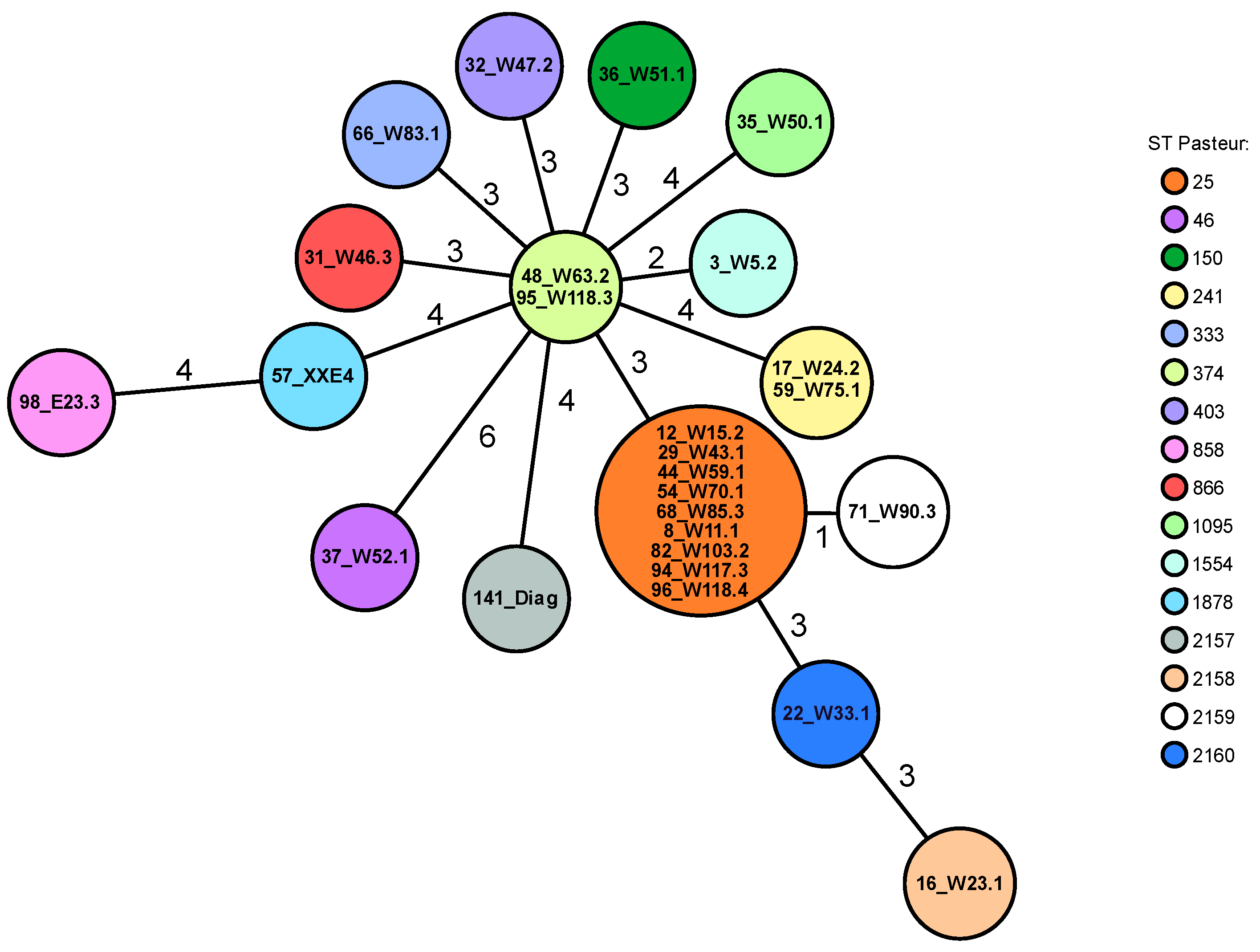
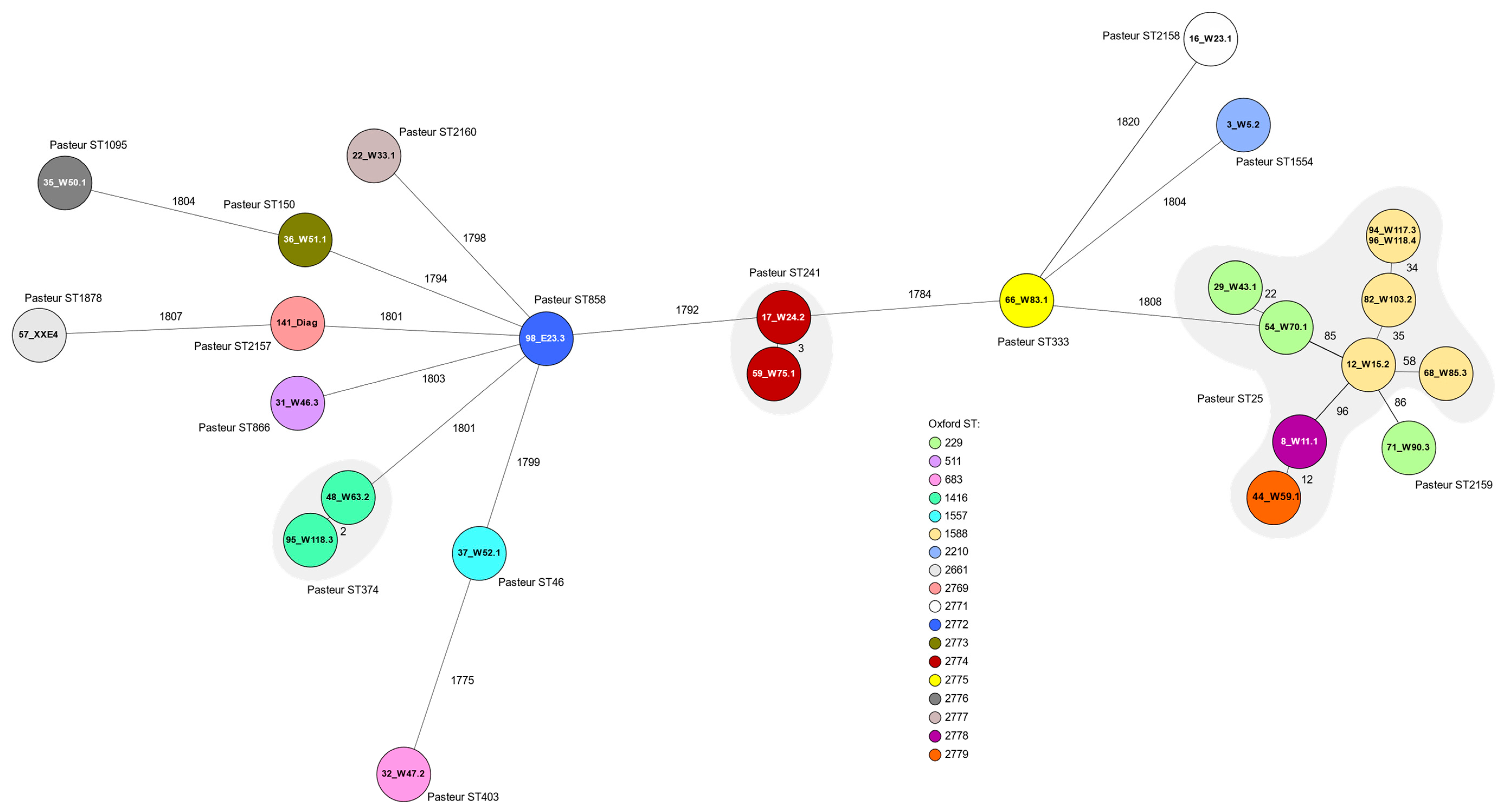
3.4. Macrorestricton Analysis with Subsequent Pulsed-Field Gel Electrophoresis

At the cut off level of ≥ 80%, there were 21 PFGE pulsotypes (A-U) containing up to 21 isolates (pulsotype P). At the cut off level ≥ 87%, there were 33 PFGE pulsotypes. These included up to 11 isolates per pulsotype and up to nine isolates with indistinguishable PFGE patterns (pulsotype B1), including isolates from different farms and different arrival dates (Figure S1).

3.5. Whole-Genome Sequencing

The whole-genome sequencing results are listed in Table 4. rMLST confirmed the assignment to the species A. baumannii with 100% support in all sequenced isolates.
Concerning the resistance genes, all sequenced isolates showed the presence of Ambler class D and Ambler class C β-lactamase genes (blaOXA-51-like and blaADC), which are intrinsic to A. baumannii [50,51,52,53]. Seventeen different blaOXA β-lactamase variants were detected. Two isolates had blaOXA-51 β-lactamase genes and the other 24 isolates had blaOXA-51-like β-lactamase genes, among which blaOXA-64 was by far the most prevalent with 38% (n = 10). All blaOXA β-lactamase genes detected were confirmed and the deduced amino acid sequences confirmed their assignment (100% identity), except isolate 22_W33.1, which had a single amino acid difference (Met84Ile) to OXA-69 (99.8% identity). Eleven different blaADC β-lactamase gene variants were detected. Here, blaADC-26 was the most common with 50% (n = 13), mostly co-located with blaOXA-64. In eight isolates, ADC β-lactamases were found, which exhibited less than 100% identity to known ADC variants. Four isolates exhibited one amino acid difference in the deduced protein sequences: isolate 54_W70.1 and isolate 29_W43.1 showed 99.7% identity to ADC-26 (Leu44Phe), respectively, isolate 31_W46.3 showed 99.7% identity to ADC-158 (Thr123Ala) and isolate 57_XXE4 showed 99.7% identity to ADC-192 (Gln2Arg). Two isolates had two amino acid differences: isolate 22_W33.1 showed 99.5% identity to ADC-159 (Glu118Lys and Ala270Thr) and isolate 98_E23.3 showed 99.5% identity to ADC-192 (Gln2Arg and Asp24Asn). Three amino acids differences were found in two isolates: isolate 32_W47.2 showed 99.2% identity to ADC-163 (Lys163Gln, Val197Ala, and Phe263Leu), and isolate 3_W5.2 showed 99.2% identity to two different ADC β-lactamases, namely ADC-158 (Thr112Lys, Pro216Ala, Arg274Lys) and ADC-274 (Ala99Gly, Pro216Ala, and Arg274Lys). The aminoglycoside nucleotidyltransferase gene ant(3″)-IIa was present in all isolates. Additional aminoglycoside O-phosphotransferase genes aph(3″)-Ib and aph(6)-Id were only identified in one isolate (35_W50.1). The tet(39) gene was found in two isolates, which were classified as tetracycline-resistant.
The sul2 gene was detected in one of the two isolates, which showed resistance to trimethoprim/sulfamethoxazole. In the ten sequenced ciprofloxacin-resistant isolates, two gene mutations were detected in gyrA and parC genes resulting in the amino acid substitutions Ser81Leu (GyrA) and Ser84Leu or Ser84Phe (ParC), respectively. Isolate 71_W90.3, which showed an elevated MIC value for nalidixic acid but not for ciprofloxacin, had only a mutation in gyrA, which resulted in the amino acid substitution Ser81Leu (Table 4).
Multilocus sequence typing (MLST) analysis using the Pasteur scheme revealed 16 different STs (Table 4). Four STs (2157, 2158, 2159, and 2160) were newly described, and a new fusA allele (detected in isolate 71_W90.3 with the new ST2159), namely Pas_fusA-407, was newly added to the PubMLST database. By far the most commonly detected ST was ST25, comprising nine isolates (35%), followed by ST241 and ST374 with two isolates each (8%), respectively. The 12 remaining isolates all showed individual allelic profiles and different STs (Figure 1).
In the MLST analysis using the Oxford scheme, 18 different STs were present. Ten of these STs (2769, 2771, 2772, 2773, 2774, 2775, 2776, 2777, 2778, and 2779) were newly described, including three new alleles, which were added to the PubMLST database. ST1588 was the most common, including five isolates (19%), followed by ST229 including three isolates (12%), and ST1416 and ST2774 with two isolates each (8%) (Table 4).
cgMLST using 1943 alleles for distance calculation showed a wide distribution of the 26 isolates tested. Most of these isolates showed a distinct allelic profile and were not closely related. They showed differences between 1775 and 1820 alleles. There was one cluster with ten related isolates (only up to 96 alleles apart). These ten isolates belonged to the Pasteur STs 25 and 2159 (new) and the Oxford STs 229, 1588, 2778 (new), and 2779 (new). The corresponding isolates all showed fluoroquinolone resistance. Otherwise, only two isolate pairs had closely related allelic profiles: isolates 48_W24.2 and 95_W75.1 with only two alleles difference, and isolates 17_W63.2 and 59_W118.3 with three alleles difference (Figure 2).

4. Discussion

In the chick-box/meconium samples from one-day-old turkey chicks, there was a very high presence of A. baumannii. Overall, 79.9% of the chick-box-papers contained A. baumannii isolates. Intriguingly, the highest detection rate in birds (25% from n = 661) up till now was also found in white stork nestlings. Other findings in goslings and chickens seem to have also been especially prevalent in younger birds [18]. Interestingly, the detection rate of A. baumannii found in boot swab samples (n = 132) taken during rearing and before slaughter was low, with only 1.5%. Our results, therefore, highlight that the presence of A. baumannii in samples from poultry can vary considerably with the age of the birds and is transient. In another study, for example, A. baumannii was not isolated in bioaerosols from a housing with 7-week-old turkeys [54]. The detection of only one A. baumannii isolate in 217 lung-heart swabs (0.5%) and in none of the yolk sac and liver samples from one-to six-day-old turkey chicks during diagnostics, additionally points towards a generally low presence of A. baumannii in fattening turkeys. The data, therefore, suggest, as in wild birds [24], that there is no evidence for a general preference of A. baumannii for avian hosts. With regard to the diagnostic samples in this study, there was also no evidence of A. baumannii playing a role in diseased turkeys.
The preliminary characterisation via PFGE revealed that in total, the A. baumannii isolates found in this study were very heterogenous, forming 21 pulsotypes at a cut off level of 80% and 33 pulsotypes at a cut off level of 87%, comprising between one and eleven isolates. Core genome MLST highlighted the diverse population of A. baumannii isolates found in this study. However, as anticipated, the PFGE results did not completely correspond with the core genome MLST data of the 26 isolates subjected to WGS. Due to the very heterogenous isolates, which were not closely related, an environmental origin as discussed in the studies on storks [18] and cattle [16] seems likely. The source of the A. baumannii isolates is not clear. To investigate possible reservoirs in future studies, the environment of one-day-old chicks, i.e., hatcheries and transport vehicles, should be analysed. In other studies, A. baumannii isolates have been found in the air of a duck hatchery [27] and non-sterile water (which is used for humidity regulation during the brood), which has been suggested to be a possible source of contamination in hatcheries [26]. Moreover, feather down has also been considered as a potential carrier [26,55]. In general, Acinetobacter spp. are widespread environmental microorganisms [56] and A. baumannii, for example, can be detected in soil [57,58].
Antimicrobial susceptibility testing revealed that the majority of isolates were susceptible to a wide range of antimicrobial agents, which is comparable with the results obtained from cattle and white storks as well [16,18]. Multidrug resistance properties were not detected. In addition to the species-specific intrinsic resistance properties, only two isolates showed acquired resistance to two different classes of antimicrobial agents, namely (fluoro)quinolones and tetracyclines. The highest resistance rates were detected for ciprofloxacin. The MIC values of the other tested quinolones, for which no clinical breakpoints exist, confirmed this finding. The detected mutations in gyrA and parC genes, respectively, are linked to fluoroquinolone selection of resistance, which suggests that the isolates have been circulating in an environment where fluoroquinolones have been used [59]. Interestingly, the isolate showing only one mutation in the gyrA gene and none in the parC gene revealed only an elevated MIC value for nalidixic acid, but not one for ciprofloxacin. The tetracycline resistance in two isolates could be attributed to the tet(39) gene, which encodes an active efflux mechanism and has been described in Acinetobacter spp. found frequently in the aquatic environment [60,61]. As A. baumannii is intrinsically resistant to trimethoprim, resistance to trimethoprim/sulfamethoxazole could only be attributed to the gene sul2, which confers resistance to sulfonamides [62] in one isolate. The cause of the resistance in the other trimethoprim/sulfamethoxazole-resistant isolate (36_W51.1) was not resolved as no further sul genes nor mutations in the genes folA and folP could be detected. The aminoglycoside nucleotidyltransferase gene ant(3″)-IIa was present in all isolates as described in other studies [63,64]. Phosphotransferase aph(3″)-Ib and aph(6)-Id, which mediate streptomycin resistance, were both detected in isolate 35_W50.1 (streptomycin MIC value 64 mg/L). The streptomycin MIC values of the remaining 25 isolates, which did not harbour these two resistance genes, varied between 4 mg/L and ≥128 mg/L. One of the three isolates resistant to cefotaxime (isolate 17_W24.2) was examined by WGS and no additional beta-lactamase gene, except the intrinsic ones, could be identified. The classification of the isolates as cefotaxime-resistant may be due to the unimodal MIC distribution of the tested A. baumannii isolates around the clinical breakpoint. In other studies, cefotaxime resistance has been described in association with the production of the CTX-M-2 extended spectrum class A β-lactamase [65,66]. In general, blaOXA genes are found on both chromosome and plasmids, and it might also be possible that more than one copy of blaOXA is on the chromosome [67]. The isolates tested in this study showed a diverse selection of intrinsic blaOXA β-lactamase genes. It is important to say that we did not detect acquired β-lactamase genes, such as blaOXA-23 or blaOXA-58, which are associated with carbapenem resistance [2] in any of the 26 sequenced isolates. The gene blaOXA-64, which was the most frequently detected blaOXA gene in this study, has previously been found in feather down and dust from turkey and goose hatcheries [18]. It correlates with the Pasteur ST25 [68], except in the case of isolate 71_W90.3, which interestingly showed a new Pasteur type, ST2159 (with a new fusA allele), but a known Oxford type ST229. Some other blaOXA β-lactamase genes found in our study have been detected in samples from other avian species as well, i.e., blaOXA-51 (white stork choana), blaOXA-68 (chicken choana, feather down and dust from a chicken hatchery), blaOXA-104 (white stork choana), blaOXA-208 (white stork pellet), and blaOXA-385 (1-day-old chicken choana) [18]. Interestingly, Wilharm et al. could assign two chicken samples with blaOXA-68 (Pasteur ST23) to the international clone 8 (IC8) [18], which includes outbreak strains in human medicine (Pasteur STs 10 and 157) [69]. Only one isolate (16_W23.1) in this study had blaOXA-68. This isolate showed new Pasteur and Oxford STs. Furthermore, a variety of different blaADC β-lactamase genes were found. The most common, blaADC-26, was mostly present in isolates that also carried blaOXA64, except for isolate 37_W52.1, which was characterised by blaOXA-104, Pasteur ST46 and Oxford ST1557, and the isolates 48_W63.2 and 95_W118.3, characterised by blaOXA-259, Pasteur ST374 and Oxford ST1416.
Biocide susceptibility testing revealed that the MIC values for the four biocides were all distributed unimodally. There is not much data concerning biocide susceptibility available for comparison. In one study, 14 A. baumannii isolates from dogs and cats were examined using the same protocol [70]. Interestingly, the MIC ranges were generally wider in our study presented here, often starting at lower dilution steps. However, it has to be kept in mind that this could be due to the number of isolates tested (n = 99 vs. n = 14).
MLST analysis revealed many different STs, which highlights the heterogenous nature of the isolates. Pasteur ST25, which was detected in 35% of the sequenced isolates, was most prevalent. In humans, ST25 is a successful lineage, which can lead to epidemics, has spread worldwide, and belongs to the international clone 7 (IC7) [68,69,71,72,73,74,75]. All ST25 isolates described here carry blaOXA-64 and were resistant to ciprofloxacin, which corresponds to the results of other studies [68,71]. Only one of the ten ciprofloxacin-resistant isolates examined by WGS (isolate 66_W83.1) belonged to Pasteur ST333, which was first described in China [76]. Two of our isolates were susceptible to ciprofloxacin and belonged to Pasteur ST374, which, according to the PubMLST database, occurs worldwide. The ST374 lineage is grouped into the clonal complex CC3 belonging to the international clone IC3 [77]. Two further isolates belonged to Pasteur ST241, which has been detected in human samples across the world according to PubMLST database. In Germany, it has been found in a cattle faecal sample [16] and in milk powder [63]. Interestingly, our two ST241 isolates, which were only three alleles apart in cgMLST, had a new Oxford sequence type ST2774, harboured blaOXA-91 and blaADC-52, and showed an elevated cefquinome MIC of ≥64 mg/L. None of the Pasteur STs in this study corresponded to those found in chicken and turkey meat in Switzerland [29]. More Oxford STs (n = 18) were found in this study in comparison to the Pasteur STs (n = 16). The Pasteur ST25 comprised four different Oxford STs (ST1588, ST229, ST2779 (new), and ST2778 (new)).
In general, it can be concluded for commercial turkeys, as it has been for cattle [16] and storks [18], that the population of A. baumannii is highly diverse and still susceptible to many antimicrobial agents. The overall occurrence of A. baumannii in samples from commercially reared turkeys seems to be very low. Only chick-box-papers were found to harbour large numbers of A. baumannii isolates. Although Acinetobacter isolates have been obtained from rhizospheric soil, tomato, and cauliflower roots [78], a transfer from these sources to the animals investigated in this study can be excluded as the turkey chicks/turkeys did not have contact to these matrices. Thus, the possible origin of the A. baumannii isolates found in this study remains to be elucidated and will be a subject for further investigation.

Supplementary Materials

The following supporting information can be downloaded at: https://www.mdpi.com/article/10.3390/microorganisms11030759/s1, Figure S1: Pulsed-field gel electrophoresis profiles from 99 A. baumannii isolates.

Author Contributions

Conceptualization, A.S., D.L. and A.T.F.; methodology, A.S., D.H. and A.T.F.; software, D.H.; validation, A.S., D.L., D.H., S.S., P.G.H. and A.T.F.; formal analysis, A.S., D.H. and A.T.F.; investigation, A.S. and A.T.F.; resources, D.L. and S.S.; data curation, A.S., D.L., D.H. and A.T.F.; writing—original draft preparation, A.S.; writing—review and editing, A.S., D.L., D.H., S.S., P.G.H. and A.T.F.; visualization, A.S.; supervision, D.L. and S.S.; project administration, D.L. and S.S. All authors have read and agreed to the published version of the manuscript.

Funding

We acknowledge support by the Open Access Publication Fund of the Freie Universität Berlin. Otherwise, this research received no external funding.

Data Availability Statement

All data presented in this study are available in the text, figures and tables of the main article and in the supplementary material. Whole-genome sequences of the A. baumannii isolates included in this study are available at DDBJ/ENA/GenBank under the bioprojekt number PRJNA904545 and the accession numbers JAPQZE010000000, JAPQZD010000000, JAPQZC010000000, JAPQZB010000000, JAPQZA010000000, JAPQYZ010000000, JAPQYY010000000, JAPQYX010000000, JAPQYW010000000, JAPQYV010000000, JAPQYU010000000, JAPQYT010000000, JAPQYS010000000, JAPQYR010000000, JAPQYQ010000000, JAPQYP010000000, JAPQYO010000000, JAPQYN010000000, JAPQYM010000000, JAPQYL010000000, JAPQYK010000000, JAPQYJ010000000, JAPQYI010000000, JAPQYH010000000, JAPQYG010000000 and JAPQYF010000000.

Acknowledgments

For excellent technical support we especially thank Petra Krienke, Elke Dyrks, Kathrin Oelgeschläger, and Gabriele Grotehenn.

Conflicts of Interest

The authors declare no conflict of interest.

References

  1. Juni, E. Interspecies transformation of Acinetobacter: Genetic evidence for a ubiquitous genus. J. Bacteriol. 1972, 112, 917–931. [Google Scholar] [CrossRef] [Green Version]
  2. Higgins, P.G.; Dammhayn, C.; Hackel, M.; Seifert, H. Global spread of carbapenem-resistant Acinetobacter baumannii. J. Antimicrob. Chemother. 2010, 65, 233–238. [Google Scholar] [CrossRef] [Green Version]
  3. Peleg, A.Y.; Seifert, H.; Paterson, D.L. Acinetobacter baumannii: Emergence of a successful pathogen. Clin. Microbiol. Rev. 2008, 21, 538–582. [Google Scholar] [CrossRef] [Green Version]
  4. Turton, J.F.; Kaufmann, M.E.; Glover, J.; Coelho, J.M.; Warner, M.; Pike, R.; Pitt, T.L. Detection and typing of integrons in epidemic strains of Acinetobacter baumannii found in the United Kingdom. J. Clin. Microbiol. 2005, 43, 3074–3082. [Google Scholar] [CrossRef] [Green Version]
  5. Sharifipour, E.; Shams, S.; Esmkhani, M.; Khodadadi, J.; Fotouhi-Ardakani, R.; Koohpaei, A.; Doosti, Z.; Golzari, S.E. Evaluation of bacterial co-infections of the respiratory tract in COVID-19 patients admitted to ICU. BMC Infect. Dis. 2020, 20, 646. [Google Scholar] [CrossRef]
  6. Shinohara, D.R.; dos Santos Saalfeld, S.M.; Martinez, H.V.; Altafini, D.D.; Costa, B.B.; Fedrigo, N.H.; Tognim, M.C.B. Outbreak of endemic carbapenem-resistant Acinetobacter baumannii in a coronavirus disease 2019 (COVID-19)—Specific intensive care unit. Infect. Control Hosp. Epidemiol. 2021, 43, 815–817. [Google Scholar] [CrossRef]
  7. Karageorgopoulos, D.E.; Falagas, M.E. Current control and treatment of multidrug-resistant Acinetobacter baumannii infections. Lancet Infect. Dis. 2008, 8, 751–762. [Google Scholar] [CrossRef] [PubMed]
  8. Howard, A.; O’Donoghue, M.; Feeney, A.; Sleator, R.D. Acinetobacter baumannii: An emerging opportunistic pathogen. Virulence 2012, 3, 243–250. [Google Scholar] [CrossRef] [PubMed] [Green Version]
  9. Vrancianu, C.O.; Gheorghe, I.; Czobor, I.B.; Chifiriuc, M.C. Antibiotic resistance profiles, molecular mechanisms and innovative treatment strategies of Acinetobacter baumannii. Microorganisms 2020, 8, 935. [Google Scholar] [CrossRef] [PubMed]
  10. Clinical and Laboratory Standards Institute. Performance Standards for Antimicrobial Susceptibility Testing, 32nd ed.; CLSI supplement M100: Wayne, PA, USA, 2022. [Google Scholar]
  11. D’Onofrio, V.; Conzemius, R.; Varda-Brkić, D.; Bogdan, M.; Grisold, A.; Gyssens, I.C.; Bedenić, B.; Barišić, I. Epidemiology of colistin-resistant, carbapenemase-producing Enterobacteriaceae and Acinetobacter baumannii in Croatia. Infect. Genet. Evol. 2020, 81, 104263. [Google Scholar] [CrossRef] [PubMed]
  12. Dahdouh, E.; Gómez-Gil, R.; Pacho, S.; Mingorance, J.; Daoud, Z.; Suárez, M. Clonality, virulence determinants, and profiles of resistance of clinical Acinetobacter baumannii isolates obtained from a Spanish hospital. PLoS ONE 2017, 12, e0176824. [Google Scholar] [CrossRef] [PubMed]
  13. Pendleton, J.N.; Gorman, S.P.; Gilmore, B.F. Clinical relevance of the ESKAPE pathogens. Expert Rev. Anti-Infect. Ther. 2013, 11, 297–308. [Google Scholar] [CrossRef]
  14. Rice, L.B. Federal funding for the study of antimicrobial resistance in nosocomial pathogens: No ESKAPE. J. Infect. Dis. 2008, 197, 1079–1081. [Google Scholar] [CrossRef] [PubMed]
  15. Wareth, G.; Neubauer, H.; Sprague, L.D. Acinetobacter baumannii—A neglected pathogen in veterinary and environmental health in Germany. Vet. Res. Commun. 2019, 43, 1–6. [Google Scholar] [CrossRef] [PubMed] [Green Version]
  16. Klotz, P.; Higgins, P.G.; Schaubmar, A.R.; Failing, K.; Leidner, U.; Seifert, H.; Scheufen, S.; Semmler, T.; Ewers, C. Seasonal occurrence and carbapenem susceptibility of bovine Acinetobacter baumannii in Germany. Front. Microbiol. 2019, 10, 272. [Google Scholar] [CrossRef] [PubMed] [Green Version]
  17. Liu, D.; Liu, Z.S.; Hu, P.; Hui, Q.; Fu, B.Q.; Lu, S.Y.; Li, Y.S.; Zou, D.Y.; Li, Z.H.; Yan, D.M.; et al. Characterization of a highly virulent and antimicrobial-resistant Acinetobacter baumannii strain isolated from diseased chicks in China. Microbiol. Immunol. 2016, 60, 533–539. [Google Scholar] [CrossRef]
  18. Wilharm, G.; Skiebe, E.; Higgins, P.G.; Poppel, M.T.; Blaschke, U.; Leser, S.; Heider, C.; Heindorf, M.; Brauner, P.; Jäckel, U.; et al. Relatedness of wildlife and livestock avian isolates of the nosocomial pathogen Acinetobacter baumannii to lineages spread in hospitals worldwide. Environ. Microbiol. 2017, 19, 4349–4364. [Google Scholar] [CrossRef] [Green Version]
  19. Rafei, R.; Hamze, M.; Pailhoriès, H.; Eveillard, M.; Marsollier, L.; Joly-Guillou, M.L.; Dabboussi, F.; Kempf, M. Extrahuman epidemiology of Acinetobacter baumannii in Lebanon. Appl. Environ. Microbiol. 2015, 81, 2359–2367. [Google Scholar] [CrossRef] [PubMed] [Green Version]
  20. Muller, M.G.; George, A.R.; Walochnik, J. Acinetobacter baumannii in localised cutaneous mycobacteriosis in falcons. Vet. Med. Int. 2010, 2010, 321797. [Google Scholar] [CrossRef] [Green Version]
  21. Zordan, S.; Prenger-Berninghoff, E.; Weiss, R.; van der Reijden, T.; van den Broek, P.; Baljer, G.; Dijkshoorn, L. Multidrug-resistant Acinetobacter baumannii in veterinary clinics, Germany. Emerg. Infect. Dis. 2011, 17, 1751–1754. [Google Scholar] [CrossRef]
  22. Ahmed, A.M.; Motoi, Y.; Sato, M.; Maruyama, A.; Watanabe, H.; Fukumoto, Y.; Shimamoto, T. Zoo animals as reservoirs of gram-negative bacteria harboring integrons and antimicrobial resistance genes. Appl. Environ. Microbiol. 2007, 73, 6686–6690. [Google Scholar] [CrossRef] [Green Version]
  23. Al Bayssari, C.; Dabboussi, F.; Hamze, M.; Rolain, J.M. Emergence of carbapenemase-producing Pseudomonas aeruginosa and Acinetobacter baumannii in livestock animals in Lebanon. J. Antimicrob. Chemother. 2015, 70, 950–951. [Google Scholar] [CrossRef] [Green Version]
  24. Łopińska, A.; Indykiewicz, P.; Skiebe, E.; Pfeifer, Y.; Trček, J.; Jerzak, L.; Minias, P.; Nowakowski, J.; Ledwoń, M.; Betleja, J.; et al. Low occurrence of Acinetobacter baumannii in gulls and songbirds. Pol. J. Microbiol. 2020, 69, 85–90. [Google Scholar] [CrossRef] [Green Version]
  25. Savin, M.; Parcina, M.; Schmoger, S.; Kreyenschmidt, J.; Käsbohrer, A.; Hammerl, J.A. Draft Genome sequences of Acinetobacter baumannii isolates recovered from sewage water from a poultry slaughterhouse in Germany. Microbiol. Resour. Announc. 2019, 8, e00553-19. [Google Scholar] [CrossRef] [Green Version]
  26. Martin, E.; Ernst, S.; Lotz, G.; Linsel, G.; Jäckel, U. Microbial exposure and respiratory dysfunction in poultry hatchery workers. Environ. Sci. Process. Impacts 2013, 15, 478–484. [Google Scholar] [CrossRef] [PubMed]
  27. Martin, E.; Jäckel, U. Characterization of bacterial contaminants in the air of a duck hatchery by cultivation based and molecular methods. J. Environ. Monit. 2011, 13, 464–470. [Google Scholar] [CrossRef] [PubMed]
  28. Carvalheira, A.; Casquete, R.; Silva, J.; Teixeira, P. Prevalence and antimicrobial susceptibility of Acinetobacter spp. isolated from meat. Int. J. Food Microbiol. 2017, 243, 58–63. [Google Scholar] [CrossRef] [PubMed]
  29. Lupo, A.; Vogt, D.; Seiffert, S.N.; Endimiani, A.; Perreten, V. Antibiotic resistance and phylogenetic characterization of Acinetobacter baumannii strains isolated from commercial raw meat in Switzerland. J. Food Prot. 2014, 77, 1976–1981. [Google Scholar] [CrossRef] [Green Version]
  30. Bitar, I.; Medvecky, M.; Gelbicova, T.; Jakubu, V.; Hrabak, J.; Zemlickova, H.; Karpiskova, R.; Dolejska, M. Complete nucleotide sequences of mcr-4.3-carrying plasmids in Acinetobacter baumannii sequence type 345 of human and food origin from the Czech Republic, the first case in Europe. Antimicrob. Agents Chemother. 2019, 63, e01166-19. [Google Scholar] [CrossRef]
  31. Tavakol, M.; Momtaz, H.; Mohajeri, P.; Shokoohizadeh, L.; Tajbakhsh, E. Genotyping and distribution of putative virulence factors and antibiotic resistance genes of Acinetobacter baumannii strains isolated from raw meat. Antimicrob. Resist. Infect. Control 2018, 7, 120. [Google Scholar] [CrossRef] [PubMed] [Green Version]
  32. Askari, N.; Momtaz, H.; Tajbakhsh, E. Prevalence and phenotypic pattern of antibiotic resistance of Acinetobacter baumannii isolated from different types of raw meat samples in Isfahan, Iran. Vet. Med. Sci. 2020, 6, 147–153. [Google Scholar] [CrossRef] [PubMed] [Green Version]
  33. Schug, A.R.; Bartel, A.; Scholtzek, A.D.; Meurer, M.; Brombach, J.; Hensel, V.; Fanning, S.; Schwarz, S.; Feßler, A.T. Biocide susceptibility testing of bacteria: Development of a broth microdilution method. Vet. Microbiol. 2020, 248, 108791. [Google Scholar] [CrossRef] [PubMed]
  34. Schug, A.R.; Scholtzek, A.D.; Turnidge, J.; Meurer, M.; Schwarz, S.; Feßler, A.T.; The Biocide Susceptibility Study Group. Development of quality control ranges for biocide susceptibility testing. Pathogens 2022, 11, 223. [Google Scholar] [CrossRef] [PubMed]
  35. Seifert, H.; Dolzani, L.; Bressan, R.; van der Reijden, T.; van Strijen, B.; Stefanik, D.; Heersma, H.; Dijkshoorn, L. Standardization and interlaboratory reproducibility assessment of pulsed-field gel electrophoresis-generated fingerprints of Acinetobacter baumannii. J. Clin. Microbiol. 2005, 43, 4328–4335. [Google Scholar] [CrossRef] [Green Version]
  36. Hamouda, A.; Evans, B.A.; Towner, K.J.; Amyes, S.G. Characterization of epidemiologically unrelated Acinetobacter baumannii isolates from four continents by use of multilocus sequence typing, pulsed-field gel electrophoresis, and sequence-based typing of blaOXA-51-like genes. J. Clin. Microbiol. 2010, 48, 2476–2483. [Google Scholar] [CrossRef] [Green Version]
  37. Andrews, S. FastQC: A Quality Control Analysis Tool for High Throughput Sequencing Data. 2019. Available online: https://www.bioinformatics.babraham.ac.uk/projects/fastqc/ (accessed on 21 February 2023).
  38. Wick, R.R.; Judd, L.M.; Gorrie, C.L.; Holt, K.E. Unicycler: Resolving bacterial genome assemblies from short and long sequencing reads. PLoS Comput. Biol. 2017, 13, e1005595. [Google Scholar] [CrossRef] [Green Version]
  39. Seemann, T. Abricate, Github. Available online: https://github.com/tseeman/abricate/ (accessed on 26 August 2022).
  40. Feldgarden, M.; Brover, V.; Haft, D.H.; Prasad, A.B.; Slotta, D.J.; Tolstoy, I.; Tyson, G.H.; Zhao, S.; Hsu, C.-H.; McDermott, P.F.; et al. Validating the AMRFinder tool and resistance gene database by using antimicrobial resistance genotype-phenotype correlations in a collection of isolates. Antimicrob. Agents Chemother. 2019, 63, e00483-19. [Google Scholar] [CrossRef] [Green Version]
  41. Bortolaia, V.; Kaas, R.S.; Ruppe, E.; Roberts, M.C.; Schwarz, S.; Cattoir, V.; Philippon, A.; Allesoe, R.L.; Rebelo, A.R.; Florensa, A.F.; et al. ResFinder 4.0 for predictions of phenotypes from genotypes. J. Antimicrob. Chemother. 2020, 75, 3491–3500. [Google Scholar] [CrossRef]
  42. Alcock, B.P.; Raphenya, A.R.; Lau, T.T.Y.; Tsang, K.K.; Bouchard, M.; Edalatmand, A.; Huynh, W.; Nguyen, A.V.; Cheng, A.A.; Liu, S.; et al. CARD 2020: Antibiotic resistome surveillance with the comprehensive antibiotic resistance database. Nucleic Acids Res. 2020, 48, D517–D525. [Google Scholar] [CrossRef]
  43. Jolley, K.A.; Bray, J.E.; Maiden, M.C.J. Open-access bacterial population genomics: BIGSdb software, the PubMLST.org website and their applications. Wellcome Open Res. 2018, 3, 124. [Google Scholar] [CrossRef]
  44. Jolley, K.A.; Bliss, C.M.; Bennett, J.S.; Bratcher, H.B.; Brehony, C.; Colles, F.M.; Wimalarathna, H.; Harrison, O.B.; Sheppard, S.K.; Cody, A.J.; et al. Ribosomal multilocus sequence typing: Universal characterization of bacteria from domain to strain. Microbiology 2012, 158, 1005–1015. [Google Scholar] [CrossRef]
  45. Diancourt, L.; Passet, V.; Nemec, A.; Dijkshoorn, L.; Brisse, S. The population structure of Acinetobacter baumannii: Expanding multiresistant clones from an ancestral susceptible genetic pool. PLoS ONE 2010, 5, e10034. [Google Scholar] [CrossRef] [PubMed] [Green Version]
  46. Bartual, S.G.; Seifert, H.; Hippler, C.; Luzon, M.A.; Wisplinghoff, H.; Rodríguez-Valera, F. Development of a multilocus sequence typing scheme for characterization of clinical isolates of Acinetobacter baumannii. J. Clin. Microbiol. 2005, 43, 4382–4390. [Google Scholar] [CrossRef] [Green Version]
  47. Wisplinghoff, H.; Hippler, C.; Bartual, S.G.; Haefs, C.; Stefanik, D.; Higgins, P.G.; Seifert, H. Molecular epidemiology of clinical Acinetobacter baumannii and Acinetobacter genomic species 13TU isolates using a multilocus sequencing typing scheme. Clin. Microbiol. Infect. 2008, 14, 708–715. [Google Scholar] [CrossRef] [Green Version]
  48. Jünemann, S.; Sedlazeck, F.J.; Prior, K.; Albersmeier, A.; John, U.; Kalinowski, J.; Mellmann, A.; Goesmann, A.; von Haeseler, A.; Stoye, J.; et al. Updating benchtop sequencing performance comparison. Nat. Biotechnol. 2013, 31, 294–296. [Google Scholar] [CrossRef] [Green Version]
  49. Naas, T.; Oueslati, S.; Bonnin, R.A.; Dabos, M.L.; Zavala, A.; Dortet, L.; Retailleau, P.; Iorga, B.I. Beta-lactamase database (BLDB) —Structure and function. J. Enzym. Inhib. Med. Chem. 2017, 32, 917–919. [Google Scholar] [CrossRef] [Green Version]
  50. Corvec, S.; Caroff, N.; Espaze, E.; Giraudeau, C.; Drugeon, H.; Reynaud, A. AmpC cephalosporinase hyperproduction in Acinetobacter baumannii clinical strains. J. Antimicrob. Chemother. 2003, 52, 629–635. [Google Scholar] [CrossRef] [PubMed] [Green Version]
  51. Segal, H.; Nelson, E.C.; Elisha, B.G. Genetic environment and transcription of ampC in an Acinetobacter baumannii clinical isolate. Antimicrob. Agents Chemother. 2004, 48, 612–614. [Google Scholar] [CrossRef] [PubMed] [Green Version]
  52. Turton, J.F.; Ward, M.E.; Woodford, N.; Kaufmann, M.E.; Pike, R.; Livermore, D.M.; Pitt, T.L. The role of ISAba1 in expression of OXA carbapenemase genes in Acinetobacter baumannii. FEMS Microbiol. Lett. 2006, 258, 72–77. [Google Scholar] [CrossRef] [Green Version]
  53. Figueiredo, S.; Poirel, L.; Papa, A.; Koulourida, V.; Nordmann, P. Overexpression of the naturally occurring blaOXA-51 gene in Acinetobacter baumannii mediated by novel insertion sequence ISAba9. Antimicrob. Agents Chemother. 2009, 53, 4045–4047. [Google Scholar] [CrossRef] [Green Version]
  54. Fallschissel, K.; Klug, K.; Kämpfer, P.; Jäckel, U. Detection of airborne bacteria in a German turkey house by cultivation-based and molecular methods. Ann. Occup. Hyg. 2010, 54, 934–943. [Google Scholar] [CrossRef] [PubMed] [Green Version]
  55. Weernink, A.; Severin, W.P.; Tjernberg, I.; Dijkshoorn, L. Pillows, an unexpected source of Acinetobacter. J. Hosp. Infect. 1995, 29, 189–199. [Google Scholar] [CrossRef]
  56. Doughari, H.J.; Ndakidemi, P.A.; Human, I.S.; Benade, S. The ecology, biology and pathogenesis of Acinetobacter spp.: An overview. Microbes Environ. 2011, 26, 101–112. [Google Scholar] [CrossRef] [Green Version]
  57. Yusuf, I.; Skiebe, E.; Wilharm, G. Evaluation of CHROMagar Acinetobacter and MacConkey media for the recovery of Acinetobacter baumannii from soil samples. Lett. Appl. Microbiol. 2022, 76, ovac051. [Google Scholar] [CrossRef]
  58. Dekic, S.; Hrenovic, J.; Durn, G.; Venter, C. Survival of extensively- and pandrug-resistant isolates of Acinetobacter baumannii in soils. Appl. Soil Ecol. 2020, 147, 103396. [Google Scholar] [CrossRef]
  59. Mohammed, M.A.; Salim, M.T.A.; Anwer, B.E.; Aboshanab, K.M.; Aboulwafa, M.M. Impact of target site mutations and plasmid associated resistance genes acquisition on resistance of Acinetobacter baumannii to fluoroquinolones. Sci. Rep. 2021, 11, 20136. [Google Scholar] [CrossRef]
  60. Agersø, Y.; Guardabassi, L. Identification of Tet 39, a novel class of tetracycline resistance determinant in Acinetobacter spp. of environmental and clinical origin. J. Antimicrob. Chemother. 2005, 55, 566–569. [Google Scholar] [CrossRef]
  61. Agersø, Y.; Petersen, A. The tetracycline resistance determinant Tet 39 and the sulphonamide resistance gene sulII are common among resistant Acinetobacter spp. isolated from integrated fish farms in Thailand. J. Antimicrob. Chemother. 2007, 59, 23–27. [Google Scholar] [CrossRef]
  62. Rådström, P.; Swedberg, G.; Sköld, O. Genetic analyses of sulfonamide resistance and its dissemination in gram-negative bacteria illustrate new aspects of R plasmid evolution. Antimicrob. Agents Chemother. 1991, 35, 1840–1848. [Google Scholar] [CrossRef] [Green Version]
  63. Wareth, G.; Brandt, C.; Sprague, L.D.; Neubauer, H.; Pletz, M.W. WGS based analysis of acquired antimicrobial resistance in human and non-human Acinetobacter baumannii isolates from a German perspective. BMC Microbiol. 2021, 21, 210. [Google Scholar] [CrossRef]
  64. Wareth, G.; Linde, J.; Nguyen, N.H.; Nguyen, T.N.M.; Sprague, L.D.; Pletz, M.W.; Neubauer, H. WGS-Based Analysis of carbapenem-resistant Acinetobacter baumannii in Vietnam and molecular characterization of antimicrobial determinants and MLST in Southeast Asia. Antibiotics 2021, 10, 563. [Google Scholar] [CrossRef] [PubMed]
  65. Nagano, N.; Nagano, Y.; Cordevant, C.; Shibata, N.; Arakawa, Y. Nosocomial transmission of CTX-M-2 beta-lactamase-producing Acinetobacter baumannii in a neurosurgery ward. J. Clin. Microbiol. 2004, 42, 3978–3984. [Google Scholar] [CrossRef] [Green Version]
  66. Celenza, G.; Pellegrini, C.; Caccamo, M.; Segatore, B.; Amicosante, G.; Perilli, M. Spread of blaCTX-M-type and blaPER-2 beta-lactamase genes in clinical isolates from Bolivian hospitals. J. Antimicrob. Chemother. 2006, 57, 975–978. [Google Scholar] [CrossRef] [PubMed] [Green Version]
  67. Pandey, D.; Singhal, N.; Kumar, M. Investigating the OXA Variants of ESKAPE Pathogens. Antibiotics 2021, 10, 1539. [Google Scholar] [CrossRef]
  68. Sahl, J.W.; Del Franco, M.; Pournaras, S.; Colman, R.E.; Karah, N.; Dijkshoorn, L.; Zarrilli, R. Phylogenetic and genomic diversity in isolates from the globally distributed Acinetobacter baumannii ST25 lineage. Sci. Rep. 2015, 5, 15188. [Google Scholar] [CrossRef] [Green Version]
  69. Tomaschek, F.; Higgins, P.G.; Stefanik, D.; Wisplinghoff, H.; Seifert, H. Head-to-head comparison of two multi-locus sequence typing (MLST) schemes for characterization of Acinetobacter baumannii outbreak and sporadic isolates. PLoS ONE 2016, 11, e0153014. [Google Scholar] [CrossRef] [PubMed] [Green Version]
  70. Feßler, A.T.; Scholtzek, A.D.; Schug, A.R.; Kohn, B.; Weingart, C.; Hanke, D.; Schink, A.K.; Bethe, A.; Lübke-Becker, A.; Schwarz, S. Antimicrobial and biocide resistance among canine and feline Enterococcus faecalis, Enterococcus faecium, Escherichia coli, Pseudomonas aeruginosa, and Acinetobacter baumannii isolates from diagnostic submissions. Antibiotics 2022, 11, 152. [Google Scholar] [CrossRef]
  71. Adams, M.D.; Pasteran, F.; Traglia, G.M.; Martinez, J.; Huang, F.; Liu, C.; Fernandez, J.S.; Lopez, C.; Gonzalez, L.J.; Albornoz, E.J.A.A.; et al. Distinct mechanisms of dissemination of NDM-1 metallo-β-lactamase in Acinetobacter species in Argentina. Antimicrob. Agents Chemother. 2020, 64, e00324-20. [Google Scholar] [CrossRef]
  72. Montaña, S.; Vilacoba, E.; Fernandez, J.S.; Traglia, G.M.; Sucari, A.; Pennini, M.; Iriarte, A.; Centron, D.; Melano, R.G.; Ramírez, M.S. Genomic analysis of two Acinetobacter baumannii strains belonging to two different sequence types (ST172 and ST25). J. Glob. Antimicrob. Resist. 2020, 23, 154–161. [Google Scholar] [CrossRef]
  73. Sennati, S.; Villagran, A.L.; Bartoloni, A.; Rossolini, G.M.; Pallecchi, L. OXA-23-producing ST25 Acinetobacter baumannii: First report in Bolivia. J. Glob. Antimicrob. Resist. 2016, 4, 70–71. [Google Scholar] [CrossRef] [Green Version]
  74. Higgins, P.G.; Prior, K.; Harmsen, D.; Seifert, H. Development and evaluation of a core genome multilocus typing scheme for whole-genome sequence-based typing of Acinetobacter baumannii. PLoS ONE 2017, 12, e0179228. [Google Scholar] [CrossRef] [PubMed] [Green Version]
  75. Cerezales, M.; Xanthopoulou, K.; Wille, J.; Bustamante, Z.; Seifert, H.; Gallego, L.; Higgins, P.G. Acinetobacter baumannii analysis by core genome multi-locus sequence typing in two hospitals in Bolivia: Endemicity of international clone 7 isolates (CC25). Int. J. Antimicrob. Agents 2019, 53, 844–849. [Google Scholar] [CrossRef] [PubMed]
  76. Wang, X.; Qiao, F.; Yu, R.; Gao, Y.; Zong, Z. Clonal diversity of Acinetobacter baumannii clinical isolates revealed by a snapshot study. BMC Microbiol. 2013, 13, 234. [Google Scholar] [CrossRef] [PubMed] [Green Version]
  77. Fedrigo, N.H.; Xavier, D.E.; Cerdeira, L.; Fuga, B.; Marini, P.V.B.; Shinohara, D.R.; Carrara-Marroni, F.E.; Lincopan, N.; Tognim, M.C.B. Genomic insights of Acinetobacter baumannii ST374 reveal wide and increasing resistome and virulome. Infect. Genet. Evol. 2022, 97, 105148. [Google Scholar] [CrossRef]
  78. Das, S.R.; Haque, M.A.; Akbor, M.A.; Abdullah-Al-Mamun, M.; Debnath, G.C.; Hossain, M.S.; Hasan, Z.; Rahman, A.; Islam, M.A.; Hossain, M.A.; et al. Organophosphorus insecticides mineralizing endophytic and rhizo-spheric soil bacterial consortium influence eggplant growth-promotion. Arch. Microbiol. 2022, 204, 199. [Google Scholar] [CrossRef]
Figure 1. Minimum spanning tree, created with the Ridom SeqSphere+ software, showing the clonal relationship of 26 A. baumannii isolates based on Pasteur sequence types (ST). Each circle represents an allelic profile and the connecting lines display the number of different alleles between the distinct profiles. The individual isolate IDs are shown within the circles. The STs are indicated by colour as shown in the legend. The isolate 141_Diagnostik has been abbreviated as 141_Diag.
Microorganisms 11 00759 g001
Figure 2. Minimum spanning tree showing the clonal relationship of 26 A. baumannii isolates based on a core genome multilocus sequence typing (cgMLST) analysis including 1943 alleles using the Ridom SeqSphere + software. Missing values were not included. Each coloured circle represents an allelic profile and the numbers adjacent to the connecting lines display the numbers of different alleles between the isolates. The individual isolate IDs are shown within the circles. The Oxford sequence types (ST) are indicated by colour as shown in the legend. The Pasteur STs are stated next to the circles, and if more than one isolate belongs to the same Pasteur ST, this is marked by grey clouds. The isolate 141_Diagnostik has been abbreviated as 141_Diag.
Microorganisms 11 00759 g002
Table 1. Overview of all study samples and the occurrence of A. baumannii.
SamplesNo.No. PositiveDetection Rate (%)No. of Isolates
chick-box-papers (meconium samples)1189479.796 *
boot swab samples during rearing50000
boot swab samples before slaughter8222.42
lung-heart swabs (diagnostics)21710.51
liver (diagnostics)88 #000
yolk sac (diagnostics)88 #000
total 6439715.199
* Two morphologically different A. baumannii isolates were recovered from each of two chick-box papers; # Liver and yolk sac were tested separately in 88 chicks.
Table 2. Minimal inhibitory concentration (MIC) distributions of 99 A. baumannii isolates tested on 18 antimicrobial agents.
Antimicrobial AgentNo. of Isolates for Which the MIC (mg/L) Is a:MIC50 (mg/L)MIC90 (mg/L)
0.0080.0150.030.060.120.250.512481632641282565121024
Colistin 17766 11
Streptomycin 12222023184 1664
Neomycin 11053332 12
Trimethoprim/
Sulfamethoxazole (1:19) b
1374415 11 0.250.5
Gentamicin 1649403 0.51
Nalidixic Acid 11938221 1 17 4≥256
Ciprofloxacin 31933243 1412 0.25≥32
Enrofloxacin 41350123 3671 0.064
Marbofloxacin 2413531 21041 0.124
Tetracycline 32751142 1 1 24
Doxycycline 1245328 11 0.120.5
Florfenicol 1206117 128256
Imipenem 37611 0.250.25
Ceftiofur 1366281 1632
Cefquinome 1 11224414142 48
Cefotaxime 142645203 1632
Cefoperazone 13167 ≥64≥64
Tiamulin 99 ≥128≥128
The black areas represent concentration steps not included in the test panels. Grey shadings mark the categories according to CLSI (dark grey for resistant, middle grey for intermediate, and light grey for susceptible). a MIC values equal to or lower than the lowest concentration tested are given as the lowest concentration tested; MIC values equal to or higher than the highest concentration tested are given as one concentration step above the highest tested concentration (white number on black background). b The MIC values of trimethoprim/sulfamethoxazole (1:19) are expressed as the MIC values of trimethoprim.
Table 3. Distribution of the MIC values for 99 A. baumannii isolates tested for four biocides.
Biocide AgentNo. of Isolates for Which the MIC (%) Is:
0.0001250.000250.00050.0010.0020.0040.008
Benzalkonium chloride--295119--
Octenidine 10503441--
Chlorhexidine 10892434131
Polyhexanide 31232251953
Table 4. Overview of the results of the 26 A. baumannii isolates which were investigated by whole-genome sequencing.
IDPFGEPasteur ST 1Oxford ST 1Resistance
Phenotype 2
blaOXA3blaADC3ant(3″)-IIaaph(3″)-Ib aph(6)-Idsul2tet(39) GyrAParCAccession
Number
68_W85.3A1251588NAL, CIP6426x 4 Ser81LeuSer84LeuJAPQZB010000000
54_W70.1A225229NAL, CIP6426 (99.7%)x Ser81LeuSer84LeuJAPQYW010000000
29_W43.1A325229NAL, CIP6426 (99.7%)x Ser81LeuSer84LeuJAPQYV010000000
17_W24.2B12412774FOT9152x JAPQYR010000000
82_W103.2C251588NAL, CIP, (TET)6426x Ser81LeuSer84LeuJAPQZA010000000
94_W117.3D2251588NAL, CIP6426x Ser81LeuSer84LeuJAPQYZ010000000
3_W5.2E15542210 424158/274 (99.2%)x JAPQYH010000000
48_W63.2F3741416 25926x JAPQYL010000000
71_W90.3G2159229NAL6426x Ser81Leu JAPQZC010000000
44_W59.1H252779NAL, CIP, TET, DOX6426x xSer81LeuSer84LeuJAPQYU010000000
8_W11.1H252778NAL, CIP, TET, (DOX)6426x xSer81LeuSer84LeuJAPQYT010000000
59_W75.1I2412774 9152x JAPQYQ010000000
12_W15.2J251588NAL, CIP6426x Ser81LeuSer84Phe JAPQYY010000000
96_W118.4K1251588NAL, CIP6426x Ser81LeuSer84LeuJAPQYX010000000
35_W50.1L10952776SXT208249xxx JAPQYM010000000
66_W83.1M13332775NAL, CIP111179x Ser81LeuSer84LeuJAPQYO010000000
22_W33.1M221602777 69 (99.8%)159 (99.5%)x JAPQYG010000000
32_W47.2N403683 263163 (99.2%)x JAPQYJ010000000
141_Diag *O21572769 51165x JAPQZE010000000
57_XXE4O18782661 863192 (99.7%)x JAPQYF010000000
16_W23.1P121582771 6876x JAPQYS010000000
36_W51.1Q21502773SXT121163x JAPQYN010000000
98_E23.3R8582772 51192 (99.5%)x JAPQZD010000000
37_W52.1S1461557(TET)10426x JAPQYP010000000
31_W46.3T866511 385158 (99.7%)x JAPQYI010000000
95_W118.3U13741416 25926x JAPQYK010000000
1 Numbers highlighted in light grey represent new STs; numbers highlighted in dark grey represent new STs including new alleles; 2 NAL: nalidixic acid; CIP: ciprofloxacin; FOT: cefotaxime; TET: tetracycline; DOX: doxycycline; SXT: trimethoprim/sulfamethoxazole; 3 100 percent identity on amino acid level (unless otherwise indicated); 4 x = present; * 141_Diag is the abbreviation for 141_Diagnostik.
Disclaimer/Publisher’s Note: The statements, opinions and data contained in all publications are solely those of the individual author(s) and contributor(s) and not of MDPI and/or the editor(s). MDPI and/or the editor(s) disclaim responsibility for any injury to people or property resulting from any ideas, methods, instructions or products referred to in the content.

