Article

Molecular and Enzymatic Responses of Chlorococcum dorsiventrale to Heavy Metal Exposure: Implications for Their Removal

1
Laboratoire de Biotechnologies des Plantes Appliquées à l’Amélioration des Cultures, Faculté des Sciences de Sfax, Université de Sfax, Sfax 3038, Tunisia
2
Laboratoire de Génie Enzymatique et Microbiologie, Equipe Biotechnologie des Algues, Ecole Nationale d’Ingénieurs de Sfax, Université de Sfax, Sfax 3038, Tunisia
3
Laboratory of Biotechnology and Plant Improvement, Center of Biotechnology of Sfax, Sfax 3038, Tunisia
4
Institut Pascal, Université Clermont Auvergne, Clermont Auvergne INP, CNRS, F-63000 Clermont-Ferrand, France
5
Chrono-Environnement Laboratory, UMR CNRS 6249, Bourgogne Franche-Comté University, F-25030 Besançon Cedex, France
*
Authors to whom correspondence should be addressed.
Appl. Sci. 2024, 14(18), 8551; https://doi.org/10.3390/app14188551
Submission received: 29 July 2024 / Revised: 18 September 2024 / Accepted: 19 September 2024 / Published: 23 September 2024
(This article belongs to the Section Applied Biosciences and Bioengineering)

Abstract

:

Featured Application

Chlorococcum dorsiventrale for effective heavy metal removal in wastewater treatment and environmental cleanup: bioremoval of cadmium, chromium, and lead.

Abstract

Heavy metals are one of the main threats to marine life and ecosystems and any remedial action in that regard is urgently required. The aim of this work is to study the bioremoval of cadmium, chromium and lead in a microalgae strain Chlorococcum dorsiventrale isolated from Tunisian coastal waters along with assessing its enzymatic and molecular responses. The microalgae were tested in artificial seawater to evaluate their capacity for phycoremediation in an aquatic environment. This strain tolerated exposure to Cd (II), Cr (VI), and Pb (II) and was able to grow for 14 days. Cd and Cr exposures elicited a decrease in chlorophyll, lipid and polysaccharide contents, whereas no damages were detected following Pb treatment. For protein content, no significant changes were seen except after Pb exposure which induced a slight increase after treatment with 5 mg/L. The assessment of stress defense-related gene expression using qRT-PCR revealed that exposure to Pb and Cr induced an up-regulation of catalase, superoxide dismutase and photosystem II protein D1 encoding genes. Moreover, heat shock protein 70 was slightly overexpressed. Removal efficiencies for Cr and Pb attained 89% and 95%, respectively. The mechanisms by which C. dorsiventrale removed Cr involved both intracellular and extracellular biosorption, while Pb was predominantly removed through membrane adsorption. This study highlights the potential of C. dorsiventrale as an efficient agent for the bioremediation of heavy metal-contaminated water, including industrial wastewater, thus paving the way for practical and environmental applications in pollution control.

1. Introduction

Heavy metals or trace metallic elements are the main hazardous chemical pollutants contaminating water, posing significant threats to marine ecosystems, seafood safety, and human health. They accumulate persistently within the food chain affecting various biological levels from cells to tissues [1,2,3,4]. They are generally toxic, even at very low concentrations, ranging from micromolar levels to even lower concentrations for heavy metals such as lead (Pb), mercury (Hg), cadmium (Cd), chromium (Cr), and arsenic (As), which induce oxidative stress [5]. Cd, Pb and Cr are among the most persistent metallic elements, posing significant health risks, including carcinogenic effects through dermal contact, ingestion, and inhalation [6]. These heavy metals are commonly present in effluents from various sources, such as industrial wastewater from electroplating, metal finishing, battery manufacturing, and electronics production [7]. Mining operations, particularly those involving metal ore extraction and processing, also contribute to heavy metal contamination in water bodies [8]. Additionally, effluents from the textile and dyeing industries and tannery operations often contain substantial amounts of Cr [9]. Traditional methods for treating water polluted by these metals include reverse osmosis, ion exchange, chemical precipitation and membrane filtration. These methods can produce secondary pollutants and are energy-intensive [10,11]. In contrast, bioremediation techniques, such as those utilizing microalgae, offer a more sustainable and cost-effective alternative for heavy metal removal. Many studies have focused on the removal of heavy metals using dead or immobilized microalgae biomass [12,13]. However, utilizing living microalgae provides a more dynamic and comprehensive assessment of bioremediation capabilities, allowing for a better understanding of their physiological responses to metal stress. In this context, several studies have demonstrated the effectiveness of living microalgae strains, particularly from Chlorophycae, in removing heavy metals [14]. For instance, Scenedesmus species have demonstrated removal efficiencies of 25% to 78% for Cr and Zn, with initial concentrations of 2 mg/L for Cr and treatment over 10 days [15]. Furthermore, Chlorella species have shown high efficiency, achieving 62% to 83% removal for Pb, Cd and Cu, with an initial concentration of 0.5 mg/L and a treatment period of 14 days [15]. In addition, microalgae strains affiliated with Dunaliella and Chlamydomonas genus have exhibited exceptional potential in removing Cr, and Cd, respectively [16]. In particular, Dunaliella achieved a removal rate of approximately 90% at concentrations of 4 mg/L and 8 mg/L for Cr over 14 days. Meanwhile, Chlamydomonas reached a similar removal efficiency toward Cd at 5 mg/L over 5 days of treatment [17]. The effectiveness of microalgae in heavy metal removal is largely attributed to a combination of extracellular and intracellular mechanisms, including biosorption, bioaccumulation and biotransformation [18]. These mechanisms are further supported by the production of antioxidants, the accumulation of protective pigments, and the synthesis of various bioproducts, all of which play a vital role in detoxification and enhance the microalgae’s tolerance to different heavy metals [1,19].
Chlorococcum species are well-known for their efficiency in heavy metal bioremediation [20,21,22,23]. Found in both freshwater and marine habitats, these green microalgae have spherical or slightly oblong cells that can exist individually or in clusters [24]. They produce different carotenoids, and during the carotenogenesis process, microalgae cells accumulate lipids [24]. This process significantly contributes to their ability to survive under stressful conditions [20]. Previously, the microalga strain Chlorococcum dorsiventrale, isolated from Mediterranean coastal waters (Mahdia, Tunisia), demonstrated high efficiency in removing heavy metals and showed considerable resistance and tolerance to the associated stress, making it a promising candidate for ecological remediation applications [20]. In the present study, we assessed its capacity to remove Cd, Pb and Cr. We investigated the effects of these metals on C. dorsiventrale growth, photosynthetic activity, bioproduct synthesis, and the expression of stress defense-related genes. Additionally, we evaluated its ability to tolerate, adsorb, and accumulate these metals.

2. Materials and Methods

2.1. Strain Isolation and Cultivation Conditions

The green microalgae Chlorococcum dorsiventrale Ch-UB5 was sampled from the Tunisian coast of Ksour-Essef (Mahdia, Tunisia) (Latitude: 35°23′32.85″ N; Longitude: 11°2′57.1704″ E). Strain isolation was made through a micromanipulation procedure after serial dilutions [25,26,27]. Cultivation was carried out in F/2 medium [28] at 22 ± 2 °C under continuous photon flux density of 80 μmol photons m−2 s−1. This medium is composed of artificial seawater (ASW), prepared using salts such as NaCl, MgSO4, and CaCl2, along with nitrogen in the form of NaNO3, phosphate as NaH2PO4, supplemented with both a sterilized metal solution and a sterilized vitamin solution.
The pH (adjusted to 7) and temperature (22 °C) were measured using a pH meter and a thermometer.
To prevent metal contamination, all glassware was washed with 1 M HCl for 24 to 48 h, rinsed three times with ultrapure Milli-Q water, and then sterilized by autoclaving at 120 °C for 20 min.

2.2. Genomic DNA Extraction, PCR, Sequencing and Phylogenetic Analysis

Genomic DNA extraction was made using the microbiome DNA purification kit “PureLinkTM” [29]. DNA quantity and quality analyses were performed using a NanoDrop 2000 spectrometer (Thermo Scientific, Waltham, MA, USA). The 18S rRNA gene was amplified by PCR according to the procedure reported by Ben Amor et al. [30]. The PCR product was purified from agarose gel using MiniElute Gel Extraction Kit (Qiagen S. A., Courtaboeuf, France) [31]. The purified product was sequenced by Sangers sequencing using 3500 Series Genetic Analyzer (Thermo Fisher Scientific, Waltham, MA, USA). The obtained sequence was compared with ones available in GenBank using the BLAST server from the NCBI website (http://www.ncbi.nlm.nih.gov/BLAST, accessed on 10 June 2024).

2.3. Heavy Metal Exposure Conditions

Metals stock solutions (10 g/L) of Cd, Pb and Cr were prepared from cadmium chloride hydrate (CdCl2, H2O), lead chloride (PbCl2) and potassium chromate (K2CrO4), respectively.
Since C. dorsiventrale has been shown to tolerate and endure high toxic metal concentrations, notably Hg [20], exposure experiments were performed in batches using two different concentrations ranges for each metal: 1.5–3 mg/L for Cd, 5–10 mg/L for Pb and 4–8 mg/L for Cr. These concentrations were selected based on previous studies [32,33,34].
Then, the strain was incubated for 14 days according to Elleuch et al. [16] in the F/2 medium supplemented with the metal stock solutions at the concentrations mentioned above. Batch experiments were conducted aseptically in topped 250 mL Erlenmeyer flasks, containing 100 mL of culture medium inoculated with exponentially growing C. dorsiventrale at 10% (v/v). The pH was adjusted to 7 and measured using a pH meter, while the temperature was maintained at 22 ± 2 °C and recorded with a thermometer. Both the pH and temperature were monitored through sampling every 48 h to ensure the accuracy and stability of these parameters.
The microalga’s strain cultivated in the medium without metals was considered a negative control. All assays were carried out in two replicates and each replicate was executedin triplicates.

2.4. Effect of Cd, Pb and Cr Ions on Growth and Biochemistry

2.4.1. Growth Measurement

The growth of C. dorsiventrale was assessed under chosen metal concentrations. The daily tracking of dry matter concentration (DW) was carried out as previously described [35]. Briefly, cells were harvested by centrifugation at 4000× g for 10 min, washed twice with sterile distilled water and oven-dried at 105 °C until obtaining constant weight. The growth curves were established by plotting the relationship between daily dried biomass concentrations on a logarithmic scale and cultivation time. Growth rates (μ) were calculated as previously reported by Jiang et al. [36] and Rugnini et al. [37]. All experiments were executed in duplicate and data were expressed as mean values.

2.4.2. Chlorophyll Content Measurement

On the last day of incubation, chlorophyll contents were determined spectrophotometrically following extraction with the ethanol-based method. This method has been widely employed for Chlorophyceae [17,25] and has proven effective in lysing their thick cell walls. Briefly, the cell pellets from 2 mL of each culture were suspended in ethanol (2 mL). The obtained mixtures were sonicated at 65 °C for 6 cycles of 5 min, then centrifuged at 10,000× g for 10 min. The supernatants were collected and used for absorbance measurements (A666 and A653). Chlorophyll contents were calculated according to Equations (1)–(3) as reported by Wellburn and Lichtenthaler [38] and Kumar, et al. [39].
[Chlorophyll a] (mg/L) = 15.65 × A666 − 7.340 × A653
[Chlorophyll b] (mg/L) = 27.05 × A653 − 11.21 × A666
[Total Chlorophyll] (mg/L) = [Chlorophyll a] + [Chlorophyll b]

2.4.3. Lipid Content Measurement

When the cultures reached the stationary phase, namely on the last day of experiments, total lipids were extracted according to Folch et al. [40]. Dried biomass was suspended in a mixture of chloroform/methanol/water (2/1/1) for 15 min at 150 rpm at room temperature [41]. The homogenate was centrifuged at 10,000× g for 10 min, and then the organic phase containing lipids was recovered. The extraction was repeated twice. The collected organic phases were combined and evaporated at 25 °C during 24 h. The lipid content was measured and expressed using the Equation (4):
Lipid Content (%) = WL/WD × 100%
where WL and WD are the weights of extracted lipid and the dried microalgae, respectively.

2.4.4. Protein Content Measurement

Samples of fresh C. dorsiventrale from a 14-day culture were used to prepare soluble-protein extracts. Fresh microalgal biomass (0.1 g) was collected and suspended in 1 mL of cold NaOH solution (0.5 M). The mixture was incubated at 80 °C for 10 min and then on ice for 10 min. These last two steps were repeated twice. The homogenate was then centrifuged at 10,000× g for 10 min at 4 °C. Supernatants containing soluble proteins were collected and the protein assay was carried out by the Bradford method [42]. Briefly, measuring the A595 allowed the protein content of the solution to be determined indirectly using a calibration curve prepared and a bovine serum albumin solution as standard [43].

2.4.5. Carbohydrate Content Measurement

On the 14th day, cell pellets from 1.5 mL of culture medium were collected after centrifugation at 10,000× g for 10 min, dissolved in 0.1 mL of PBS (1×) and supplemented with 0.1 mL of 5% (v/v) phenol. The homogenate was incubated on ice for 5 min. Subsequently, 0.5 mL of concentrated sulfuric acid was added. After agitation, the mixture was incubated in darkness at 100 °C for 5 min. Finally, the A492 was measured. The concentration of carbohydrates was determined based on a D-glucose standard curve [44]. All assays were set up in triplicate.

2.5. Determination of Metals Concentration

On day 14, the concentrations of dissolved, intracellular and/or extracellular ion metals in all samples were determined according to the protocol reported by Elleuch et al. [25].
Briefly, the microalgal culture was centrifuged at 6000× g for 10 min to separate the supernatant and pellet. The supernatant was filtered (0.22 μm), treated with 8 μL of a concentrated HNO3, and used to measure the metal dissolved. The pellets were resuspended in 0.02 M EDTA and centrifuged again. The resulting supernatant was treated for extracellular metal analysis while the pellet was acid-digested with HNO3 for intracellular metal analysis.
The ion metals concentrations were determined using an Atomic Absorption Spectrometer (Thermo Scientific iCE™ 3300).
The percentage removal of metal ions was calculated using the Equation (5):
% METAL REMOVED = (CI − CF)/CI × 100
where CI is metal’s initial concentration in residual medium culture and CF is the metal’s final concentration.

2.6. Gene Expression Analysis

2.6.1. RNA Extraction Method

C. dorsiventrale cells aged 3 days from each culture were harvested by centrifugation at 10,000× g for 10 min at 4 °C and used for total RNA extraction with TRIzol phenol-chloroform classic method described and optimized by Toni et al. [45]. The resulting total RNA was analyzed by agarose gel electrophoresis (2% (w/v)) and their purity and quantity were determined spectrometrically using a NanoDrop 2000 spectrophotometer (Thermo Scientific) [46].

2.6.2. RNA Reverse Transcription and cDNA Generation

RNA samples were converted into complementary DNA using PrimeScriptTM RT Reagent kit with gDNA eraser (TaKaRa, Shiga, Japon) according to the manufacturer’s protocol. Briefly, 1 μg of each total RNA sample was used for cDNA synthesis using prime script reverse transcriptase enzyme [47].

2.6.3. Real-Time qPCR

Real-time qPCR was conducted for four target genes related to oxidative stress defense: genes coding for superoxide dismutase (SOD), catalase (CAT), photosystem II protein D1 (psbA) and heat shock protein 70 (HSP70) (Table 1). All qPCR assays were carried out on a StepOnePlus™ PCR cycler (Applied Biosystems, Foster City, CA, USA).
Amplification reactions were performed in a 20 µL reaction mix containing 10 µL of SYBR Premix Ex Taq II (2×) (Takara, Kyoto, Japan), 1 μM of each primer (Bio Basic Canada Inc., Markham, ON, Canada) and 2 µL of each appropriate cDNA sample. The used cycling conditions were 30 s at 95 °C, followed by 40 cycles of 5 s at 95 °C and 30 s at 60 °C. At the end of the qPCR cycles, the amplification specificity of each primer pair was verified through a fusion step performed by heating from 60 °C to 95 °C. The gene coding for β-tubulin was used as a reference gene. Relative mRNA expression values were determined with the 2−ΔΔCt method [47].

2.7. Fourier Transform Infrared (FTIR) Spectroscopy

The C. dorsiventrale dry biomass was analyzed before and after Cr and Pb removal using FTIR to confirm the presence of functional groups in samples potentially involved in this biosorption.
Then, the FTIR spectra were performed using Agilent Technologies Spectrometer (Cory 630 FTIR, Agilent Technologies, Santa Clara, CA, USA) with ATR within the range of 800–4000 cm−1 using 10 scans and 4 cm−1 resolutions.

2.8. Statistical Analysis

Data were tested for normality and homogeneity of variance. Tests for significance between treatments were determined using a one-way analysis of variance (ANOVA) followed by Tukey’s multiple comparisons as a post-hoc test for the detection of differences between groups. Differences were considered significant at p < 0.05. All analyses were performed using SPSS 28.0 software for Windows. All measurements were made in triplicate and the results were expressed as means.

3. Results

3.1. Isolation and Identification of the Microalga Strain

Microscopic observation indicates that the isolated strain is affiliated with the Chlorococcum genus characterized by the presence of chloroplasts with two envelopes and thylakoids stacked (grana).
The 18S rRNA-encoding gene was successfully amplified using the universal primers EukA and EukB and subsequently sequenced. Through sequence comparison with DNA sequences available in the GenBank database, utilizing the BLAST web interface (http://blast.ncbi.nlm.nih.gov/Blast.cgi, 10 June 2024), the isolated microalgae strain was conclusively identified as Chlorococcum dorsiventrale [20].

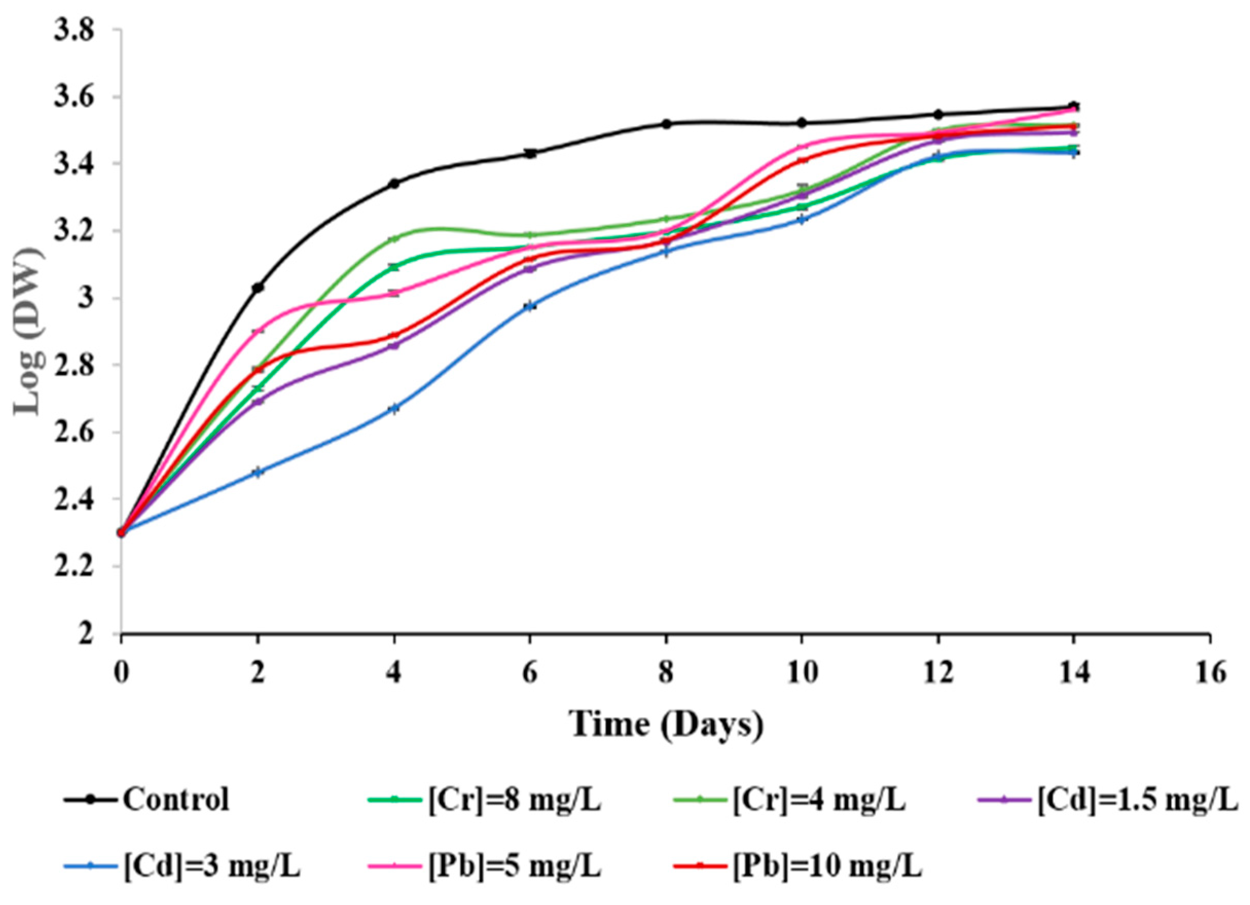
3.2. Effects of Metal Ions on the Microalga Growth

The growth kinetics of C. dorsiventrale in the presence of single metallic ions (Cd, Cr and Pb), are displayed in Figure 1. The results showed that exposition to Cd induced a slowing growth rate at the beginning of the culture, especially at the highest administered concentration (Figure 1).
The same pattern was observed for cultures exposed to Cr on the 4th day of culture (Figure 1). However, on the last day (14th day), biomass yields from cultures carried out in the presence of Cd, Pb or Cr were not significantly different from the controls. Growth rates were also determined and are summarized in Table 2. Using the lowest concentrations for Cd and Cr, inhibition growth rates attained 6.2% and 4.23%, respectively. However, Pb also induced weak growth inhibitory effects for C. dorsiventrale (Figure 1). Growth rate inhibition was clearly dose-dependent ranging from 1 to 4.6% (Table 2).

3.3. Effects of Metal Ions on Bioproducts Synthesis

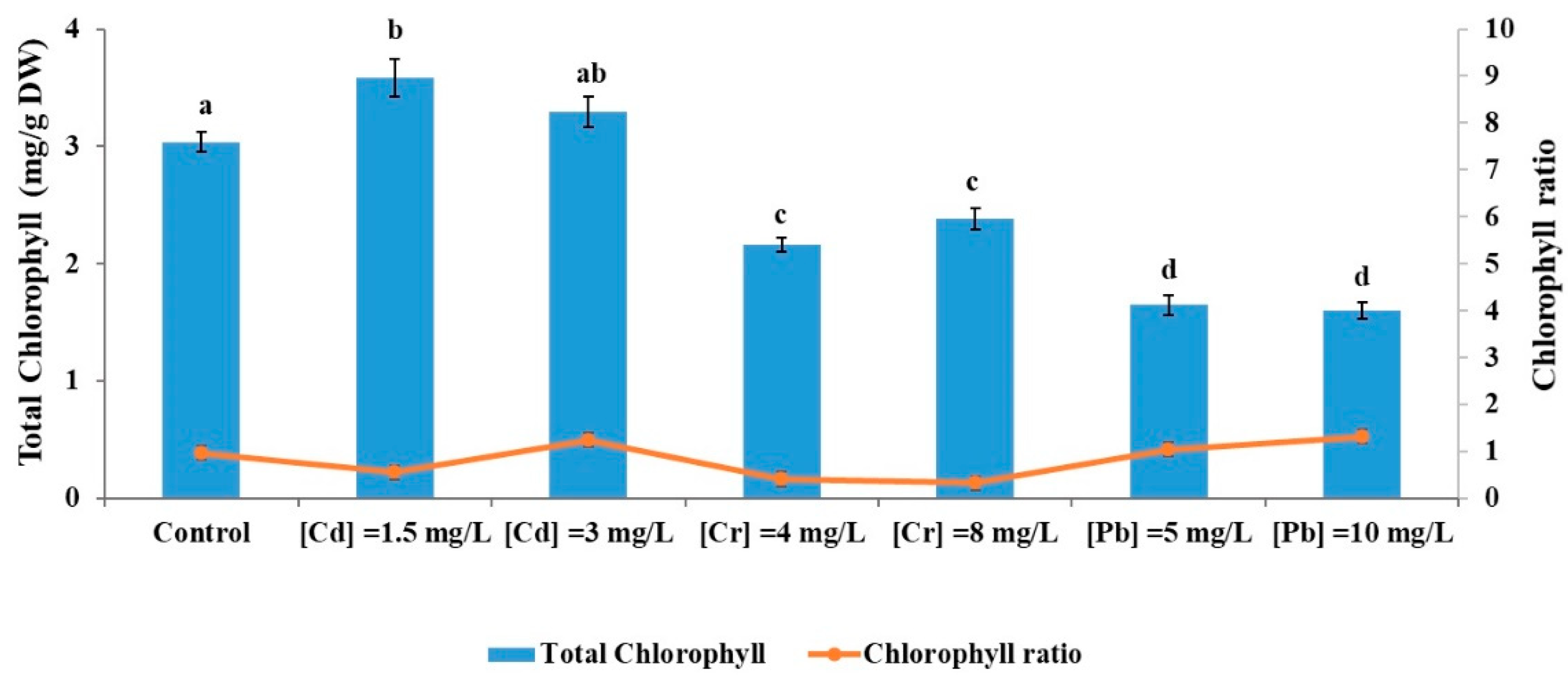
3.3.1. Impact of Metal Ions on Chlorophyll Content

At the end of experiments, total chlorophyll concentrations along with chlorophyll ratios (chlorophyll a/chlorophyll b) were determined and are shown in Figure 2.
Exposure to Cd induced a significant increase in total chlorophyll content ranging from 8.33% to 15.23% in the presence of 3 and 1.5 mg/L of Cd, respectively. However, for Cr and Pb, total chlorophyll concentrations were lower than control, particularly for the highest Pb tested concentration. Chlorophyll ratios remained constant in the presence of 10 mg/L of Pb and 3 mg/L of Cd, but decreased under all other conditions.

3.3.2. Impact of Metal Ions on Soluble-Protein Content

Soluble-protein content under the three metals treatments exhibited no significant differences compared to the control except when exposed to 5 mg/L of Pb which increased the production up to 11.6% (Figure 3A).

3.3.3. Impact of Metal Ions on Lipid Content

Exposure to metal ions decreased the lipid content of C. dorsiventrale, particularly after treatment with Cd (1.5 and 3 mg/L) and Cr (4 and 8 mg/L) resulting in reductions of 24.66% to 31.17% and from 15.33% to 16.26%, respectively. However, the treatment with Pb showed no significant effect on lipid content, compared to the negative control, especially at the lowest metal concentration (Figure 3B).

3.3.4. Impact of Metal Ions on Polysaccharide Content

The results are summarized in Figure 3C, indicating that exposure of C. dorsiventrale to the highest concentrations of Cd, Cr or Pb decreased its carbohydrate content. This reduction was more pronounced for Cd and Cr than for Pb. No significant effects were observed at the lowest concentrations for any of the tested metals.

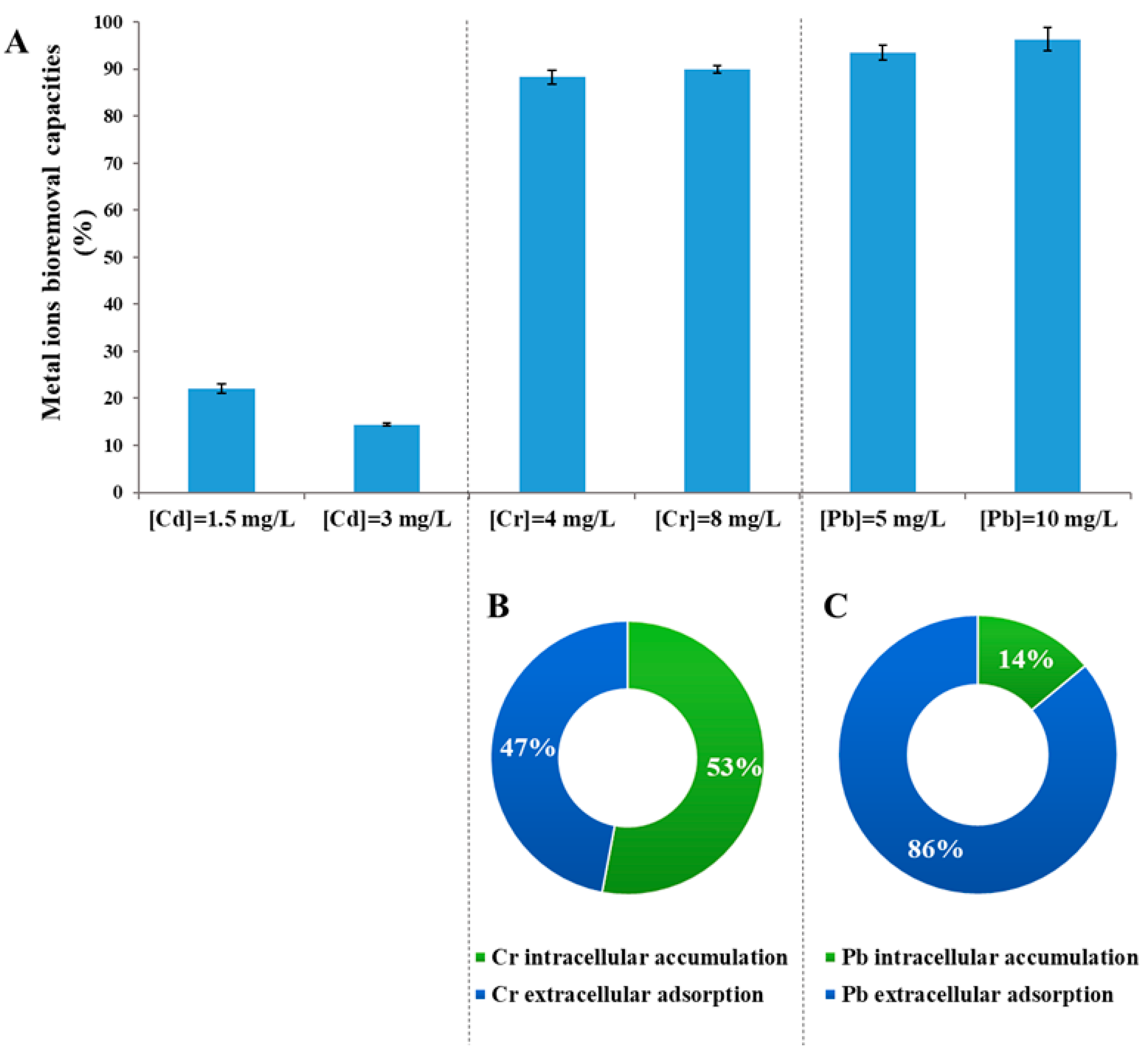
3.4. Metal Removal Rates

The ability of C. dorsiventrale to remove Cd, Cr and Pb was assessed after 14 days of exposure. Amounts of residual ions metals were determined and then metal-removed percentages were calculated (Figure 4A).
For Cd, removal efficiency did not exceed 22%, whereas those observed for both Cr (89%) and Pb (95%) were much higher. We therefore investigated Cr and Pb uptake mechanisms through measuring intracellular and extracellular ion accumulation (Figure 4B,C). The results showed that C. dorsiventrale removed around 89% of Cr, of which 53% were accumulated intracellularly and 47% were adsorbed on the cell surface. An even higher percentage (nearly 95%) was quoted for Pb with 86% adsorbed at the microalgae cell surface.

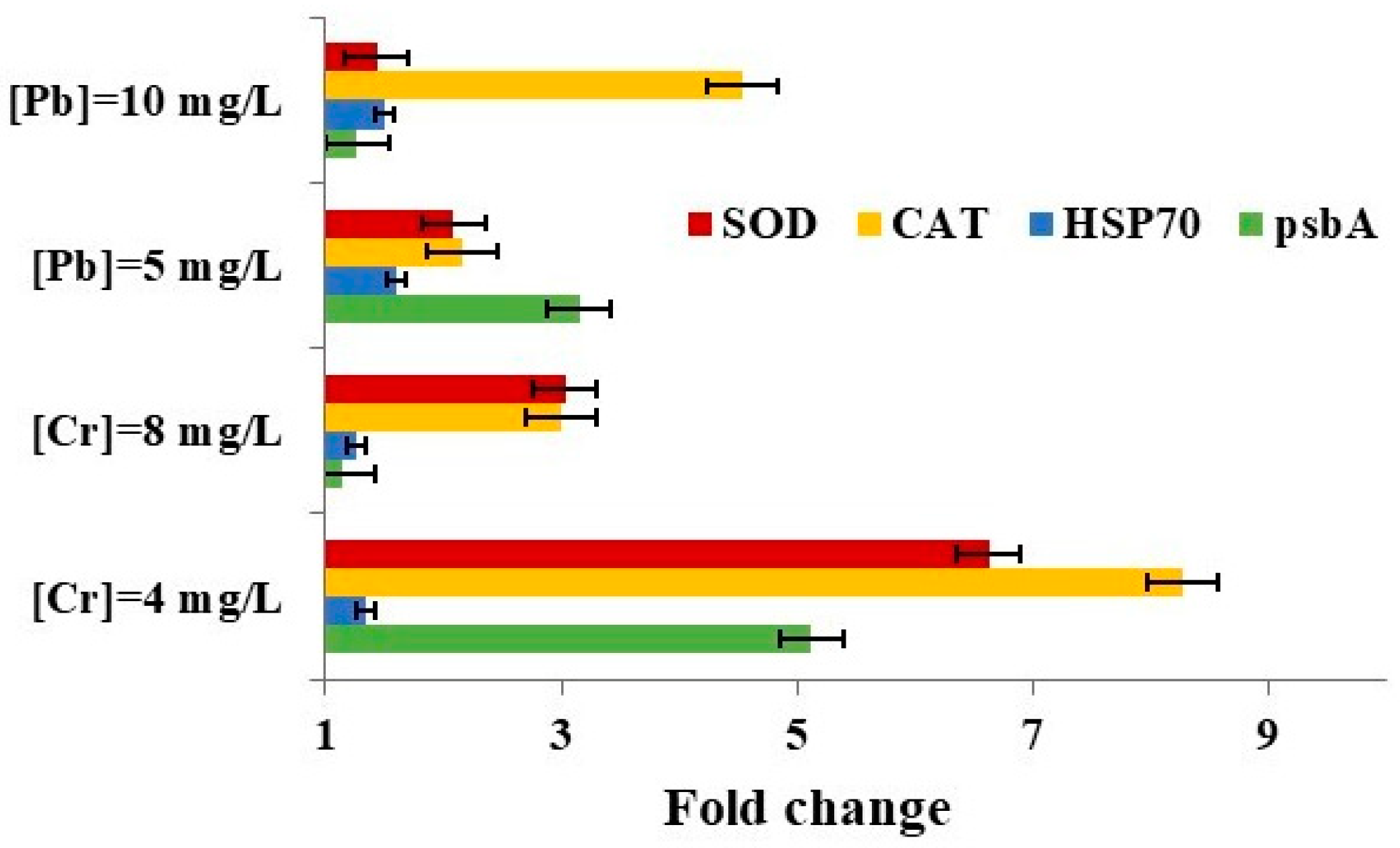
3.5. Impact of Metal Ions on Gene Expression

The strong removal capacities for Cr and Pb prompted us to investigate the molecular mechanisms by which C. dorsiventrale responded specifically to these two metals putting those for Cd aside since their removal rates were not very encouraging.
The effects of Cr and Pb on oxidative stress defenses were assessed by quantifying the expression of superoxide dismutase (SOD), catalase (CAT), photosystem II protein D1 (psbA) and heat shock protein 70 (HSP70) coding genes. After 72 h of exposure to Cr and Pb, the relative expression levels of these genes (Figure 5) were measured and compared to the reference gene coding for β-tubulin (β-TUB).
The results showed a significant increase in the expression of the catalase coding gene after exposure to Pb and especially to Cr, with an eightfold increase. An overexpression of the superoxide dismutase-coding gene was also observed. The expression of the psbA gene increased fivefold at 4 mg/L of Cr and threefold at 5 mg/L of Pb. A non-significant increase in the expression of the HSP70 coding gene was observed under Cr exposure, whereas a slight overexpression was noted with Pb treatment.

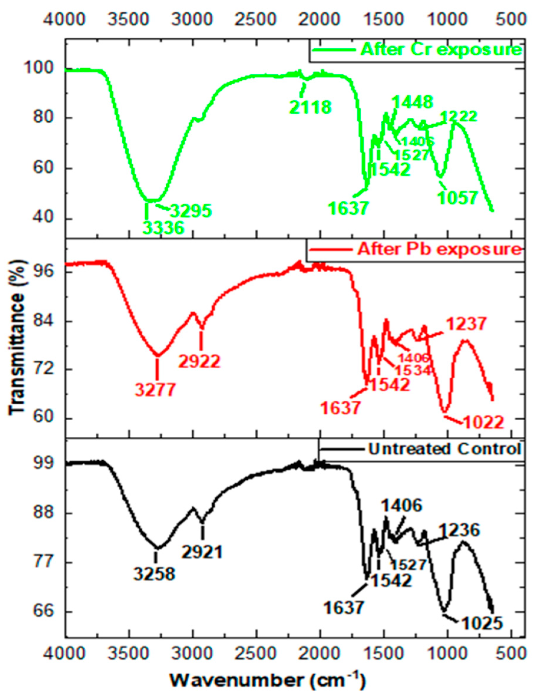
3.6. FTIR Band Assignment and Analysis

Cellular mechanisms associated with the removal capacities of Pb and Cr in C. dorsiventrale were also investigated using FTIR analysis. The FTIR spectra of C. dorsiventrale biomass were analyzed before and after exposure to Cr and Pb in order to detect, identify and characterize potential differences due to interactions of these metallic ions with functional groups of the microalgae (Figure 6).
The obtained spectra of C. dorsiventrale biomass after exposure to Cr and Pb revealed changes in functional groups (hydroxyl, carboxyl, phosphate, amino, sulfate, etc.) compared to the control. Those changes were represented by different peaks mainly characteristic of lipids, polysaccharides, and proteins. Shifts in many groups’ positions were observed after treatments with Pb and especially with Cr (Table 3).
The difference in the absorption peaks at the region 3639–3029 cm−1 revealed the existence of stretching vibration of hydroxyl (-OH) and amino (-NH2) groups. Profile modifications detected in the peaks at the region 3012–2809 cm−1 showed the stretching vibration of methyl and methylene functional groups (-CH3, -CH2-). However, this peak was absent after Cr exposure. A band at 2118 cm−1 obtained after Cr treatment and related to the carbodiimide stretching group was absent in untreated control spectra as well as after Pb exposure. Moreover, the peak at the region 1460–1420 cm−1, related to carbonate carboxylate or methyl groups, existed only after Cr treatment. Focusing on the 1680–1600 cm−1, 1545–1540 cm−1 and 1425–1330 cm−1 regions, all associated with proteins and assigned to Amide I (C=O), Amide II (N-H, C-N) and carboxylate (-COO), respectively. The obtained peaks presented no significant differences between untreated and treated biomass with both ions metal Cr or Pb. Peaks around 1356 and 1191 cm−1 may be attributed to the phosphorous (-PO-) and sulfur (-SO-) groups of membrane phospholipids. In this region, there were no significant changes before and after Pb treatment, contrary to Cr exposure.
Finally, bands in the region between 1072 and 980 cm−1 showed a more important peak after Cr treatment which could be due to the elongation of bonds C-C and C-O abundant in polysaccharides.

4. Discussion

The present study provides an evaluation of the toxic effects of Cd, Cr, and Pb on C. dorsiventrale, highlighting distinct physiological and molecular responses, as well as their bioremediation potential. Cd and Cr were more toxic to C. dorsiventrale than Pb, particularly during the lag and exponential phases. This finding is in agreement with a previous study showing the negative impact of Cd and Cr on the growth of the green marine microalga Dunaliella salina [16]. Other studies have indicated a tolerance to these two metals in plants, green microalgae and diatoms [52,53]. In this sense, we infer that C. dorsiventrale faced metal stress by exploiting the nutrients released by decomposing dead cells [54]. Regarding exposition to Pb, C. dorsiventrale growth did not seem significantly affected, which may be explained by the importance of Pb as an inducer of peroxidase enzyme activity, which induces the Indole Acetic Acid (IAA) hormone known to stimulate growth and cell division in microalgae [55]. Chlorococcum aquaticum exhibited significant tolerance to Pb2+, evidenced by an LC50 value of 100 mg L−1, which underscores its potential as an effective bioremediation agent for lead-contaminated water [23].
In regards to algal metabolic activity, we observed a reduction in total chlorophyll concentration in C. dorsiventrale after Cr and Pb exposures. The decrease in chlorophyll content has been documented as a common stress response in microalgae cells [25,56]. Upadhyay et al. [57] reported that Chlorococcum sp. treated with 10 µM As(III) exhibited a minimal reduction of 21% in chlorophyll concentration. In concordance, Chlorella vulgaris, exposure to Zn and Hg resulted in a dose-dependent reduction in photosynthetic pigments, with decreases ranging from 32% to 100% as metal concentrations increased [58]. This decrease may be attributable to the substitution of magnesium within the chlorophyll molecule by trace metals which obviously threatens photosynthesis [49,59,60]. Metal toxicity is also reflected by the fluctuations of Chl-a/Chl-b ratios according to metal concentrations with increased levels after Pb exposures while a decrease was registered after Cr exposure. We infer that this shift between these two pigments may be explained by their complementary defense mechanisms against reactive oxygen species due to metallic stress [61]. Despite pigment adjustment to ensure the removal of reactive oxygen species, we observed a decrease in lipid content in C. dorsiventrale under Cd and Cr treatments along with that of polysaccharides after exposure to the highest concentrations of all tested metals. Clearly, this persistent oxidative stress will affect the integrity of cell macromolecules such as lipids, proteins, polysaccharides and nucleic acids. Indeed, FTIR analysis suggested an interaction of Pb and Cr metal ions with polysaccharides and phospholipids. Previous studies showed a decrease in carbohydrate content in Monoraphidium sp. exposed to 40 μM Cd [62]. It is worth noting that Pb did not affect C. dorsiventrale lipid content, inducing only a slightly lower reduction in its carbohydrates which is illustrated through FTIR analysis by similar spectra for untreated and treated cells by Pb and Cr. Moreover, soluble-protein content seemed to be non-affected in C. dorsiventrale cells treated with Cd or Cr. Scenedesmus sp. exposure to Pb induced a rise in protein amount by 11.6% at 5 mg/L of metal, while a significant inhibition of soluble-protein content was observed in Chlorella sorokiniana treated with 52 mg/L of Pb [63].
At a molecular level, CAT and SOD encoded genes were overexpressed after Pb and Cr exposures, representing an up-regulation triggered by metals as previously reported in the literature [16,64,65,66]. Briefly, SOD constitutes the first barrier of defense against O2•− by quickly converting it to O2 and H2O2. The H2O2 generated is highly toxic and CAT catalyzes its transformation into H2O [67,68]. Regarding the psbA gene, it was up-regulated under Cr and Pb exposures, which illustrates the role played by the D1 protein of the photosystem II (PSII) in combatting oxidative stress [69,70]. For HSP70 gene expression, we observed a slight increase after Cr and Pb exposures. Similar results have been observed previously in Tetraselmis suecica exposed separately to 5 mg/L of CdCl2, CuCl2 and PbCl2 during 24 h [71]. This trend may be explained by the nature of HSP70 responsiveness to environmental stress. Indeed, HSP70 has been considered a good biomarker of toxicity only after long-term exposure to environmental pollutants [72,73,74]. Concerning C. dorsiventrale, removal capacities differed according to the metal. For the lowest concentration of Cd, the removal reached a maximum of 22.1%. Therefore, C. dorsiventrale did not seem to represent a promising candidate for Cd removal despite its tolerance observed towards this metal. Conversely, Cr removal efficiency attained 89% and is equally distributed between intracellular and extracellular compartments. The most spectacular results, however, were obtained with Pb since its removal rate reached 95%. This result was higher than those of previous studies on the capacity of the microalgae Chroococcus minutus, Chlorococcum aegyptiacum and Chlorella vulgaris in removing heavy metals, including Pb [74,75,76,77]. Interestingly, 86% of removed Pb was adsorbed in microalgae cell surfaces. This opens up some new research avenues that could track the regeneration of algal biomass and the release of intact metals.
Despite these promising results, the study’s use of artificial seawater and batch cultures might limit the applicability of C. dorsiventrale to real-world conditions. Future research should address these limitations by exploring large-scale cultivation and metal recycling to improve scalability and practical application.

5. Conclusions

The present study underscores the significant potential of C. dorsiventrale as an effective tool for bioremediation of heavy metals in contaminated environments. The microalgae demonstrated notable tolerance and growth in the presence of Cd, Cr, and Pb, achieving removal efficiencies of 89% for Cr and 95% for Pb. Cd exposure increased total chlorophyll by 8.33% to 15.23%, while Cr and Pb reduced chlorophyll levels, especially at higher Pb concentrations. Protein content increased by 11.6% at 5 mg/L Pb. Lipid content decreased by 24.66% to 31.17% with Cd and 15.33% to 16.26% with Cr, but Pb had no significant effect. Polysaccharide content also decreased, notably with Cd and Cr. Molecular analysis showed significant increases in catalase and superoxide dismutase coding genes, with an eightfold rise in catalase expression after Cr exposure, highlighting the alga’s strong oxidative stress response and potential for managing heavy metal pollution.
These findings suggest that C. dorsiventrale could be a valuable asset in developing sustainable strategies for managing heavy metal pollution and protecting marine ecosystems.

Author Contributions

Conceptualization, J.E., P.M., L.A., S.A. and I.F.; methodology, R.H., F.E. and M.D.; software, J.E.; validation, J.E., P.M., L.A., S.A. and I.F.; formal analysis, R.H., J.E. and F.E.; investigation, R.H., J.E., F.E. and I.F.; data curation, R.H., J.E., F.E., S.A. and I.F.; writing—original draft preparation, R.H.; writing—review and editing, J.E., P.M., L.A., S.A. and I.F.; supervision P.M., S.A. and I.F.; project administration, S.A. and I.F.; funding acquisition, J.E. and S.A. All authors have read and agreed to the published version of the manuscript.

Funding

This research was funded by the Tunisian–Moroccan Project 20/PR&D-02 (2020–2022) with the financial support of Tunisian Ministry of Higher Education and Scientific Research.

Institutional Review Board Statement

Not applicable.

Informed Consent Statement

Not applicable.

Data Availability Statement

The data presented in this study are available on request from the corresponding author.

Acknowledgments

This work was supported by grants from the Tunisian Ministry of Higher Education and Scientific Research.

Conflicts of Interest

The authors declare no conflict of interest.

References

  1. Balzano, S.; Sardo, A.; Blasio, M.; Chahine, T.B.; Dell’Anno, F.; Sansone, C.; Brunet, C. Microalgal metallothioneins and phytochelatins and their potential use in bioremediation. Front. Microbiol. 2020, 11, 517. [Google Scholar] [CrossRef] [PubMed]
  2. Li, C.; Yu, Y.; Fang, A.; Feng, D.; Du, M.; Tang, A.; Chen, S.; Lial, A. Insight into biosorption of heavy metals by extracellular polymer substances and the improvement of the efficacy: A review. Lett. Appl. Microbiol. 2022, 75, 1064–1073. [Google Scholar] [CrossRef] [PubMed]
  3. Shen, Z.; Wang, F.; Liang, Y.; Li, Y.; Liu, Q.; Liu, F. Diversity and functions of microbes in surface sediments under heavy metal pollution of western Chaohu Lake. Lett. Appl. Microbiol. 2022, 75, 1093–1102. [Google Scholar] [CrossRef] [PubMed]
  4. Zaynab, M.; Al-Yahyai, R.; Ameen, A.; Sharif, Y.; Ali, L.; Fatima, M.; Ali Khan, K.; Li, S. Health and environmental effects of heavy metals. J. King Saud Univ. Sci. 2022, 34, 101653. [Google Scholar] [CrossRef]
  5. Sall, M.L.; Diaw, A.K.D.; Gningue-Sall, D.; Efremova, A.S.; Aaron, J.J. Toxic heavy metals: Impact on the environment and human health, and treatment with conducting organic polymers, a review. Environ. Sci. Pollut. Res. 2020, 27, 29927–29942. [Google Scholar] [CrossRef]
  6. Aendo, P.; Netvichian, R.; Thiendedsakul, P.; Khaodhiar, S.; Tulayaku, P. Carcinogenic risk of Pb, Cd, Ni, and Cr and critical ecological risk of Cd and Cu in soil and groundwater around the municipal solid waste open dump in central Thailand. J. Environ. Public Health 2022, 2022, 3062215. [Google Scholar] [CrossRef]
  7. Ayele, A.; Haile, M.; Setegn, S.; Alemu, H.; Digafe, S.; Kamaraj, M. Comparative utilization of dead and live fungal biomass for the removal of heavy metal: A concise review. Sci. World J. 2021, 2021, 5588111. [Google Scholar] [CrossRef]
  8. Cao, J.; Xie, C.; Hou, Z. Ecological evaluation of heavy metal pollution in the soil of Pb-Zn mines. Ecotoxicology 2022, 31, 259–270. [Google Scholar] [CrossRef]
  9. Ren, Y.; Lin, M.; Liu, Q.; Zhang, Z.; Fei, X.; Xiao, R.; Lv, X. Contamination assessment, health risk evaluation, and source identification of heavy metals in the soil-rice system of typical agricultural regions on the Southeast Coast of China. Environ. Sci. Pollut. Res. 2021, 28, 12870–12880. [Google Scholar] [CrossRef]
  10. Carmona, B.; Abejón, R. Innovative membrane technologies for the treatment of wastewater polluted with heavy metals: Perspective of the potential of electrodialysis, membrane distillation, and forward osmosis from a bibliometric analysis. Membranes 2023, 13, 385. [Google Scholar] [CrossRef]
  11. Chaemiso, T.D.; Nefo, T. Removal methods of heavy metals from laboratory wastewater. J. Nat. Sci. Res. 2019, 9, 36–42. [Google Scholar] [CrossRef]
  12. Sarma, U.; Hoque, M.E.; Thekkangil, A.; Venkatarayappa, N.; Rajagopal, S. Microalgae in removing heavy metals from wastewater—An advanced green technology for urban wastewater treatment. J. Hazard. Mater. Adv. 2024, 15, 100444. [Google Scholar] [CrossRef]
  13. Chen, Z.; Osman, A.I.; Rooney, D.W.; Oh, W.-D.; Yap, P.-S. Remediation of heavy metals in polluted water by immobilized algae: Current applications and future perspectives. Sustainability 2023, 15, 5128. [Google Scholar] [CrossRef]
  14. Chugh, M.; Kumar, L.; Shah, M.P.; Bharadvaja, N. Algal bioremediation of heavy metals: An insight into removal mechanisms, recovery of by-products, challenges, and future opportunities. Energy Nexus 2022, 7, 100129. [Google Scholar] [CrossRef]
  15. Goswami, R.K.; Agrawal, K.; Shah, M.P.; Verma, P. Bioremediation of heavy metals from wastewater: A current perspective on microalgae-based future. Lett. Appl. Microbiol. 2022, 75, 701–717. [Google Scholar] [CrossRef]
  16. Elleuch, J.; Hmani, R.; Drira, M.; Michaud, P.; Fendri, I.; Abdelkafi, S. Potential of three local marine microalgae from Tunisian coasts for cadmium, lead and chromium removals. Sci. Total Environ. 2021, 799, 149464. [Google Scholar] [CrossRef]
  17. Ben Mohamed, J.; Elleuch, J.; Drira, M.; Esteban, M.Á.; Michaud, P.; Abdelkafi, S.; Fendri, I. Characterization and biotechnological potential of two native marine microalgae isolated from the Tunisian coast. Appl. Sci. 2021, 11, 5295. [Google Scholar] [CrossRef]
  18. Danouche, M.; El Ghachtouli, N.; El Arroussi, H. Phycoremediation mechanisms of heavy metals using living green microalgae: Physicochemical and molecular approaches for enhancing selectivity and removal capacity. Heliyon 2021, 7, e07609. [Google Scholar] [CrossRef]
  19. Vidya, D.; Nayana, K.; Sreelakshmi, M.; Keerthi, K.V.; Sneha Mohan, K.; Sudhakar, M.P.; Arunkumar, K. A sustainable cultivation of microalgae using dairy and fish wastes for enhanced biomass and bio-product production. Biomass Convers. Biorefinery 2023, 13, 6859–6873. [Google Scholar] [CrossRef]
  20. Thabet, J.; Elleuch, J.; Martínez, F.; Abdelkafi, S.; Hernández, L.E.; Fendri, I. Characterization of cellular toxicity induced by sub-lethal inorganic mercury in the marine microalgae Chlorococcum dorsiventrale isolated from a metal-polluted coastal site. Chemosphere 2023, 338, 139391. [Google Scholar] [CrossRef]
  21. Subashchandrabose, S.R.; Megharaj, M.; Venkateswarlu, K.; Naidu, R. Interaction effects of polycyclic aromatic hydrocarbons and heavy metals on a soil microalga, Chlorococcum sp. MM11. Environ. Sci. Pollut. Res. 2015, 22, 8876–8889. [Google Scholar] [CrossRef]
  22. Nam, S.-H.; Kim, D.; An, S.; An, Y.-J. Validation of the paper-disc soil method using soil alga Chlorococcum infusionum to quantitatively determine the toxicity of heavy metals. Comp. Biochem. Physiol. Part C Toxicol. Pharmacol. 2022, 258, 109380. [Google Scholar] [CrossRef]
  23. Liyanage, L.M.M.; Lakmali, W.G.M.; Athukorala, S.N.P.; Jayasundera, K.B. Application of live Chlorococcum aquaticum biomass for the removal of Pb(II) from aqueous solutions. J. Appl. Phycol. 2020, 32, 4069–4080. [Google Scholar] [CrossRef]
  24. Shiels, K.; Tsoupras, A.; Lordan, R.; Nasopoulou, C.; Zabetakis, I.; Murray, P.; Saha, S.K. Bioactive lipids of marine microalga Chlorococcum sp. SABC 012504 with anti-inflammatory and anti-thrombotic activities. Mar. Drugs 2021, 19, 28. [Google Scholar] [CrossRef] [PubMed]
  25. Elleuch, J.; Ben Amor, F.; Chaaben, Z.; Frikha, F.; Michaud, P.; Fendri, I.; Abdelkafi, S. Zinc biosorption by Dunaliella sp. AL-1: Mechanism and effects on cell metabolism. Sci. Total Environ. 2021, 773, 145024. [Google Scholar] [CrossRef]
  26. Mehra, A.; Zafar, S.U.; Jutur, P.P. Optimization of biomass production by Chlorella saccharophila UTEX 247 employing response surface methodology. Biomass Convers. Biorefinery 2024, 14, 8549–8561. [Google Scholar] [CrossRef]
  27. Wang, L.; Wang, L.; Manzi, H.P.; Yang, Q.; Guo, Z.; Zheng, Y.; Liu, X.; Salama, E.S. Isolation and screening of Tetradesmus dimorphus and Desmodesmus asymmetricus from natural habitats in Northwestern China for clean fuel production and N, P removal. Biomass Convers. Biorefinery 2022, 12, 1503–1512. [Google Scholar] [CrossRef]
  28. Guillard, R.R.L. Culture of phytoplankton for feeding marine invertebrates. In Culture of Marine Invertebrate Animals; Smith, W.L., Chanley, M.H., Eds.; Springer US: New York City, NY, USA, 1975; pp. 29–60. [Google Scholar] [CrossRef]
  29. Baccari, O.; Elleuch, J.; Barkallah, M.; Boukedi, H.; Ayed, N.B.; Hammami, A.; Fendri, I.; Abdelkafi, S. Development of a new TaqMan-based PCR assay for the specific detection and quantification of Simkania negevensis. Mol. Cell. Probes 2020, 53, 101645. [Google Scholar] [CrossRef]
  30. Ben Amor, F.; Elleuch, F.; Ben Hlima, H.; Garnier, M.; Saint-Jean, B.; Barkallah, M.; Pichon, C.; Abdelkafi, S.; Fendri, I. Proteomic analysis of the chlorophyta Dunaliella new strain AL-1 revealed global changes of metabolism during high carotenoid production. Mar. Drugs 2017, 15, 293. [Google Scholar] [CrossRef]
  31. Elleuch, J.; Tounsi, S.; Belguith Ben Hassen, N.; Noël Lacoix, M.; Chandre, F.; Jaoua, S.; Zribi Zghal, R. Characterisation of novel Bacillus thuringiensis isolates against Aedes aegypti (Diptera: Culicidae) and Ceratitis capitata (Diptera: Tephridae). J. Invertebr. Pathol. 2015, 124, 90–97. [Google Scholar] [CrossRef]
  32. Ahmad, S.; Pandey, A.; Pathak, V.V.; Tyagi, V.V.; Kothari, R. Phycoremediation: Algae as eco-friendly tools for the removal of heavy metals from wastewaters. In Bioremediation of Industrial Waste for Environmental Safety: Volume II: Biological Agents and Methods for Industrial Waste Management; Springer: Singapore, 2020; pp. 53–76. [Google Scholar] [CrossRef]
  33. Cameron, H.; Mata, M.T.; Riquelme, C. The effect of heavy metals on the viability of Tetraselmis marina AC16-MESO and an evaluation of the potential use of this microalga in bioremediation. PeerJ 2018, 6, e5295. [Google Scholar] [CrossRef] [PubMed]
  34. Duque, D.; Montoya, C.; Botero, L.R. Cadmium (Cd) tolerance evaluation of three strains of microalgae of the genus Ankistrodesmus, Chlorella, and Scenedesmus. Rev. Fac. Ing. Univ. Antioq. 2019, 92, 60–69. [Google Scholar] [CrossRef]
  35. Elleuch, J.; HadjKacem, F.; Ben Amor, F.; Hadrich, B.; Michaud, P.; Fendri, I.; Abdelkafi, S. Extracellular neutral protease from Arthrospira platensis: Production, optimization and partial characterization. Int. J. Biol. Macromol. 2021, 167, 1491–1498. [Google Scholar] [CrossRef] [PubMed]
  36. Jiang, L.; Luo, S.; Fan, X.; Yang, Z.; Guo, R. Biomass and lipid production of marine microalgae using municipal wastewater and high concentration of CO2. Appl. Energy 2011, 88, 3336–3341. [Google Scholar] [CrossRef]
  37. Rugnini, L.; Costa, G.; Congestri, R.; Bruno, L. Testing of two different strains of green microalgae for Cu and Ni removal from aqueous media. Sci. Total Environ. 2017, 601–602, 959–967. [Google Scholar] [CrossRef]
  38. Wellburn, A.R.; Lichtenthaler, H. Formulae and program to determine total carotenoids and chlorophylls a and B of leaf extracts in different solvents. In Advances in Photosynthesis Research; Sybesma, C., Ed.; Springer: Dordrecht, The Netherlands, 1984; Volume 2, pp. 1–12. [Google Scholar] [CrossRef]
  39. Kumar, P.; Ramakritinan, C.M.; Kumaraguru, A.K. Solvent extraction and spectrophotometric determination of pigments of some algal species from the shore of Puthumadam, Southeast Coast of India. Int. J. Ocean. Oceanogr. 2010, 4, 29–34. Available online: https://www.researchgate.net/publication/229148336 (accessed on 5 August 2024).
  40. Folch, J.; Lees, M.; Sloan-Stanley, G.H. A simple method for the isolation and purification of total lipids from animal tissues. J. Biol. Chem. 1957, 226, 497–509. [Google Scholar] [CrossRef]
  41. Fendri, I.; Chaari, A.; Dhouib, A.; Jlassi, B.; Abousalham, A.; Carrière, F.; Sayadi, S.; Abdelkafi, S. Isolation, identification and characterization of a new lipolytic Pseudomonas sp., strain AHD-1, from Tunisian soil. Environ. Technol. 2010, 31, 87–95. [Google Scholar] [CrossRef]
  42. Bradford, M.M. A Rapid and sensitive method for the quantitation of microgram quantities of protein utilizing the principle of protein-dye binding. Anal. Biochem. 1976, 72, 248–254. [Google Scholar] [CrossRef]
  43. Elleuch, J.; Zghal, R.Z.; Jemaà, M.; Azzouz, H.; Tounsi, S.; Jaoua, S. New Bacillus thuringiensis toxin combinations for biological control of lepidopteran larvae. Int. J. Biol. Macromol. 2014, 65, 148–154. [Google Scholar] [CrossRef]
  44. Elleuch, J.; Drira, M.; Ghribi, I.; Hadjkacem, F.; Pierre, G.; Khemakhem, H.; Abdelkafi, S. Lead removal from the aqueous solution by extracellular polymeric substances produced by the marine diatom Navicula salinicola. Environ. Technol. 2024, 1–13. [Google Scholar] [CrossRef] [PubMed]
  45. Toni, L.S.; Garcia, A.M.; Jeffrey, D.A.; Jiang, X.; Stauffer, B.L.; Miyamoto, S.D.; Sucharov, C.C. Optimization of phenol-chloroform RNA extraction. Methods 2018, 5, 599–608. [Google Scholar] [CrossRef] [PubMed]
  46. Derbel, H.; Elleuch, J.; Tounsi, L.; Nicolo, M.S.; Rizzo, M.G.; Michaud, P.; Fendri, I.; Abdelkafi, S. Improvement of biomass and phycoerythrin production by a strain of Rhodomonas sp. isolated from the Tunisian coast of Sidi Mansour. Biomolecules 2022, 12, 885. [Google Scholar] [CrossRef] [PubMed]
  47. Drira, M.; Elleuch, J.; Hadjkacem, F.; Hentati, F.; Drira, R.; Pierre, G.; Gardarin, C.; Delattre, C.; El Alaoui-Talibi, Z.; El Modafar, C.; et al. Influence of the sulfate content of the exopolysaccharides from Porphyridium sordidum on their elicitor activities on date palm vitroplants. Plant Physiol. Biochem. 2022, 186, 99–106. [Google Scholar] [CrossRef] [PubMed]
  48. Elleuch, F.; Ben Hlima, H.; Barkallah, M.; Baril, P.; Abdelkafi, S.; Pichon, C.; Fendri, I. Carotenoids overproduction in Dunaliella sp.: Transcriptional changes and new insights through lycopene β cyclase regulation. Appl. Sci. 2019, 9, 5389. [Google Scholar] [CrossRef]
  49. Chen, X.J.; Zhang, X.H.; Hu, L.D.; Zhang, J.Q.; Jiang, Y.; Yang, Y.; Yan, Y.B. DsCaf1 is involved in environmental stress response of Dunaliella salina. Int. J. Biol. Macromol. 2016, 82, 369–374. [Google Scholar] [CrossRef]
  50. Li, M.; Hu, C.; Zhu, Q.; Chen, L.; Kong, Z.; Liu, Z. Copper and zinc induction of lipid peroxidation and effects on antioxidant enzyme activities in the microalga Pavlova viridis (Prymnesiophyceae). Chemosphere 2006, 62, 565–572. [Google Scholar] [CrossRef]
  51. El-Naggar, N.E.; Hussein, M.H.; Shaaban-Dessuuki, S.A.; Dalal, S.R. Production, extraction and characterization of Chlorella vulgaris soluble polysaccharides and their applications in AgNPs biosynthesis and biostimulation of plant growth. Sci. Rep. 2020, 10, 3011. [Google Scholar] [CrossRef]
  52. Vo, M.T.; Nguyen, V.T.; Dao, T.S. Responses of green algae and diatom upon exposure to chromium and cadmium. Vietnam J. Sci. Technol. Eng. 2020, 62, 69–73. [Google Scholar] [CrossRef]
  53. Zhu, G.; Xiao, H.; Guo, Q.; Zhang, Z.; Zhao, J.; Yang, D. Effects of cadmium stress on growth and amino acid metabolism in two compositae plants. Ecotoxicol. Environ. Saf. 2018, 158, 300–308. [Google Scholar] [CrossRef]
  54. Soeprobowati, T.R.; Hariyati, R. Phycoremediation of Pb, Cd, Cu, and Cr by Chaetoceros calcitrans (Paulsen) Takano. Int. J. Adv. Chem. Eng. Biol. Sci. 2014, 1, 37–40. [Google Scholar] [CrossRef]
  55. Lamai, C.; Kruatrachue, M.; Pokethitiyook, P.; Upatham, E.S.; Soonthornsarathool, V. Toxicity and accumulation of lead and cadmium in the filamentous green alga Cladophora fracta (O.F. Muller ex Vahl) Kutzing: A laboratory study. Sci. Asia 2005, 31, 121–127. [Google Scholar] [CrossRef]
  56. Elleuch, J.; Thabet, J.; Ghribi, I.; Jabeur, H.; Hernández, L.E.; Fendri, I.; Abdelkafi, S. Responses of Dunaliella sp. AL-1 to chromium and copper: Biochemical and physiological studies. Chemosphere 2024, 364, 143133. [Google Scholar] [CrossRef] [PubMed]
  57. Upadhyay, A.K.; Singh, L.; Singh, R.; Singh, D.P.; Saxena, G. Bioaccumulation and toxicity of As in the alga Chlorococcum sp.: Prospects for As bioremediation. Bull. Environ. Contam. Toxicol. 2022, 108, 500–506. [Google Scholar] [CrossRef]
  58. Ajitha, V.; Sreevidya, C.P.; Sarasan, M.; Park, J.C.; Mohandas, A.; Singh, I.S.; Puthumana, J.; Lee, J.S. Effects of zinc and mercury on ROS-mediated oxidative stress-induced physiological impairments and antioxidant responses in the microalga Chlorella vulgaris. Environ. Sci. Pollut. Res. 2021, 28, 32475–32492. [Google Scholar] [CrossRef]
  59. Čypaitė, A.; Žaltauskaitė, J.; Venclovienė, J. Assessment of chlorophyll-a, chlorophyll-b and growth rate in freshwater green algae Pseudokirchneriella subcapitata exposed to cadmium and copper. In Proceedings of the 9th International Conference Environmental Engineering (9th ICEE)—Selected Papers, Vilnius, Lithuania, 22–23 May 2014. [Google Scholar] [CrossRef]
  60. Zamani-Ahmadmahmoodi, R.; Malekabadi, M.B.; Rahimi, R.; Johari, S.A. Aquatic pollution caused by mercury, lead, and cadmium affects cell growth and pigment content of marine microalga Nannochloropsis oculata. Environ. Monit. Assess. 2020, 192, 330. [Google Scholar] [CrossRef]
  61. Qian, L.; Qi, S.; Cao, F.; Zhang, J.; Zhao, F.; Li, C.; Wang, C. Toxic effects of boscalid on the growth, photosynthesis, antioxidant system and metabolism of Chlorella vulgaris. Environ. Pollut. 2018, 242, 171–181. [Google Scholar] [CrossRef]
  62. Zhao, Y.; Song, X.; Yu, L.; Han, B.; Li, T.; Yu, X. Influence of cadmium stress on the lipid production and cadmium bioresorption by Monoraphidium sp. QLY-1. Energy Convers. Manag. 2019, 188, 76–85. [Google Scholar] [CrossRef]
  63. Dao, L.H.T.; Beardall, J. Effects of lead on two green microalgae Chlorella and Scenedesmus: Photosystem II activity and heterogeneity. Algal Res. 2016, 16, 150–159. [Google Scholar] [CrossRef]
  64. Chia, M.A.; Chimdirim, P.K.; Japhet, W.S. Lead induced antioxidant response and phenotypic plasticity of Scenedesmus quadricauda (Turp.) de Brébisson under different nitrogen concentrations. J. Appl. Phycol. 2015, 27, 293–302. [Google Scholar] [CrossRef]
  65. Danouche, M.; El Ghachtouli, N.; El Baouchi, A.; El Arroussi, H. Heavy metals phycoremediation using tolerant green microalgae: Enzymatic and non-enzymatic antioxidant systems for the management of oxidative stress. J. Environ. Chem. Eng. 2020, 8, 104460. [Google Scholar] [CrossRef]
  66. Venkatachalam, P.; Jayaraj, M.; Manikandan, R.; Geetha, N.; Rene, E.R.; Sharma, N.C.; Sahi, S.V. Zinc oxide nanoparticles (ZnONPs) alleviate heavy metal-induced toxicity in Leucaena leucocephala seedlings: A physiochemical analysis. Plant Physiol. Biochem. 2017, 110, 59–69. [Google Scholar] [CrossRef]
  67. Ighodaro, O.M.; Akinloye, O.A. First line defence antioxidants—Superoxide dismutase (SOD), catalase (CAT) and glutathione peroxidase (GPX): Their fundamental role in the entire antioxidant defence grid. Alex. J. Med. 2018, 54, 287–293. [Google Scholar] [CrossRef]
  68. Miazek, K.; Iwanek, W.; Remacle, C.; Richel, A.; Goffin, D. Effect of metals, metalloids and metallic nanoparticles on microalgae growth and industrial product biosynthesis: A review. Int. J. Mol. Sci. 2015, 16, 23929–23969. [Google Scholar] [CrossRef] [PubMed]
  69. Kós, P.B.; Deák, Z.; Cheregi, O.; Vass, I. Differential regulation of psbA and psbD gene expression, and the role of the different D1 protein copies in the cyanobacterium Thermosynechococcus elongatus BP-1. Biochim. Biophys. Acta (BBA)-Bioenerg. 2008, 1777, 74–83. [Google Scholar] [CrossRef]
  70. Mu, W.; Chen, Y.; Liu, Y.; Pan, X.; Fan, Y. Toxicological effects of cadmium and lead on two freshwater diatoms. Environ. Toxicol. Pharmacol. 2018, 59, 152–162. [Google Scholar] [CrossRef] [PubMed]
  71. Sathasivam, R.; Ki, J.S. Heat shock protein genes in the green alga Tetraselmis suecica and their role against redox and non-redox active metals. Eur. J. Protistol. 2019, 69, 37–51. [Google Scholar] [CrossRef] [PubMed]
  72. Bierkens, J.G. Applications and pitfalls of stress-proteins in biomonitoring. Toxicology 2000, 153, 61–72. [Google Scholar] [CrossRef]
  73. Köhler, H.R.; Eckwert, H.; Triebskorn, R.; Bengtsson, G. Interaction between tolerance and 70 kDa stress protein (hsp70) induction in collembolan populations exposed to long-term metal pollution. Appl. Soil Ecol. 1999, 11, 43–52. [Google Scholar] [CrossRef]
  74. Rößner, P.; Binková, B.; Šrám, R.J. Heat shock proteins hsp32 and hsp70 as biomarkers of an early response? In vitro induction of heat shock proteins after exposure of cell culture to carcinogenic compounds and their real mixtures. Mutat. Res.-Genet. Toxicol. Environ. Mutagen. 2003, 542, 105–116. [Google Scholar] [CrossRef]
  75. Al-Enazi, N.M. Biosorption of Pb2+ from water solution by using Chroococcus minutus and Chlorococcum aegyptiacum as biosorbents. Int. J. Pharm. Res. Allied Sci. 2017, 6, 247–254. [Google Scholar]
  76. Alharbi, R.M. Toxicity and bioaccumulation of lead, cadmium and zinc in Chroococcus minutus and Chlorococcum aegyptiacum. Int. J. Pharm. Res. Allied Sci. 2017, 6, 290–300. [Google Scholar]
  77. Shaker, I.M.; Islam, M.; Ahmed, M.H.; Salama, M.M. Potentiality of Chroococcus minutus (Cyanophyta) and Chlorella vulgaris (Chlorophyta) isolated from Manzala Lake for bioremediation of cadmium and lead. Int. J. Pharm. Res. Allied Sci. 2019, 12, 207–230. [Google Scholar]
Figure 1. C. dorsiventrale cell growth under Cd, Cr and Pb single metal ion exposure.
Applsci 14 08551 g001
Figure 2. Effect of Cd, Cr and Pb single metal ion exposure on chlorophyll content and chlorophyll ratio in C. dorsiventrale after 14 days of exposure. Data are means ± SE. Columns labeled with different lower-case letters indicate statistically significant differences (p < 0.05, ANOVA, Tukey’s multiple comparison test).
Applsci 14 08551 g002
Figure 3. Effect of Cd, Cr and Pb single metal ion exposure on soluble-protein content (A), lipids content (B) and polysaccharides content (C) in C. dorsiventrale after 14 days of exposure. Data are means ± SE. Columns labeled with different lower-case letters indicate statistically significant differences (p < 0.05, ANOVA, Tukey’s multiple comparison test).
Applsci 14 08551 g003
Figure 4. Metal ions bioremoval capacities (A) and intracellular/extracellular bioremoval rate of Cr (B) and Pb (C) using C. dorsiventrale. Data are means ± SE.
Applsci 14 08551 g004
Figure 5. Fold change of mRNA expression in C. dorsiventrale cells, treated with Pb and Cr single metal ion during 72 h, relative to untreated cells for SOD, CAT, psbA and HSP70 coding genes. Columns, mean of two different RT-PCR analyses; bars, SE.
Applsci 14 08551 g005
Figure 6. FTIR spectra of C. dorsiventrale biomass before and after Cr and Pb single metal ion exposure for 14 days.
Applsci 14 08551 g006
Table 1. Specific primers used for qRT-PCR.
Gene
Abbreviation
DescriptionPrimer Sequence (F = Forward; R = Reverse)References
SODSuperoxide dismutaseF: 5′ CGCATGATCCTTTGGCTTCG 3′
R: 5′ TCCTGGTTGGCTGTGGTTTC 3′
[16]
CATCatalaseF: 5′ TCCTGTTATCGTTCGTTTCTCA 3′
R: 5′ CAAAGTTCCCCTCTCTGGTGTA 3′
[16]
psbAPhotosystem II protein D1F: 5′ ACTTCTTCTTAGCTGCTTGGC 3′
R: 5′ GACCTTGTGAGTCTACTACTG 3′
This study
HSP70Heat shock protein 70F: 5′ TGCAGGCTGGTGTGCTGTCT 3′
R: 5′ AGGGTGGTGTTGCGGGTGAT 3′
[48]
β-TUBBeta tubulinF: 5′ ATCTGCTTCCGCACCCTGAA 3′
R: 5′ AGCCGACCATGAAGAAGTGC 3′
This study
18S18S RNAF: 5′ TTGGGTAGTCGGGCTGGTC 3′
R: 5′ CGCTGCGTTCTTCATCGTT 3′
[49]
Table 2. Growth rates (μ) of C. dorsiventrale incubated at different concentrations of Cd, Cr or Pb for 14 days.
Metal IonConcentration (mg/L)Growth Rates μ (Day−1)
Control00.2090 ±0.0033 a*
Cd1.50.1962 ±0.0033 c
30.1862 ±0.0006 d
Cr40.1999 ±0.0004 b
80.1883 ±0.0011 d
Pb50.2073 ±0.0011 a
100.1992 ±0.0002 b
* Different lower-case letters in the third column indicate statically significant differences among growth rates in the series of Cd, Cr or Pb exposures. Values followed by the same letter(s) are not significantly different (p < 0.05, ANOVA, Tukey’s multiple comparison test) (Mean ± S.E.).
Table 3. Summary of wavenumbers and corresponding functional groups.
Untreated Control
(cm−1)
After Pb Exposure
(cm−1)
After Cr Exposure
(cm−1)
Wavenumber Range
(cm−1)
Functional Groups a
3258326632953639–3029υO-H/υ N-H
292129223012–2809υas CH2
21182270–1950X=C=Y
1637163716371680–1600υC=O
1542154215421545–1540δ N-H, υC-N
14481460–1420CH, C=O
1406140614061425–1330υ COO
1236123712221356–1191υas P=O, S=O
1025102210571072–980υ C-O
a Band assignments based on Liu et al. [50] and El-Naggar et al. [51]. υ = symmetric stretch; υas = asymmetric stretch; δ = symmetric deformation (bend).
Disclaimer/Publisher’s Note: The statements, opinions and data contained in all publications are solely those of the individual author(s) and contributor(s) and not of MDPI and/or the editor(s). MDPI and/or the editor(s) disclaim responsibility for any injury to people or property resulting from any ideas, methods, instructions or products referred to in the content.

