1. Introduction
Environmental pollutants, and, in particular, emerging environmental pollutants, such as agricultural contaminants (pesticides, herbicides,
etc.) and veterinary pharmaceutical by-products, represent one of the largest and most challenging classes of pollutant chemicals that can have a significant, detrimental impact on human health and the environment. For example, pesticides and herbicides are classified by Environmental Protection Agency as two of the most toxic classes of environmental pollutants [
1,
2,
3]. One of the most dangerous effects of these pollutants is that they are capable of causing serious health problems, such as bone marrow disease, increased infertility, and increased instances of immunological and respiratory diseases, at relatively low prevalence [
2,
3,
4,
5]. Hence, it is essential to have an accurate method that is capable of trace detection of these pollutants in food and water systems. Currently, the primary method for detecting and identifying these pollutants is chromatography, including GC-MS and HPLC, which can accurately detect and identify these compounds from water samples extracted from the environment [
6]. However, there are significant disadvantages to these methods that can interfere with adequate monitoring: for instance, the requirement for a library of data from high purity standards of all possible pollutants, the extensive sample preparation time needed, and the high equipment costs, in addition to the general lack of portability of these methods. Therefore, significant improvement is needed to address the ever-increasing complexity and pervasiveness of environmental pollutants in food and water systems.
Recently, biosensors, and in particular, optical biosensors, have been suggested as a possible alternative to chromatography-based techniques [
7,
8,
9]. Optical biosensors have shown tremendous promise for numerous applications in medical diagnostics, and more recently, national security, and environmental monitoring applications [
10,
11,
12]. In general, their utility arises from their non-destructive sample interrogation methods, their ability to perform extremely sensitive detection in liquid environments, and their high signal-to-noise ratios compared to other signal transduction methods, due to their relative immunity to the environment or system noise from electrical and mechanical sources [
13]. Traditional optical sensors are typically labeled devices, which detect a label on the target of interest (such as fluorophores, enzymes, nanoparticles,
etc.), rather than the target itself [
14]. Label-free optical sensors can offer better device stability, quicker response, and potentially enhanced performance, particularly when tracking molecules whose behavior may change due to the presence of the label. Labels are typically used to increase the signal-to-noise ratio; label-free sensors have an inherently high signal-to-noise ratio, negating the need for a label. Many label-free optical sensors are refractometric sensors; that is, they function by detecting small changes in the effective refractive index of their optical field, which allows them to perform fast detection of minute changes in the refractive index of the optical field at the sensor surface/sample interface. These sensors, which include optical fiber sensors, Surface Plasmon Resonance (SPR) devices, and Whispering Gallery Mode (WGM) devices, have become increasingly popular due their speed, sensitivity, and label-free sensing properties [
15,
16]. Recently, a biosensing platform using an ultrahigh-Q microcavity has been reported for the detection of the Influenza A virus and polystyrene nanobeads. Although this method used a microtoroid platform, it can be applied to other type of WGM devices, such as silica microdisks and microspheres [
17]. In this study, we focus specifically on silica microsphere WGM optical microresonators as the device of interest.
WGM optical microresonators are a type of circular resonant cavity device that confines light around their periphery via total internal reflection. WGM optical microresonators can be fabricated in a number of geometries, such as spheres, toroids, disks, and rings, both on-chip and free-standing [
18]. In this study, we use silica microspheres of approximately 100 μm in diameter as the optical microresonator. Because of the low sorption of light by silica and their atomically smooth surface, these devices typically have very low loss, so that when light is coupled into these devices, it can circulate around these microspheres over 100,000 times [
19,
20]. While the light is reflected around the periphery of the device, it is not totally confined, but instead extends into the surrounding environment through the process of evanescence, allowing the field to interact with both the device surface and the surrounding environment [
20]. Each time the optical field circulates around the device, it has an opportunity to interact with the environment, resulting in the amplification of the single-pass sensitivity that would be present if the device was linear. The photon lifetime in the resonator is measured by the Quality (Q) Factor of the device. A longer photon lifetime results in higher Q Factors. In sensing, the adsorption of an analyte onto the device’s surface causes the circulating optical field to undergo a change in its effective refractive index, thus inducing a rapid and measurable shift in the resonant frequency of the circulating field. This is the fundamental basis for the sensing capabilities of WGM optical microresonators, as well as their inherently high sensitivity [
21].
When these devices are paired with an appropriate recognition element, they are also capable of high
selectivity detection and target identification [
21,
22,
23,
24]. Typically, these recognition elements will target a compound of interest through biological molecular recognition processes, such as antibody-antigen binding, hormone-receptor binding,
etc. Previously, a number of routes to adding selectivity to label-free optical biosensors, such as WGM optical microresonators, has been explored. These routes include physical adsorption, the generation of self-assembled monolayers, the use of covalent binding (or other grafting-type approaches, including the use of inorganic-organic coupling agents, such as silane molecules, polymer brushes,
etc.), and the generation of sandwich-type attachment using the streptavidin- biotin complex [
21,
24]. These have resulted in surfaces modified to present a number of different receptors, including nanoparticles [
25,
26,
27,
28], polymers [
19], and a host of biological substances, including proteins, antibodies, oligonucleotides,
etc. [
21,
29,
30,
31,
32]. These devices, when integrated with the appropriate recognition element, have performed ultra-low detection of trypsin, thrombin, DNA, and streptavidin, among many others [
24,
33]. Recently, another method that includes a WGM-nanoshell hybrid resonator has been used to detect MS2, which is the smallest RNA virus. Further, the detection of the thyroid cancer marker, Thyroglobulin, and bovine serum albumin has been demonstrated with WGM-h microcavities that were created by adding a single gold nanoshell to the equator of the WGM resonator [
34].
However, many biologically relevant compounds, including many emerging environmental pollutants, do not have correspondingly specific biorecognition elements. Fortunately, biomimetic chemistries, such as molecular imprinting, allow for the design of nanostructured, artificial receptors, based on shape, size, and functional group selectivity, that have greater stability than most biological recognition elements and the potential for very high selectivity [
35,
36]. The molecular imprinting technique offers a promising alternative to the use of traditional biorecognition elements, and has been used extensively for the development of high selectivity optical biosensors [
23,
35,
37]. In general, artificial receptors created via the molecular imprinting of a target molecule are stable at a wide range of temperatures and pH, are typically less costly to create than natural biorecognition elements and easier to process, can be produced for almost any compound, and are significantly more compatible with the typical inorganic surfaces that comprise most label-free optical sensor surfaces than traditional biorecognition elements [
36].
The first attempt of using Molecularly Imprinted Polymers (MIPs) as a recognition element for biosensors was made in 1992 [
36]. Since that time, MIPs have become an important class of synthetic, nanostructured material whose ability to selectively adsorb and otherwise interact with a variety of compounds has seen use not only in sensing applications but also in chemical and biological separations [
38,
39]. The term molecular imprinting refers to Fischer’s ‘lock and key concept’ [
40], which explains the specific action of an enzyme with a single substrate, where the lock is the enzyme and key is the substrate. This theory helps to illustrate the specificity of enzymes towards a certain substrate. In addition, Pauling’s ‘production of antibodies
in vitro’ [
41] and Dickey’s ‘specific adsorbents’ [
28] influenced the initial design of MIPs structures. However, “modern molecular imprinting was clearly established by Wulff and Mosbach, and their pioneering work has led to the current flourishing of molecular imprinting” [
42]. Since then, the molecular imprinting technique has proven to be an effective technique for the creation of specific recognition sites in polymeric matrices.
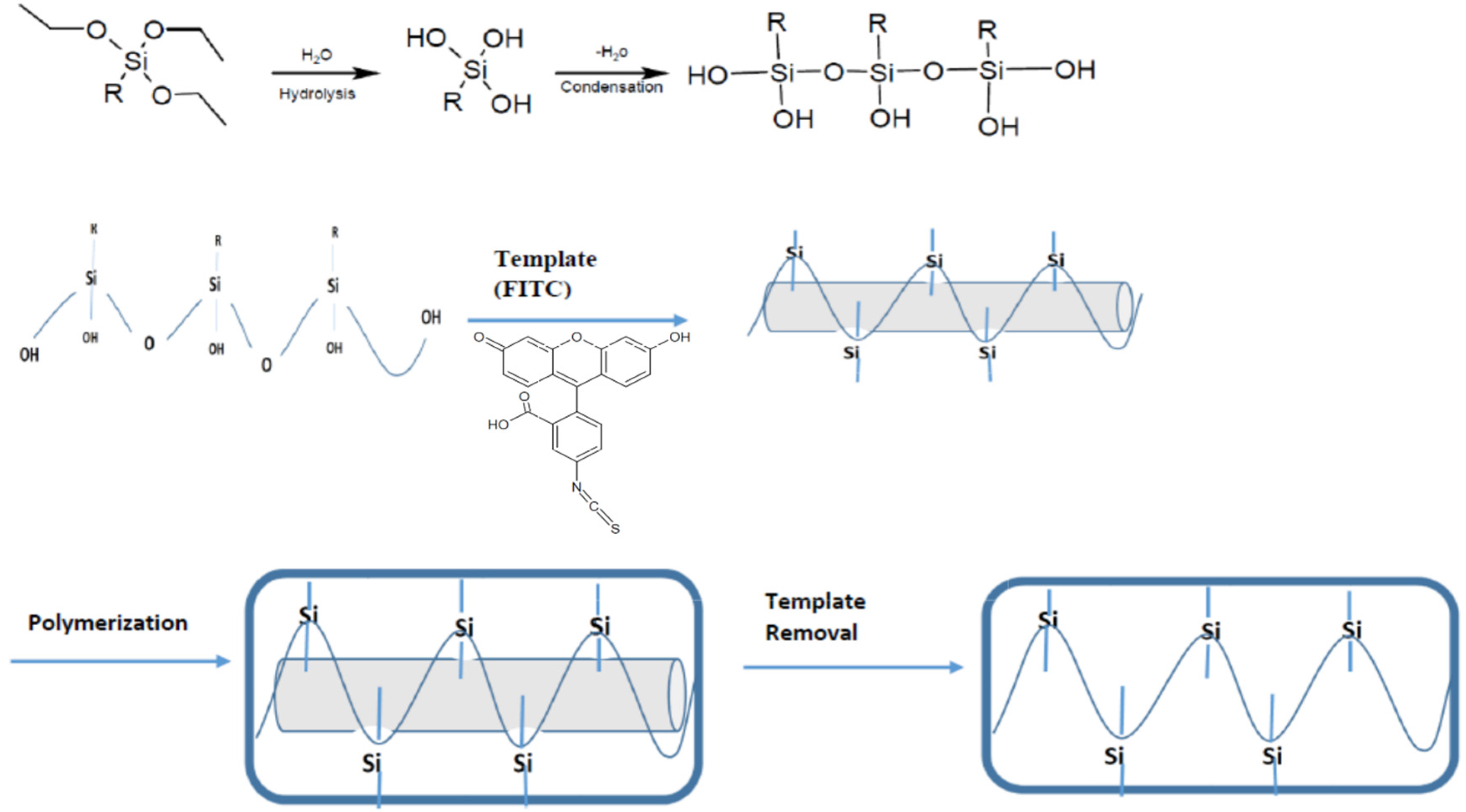
The molecular imprinting technique is accomplished by polymerizing (typically organic) monomers in the presence of the target compound, which acts as a template around which the polymer network forms, and with which the network interacts. After polymerization, the template is removed from the polymer host network, resulting in a three-dimensional, nanostructured, porous polymeric network that theoretically is capable of reabsorbing the template molecule—and only the template molecule, due to the strong interactions between the template and host matrix. This process creates artificial recognition sites that have a high affinity for the template molecule [
43].
MIPs created in this fashion can then be prepared in a variety of physical forms [
44]. The traditional approach is to synthesize MIPs in bulk, then grind the resulting polymer and successively sieve the particles into the desired size ranges according to the specific application. Although this method is simple, the overall process is time-consuming and often produces non-uniform particles in terms or size or shape. Moreover, the grinding process might result in the destruction of some interaction sites, which reduces the material’s loading capacity [
45]. Generally, macro, micro, and nano-sized interaction sites are formed from the monomers simultaneously as a result of this process. Therefore, it is hard to optimize the binding properties of bulk MIPs [
46]. An alternative approach is the creation of MIPs in thin layers (such as conformal coatings or thin films). Although both methods provide an imprinting effect, and result in reasonable specificity, and selectivity, thin layer MIPs result in significantly better outcomes in all areas [
47]. Recently, the literature has reported the creation of membranes,
in situ prepared monoliths, surface imprinting, and monolayers through a thin layer approach [
48]. Additionally, bulk MIPs are challenging to appropriately integrate with a high-sensitivity optical signal transducer, which is usually quite sensitive to the impact of surface treatments on its overall performance. Therefore, if MIPs are to be integrated with a high sensitivity, label-free optical signal transducer, such as a Whispering Gallery Mode optical microresonator, a thin layer MIPs procedure will be required to obtain a well-adhered, conformal coating that does not negatively impact the underlying device performance. Previously, there have been studies demonstrating the coating of single layer of molecular gain media on WGM optical resonators [
49,
50]. However, this study represents a coating that is nanostructured and theoretically able to act as an artificial receptor and therefore able to respond selectively to various target molecules.
However, there are some challenges that exist in terms of integrating surface coatings with micro/nanoscale, often fragile, optical devices. Surface coatings applied to WGM optical microresonators may decrease the sensitivity due to damage the coating process may cause to their glass structure. In general, these devices do not respond well to traditional wet chemistry techniques, which significantly limits the approaches that may be taken to create a conformal coating of a nanostructured, polymeric network that can act as a source of artificial receptors. Furthermore, they often require inorganic-organic linkers in order to be bound to the surface to ensure good adhesion between the surface and the recognition element or the coating. Hence, in this study, the possibility of creating of an artificial receptor via the molecular imprinting technique using inorganic silane polymer precursors, which can covalently bond to the underlying silica device surface, is examined. The expected (and achieved) result is the creation of nanostructured, inorganic polymer that is compatible with and capable of good adhesion to the underlying device.
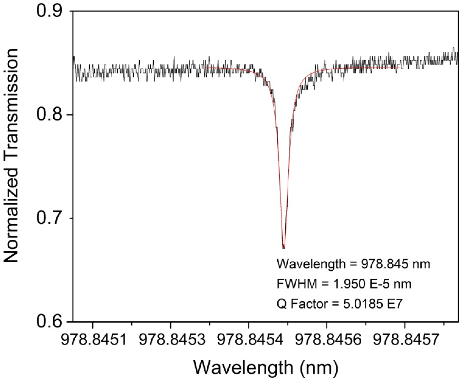
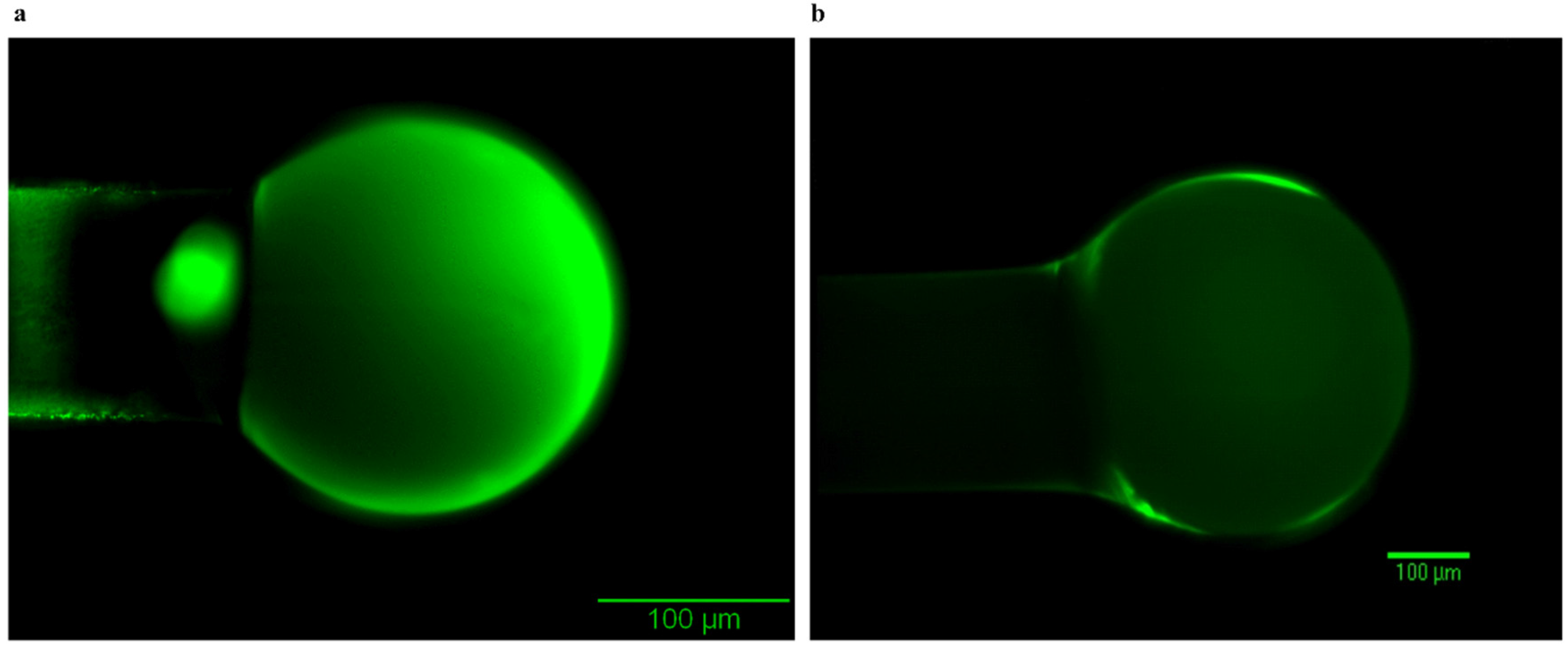
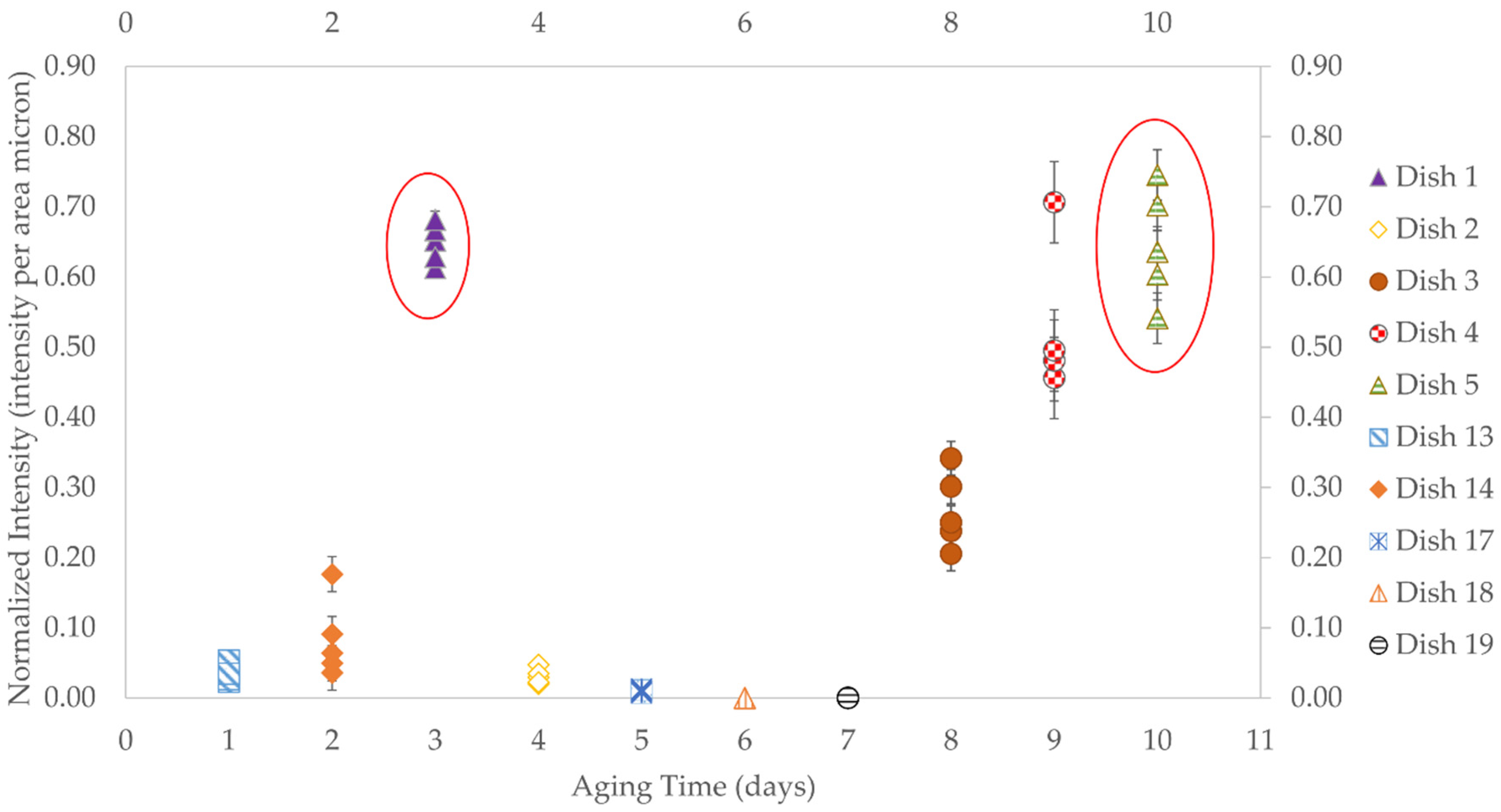
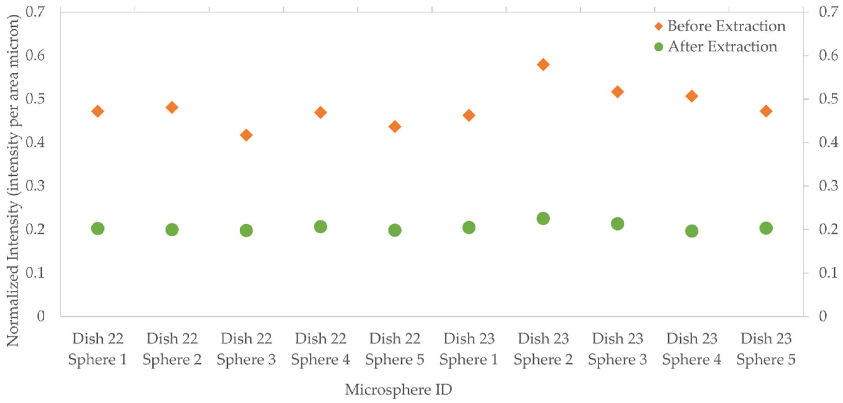
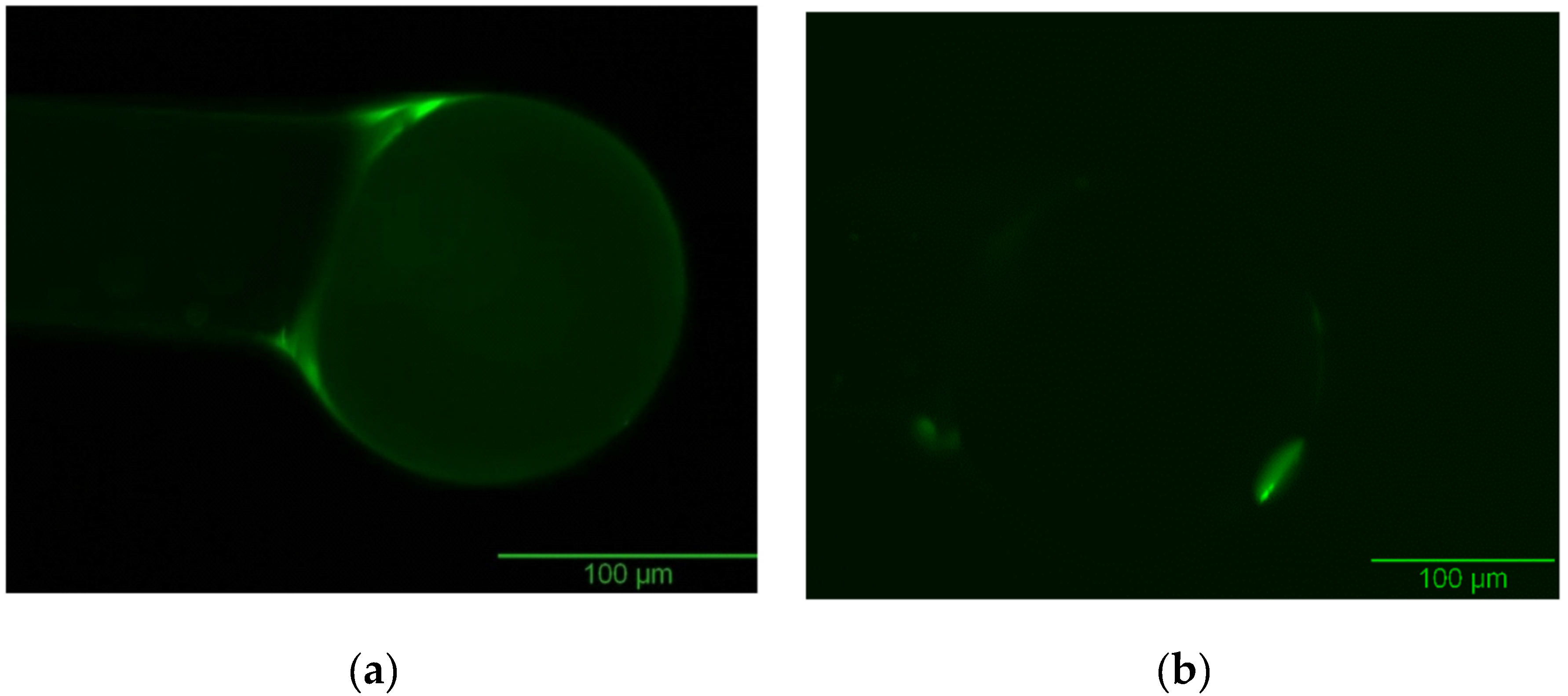
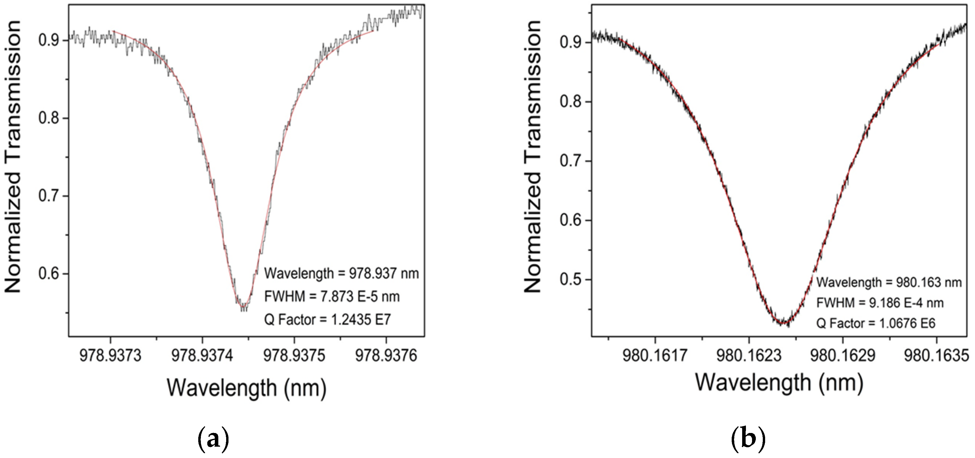
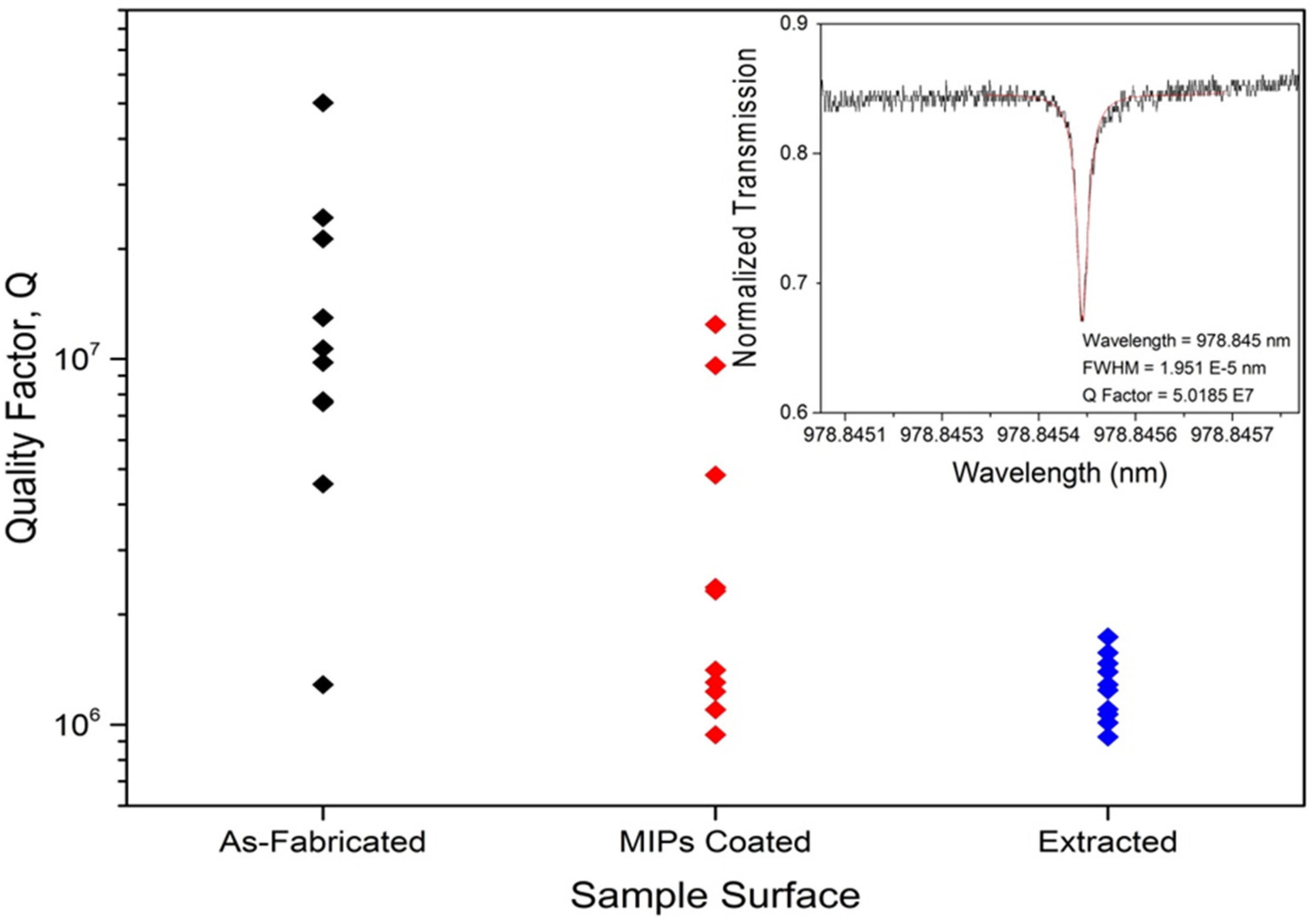
The choice of a target molecule (and, therefore, MIPs templating agent) was based upon the need to easily confirm its presence or absence using well-accepted techniques, such as microscopy and spectroscopy, and upon the requirement that it be reasonably sized, such that it can act as a model target for common environmental pollutants. Therefore, fluorescein isothiocyanate (FITC) was chosen as the target molecule, as it satisfies the aforementioned conditions. In order to promote adhesion and conformal coating to form an inorganic polymer network around the template, silane precursors were used. To ensure high quality coatings, different types of coating methods were examined, including dip coating by hand and via automation. In addition, different types of extraction methods were studied to determine an appropriately effective route to template removal. Optical performance of the devices was measured in term of Q factors, both before and after coating and template removal, which allowed the quantification of the impact of the coatings on the devices. This study represents the first example of pairing silica microsphere WGM optical resonators with molecularly imprinted polymers, and, in particular, an inorganic polymeric network. Therefore, it should serve as an initial guide for designing appropriate solutions for the detection of emerging environmental pollutants using artificial receptors.


