Article

Determination of the Size of Complex Iron Oxide Nanoparticles Using Various Physical Experimental Methods

by
Airat G. Kiiamov
1,*,
Anna G. Ivanova
1,
Alexander N. Solodov
2,
Mikhail A. Cherosov
1,
Dmitrii A. Tayurskii
1 and
Artur Khannanov
2
1
Quantum Simulators Research Laboratory, Institute of Physics, Kazan Federal University, Kremlevskaya Str. 18, Kazan 420008, Russia
2
A.M. Butlerov Chemical Institute, Kazan Federal University, Kremlevskaya Str. 18, Kazan 420008, Russia
*
Author to whom correspondence should be addressed.
Coatings 2023, 13(9), 1589; https://doi.org/10.3390/coatings13091589
Submission received: 28 July 2023 / Revised: 26 August 2023 / Accepted: 8 September 2023 / Published: 12 September 2023

Abstract

:
A series of organically coated iron oxide nanoparticles obtained via the thermal decomposition of iron–oleate complexes via a “heating-up” process were investigated using the methods of transmission electron microscopy, X-ray diffraction and fine magnetometry, accompanied by elaborate mathematical analysis. The analysis of dependencies of field dependencies on the magnetization of the shape and broadening of maxima of X-ray diffraction patterns and fine refinement of transmission electron microscopy data allowed us to demonstrate that all of the samples under consideration had a tripartite structure: (i) the magnetic crystalline core of iron oxide, (ii) the paramagnetic stratum of amorphous iron oxide and (iii) the organic coater. The new approach toward synthesis for organic coated iron oxide shows that it could be applied to the preparation of magnetic nanoparticles with different and controlled magnetic properties and sizes depending on necessary applications, especially biomedical.

Graphical Abstract

1. Introduction

To date, magnetic nanoparticles (MNPs) have been considered as the most widely used and studied nanomaterials [1,2]. This is because MNPs offer many advantages over other nanoparticles: (i) the adjustable sizes and morphology within limits of from approximately three to several hundred nanometers, (ii) the tailor-made surface coating, which can be adapted in a way so that the particles can selectively bind to a defined biologic entity [3], (iii) low toxicity [4] and (iv) the ability to convert magnetic field energy into thermal and/or mechanical energy [5,6]. The combination of these properties opens up great possibilities for using MNPs in various applications such as multi-terabit storage data [7], catalysis [8], targeted drug delivery [1], magnetic hyperthermia [9,10,11], special high-contrast agents for magnetic resonance imaging (MRI) [12], biosensors and bioseparation [13], thermoablation and the development of different smart materials [14,15,16,17,18]. The properties of MNPs heavily depend on their size and shape. For example, for the use of MNPs as a contrast agent in MRI, particle size plays a key role; thus, large-size MNPs are used as T2 contrast agents, and smaller particles are used as T1 contrast agents [19]. Magnetic hyperthermia, one of the most important applications of MNPs, is considered as a promising cancer treatment due to its low rate of side effects. In magnetic hyperthermia, heat generation by MNPs is based on their capability of dissipating magnetic energy to heat [20,21]. This effect is influenced by parameters such as size and concentration of MNPs in the fluid, as well as magnetic parameters of MNPs such as saturation magnetization (Ms) and magnetic anisotropy constant. Among these parameters, the particle size plays a key role, because it specifies the magnetic behavior type and scope of application of MNPs.
Often, the size of a spherical MNP is understood as its diameter, determined by images obtained using bright-field transmission electron microscopy [22].
The size of a nanoparticle determined using TEM is the physical diameter of MNPs, which does not always reflect its magnetic properties. It can be expected that MNPs of the same size should all have the same magnetic moment; nevertheless, due to crystal defects, the magnetic polydispersity is likely to be greater than expected from the geometric size polydispersity [23,24]. Such effects include a “magnetically dead” surface layer and internal defects of the crystal structure [25,26].
Biomedicine is a promising field of application of MNPs, and so the size of nanoparticles plays a key role. This is important to note as the biodistribution, and ultimately biological responses, for example, of the material can be correlated sizes [27]. To this end, the ability to characterize sizes of particles is the most widely accepted critical quality attribute [28]. However, the results of the sizes of MNPs obtained via the only approach from the TEM, for instance, are generally not applicable to the comprehensive characterizing of all of the properties of the samples, especially in the case of multilayer particles. So, in the case of particles made of strong magnetic material cores shelled by the nonmagnetic coating, the TEM approaches are only able to measure the outward diameter of the particle, whereas the magnetic properties of the particles ought to depend on the size of the magnetic core. Hence, in this case, usage based on TEM measurements of particle sizes for the analysis of the magnetic properties provides the systematic miscalculation.
So, in the present study, we use three experimental methods to measure the sizes: (i) TEM to measure the full outer size, (ii) fine analysis of X-ray diffraction data in order to estimate the crystalline size and (iii) utilizing of magnetization field dependencies to obtain the size of the magnetic part of the particles. And, running ahead, it could be said that all of the sizes obtained via each method for each sample are presented in Table 1.
Nanoparticles with low geometric size polydispersity can be prepared via the thermal decomposition of organometallic precursors [29,30,31]. Obtaining uniformity of the size of MNPs in this synthesis method is achieved by separating the processes of nucleation and growth. A brief nucleation event yields initial particles that subsequently all grow at the same rate under conditions where no new nuclei are formed. However, recent work has uncovered that the thermal decomposition routes may actually proceed via the formation of a nonmagnetic Wüstite phase, and that the formation of the magnetic magnetite/maghemite phases proceed afterward due to oxidation upon exposure to atmospheric oxygen [32,33]. Therefore, the geometric size and magnetic size for MNPs may differ.
In this paper, we present three approaches toward the synthesis of MNPs based on iron oxides via thermal decomposition: (i) the classical approach of obtaining MNPs via thermal decomposition, proposed by Park [31], (ii) using seed crystals and (iii) the joint thermal decomposition of iron (II) and (III) precursors. We also compare the dimensions obtained via transmission electron microscopy, magnetometry and XRD.

2. Materials and Methods

Materials.
Sodium hydroxide, n-hexane and ethanol were supplied by JSC “TatKhim Product” LLC (Kazan, Republic of Tatarstan, Russia); iron(III) chloride hexahydrate (FeCl3·6H2O, 99%), iron(II) chloride tetrahydrate (FeCl2·4H2O, 99%), oleic acid (C18H34O2, 90%) and 1-octadecene (C18H36, 90%) were obtained from Sigma-Aldrich (Burlington, MA, USA).
Methods.
Synthesis of iron–oleate complex. The metal–oleate complex was prepared by reacting metal chlorides and sodium oleate. Deionized water (60 mL) was mixed with ethanol (80 mL) and divided into two equal portions. Sodium hydroxide (9.6 g) was added to the first portion (the first solution), and FeCl3·6H2O (21.6 g) for the iron(III)–oleate complex or FeCl2·4H2O (23.9 g) for the iron(II)–oleate complex were added to the second portion (the second solution). Both solutions were successively added to a solution of oleic acid (67.8 g) in 280 mL of n-hexane. The mixture was magnetically stirred and refluxed for 4 h at 70 °C. After the reflux, the flask was removed from the oil bath and cooled to room temperature. Next, the upper organic layer containing the iron–oleate complex was washed three times with 100 mL distilled water in a separatory funnel. After washing, hexane was evaporated off. The product yield was 98%.
Synthesis of iron oxide nanocrystals. (i) The MNPs (Nos. 1, 3, 5 and 6) were synthesized according to the Park method [31,34]. We used the same synthesis procedure and materials as declared in our previous study on a similar kind of sample [34]. In a typical synthesis of MNPs (No. 1), 3.6 g of iron(III)–oleate complex was dissolved in 25 g of 1-octadecene at room temperature. The reaction mixture was heated to 320 °C with a constant heating rate of 3.3 °C min−1, and then kept at that temperature for 30 min. The resulting solution containing the nanocrystals was then cooled to room temperature, and 500 mL of ethanol was added to the solution to precipitate the nanocrystals. The nanocrystals were separated via centrifugation.
Sample Nos. 3, 5 and 6 were obtained via the method described above. For particle No. 3, the solution contained 3.6 g of iron(III)–oleate complex and 35 g of 1-octadecene; for particle No. 5, the solution contained 3.6 g of iron(III)–oleate complex, 0.57 g of oleic acid and 25 g of 1-octadecene; for particle No. 6, the solution contained 3.6 g of iron(III)–oleate complex, 0.57 g of oleic acid and 15 g of 1-octadecene.
(ii) For the synthesis of MNP Nos. 2, 4 and 9, the thermal decomposition method was used, as in the Park method, but seed crystals were additionally used. For particle No. 2, the solution contained 3.6 g of iron(III)–oleate complex, 0.1 g of MNPs (No. 1) and 25 g of 1-octadecene; for particle No. 4, the solution contained 3.6 g of iron(III)–oleate complex, 0.58 g of oleic acid, 0.5 g of MNPs (No. 1) and 25 g of 1-octadecene; and for particle No. 9, the solution contained 3.6 g of iron(III)–oleate complex, 0.57 g of oleic acid, 0.1 g of MNPs (No. 1) and 25 g of 1-octadecene.
For the synthesis of particle Nos. 7, 8 and 10, the thermal decomposition method was used, as in the Park method, but the addition of iron(II)–oleate complex was additionally used. For particle No. 7, the solution contained 2.4 g of iron(III)–oleate complex, 1.2 g of iron(II)–oleate complex, 0.57 g of oleic acid and 25 g of 1-octadecene; for particle No.8, the solution contained 2.4 g of iron(III)–oleate complex, 1.2 g of iron(II)–oleate complex and 25 g of 1-octadecene; for particle No. 10, the solution contained 2.4 g of iron(III)–oleate complex, 1.2 g of iron(II)–oleate complex, 1.20 g of oleic acid and 25 g of 1-octadecene.
We used the transmission electron microscope Hitachi HT7700 Excellence to provide transmission electron microscopy imaging (with an accelerated voltage value of 100 kV).
The powder XRD (X-ray diffraction) measurements were taken using a Bruker D8 Advance diffractometer (Cu Kα radiation λ = 1.5418 Å) with symmetric measurement geometry; the measurement time was 4 s per step and the diffraction angle range was of 7°–100°. We used the Scherrer equation to estimate the size of the crystalline iron oxide particles [34].
The magnetization measurements were taken using a commercial PPMS-9 machine and a commercial vibrational magnetometer at a temperature of 300 K with a magnetic field value up to 9 T. The obtained magnetization curves were used to determine the size of magnetic particles via the Langevin function. The details of the fitting procedure and data analysis are given below in the next section.
We conducted the Fourier transform infrared spectroscopy measurements with 4 cm−1 step length and wave numbers ranging from 400 to 4000 cm−1 using an FT-801 spectrometer with attenuated total reflection.

3. Results

We obtained 10 types of MNPs via the thermal decomposition (Park’s method) of iron–oleate complexes in an oleic acid/octadecene solution at a temperature of 320 °C. In addition to the thermal decomposition of iron–oleate complexes to the corresponding oxides, the resulting oxides were partially reduced, resulting in magnetite or iron oxide nanoparticles (II/III). In this regard, the presence of both iron(II) and iron(III) in the system should facilitate the formation of nanoparticles and improve their magnetic properties.
TEM images of the MNP samples and the corresponding particle size distribution obtained via the statistical analysis of over ∼3500 particles are shown in Figure 1. The nanocrystals exhibit a well-defined spherical morphology, are faceted and are nearly monodispersed in size and shape. The particle size of the spherical samples increases from sample No. 1 to No. 10; the average diameter of the samples is shown in Figure 1.
The FT-IR spectra of the MNPs are presented in Figure 2. A typical MNP’s spectrum shows a peak at 554 cm−1 which corresponds to the Fe-O bond valence vibration range. The absorbance bands of the organic surfactant on the surface of the particles were clearly observed at higher wavenumbers.
The iron oxide nanocrystals were characterized by X-ray powder diffraction (XRD), and the XRD patterns (Figure 3) of the nanocrystals were assigned to the (220), (311), (400), (422), (511) and (440) reflections of the inverse spinel structure iron oxide. In all of the XRD patterns, a clear line broadening can be observed, which results from size effects. This peak broadening can be utilized to estimate the mean crystallite size and the lattice strain in particles due to crystal imperfection and distortion. As the particle size diminishes, the XRD peaks become wider.
The Scherer formula approach was used to determine the crystallite size and estimate the shape of iron oxide nanoparticles from the corresponding X-ray diffraction patterns. In accordance with the Scherrer formula, the particle size D is inversely proportional to the peak width β:
D = K λ β   c o s θ ,
where K means the shape factor (we used K = 0.9), θ is a half of the diffraction angle 2θ corresponding to the diffraction maximum under consideration and λ means the used X-ray radiation wavelength (1.5418 Å). The angular positions 2θ and full width at half maximum β for all of the diffraction maxima of each sample were determined using Wolfram Mathematica 6.0 software, and each diffraction maximum was described using the Lorentzian line shape.
For each sample, the particle sizes along different crystallographic directions were estimated using an analysis of the diffraction maxima from the corresponding set of crystallographic planes. As seen via the comparison of particle sizes along various spatial dimensions, the shapes of particles were characterized as being spherical or non-spherical. In order to formularize the criteria for the sphericity of a particle, we propose to consider a nanoparticle as being spherical if its average size, obtained as the arithmetic mean of all measured crystallite sizes along different crystallographic directions, differs from the crystallite size along any measured crystallographic direction by no more than 10 percent.
Thus, the shapes of iron oxide nanoparticles of only two samples (3 and 4) were estimated as being spherical. The iron oxide nanoparticles of the sample numbers 5, 7 and 9 are relatively elongated along the (511) or (440) crystallographic directions, which allows us to make assumptions about the cigar shape of the crystallites in the case of these samples.
The field dependencies of the magnetization M H of the samples under consideration measured via sample temperature fine stabilization with the temperature T of 300 K are shown in Figure 4. The Langevin function [L(x) = coth(x) − 1/x] describing the degree of alignment of a magnetic dipole in an external field H was used to approximate the measured field dependencies of magnetization.
M H = 0 p ( D m ; μ ; σ ) M s L x + χ H d D m x = π μ 0 M s D m 3 H 6 k B T
where
x =   π μ 0 M s D m 3 H 6 k B T
The Boltzmann constant is written as kB, whereas Ms is the saturated magnetization and μ 0 is the magnetic constant. The function was weighted using a log-normal size distribution p D m ; σ = 1 2 π σ D m e x p ( l n 2 ( D m / D M ) 2 σ 2 with a dispersion of σ in order to take into account the non-uniformity of particle sizes Dm, as has been proposed in [35]. Finally, the simple paramagnetic term χ H ( χ is a paramagnetic susceptibility) was also used in order to take into account the contribution of amorphous iron oxide which surrounded the crystalline superparamagnetic core. As a result of the analysis of magnetic data, the size of each sample of magnetic core DM was estimated. It deserves to be noted that we used the Wolfram Mathematica 6.0 software for approximation.

4. Discussion

The estimated sizes of particles, crystalline parts and magnetic cores are collected in Table 1. The full particle sizes were obtained using TEM methods, while the crystalline size was found as a result of the approximation of XRD data. It should be reinstated that by means of X-ray diffraction analysis methods, we can only estimate the size of the crystalline part of the particles. So, Table 1 compares three differently estimated sizes of the particles: (i) the real particle size measured using TEM approaches, (ii) the crystalline particle size obtained via means of XRD and (iii) the magnetic core size estimated from the fine analysis of field dependencies of magnetization.
The results presented above confirm that the synthesis of shelled iron oxide magnetic nanoparticles was performed well using ten different procedures. The good agreement and consistency between the particle size estimations obtained via three different methods are clearly visible.
It was proven that particles have a multilayer structure with crystalline cores of iron oxide coated by the organic shells. These iron oxide cores are stand-alone magnets and, in unison, act as a superparamagnetic conglomerate. Nevertheless, we note the kind of minor divergence of the size of data obtained within XRD and magnetometry approaches. We argue that this disagreement is not a result of experimental error; we believe that it could be due to the fact that the upper-bound part of an iron oxide core gives no contribution to the total magnetic moment, since the surface spins are not exchange interactions coupled with the spins in the volume of the magnetic cores of the nanoparticles.
So, it could be confidently argued that the particles have a tripartite structure: (i) the magnetic crystalline core of iron oxide, (ii) the paramagnetic stratum of amorphous iron oxide and (iii) the organic coater.

5. Conclusions

We conclude that it has been proven that directly measured TEM particle sizes are not equal to the crystalline size obtained via XRD approaches nor the effective magnetic particle size. An effective magnetic particle size means that the magnetic behavior of the particles resembles a behavior of a superparamagnetic assembly with the particles of the same size as effective magnetic particle size. However, the difference obtained between the TEM particle sizes and the data obtained via XRD evidence the fact that the crystalline particles are covered with a non-crystalline organic oleate shell.
It could be concluded that the presented synthesis procedures allow for preparing MNPs with different and controlled magnetic properties and sizes depending on necessary applications, especially biomedical, as, as considered in the present study, nanoparticles are coated with a bio-friendly shell. Nevertheless, it should be noted that further possible medical application of MNPs should be prefaced with reliable and strict medical verification.

Author Contributions

Conceptualization, A.K.; Methodology, A.G.K. and A.N.S.; Software, A.G.K.; Formal analysis, A.G.K. and A.G.I.; Investigation, A.G.K. and M.A.C.; Resources, A.N.S. and A.K.; Data curation, A.G.K., A.G.I. and A.N.S.; Writing—original draft, A.G.K. and A.N.S.; Writing—review & editing, A.G.K.; Supervision, D.A.T.; Project administration, D.A.T. and A.K.; Funding acquisition, A.K. All authors have read and agreed to the published version of the manuscript.

Funding

The study was supported by a grant from the Russian Science Foundation, No. 22-73-10036 (https://rscf.ru/project/22-73-10036/ accessed on 11 September 2023). This work was supported by the Kazan Federal University Strategic Academic Leadership Program (PRIORITY-2030).

Institutional Review Board Statement

Not applicable.

Informed Consent Statement

Not applicable.

Data Availability Statement

Not applicable.

Conflicts of Interest

The authors declare no conflict of interest.

References

  1. Laurent, S.; Forge, D.; Port, M.; Roch, A.; Robic, C.; Vander Elst, L.; Muller, R.N. Magnetic iron oxide nanoparticles: Synthesis, stabilization, vectorization, physicochemical characterizations, and biological applications. Chem. Rev. 2008, 108, 2064–2110. [Google Scholar] [CrossRef] [PubMed]
  2. Sezer, N.; Arı, İ.; Bicer, Y.; Koc, M. Superparamagnetic nanoarchitectures: Multimodal functionalities and applications. J. Magn. Magn. Mater. 2021, 538, 168300. [Google Scholar] [CrossRef]
  3. Veiseh, O.; Gunn, J.W.; Zhang, M. Design and fabrication of magnetic nanoparticles for targeted drug delivery and imaging. Adv. Drug Deliv. Rev. 2010, 62, 284–304. [Google Scholar] [CrossRef] [PubMed]
  4. Mahmoudi, M.; Hofmann, H.; Rothen-Rutishauser, B.; Petri-Fink, A. Assessing the in vitro and in vivo toxicity of superparamagnetic iron oxide nanoparticles. Chem. Rev. 2012, 112, 2323–2338. [Google Scholar] [CrossRef]
  5. Nandhini, G.; Shobana, M. Role of ferrite nanoparticles in hyperthermia applications. J. Magn. Magn. Mater. 2022, 552, 169236. [Google Scholar] [CrossRef]
  6. Zhang, C.; Li, X.; Jiang, L.; Tang, D.; Xu, H.; Zhao, P.; Fu, J.; Zhou, Q.; Chen, Y. 3D printing of functional magnetic materials: From design to applications. Adv. Funct. Mater. 2021, 31, 2102777. [Google Scholar] [CrossRef]
  7. Weller, D.; Moser, A. Thermal effect limits in ultrahigh-density magnetic recording. IEEE Trans. Magn. 1999, 35, 4423–4439. [Google Scholar] [CrossRef]
  8. Weiss, W.; Ranke, W. Surface chemistry and catalysis on well-defined epitaxial iron-oxide layers. Prog. Surf. Sci. 2002, 70, 1–151. [Google Scholar] [CrossRef]
  9. Rytov, R.; Bautin, V.; Usov, N. Towards optimal thermal distribution in magnetic hyperthermia. Sci. Rep. 2022, 12, 3023. [Google Scholar] [CrossRef]
  10. Usov, N.A.; Gubanova, E.M. Application of magnetosomes in magnetic hyperthermia. Nanomaterials 2020, 10, 1320. [Google Scholar] [CrossRef]
  11. Tong, S.; Quinto, C.A.; Zhang, L.; Mohindra, P.; Bao, G. Size-dependent heating of magnetic iron oxide nanoparticles. ACS Nano 2017, 11, 6808–6816. [Google Scholar] [CrossRef] [PubMed]
  12. Na, H.B.; Song, I.C.; Hyeon, T. Inorganic nanoparticles for MRI contrast agents. Adv. Mater. 2009, 21, 2133–2148. [Google Scholar] [CrossRef]
  13. Khizar, S.; Ahmad, N.M.; Zine, N.; Jaffrezic-Renault, N.; Errachid-el-salhi, A.; Elaissari, A. Magnetic nanoparticles: From synthesis to theranostic applications. ACS Appl. Nano Mater. 2021, 4, 4284–4306. [Google Scholar] [CrossRef]
  14. Xu, Z.; Xu, Q. Collective behaviors of magnetic microparticle swarms: From dexterous tentacles to reconfigurable carpets. ACS Nano 2022, 16, 13728–13739. [Google Scholar] [CrossRef]
  15. Khamidullin, T.; Lounev, I.; Solodov, A.; Vakhitov, I.; Hashemi, S.A.; Dimiev, A.M. Graphene Oxide–Epoxy Composites with Induced Anisotropy of Electrical Properties. J. Phys. Chem. C 2021, 125, 26823–26831. [Google Scholar] [CrossRef]
  16. Solodov, A.N.; Balkaev, D.; Amirov, R.; Gataullina, R.; Nurtdinova, L.; Yusupov, R.; Kharintsev, S.S.; Dimiev, A.M. Polymer Composites with Magnetically Tunable Optical Anisotropy. ACS Appl. Polym. Mater. 2023, 5, 6338–6345. [Google Scholar] [CrossRef]
  17. Solodov, A.; Neklyudov, V.V.; Shayimova, J.; Amirov, R.R.; Dimiev, A.M. Magneto-optical properties of the magnetite-graphene oxide composites in organic solvents. ACS Appl. Mater. Interfaces 2018, 10, 40024–40031. [Google Scholar] [CrossRef]
  18. Solodov, A.N.; Balkaev, D.A.; Shayimova, J.R.; Vakhitov, I.R.; Gataullina, R.M.; Sukhov, A.V.; Burilova, E.A.; Amirova, L.M.; Zhuravleva, Y.I.; Amirov, R.R. Tribological properties of an epoxy polymer containing a magnetically oriented graphene oxide/iron oxide nanoparticle composite. Diam. Relat. Mater. 2023, 138, 110211. [Google Scholar] [CrossRef]
  19. Zhou, Z.; Yang, L.; Gao, J.; Chen, X. Structure–relaxivity relationships of magnetic nanoparticles for magnetic resonance imaging. Adv. Mater. 2019, 31, 1804567. [Google Scholar] [CrossRef]
  20. Tang, Y.; Flesch, R.C.; Jin, T.; Gao, Y.; He, M. Effect of nanoparticle shape on therapeutic temperature distribution during magnetic hyperthermia. J. Phys. D Appl. Phys. 2021, 54, 165401. [Google Scholar] [CrossRef]
  21. Nemati, Z.; Alonso, J.; Rodrigo, I.; Das, R.; Garaio, E.; García, J.Á.; Orue, I.; Phan, M.-H.; Srikanth, H. Improving the heating efficiency of iron oxide nanoparticles by tuning their shape and size. J. Phys. Chem. C 2018, 122, 2367–2381. [Google Scholar] [CrossRef]
  22. Lim, J.; Yeap, S.P.; Che, H.X.; Low, S.C. Characterization of magnetic nanoparticle by dynamic light scattering. Nanoscale Res. Lett. 2013, 8, 381. [Google Scholar] [CrossRef]
  23. Unni, M.; Uhl, A.M.; Savliwala, S.; Savitzky, B.H.; Dhavalikar, R.; Garraud, N.; Arnold, D.P.; Kourkoutis, L.F.; Andrew, J.S.; Rinaldi, C. Thermal decomposition synthesis of iron oxide nanoparticles with diminished magnetic dead layer by controlled addition of oxygen. ACS Nano 2017, 11, 2284–2303. [Google Scholar] [CrossRef] [PubMed]
  24. Luigjes, B.; Woudenberg, S.M.; de Groot, R.; Meeldijk, J.D.; Torres Galvis, H.M.; de Jong, K.P.; Philipse, A.P.; Erné, B.H. Diverging geometric and magnetic size distributions of iron oxide nanocrystals. J. Phys. Chem. C 2011, 115, 14598–14605. [Google Scholar] [CrossRef]
  25. Kodama, R. Magnetic nanoparticles. J. Magn. Magn. Mater. 1999, 200, 359–372. [Google Scholar] [CrossRef]
  26. Roca, A.; Morales, M.; O’Grady, K.; Serna, C. Structural and magnetic properties of uniform magnetite nanoparticles prepared by high temperature decomposition of organic precursors. Nanotechnology 2006, 17, 2783. [Google Scholar] [CrossRef]
  27. Hassellöv, M.; Readman, J.W.; Ranville, J.F.; Tiede, K. Nanoparticle analysis and characterization methodologies in environmental risk assessment of engineered nanoparticles. Ecotoxicology 2008, 17, 344–361. [Google Scholar] [CrossRef]
  28. Maguire, C.M.; Rösslein, M.; Wick, P.; Prina-Mello, A. Characterisation of particles in solution–a perspective on light scattering and comparative technologies. Sci. Technol. Adv. Mater. 2018, 19, 732–745. [Google Scholar] [CrossRef] [PubMed]
  29. Park, J.; Lee, E.; Hwang, N.M.; Kang, M.; Kim, S.C.; Hwang, Y.; Park, J.G.; Noh, H.J.; Kim, J.Y.; Park, J.H. One-nanometer-scale size-controlled synthesis of monodisperse magnetic Iron oxide nanoparticles. Angew. Chem. 2005, 117, 2932–2937. [Google Scholar] [CrossRef]
  30. Sun, S.; Zeng, H.; Robinson, D.B.; Raoux, S.; Rice, P.M.; Wang, S.X.; Li, G. Monodisperse mfe2o4 (m = fe, co, mn) nanoparticles. J. Am. Chem. Soc. 2004, 126, 273–279. [Google Scholar] [CrossRef]
  31. Park, J.; An, K.; Hwang, Y.; Park, J.-G.; Noh, H.-J.; Kim, J.-Y.; Park, J.-H.; Hwang, N.-M.; Hyeon, T. Ultra-large-scale syntheses of monodisperse nanocrystals. Nat. Mater. 2004, 3, 891–895. [Google Scholar] [CrossRef]
  32. Chalasani, R.; Vasudevan, S. Form, content, and magnetism in iron oxide nanocrystals. J. Phys. Chem. C 2011, 115, 18088–18093. [Google Scholar] [CrossRef]
  33. Casula, M.F.; Jun, Y.-W.; Zaziski, D.J.; Chan, E.M.; Corrias, A.; Alivisatos, A.P. The concept of delayed nucleation in nanocrystal growth demonstrated for the case of iron oxide nanodisks. J. Am. Chem. Soc. 2006, 128, 1675–1682. [Google Scholar] [CrossRef] [PubMed]
  34. Solodov, A.N.; Shayimova, J.R.; Burilova, E.A.; Shurtakova, D.V.; Zhuravleva, Y.I.; Cherosov, M.A.; Tian, Y.; Kiiamov, A.G.; Amirov, R.R. Understanding the Nucleation and Growth of Iron Oxide Nanoparticle Formation by a “Heating-Up” Process: An NMR Relaxation Study. J. Phys. Chem. C 2021, 125, 20980–20992. [Google Scholar] [CrossRef]
  35. Chen, D.-X.; Sanchez, A.; Taboada, E.; Roig, A.; Sun, N.; Gu, H.-C. Size determination of superparamagnetic nanoparticles from magnetization curve. J. Appl. Phys. 2009, 105, 083924. [Google Scholar] [CrossRef]
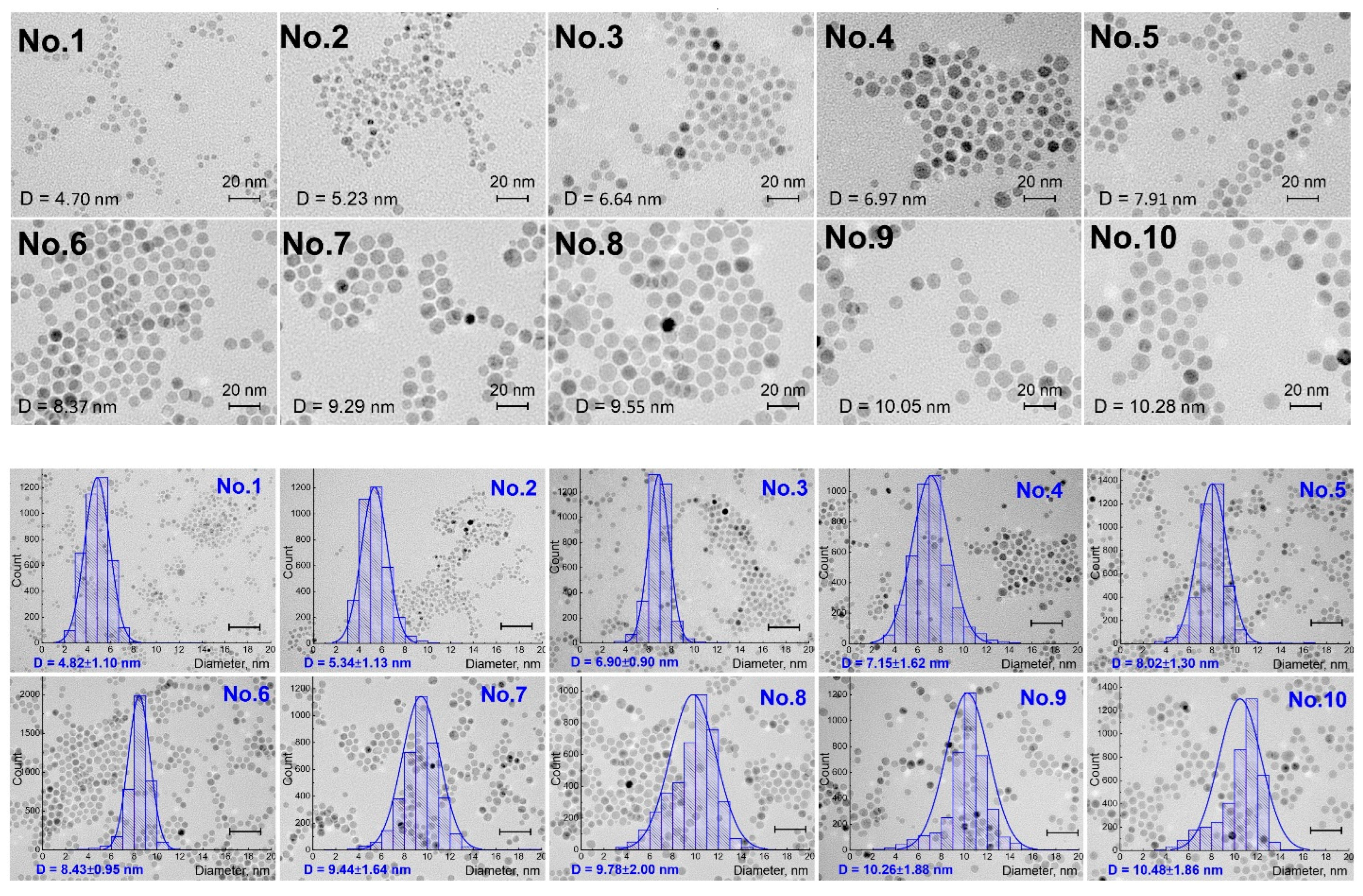
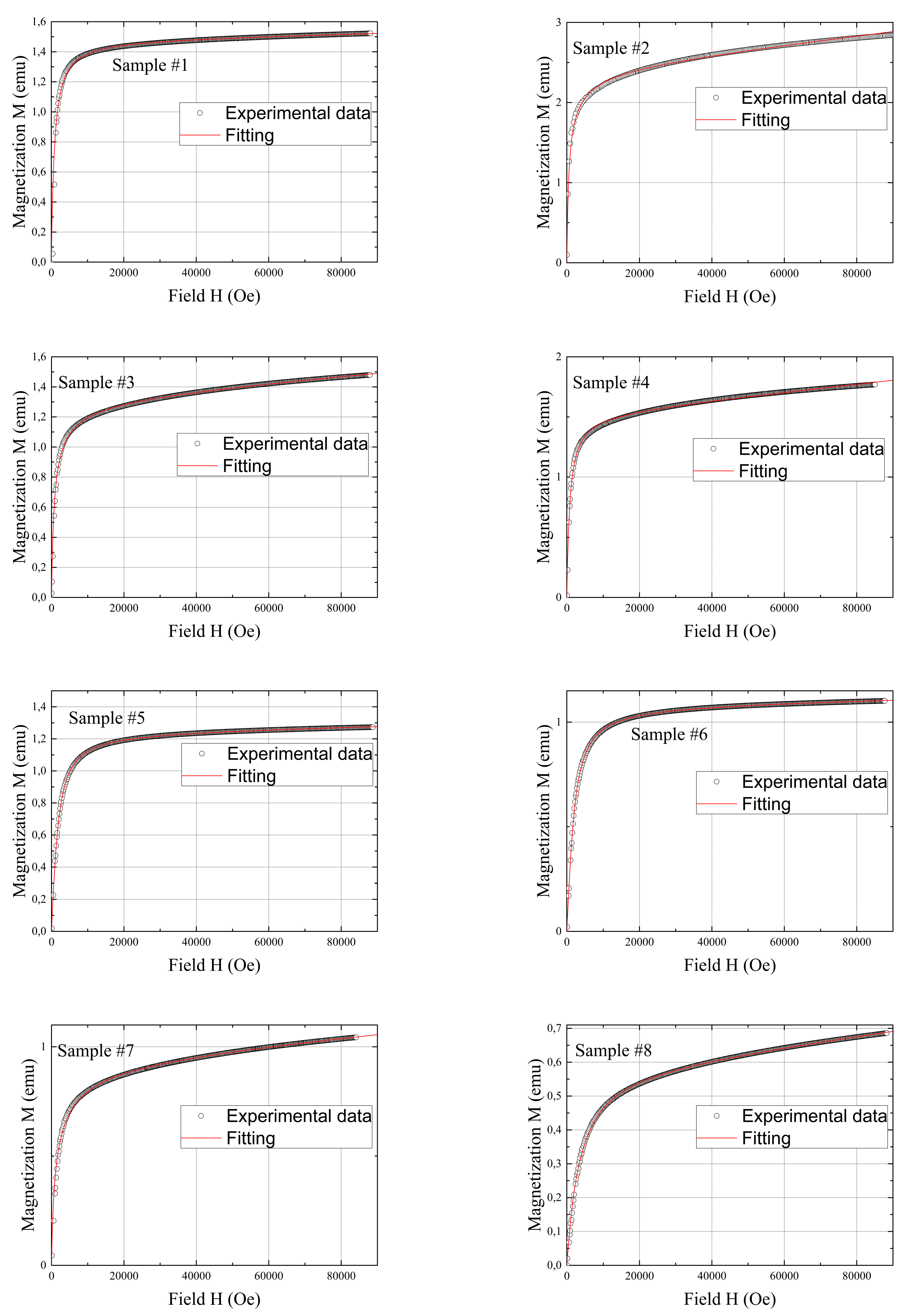
Figure 1. TEM images (top section) and the corresponding particle size distribution (bottom section) obtained via statistical analysis of over ~3500 particles. In the (bottom section), all scale bars are 40 nm.
Coatings 13 01589 g001
Figure 2. (a) FT-IR spectra of the synthesized MNPs (Nos. 1–10). (b) FT-IR spectra of MNP No. 1.
Coatings 13 01589 g002
Figure 3. Powder XRD patterns of the synthesized MNPs: (a) Nos. 1–5, (b) Nos. 6–10.
Coatings 13 01589 g003
Figure 4. Field dependencies of magnetization M (emu) of MNPs (open black circles) and its approximation (thin red line) within the Langevin function weighted using a log-normal size distribution (see details in the text).
Coatings 13 01589 g004aCoatings 13 01589 g004b
Table 1. The particle sizes obtained via different methods.
Sample ID NumberObtained via TEM, nmObtained via XRD, nmObtained from the Field Dependencies of Magnetization, nm
14.82 ± 1.104.73.50
4.3
4.5
4.7
25.34 ± 1.13 5.63.33
4.0
5.3
36.90 ± 0.906.61.80
7.4
6.5
47.15 ± 1.626.84.50
6.6
58.02 ± 1.304.64.33
7.1
4.9
68.43 ± 0.954.94.25
8.0
7.6
4.8
79.44 ± 1.645.44.00
7.0
5.3
89.78 ± 2.006.33.30
8.2
6.5
910.26 ± 1.886.33.33
10.1
7.4
8.0
1010.48 ± 1.867.93.33
10.3
10.0
Disclaimer/Publisher’s Note: The statements, opinions and data contained in all publications are solely those of the individual author(s) and contributor(s) and not of MDPI and/or the editor(s). MDPI and/or the editor(s) disclaim responsibility for any injury to people or property resulting from any ideas, methods, instructions or products referred to in the content.

