Article

E-textiles in Clinical Rehabilitation: A Scoping Review

1
Bloorview Research Institute, Holland Bloorview Kids Rehabilitation Hospital, Toronto, ON M4G-1R8, Canada
2
Institute of Biomaterials and Biomedical Engineering, University of Toronto, Toronto, ON M5S-3G9, Canada
*
Author to whom correspondence should be addressed.
Electronics 2015, 4(1), 173-203; https://doi.org/10.3390/electronics4010173
Submission received: 16 November 2014 / Revised: 16 January 2015 / Accepted: 3 February 2015 / Published: 25 February 2015
(This article belongs to the Special Issue Wearable Electronics)

Abstract

:
Electronic textiles have potential for many practical uses in clinical rehabilitation. This scoping review appraises recent and emerging developments of textile-based sensors with applications to rehabilitation. Contributions published from 2009 to 2013 are appraised with a specific focus on the measured physiological or biomechanical phenomenon, current measurement practices, textile innovations, and their merits and limitations. While fabric-based signal quality and sensor integration have advanced considerably, overall system integration (including circuitry and power) has not been fully realized. Validation against clinical gold standards is inconsistent at best, and feasibility with clinical populations remains to be demonstrated. The overwhelming focus of research and development has been on remote sensing but the opportunity for textile-mediated feedback to the wearer remains unexplored. Recommendations for future research are provided.

1. Introduction

The term “e-textiles” (short for electronic textiles) is often used interchangeably with “smart fabrics”, “functional apparel” and even “wearable technology”. “Wearable technology” broadly refers to the category of electronics that are carried on the body, while “e-textiles” refers specifically to garments or textiles with some sort of technology, such as wiring and sensors, embedded directly into the fabric.
As early as 1998, researchers have been working on garments to monitor physiological signals for healthcare, military and space exploration [1]. In the pioneering Wearable Motherboard garment [1], rigid electronic components for sensing and processing were attached to a fabric substrate. This work catalyzed a vision in which clothing serves as a platform for computing. Over the last 10 years, e-textile technologies have evolved from hobby crafts and fashion innovations to healthcare solutions. In parallel, there has been a surge of related research, with a Scopus search yielding some 100 papers in 2014 alone with the key word “e-textile”, compared to only 6 in 2002. This growth of e-textile research and clinical application can be attributed to a number of technological advances as detailed below.
1
Sewable and washable microcontrollers like the commercially available, Arduino Lilypad (lilypadarduino.org) offer researchers affordable and fabric-friendly embedded platforms for quickly developing robust prototypes [2].
2
Flexible circuits have allowed the textile integration of advanced electronics capable of sensing and information transmission, without compromising the comfort of the wearer. Likewise, smaller and flexible power sources for e-textile applications have emerged [3] while energy-harvesting power electronics are already under development by companies like PowerLeap.(www.powerleap.com).
3
Conductive threads and yarns, usually composed of stainless steel or conductive silver with a nylon core, have become widely available. The properties of such threads and fabric transmission lines for textile computing applications have been documented [4,5,6], facilitating their selection and implementation in e-textile solutions.
4
Fabric-based components including capacitors, resistors, and transistors have been devised [4,7,8,9], allowing full integration of simple circuits onto a fabric substrate. Piezoelectric textiles have also emerged, promoting the development of a full range of fabric-based sensors [10,11].
With a globally aging population, it is not surprising that the majority of applied research on e-textiles has focused primarily on the unobtrusive, remote monitoring of older persons [12,13], particularly post-myocardial infarction or post-stroke. Increasingly, these wearable monitoring systems are capable of tracking complex movement patterns [14]. Against this backdrop, this paper provides a timely and critical appraisal of recent developments in textile-based sensors with either demonstrated or potential applications in clinical rehabilitation. Unlike previous e-textile reviews (e.g., [12]), our appraisal is not limited to physiological or behavioral monitoring applications. Before presenting our review methodology and findings, we introduce an integration rating. These introductory remarks will help to contextualize the ensuing review.

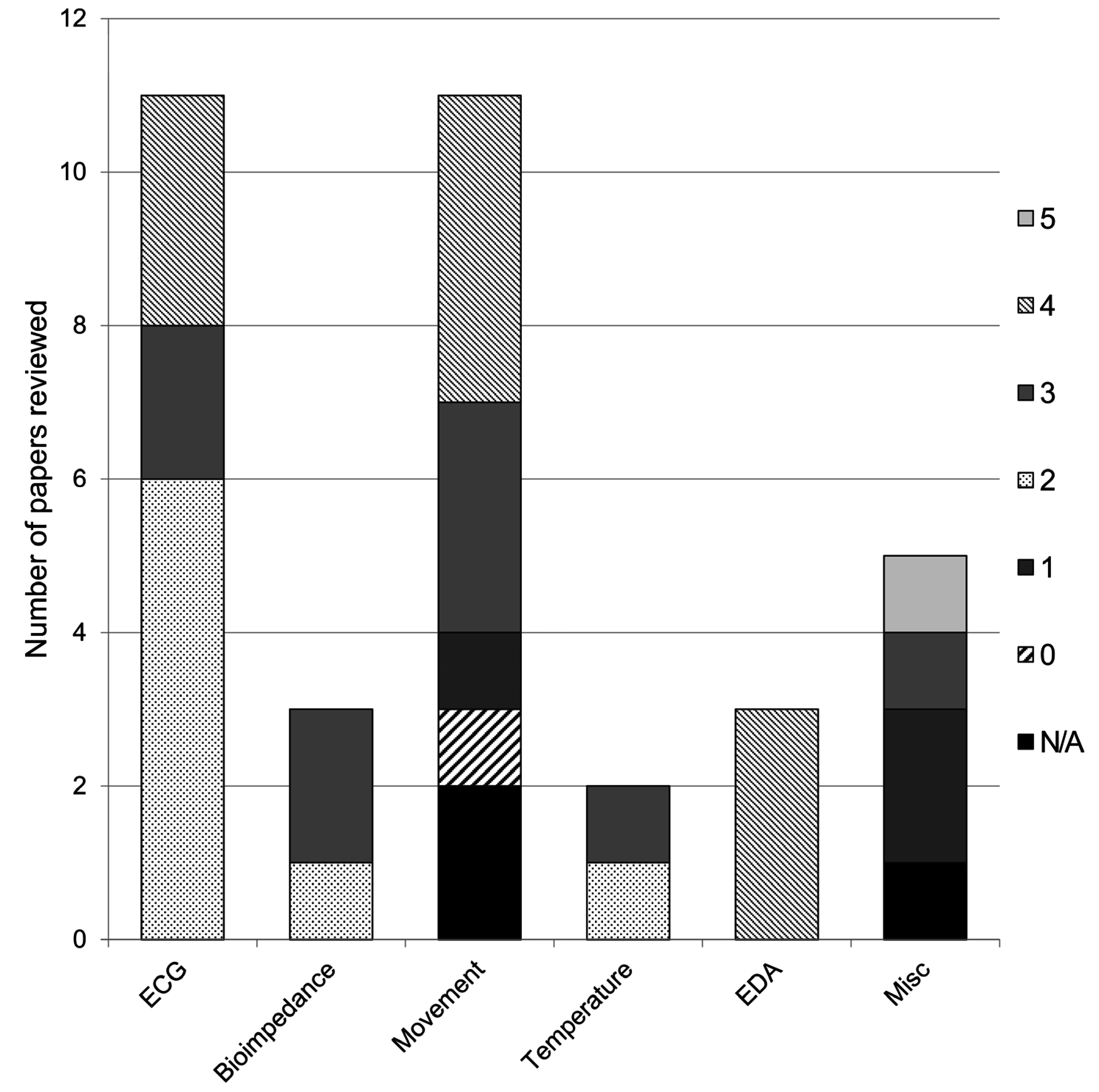
1.1.Degree of Integration

Fundamental to the concept of an e-textile, is the degree to which electronic components are integrated into the fabric. In this paper, we consider a fully integrated sensor as one that is woven directly into the fabric, in the absence of any rigid components. Conversely, an unintegrated sensor is one that is independently packaged and simply attached to the user. Table 1 proposes a rating system to describe the degree of integration.
Table 1. Degree of integration.
RatingDescription
0→Wearable computer: no textile integration
1→Superficial integration: components in pockets or connected to fabric with snaps
2→Partial integration: some sensing components incorporated (e.g., woven, knit, printed, embroidered etc.) into fabric
3→Partial integration: all sensing components incorporated (e.g., woven, knit, printed, embroidered etc.) into fabric
4→Partial integration: wiring and sensing woven into fabric
5→Towards full integration: sensing, wiring and power supply woven into fabric
The underlying aim of this review is to explore literature relating to the application of e-textile technology. A scoping review will allow us to survey the existing literature in an inclusive way while facilitating the identification of gaps and topics for further research.

2. Methods

We searched the literature for recent studies related to the use of e-textiles in rehabilitation. The search was conducted using electronic databases including IEEE, PubMed, Scopus, and ISI Web of Science. We used various combinations of the terms outlined in Table 2.
Table 2. Terms used in literature search.
TextileElectronicRehab
fabric clothingsmart intelligent sensingphysiology biomechanics bio-
We included all English-language, peer-reviewed journal articles that directly examined the development or implementation of e-textile sensors and actuators with applications in healthcare and rehabilitation, in the last five years (2009–2013). With a specific interest in integrated biomechanical and physiological sensors, we excluded all chemical sensors, and sensors which primarily fall into the category of “wearable technology”; that is, lacking any integration.

3. Results

Our search yielded 100 unique articles, 35 of which were found to meet the aforementioned criteria. Various characteristics of the studies are summarized in Table 3. We were specifically interested in the intended application within clinical rehabilitation, the stage of development (e.g., conceptual design, laboratory characterization without human participants, or empirical testing with human participants), sensor type (e.g., electrical, optical, inertial, thermal), the degree of integration (Table 1) and the status of sensor validation (e.g., from unvalidated to validation against a gold standard).
Table 3. Summary of recently published e-textile research (DoI = degree of integration; BP = blood pressure; SpO2 = saturation of peripheral oxygen; VOP = venous occlusion plethysmography).
ReferenceStage of Development# Study ParticipantsParticipant TypeSensed phenomenonSensor validationDoIArea Application of
Adnane et al. [15]Empirical testing1TargetECG, respirationNon-fabric alternative: 3-lead ECG; pneumography2Sleep disorders
Angelidis [16]Conceptual designN/AN/AECG, BP, SpO2, temperature, sweatN/A5Healthcare: general
Baek et al. [17]Empirical testing5Adult, male, healthyECG, pulse, BPNon-fabric alternative: Biopac ECG& PPG, Finometerpro blood pressure2Hospital monitoring, Remote monitoring
Bianchi et al. [18]Empirical testing24Adult, healthyECGGold standard; clinical polysomnography4Sleep disorders
Empirical testing50Target, databaseECG
Cho et al. [19]Empirical testing2Adult, male, healthyECGNone4Remote monitoring
Fletcher et al. [20]Lab testingN/AN/AEDA, pulseGold standard4Emotion
Empirical testing12Adult, healthyEDA, acceleration, tempreatureGold standard
Gioberto and Dunne [21]Lab testingN/AN/AStrainNone3Monitoring: general
Giorgino et al. [22]Empirical testing3UnknownStrainHuman expert4Motor rehabilitation
Goy et al. [23]Lab testingN/AN/AVOPN/A2Remote monitoring
Empirical testing5Adult, healthyVOPGold standard; 4 Ag/AgCl electrodes
Hannikainen et al. [24]Empirical testing9Adult, healthy; TargetBioimpedanceNone3Remote monitoring
Harms et al. [25]Computer model5Adult, healthyAccelerationNoneN/AMotor rehabilitation
Hong et al. [26]Empirical testing18Adult, healthyECGNon-fabric alternative: 3 lead ECG4Remote monitoring
Kim and Cho [27]Empirical testing12TargetBP, HRN/A2Treatment
Lanata et al. [28]Lab testingN/AN/AEDAPlatinum electrodesN/AEmotion detection
Empirical testing35Adult, healthyEDAAg/AgCl electrode4
Lee et al. [29]Empirical testingUnknownUnknownECG, respiration, pulse wave velocity, EMG, pressureNon-fabric alternatives: commercial sensors3Remote monitoring
Lee et al. [30]Empirical testing15Adult, male, healthyKnee joint movements via bioimpedanceNon-fabric alternative: tilt sensor3Motor rehabilitation
Lee et al. [31]Empirical testing1Adult, healthyEDA, pulse waveNone4Remote monitoring
Lee and Chung [32]Empirical testing1Adult, healthyECG, accelerationNone2Remote monitoring
Li et al. [33]Lab testing (mathematical modeling)N/AN/ATemperatureMathematical model2Remote monitoring; diagnostic tool
Lofhede et al. [34]Empirical testing5Adult, healthyEEGStandard EEG electrodes3Neonatal monitoring
Lopez et al. [35]Lab testingN/AN/AECG, temperature, acceleration, positionSimulated signals2Hospital monitoring
Empirical testing5TargetECG temperature, acceleration, positionNone
Lorussi et al. [36]Lab testingN/AN/ABend angleNon-fabric alternative: electrogoniometer0Motor rehabilitation
Empirical testing1
Marquez et al. [37]Empirical testing3Adult, male, healthyBioimpedanceGold standard: clinical bioimpedance spectrometer3Remote monitoring
Preece et al. [38]Empirical testing20Adult, healthyStrainNon-fabric alternative: AMTI force platforms4Motor rehabilitation
Di Renzo et al. [39]Lab testingN/AN/APostureNon-fabric alternative: “traditional” ECG (no further details provided)N/AGeneral e-textiles
Empirical testingTargetECG, respiration4
Schwarz et al. [40]Lab testingN/AN/AElectroconductivityMathematical modelN/AGeneral e-textile
Shu et al. [41]Empirical testing8Adult, male, healthy & TargetPressureGold standard: force platform; commercial in-sole pressure system4Hospital monitoring
Song et al. [42]Empirical testing3Adult, healthyECGNone3Healthcare: general
Tormene et al. [43]Empirical testing1Adult, male, healthyStrainNon-fabric alternative: triaxial accelerometer & magnetometer4Motor rehabilitation
Vuorela et al. [44]Empirical testing1Adult, healthyECG, respirationGold standard: pneumotachograph; clinical ECG (# leads not specified)2Remote monitoring
Yamada et al. [45]Lab & empirical testing1N/AStrainNone1Motor rehabilitation
Zhang et al. [46]Lab testingN/AN/AECG, respiration, SpO2Signal database, lung simulator & patient simulator2Remote monitoring
Empirical testing15Adult, male, healthyRespirationGold standard: clinical ventilator tester
Empirical testing10Adult, male, healthyECGNon-fabric alternative: Polar HR monitor
Zheng et al. [47]Lab testingN/AN/APowerConventional discharge policies1General e-textiles
Zysset et al. [48]Empirical testingN/AN/ATemperature, accelerationNone3General e-textiles
A variety of physiological and behavioral measurements, relevant to clinical rehabilitation, have been enabled through the integration of sensors within a fabric substrate. Some sensors, such as electrocardiogram (ECG) electrodes, are commonly used and the degree of integration is such that an acceptable quality signal is possible from what looks and feels like simple fabric. Other sensors are in their infancy, little more than proof-of-concept, and are therefore often as bulky and obtrusive as their standard non-textile counterparts. The results of our search are presented below by the phenomenon measured. Each of the ensuing sections follows a common format: introduction to the phenomenon of interest, current measurement practice, textile innovations, and, merits and limitations of the current textile technology. The review closes with some recommendations for future research. Figure 1 summarizes the number of studies appraised by phenomenon (horizontal axis) and degree of integration (shading).
Figure 1. Summary of the reviewed studies by phenomenon measured (horizontal axis) and degree of textile integration (shading of the stacked bars).
Electronics 04 00173 g001

3.1. Electrocardiogram (ECG)

3.1.1. Phenomenological Background

The heart continuously circulates blood throughout blood vessels of the entire cardiovascular system by means of systematic muscular contractions. A specialized group of pacemaker cells known as the sinoatrial node (SA node) spontaneously generates rhythmic electrical impulses that initiate myocyte contraction [49]. Heart rate (HR) is determined by the firing rate of the SA node, which spreads the electrical signal in the form of cell membrane depolarization events throughout the atrium. Upon reaching the atrioventricular node (AV node), the depolarization wave then propagates to the ventricles. The contraction of the heart muscle creates a pressure gradient that ejects blood throughout the body.
The SA node is innervated by both sympathetic and parasympathetic nervous system fibers and coordinates efferent responses that meet the body’s variable metabolic demands and mediate responses to physiological stress. Stress-induced sympathetic stimulation is known to increase the firing rate of electrical signals in the SA node whereas “rest and digest” parasympathetic activity is known to decrease SA node activity. Conditions where heart rate is too fast, too slow, or irregular are known as arrhythmias. Heart rate variability (HRV), specifically, refers to beat-to-beat variations in the interval between heartbeats and may be used diagnostically in patients post-myocardial infarction [50].

3.1.2. Current Practice

Methods of heart rate measurement involve detection of cardiac pulse and frequency, as signified by periodic physiological changes. A tactile palpation of the pulse may be used clinically to determine HR, but many other technologies provide detailed recordings of various circulatory system characteristics.
In electrocardiography (ECG), electrical potential impulses initiated by the SA node are conducted by ionic currents and detected by electrodes on the skin’s surface. Traditionally, an electrolyte solution is applied between the electrode and the skin to facilitate the conversion of ionic currents into electrical currents by oxidation-reduction reactions [51]. Each cardiac cycle reveals typical waves of depolarization and repolarization that are used to calculate the frequency of heart muscle contractions. Typically the normal-to-normal (NN) interval is used to measure heart rate, which refers to the time interval between successive sinus node depolarizations.
Electrodes are positioned on either side of the heart in pairs and conventionally require a conducting gel. Figure 2 depicts an electrical model of a wet electrode-skin interface [52]. In this model, Vh,Re and Ce are the half-cell potential of the electrode (dependent on type of material), its resistance and its capacitance. Rg is the effective resistance of the gel; Vs is the potential due to the semi-permeability of the stratum corneum, the top layer of the epidermis; Rs and Cs are the resistance and capacitance of the epidermal layer and Rd is the resistance of the deeper skin layers. While the number of electrodes may vary, 12-lead ECG monitoring is typically accepted as the “gold standard” in arrhythmia diagnosis [53]. Classic ECG monitoring requires that the patient remain stationary in the supine position and is thus not conducive to continuous ambulatory monitoring. Wearable Holter devices facilitate long-term and ambulatory monitoring of general wellness, sport, sleep, and numerous heart irregularities, often providing data for heart rate variability analyses [18,19,39].
The balancing actions of both sympathetic and parasympathetic branches of the ANS are represented in measurements of heart rate. Moreover, changes in heart rate may demonstrate arousal in the sympathetic or parasympathetic nervous system. It may also be possible for ECG to distinguish the contributions of both ANS branches by detecting different frequencies [54]. Heart rate variability (HRV), therefore, can be used as an indicator of specific physiological or emotional changes [55].

3.1.3. Textile Innovations

3.1.3.1. Sensor development

ECG is typically detected in a clinical setting using pre-gelled electrodes (Ag/AgCl) attached by adhesive to the skin. Textile electrodes, in contrast, are usually composed of conductive threads woven directly into fabric or by applying a metallic conductive coating to conventional fabrics. Both gelled and ungelled textile electrodes have been proposed. Gel-based electrodes are said to have better signal quality [56,35], while gel-free electrodes have been touted as being more comfortable and less irritating over time [57].
Figure 2. Electrical model of biopotential measurement at the skin surface.
Electronics 04 00173 g002
Recent advancements in signal processing techniques, electrode specifications, and garment design have attempted to reduce the disparity between the two signals.

Improving fabric-based ECG signals

Lee and Chung [32] addressed common signal quality issues with textile electrodes during activity (walking and running) by using adaptive filtering techniques. Data from an on-board three-axis accelerometer were used to remove motion artifact, resulting in improved ECG signal quality, although observations were based primarily on visual inspection of the signals. Similarly, the accelerometer signals were used as a measure of physical activity, but the ability to distinguish between rest, walking, and running was not verified quantitatively. In a subsequent experiment, Hong et al. [26] presented a quantitative comparison of their single-lead fabric-based ECG with a conventional 3-lead system for treadmill walking/running at variable speeds (3 km/h, 6 km/h, and 9 km/h). In an experiment with 18 healthy adult male participants, the authors found high correlations between the two systems on all measures, including HRV (>0.96), R-peak detection, and wave duration, suggesting the suitability of fabric-based ECG for ambulatory monitoring.

PVDF

Lee et al. [29] turned to sensor design, exploring a different type of sensor for physiological measurement, namely polyvinylidene fluoride (PVDF) thin films, which change their electrical properties in response to applied physical stress. The authors investigated the use of PVDF and thermoplastic polyurethane (TPU) hybrids to measure respiration, ECG, pulse wave velocity, and muscle activity, among other signals. Qualitative results were presented for a number of experiments with different sensors, but ECG signals from PVDF sensors appeared similar to those obtained from standard Ag/AgCl electrodes. Baek et al. [17] also employed PVDF sensors to measure the ballistocardiogram (BCG; a measure of the ballistic force caused by the ejection of blood from the heart during the cardiac cycle) in an office chair designed for unconstrained health monitoring during daily activities.

Active electrodes

In addition to using redundant PVDF sensors, Baek et al. [17] sought to improve ECG signal acquisition by using active electrodes, which have high input impedance and perform signal amplification as close to the sensor as possible. The authors quantitatively compared the results from ECG measurements through a driven-capacitive-ground circuit (composed of two active electrodes embedded in the back rest of an office chair and a single fabric-based electrode in the seat) to those using a conventional ECG measurement system, reporting mean beat-to-beat errors of less than 3.0%. Similarly, Zhang et al. [46] proposed a shirt with three integrated active Ag/AgCl button electrodes for single-lead ECG monitoring. In an offline analysis with 10 adult male participants, ECG signals from the sensing shirt were compared with those from a commercially available HR monitor (Polar Electro, Finland). Good agreement between textile HR estimates and Polar reference measurements (mean difference of 0.24 beats per minute [46]) was achieved. Both Baek et al. [17] and Zhang et al. [46] reported accurate QRS detection using their textile systems.

Improving skin-electrode interface

Several advancements have also been made in garment design and fit to improve skin-electrode contact and hence signal quality, as well as to reduce motion artifact during ambulatory monitoring of ECG. Focusing specifically on the properties of jacquard woven electrodes, Song et al. [42] argued that woven textiles can be manufactured more easily and consistently than knits, and with more uniform properties. However, the jacquard woven electrodes were not quantitatively compared to either knits or standard Ag/AgCl electrodes. The authors compared different weaves and concluded that woven electrodes with a reduced number of conductive warp yarns and conductive paste between connections provide the best signal-to-noise ratio. In contrast, Cho et al. [19] investigated several different garment designs to decrease motion artifact of ECG electrodes in a knit shirt, relying on the convex shape of the embroidered electrodes and the proximity of the electrodes to the body as facilitated by the stretchable knit. Their different garment designs involved lines of less elastic fabric (8% polyurethane vs. 20% in the rest of the garment) in various configurations to stabilize the shirt. The authors concluded that the cross-type garment, with lines of stiffer fabric in a cross shape over the heart, offered the smallest displacement of the textile electrodes during prescribed movements and the best signal to noise ratio during a 5 min. walking task.

Sensing shirt/belt for sleep measurements

In several independent studies, wearable ECG has been investigated for use in sleep disorder diagnosis. The current standard in sleep disorder diagnosis and assessment is polysomnography (PSG), which requires multiple sensors (EEG, ECG, EMG, and respiration, among others) and expert interpretation. With an interest in home-based sleep monitoring, several groups have investigated the possibility of reducing the sleep parameters to heart rate variability alone [18,58], or ECG in combination with a respiration signal [15]. Using a previously validated smart shirt (www.smartex.it) equipped with e-textile ECG sensors, Bianchi et al. [18] investigated the possibility of assessing sleep quality using ECG alone. Using signals from a database, authors were able to detect apnea events with accuracies greater than 86% using several features of the ECG waveform. Although the implications for textile sensing were promising, comparisons between textile sensing and standard PSG were not disclosed. Seeking a simple method to detect apnea and hypopnea in at-risk patients, Adnane et al. [15] used a cardiorespiratory monitoring belt containing conductive fabric electrodes for ECG and PVDF thin films for respiration. The authors presented results from a single subject, demonstrating the ability of the system to detect apnea/hypopnea events using a ratio of the low and high frequency components of the respiration signal (similar to HRV).

Monitoring

There has also been interest in wearable technology and fabric-based sensors that provide patient comfort and ease of movement within a hospital-based environment. In this context, mobility is less important since patients generally move within a specific area, and health care professionals expect a higher signal quality than that currently attainable in mobile applications. The user interface is especially critical in a hospital environment to support workflow and minimize errors [35]. Lopez et al. [35] presented a system overview of an in-hospital patient monitoring system, termed LOBIN. The system included a wireless monitoring system, data acquisition and processing, and indoor location awareness for multiple patients. The authors reported unit tests of the individual components in a laboratory and systems integration tests in a hospital environment with cardiac patients. The system measured a variety of signals including ECG, respiration, and temperature, but only the electrodes for ECG were textile-based. Unfortunately, textile results were purely qualitative. The authors acknowledged that signal quality and robustness to motion were improved by the use of a conductive substance on the electrodes (water or gel), although these were prone to dry out over time.

3.1.4. Merits and Limitations
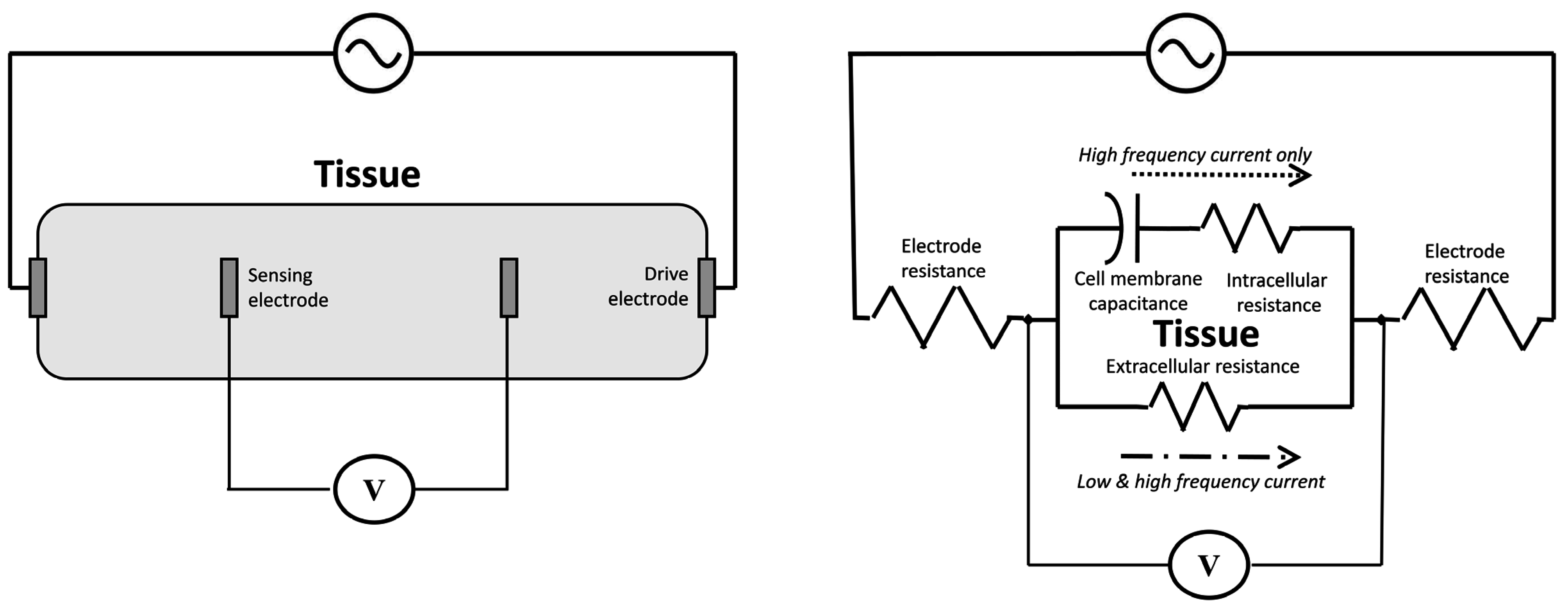
In commercial applications, fabric-based HR monitoring technology has been available in relatively inexpensive sports bras and chest belts from companies like Lululemon (www.lululemon.com) and Adidas (www.adidas.com), with a focus on personal feedback during training activities. E-textile ECG sensors may be able to fill the gap between commercial technologies (which often only display heart rate and possibly respiration rate to the user), and clinical systems (which are time consuming to apply and generally require an expert for placement of the electrodes and/or analysis of the results). Unfortunately many of the ECG systems mentioned here do away with feedback entirely and the results are intended to be sent directly to a clinician or health care professional for interpretation.
Rather than trying to replicate clinical testing or results, some researchers [18,15] have focused on deriving acceptable quality signals from body worn e-textile sensors for practical monitoring and diagnosis. By producing tools capable of screening and monitoring as opposed to a definitive diagnosis, researchers have been able to reduce the multiplicity of necessary sensors or electrodes, alleviating the burden on the user and perhaps minimizing the need for highly trained personnel for simple screening and monitoring applications.
Although applications in home or ambulatory monitoring generally do not require medical grade signals, textile measurements would be more meaningful upon contextualization against a gold standard. In many cases, textile ECG electrodes have only been assessed qualitatively, primarily via visual inspection of ECG waveforms [15,19,29,32,35].

3.2. Bioimpedance

3.2.1. Phenomenological Background

Bioimpedance is the opposition of current flow through biological tissue. In particular, cell membranes act as high impedance (i.e., capacitance) insulators at DC and low frequency currents. As a result, ions flow exclusively through extracellular space and thus the associated impedance is largely resistive [59]. Generally, the greater the cell concentration, the higher the low frequency impedance. High frequency currents on the other hand pass through cell membranes, and thus the ensuing impedance is a combination of capacitance and resistance, reflecting both intra- and extracellular conductivity [60]. Bioimpedance is thus frequency-dependent. In the simplest model of tissue impedance, ion-rich extracellular and intracellular spaces are represented as separate resistors while cell membranes are depicted as a capacitor, as shown in Figure 3. The human body can be modeled as five cylindrical conductors (2 arms, 2 legs and trunk) [61] each with its own segmental impedance.
Figure 3. Tetrapolar measurement of tissue impedance (left); and equivalent electric circuit model (right).
Electronics 04 00173 g003

3.2.2. Current Practice

The aforementioned biochemical currents in body tissue can be generated by or transformed into electrical currents in metallic conductors via electrodes and an accompanying electrolyte [62]. Given that its DC potential is relatively independent of DC current flow, the silver–silver chloride (Ag/AgCl) electrode is preferred in many clinical applications.
Conventional bioimpedance measurement deploys a tetrapolar arrangement of pre-gelled electrodes, where current is delivered through two drive electrodes and voltage is measured via two sensing electrodes (Figure 3). With this configuration, the impedances of the drive electrodes do not factor into the voltage measurement. The location of drive and sensing electrodes varies with different bioimpedance analysis (BIA) methods. For whole-body impedance, the drive and sensing electrodes are co-located on the hand and foot [61]. For segmental BIA (separately estimating appendicular and trunk bioimpedances), the current drive electrodes are situated on the right hand and foot, while the sensing electrodes are placed over the shoulders, clavicle, left elbow and wrist, and, right ankle and thigh [63]. Localized BIA, which aims to detect changes in soft tissue hydration and cell membrane integrity in a specific area of the body, situates electrodes over the segment of interest [64].
Bioimpedance measurements are typically made with an AC drive current. Single-frequency BIA typically uses a 50 kHz current. Multi-frequency BIA deploys a small set of frequencies while bioelectric spectroscopy sweeps through a wide range of frequencies [60].
The measured impedance values can be used to estimate total body water, intracellular and extracellular fluid volumes, fat free mass, and body cell mass [60]. These estimates can be useful for body composition analysis [65], assessing pulmonary function [66], and monitoring nutritional status of patients undergoing hemodialysis [67,68], among other applications.

3.2.3. Textile Innovations

Textile-based bioimpedance measures have focused on body composition [24,37] and segmental volume estimation [23,44]. Hannikainen et al. [24] proposed a total body water estimate using textile electrodes (material not disclosed), each connected by conductive yarn to a metallic snap fastener. The electrodes were sewn into elastic bands, which were worn around the user’s wrist and ankle. With this tetrapolar configuration, the authors made multifrequency (5 to 50 kHz) impedance recordings during walking, cycling and running in nine participants. Although they did not validate their measurements against a clinical gold standard, the authors qualitatively demonstrated the ability to track changes in total body water levels during exercise conditions.
In a more rigorous quantitative study, Marquez et al. [37] estimated 8 body composition parameters in 3 male subjects using “textrodes” made from silver-plated, stretchable and highly conductive knitted fabric (78% polyamide, 22% elastomer; plated with 99% conductive silver) layered with a thin foam and an outer knit fabric to improve surface contact. Like Hannikainen et al., Marquez et al. also invoked a hand-foot tetrapolar arrangement, but swept through a broader range of frequencies (3 to 500 kHz) and validated their textrode measures against those of traditional Ag/AgCl electrodes. Encouragingly, Marquez et al. found that body composition estimates obtained from textrode-measured impedances were not statistically different than those derived via traditional metal electrodes.
As in body composition analysis, there have also been advances in impedance-based segmental volume estimation using textile electrodes. Vuorela et al. [44] created 225 mm2 square-shaped textile electrodes from conductive silver yarn. Deploying a tetrapolar configuration, they qualitatively compared bioimpedance-based estimates of thoracic volume to those obtained with a pneumotachograph, during quiet sitting, arm swinging and light exercise. The authors qualitatively ascertained a maximum difference of 50 mL in tidal volume estimates between the two methods. Turning to the lower extremity, Goy et al. [23] systematically investigated the utility of five different conductive textile materials (formed into 2 cm × 21 cm surface electrodes) for impedance-based estimation of blood volume changes, both in vitro (agar-agar synthetic skin) and in 5 healthy volunteers. Compared to standard Ag/AgCl electrodes, Goy et al. obtained the strongest signal via two-way stretch knit fabric made of silver coated nylon/elastomer thread (76% nylon, 24% elastic fibre; 100% Ag coating) and deemed textile electrodes as suitable for lower extremity venous occlusion plethysomography.

3.2.4. Merits and Limitations

Although textile electrodes have historically had signal quality issues due to poor skin contact resulting in an increased sensitivity to disturbances [44], recent advances in electrode characteristics and garment design have improved signal quality [37]. Similarly, wearability and data quality have advanced from contradictory bioimpedance measures using a jogging suit with bulky attachments for electronic components [24] to a streamlined system of textile electrode-bearing wrist and ankle straps, which yield impedance-based body composition parameters comparable in value to those derived via standard methods [37].
While it is possible to measure changes in total body water, fat-free body mass, or respiration rate with compelling accuracies (compared to clinical gold standards) using bio-impedance analysis, it is not possible to obtain absolute values. Further research is needed in particular with clinical populations (e.g., hemodialysis patients) to verify results obtained from healthy volunteers.
Finally, some lingering technical challenges seem apparent. Hannikainen et al. did not explain suspect findings for both cycling and walking conditions while Vuorela et al. vaguely alluded to data loss due to instrumentation “malfunction” during longer-term recordings. Goy et al. reported that to avoid errors due to instabilities of the polarization potential, the textile electrodes must rest on skin for 500 s prior to measurement, a constraint which may preclude practical clinical application.

3.3. Movement and Posture

3.3.1. Phenomological Background

Human movement is governed by the hierarchical control of the primary motor, premotor and supplemental motor cortices, basal ganglia, cerebellum, brainstem and spinal pattern generators, and modulated by feedback from the vestibular and sensory systems [69]. Likewise, postural maintenance integrates the control of higher brain centers (cerebellum and basal ganglia), brain stem and spinal cord with feedback from various sensory receptors to stabilize one’s center-of-mass via muscle contractions [70]. The measurement of functional movement and posture can be informative in rehabilitation science and practice. For example, such quantification can reveal changes in movement performance due to development, injury, recovery, therapy or assistive technology. The measurement of movement can also be used to decode communicative intent in non-verbal individuals [71] and to predict adverse events, such as falls or loss of balance [72]. Long-term compliance to therapy, particularly in the community, may benefit from remote monitoring through wearable systems that sense movement. Such portable systems enable therapists to track the frequency and quality of exercises performed outside of a clinic environment.

3.3.2. Current Practice

While expensive multi-camera-based systems that track body markers, such as the VICON system, are the gold standard for quantifying movement and body position (kinematics), their use is generally restricted to a finite volume within an institutional setting [73]. Mobile applications often invoke a combination of accelerometers, gyroscopes and goniometers to achieve the same purpose, but these sensors are unable to detect postural changes.
In similar vein, the kinetic characterization of movement has relied on floor-embedded force plates, which have a high degree of accuracy and precision, but are limited in size and flexibility. In-shoe pressure-sensing systems offer more flexible measurement possibilities but are still costly and unless wireless, may require tethering the footwear to wearable data acquisition units. With fabric-based sensing, there is an opportunity to explore alternative mobile monitoring of movement for motor rehabilitation and assessment.

3.3.3. Textile Innovations

While accelerometers and gyroscopes are appropriate for ambulatory measurement of limb and body movements, they are typically deployed as rigid sensors on the body or sewn into textiles, rather than fully integrated into fabric [74]. Early textile-based movement sensors used various conductive [73] or optical fibers [75,76] to estimate joint angles or limb position, but were not wearable for physical activity.
Recent advancements in textile-based measurement of movement have come primarily from the development of conductive elastomer (CE) sensors. Lorussi et al. [77] present a CE electrogoniometer, realized by applying two thin CE films (1 mm thick) to each side of a flexible and inextensible substrate. Their mathematical model indicated that the angle estimation error within the normal physiological range of bending is significantly reduced by the double layer configuration (<1% at a curvature of approximately 1 rad as opposed to approximately 3% with the single layer configuration). Although the substrate in this case was not fabric, Lorussi et al. [77] demonstrated that small, local deformations of the sensor do not significantly affect the overall angle measurement.
Lorussi et al. [36] subsequently employed the sensors on an extensible substrate—elastic woven cotton “Kinesio” tape capable of up to 40% elongation from its resting length. The final sensor consisted of three insulating layers of Kinesio tape and two CE sensing layers. Sensor readings were quantitatively compared to a traditional goniometer in lab tests as well as in a briefly described in vivo experiment measuring wrist flexion. In lab tests, predicted angles using CE sensors were generally within 1° of the imposed angle, as verified by the standard goniometer. Angles were not significantly different for small perturbations to local curvature.
Adopting a different manufacturing approach, Giorgino et al. [22] screen-printed 19 CE stretch sensors on a garment over the arm, shoulder, and chest areas to classify a specific set of upper limb postures (sagittal flexion, lateral abduction and external rotation of the shoulder) relevant to rehabilitation exercises. The study aimed at evaluating the contribution (measured by information gain [78]) of each sensor in order to inform optimization of sensor number and placement in later prototypes. Results predictably showed that sensors over locations with the most stretch during specific movements were most informative and that the information gain of each sensor depended on the exercise being performed. Nonetheless, information gain was consistent across the three subjects tested. Tormene et al. [43] later applied 13 similar sensors to the back of a fabric corset in superior-inferior and lateral directions to measure trunk motion in a single subject. While the sensors were not able to measure extension or lateral bending due to sensor buckling, for trunk flexion the fabric sensors correlated well with an accelerometer and magnetometer combination MEMS inertial sensor (Spearman’s rank correlation coefficient = 0.88). Using a small training set of one example from each class (30, 60, 90 degrees of flexion), a strong level of agreement (Cohen’s Kappa 0.85 ± 0.12) was reported between fabric-based and inertial sensor-derived estimates of flexion angles.
Yamada et al. [45] critiqued conductive elastomers and composites used to date as strain gauges, citing the slow response and creep as major concerns. Instead, the authors introduced a sensor composed of stretchable carbon nanotubes with desirable material properties including a fast response (14 ms), low overshoot (3%) and a fast recovery (5 s). The sensors maintained these properties when stretched to 150% for up to 10,000 cycles. While the sensors were insensitive to other types of deformation (twisting and applied force), the properties did change with temperature and the presence of certain gases, requiring that the sensors be sealed before use.
Shu et al. [41] employed similar strain sensors (knit fabric with a silicon coating doped with conductive carbon black) in an in-shoe plantar pressure measurement system. The strain sensors were sandwiched between two textured pieces of rubber, allowing an applied pressure from the foot to be measured as a change in length of the sensor. Six individual sensors were arranged in a shoe insole to measure pressures at the heel and metatarsals. In the 8 subjects tested, the sensors performed well in static measures of center of pressure (COP), with less then 5% measurement difference from the ANSI force platform used for comparison. With increased comfort, sensitivity, and fatigue resistance, this in-shoe measurement system may prove to be extremely useful for ambulatory and dynamic measurement outside of a clinic environment where expensive integrated force plates are impractical. Unfortunately, although the authors described a dynamic walking test, no quantitative results were reported. On a related front, Preece et al. [38] created an instrumented sock to identify heel strike, heel lift, and toe off during the gait cycle. The sock contained knitted strain sensors formed using the characteristics of the knitting pattern, which allowed loops of conductive yarn to come into or out of contact with each other as the fabric stretched, decreasing or increasing the effective length of the conductor and proportionally, the resistance. Although the sock solution bears clinical appeal, it was not designed for the measurement of absolute static or dynamic pressures. Nonetheless, it faired well at its intended purpose of event detection during the gait cycle.
Other fabric-based strain sensors have been implemented using conductive thread and specific machine stitches including cover stitch [79] and overlocked sensors [21]. In these sensors, the nature of the stitch allows loops of thread to come into or out of contact with each other as the fabric stretches, decreasing or increasing the effective length of the conductor and proportionally, the resistance. Using an overlock stitch, sensors produced a repeatable and approximately linear response up to 29% elongation. While these types of sensors are promising in their ease of application and repeatable response, the sensor properties, including baseline drift and recovery times, appear to be dominated by substrate characteristics.
Moving away from strain sensors, Lee et al. [30] investigated a system that measures joint motion by using bioimpedance to measure the volume changes in the muscle surrounding a joint, which can be associated with joint movement. Standard systems use disposable electrodes, but fabric-based electrodes have the advantage of being able to cover a larger surface area (1 cm × 25 cm in this case) and to be used for longer periods of time. The electrode in this case was polyester with Ni-Cu-Ni applied via electroless plating. The authors aimed to determine the optimum placement of fabric electrodes for discerning various joint angles, but did not compare their results to those with standard disposable Ag/AgCl electrodes. While the authors recommend the sensor pair most distal to the knee joint, their analysis is based on the ranking of each pair according to bioimpedance changes and signal-to-noise ratio (SNR), separately for hip and ankle movement. The three rank values are then added together to determine the best pairing. This type of analysis ignores the absolute values of the bioimpedance and SNR changes, which might suggest a more robust pairing.
Harms et al. [25] proposed a wrinkle modeling method for estimating the impact of loose-fitting garments on the automatic recognition of 10 shoulder rehabilitation exercise postures via a ‘smart shirt’ outfitted bilaterally with tri-axial accelerometers in its sleeves. The algorithm was shown to be a valuable tool for rapid prototyping, providing a prediction of the performance of textile-integrated, soft silicon-packaged electronics and thereby facilitating the optimization of smart garments before implementation. Di Rienzo et al. [39] developed an approach to quantify garment behavior during trunk motion with the aim of reducing motion artifact during high levels of activity. Their approach relied on a system of cameras and reflective markers (a total of 65 over the front and back of the torso) tracking changes in the relative positions of markers during movement with and without the garment. The final stage involved textile experts interpreting results to propose changes to fabric structure, electrode position, and garment cut.

3.3.4. Merits and Limitations

In the garments described by Giorgino et al. [22] and Tormene et al. [43], the sensing components and much of the wiring were integrated using a conductive elastomer. This produces a comfortable and fitted garment with only the battery and electronics as hard components, but CE sensors tend to exhibit creep, and languid response and recovery times. Sensors that yielded high sensitivity and repeatability, unfortunately have yet to be implemented on a fabric substrate [36,45,77].
In many cases, the effectiveness of posture recognition technology has been evaluated on its ability to correctly identify the posture of the participant, and not necessarily against standardized kinematic measurement tools [22,43,45]. As a result, no full garments have yet been described in the literature with the ability to discern joint angle with a high degree of accuracy. In addition, none of the studies described here test sensors with participants with disabilities; testing has occurred exclusively with healthy volunteers.

3.4. Temperature

3.4.1. Phenomenological Background

While heat generated by metabolic activity is distributed evenly throughout bodily tissues by circulating blood, gradients in temperature exist between core and periphery [80]. One mechanism of homeostatic thermoregulation involves microcirculatory adjustments that control conductive and convective heat loss between the skin and the external environment. Blood flow to the skin surface, where heat exchange takes place, can be adjusted through vasoconstriction and vasodilation of arteriovenous anastomoses (AVAs) [81]. Vasoconstriction of AVAs shunts blood toward, while vasodilation of AVAs directs blood away from, cutaneous vasculature. Therefore, the anatomy of the underlying cutaneous vasculature determines the degree to which heat exchange can take place.
AVAs are innervated by sympathetic nerve fibers, which receive and integrate thermoregulatory signals from the hypothalamus. The direction and magnitude of the sympathetic response is dependent on baseline body temperature, therefore it is possible that a certain stimulus can elicit either a vasodilation or vasoconstriction response [82]. Temperature variations among skin surface locations exist due to variations in circulatory anatomy, the degree of nervous system innervation and proximity to signaling organs [83]. Moreover, different skin surface locations respond more or less markedly to sympathetic stimulation. For instance, studies using infrared thermography of the face have shown that nasal and periorbital regions exhibit the most discriminatory skin temperature changes when sympathetically stimulated [84].
Transient thermoregulatory changes in skin blood flow occur in response to sympathetic stimuli while longer-term thermal homeostasis acts to maintain an individual’s core body temperature within an optimal range. Deviation from this ‘normal’ temperature set point, such as in fever, reveals important diagnostic information [81].

3.4.2. Current Practice

The simplest measurements of temperature can be taken with technologies such as thermometers that equate thermal expansion in liquids and solids with numerical values, and require materials that are highly sensitive to heat changes. Resistance temperature detectors are highly sensitive as they exploit the repeatable, heat-induced rise in electrical resistance of a metal (e.g., copper) to sense temperature. A thermocouple is a rugged alternative that senses temperature as the voltage difference between a junction of two dissimilar metals at the probe tip and a reference junction (the same metals at a known temperature). A thermistor is typically a ceramic or polymer resistor whose resistance varies measurably and repeatedly with temperature. Generally, measurement range is greatest with thermocouples and most restricted with thermistors. A microbolometer consists of an array of uncooled thermal sensitive detectors (e.g., indium gallium arsenide) and are used in contemporary infrared thermal cameras.
In hospitals and clinics, thermometers are commonly used to collect oral, rectal, tympanic and axillary body temperatures. However, more recent research has focused on non-invasive methods of body temperature measurement for application in long-term monitoring. For example, thermal imaging captures the infrared radiation emitted by an individual, and is able to provide a detailed illustration of the body’s heat distribution. Similarly, infrared thermometers detect infrared heat emitted at specific body sites such as the temporal artery [85].
Specific external measurement sites are known to provide accurate estimations of core body temperature. One such site is the tympanum, which receives a blood supply directly from the hypothalamus via the carotid artery [83]. However, representations of temperature distributions at the skin’s surface are valuable as they reflect microcirculatory adjustments made in response to sympathetic stimuli.
For instance, digital temperature fluctuations are especially useful as indications of sympathetic nervous system activity because they have exclusive sympathetic innervation and dense cutaneous vasculature [86]. Transient skin temperature variations can be used to detect an individual’s emotional state; for example, using infrared thermography, facial skin temperature is highly labile and has potential to serve as an access pathway in rehabilitation medicine [84].

3.4.3. Textile Innovations

Interested in determining optimal sensor placement for accurate mobile measurement of human body temperature, Li et al. [33] presented a novel wearable sensor and a finite element model of body temperature. Sensors were located over the left and right chest, at the axilla and upper back. The sensors consisted of short optical fibers embedded in polymer resin strips and based on Bragg grating (FBG) principles, reflecting light only at a specific wavelength while transmitting all others. The FBG wavelength changed linearly with temperature and the fiber optics were easily integrable in textiles. Although it is not clear whether or not a final device was tested on human subjects, the authors developed a weighted model of the five measurement locations to derive estimates of human body temperature, reportedly, to within 0.1 °C.
Shifting the focus away from the sensors themselves, Zysset et al. [48] introduced methods of better integrating electronics with textiles via gold- or copper-coated flexible plastic strips, which can act as a platform for surface mount devices (SMDs). Three methods of integration were investigated, namely, (a) wrapping plastic strips in cotton yarn using a roving frame; (b) weaving plastic strips into textiles; and (c) embroidering strips onto the fabric surface. Mechanical strain in the sensors was tested and embroidery was found to introduce the least strain into the plastic strips during fabrication, which is important to maintain the integrity of the circuits and any solder used to attach SMDs. A resistive temperature detector (RTD) consisting of a 100 nm thick gold layer on a plastic substrate exhibited a linear response between 30 °C and 90 °C as measured in a climate chamber. The response was not adversely affected by the weaving process.

3.4.4. Merits and Limitations

While significant advancements have been made in the integration of temperature sensors for non-invasive monitoring, these novel sensors have not yet been tested on human subjects. The sensors perform well theoretically and under controlled conditions, but without human testing it is impossible to determine whether they will be robust and accurate enough for long-term ambulatory monitoring. That being said, clinical methods of temperature measurement can be relatively non-invasive and there is limited evidence to suggest that long-term daily monitoring of temperature is valuable, making the intended application of a wearable temperature monitor unclear. Although Zysset et al. [48] address the issue of strain during manufacturing, it is unclear how fabric-based temperature sensors will respond to the stress of repeated washing and wearing.

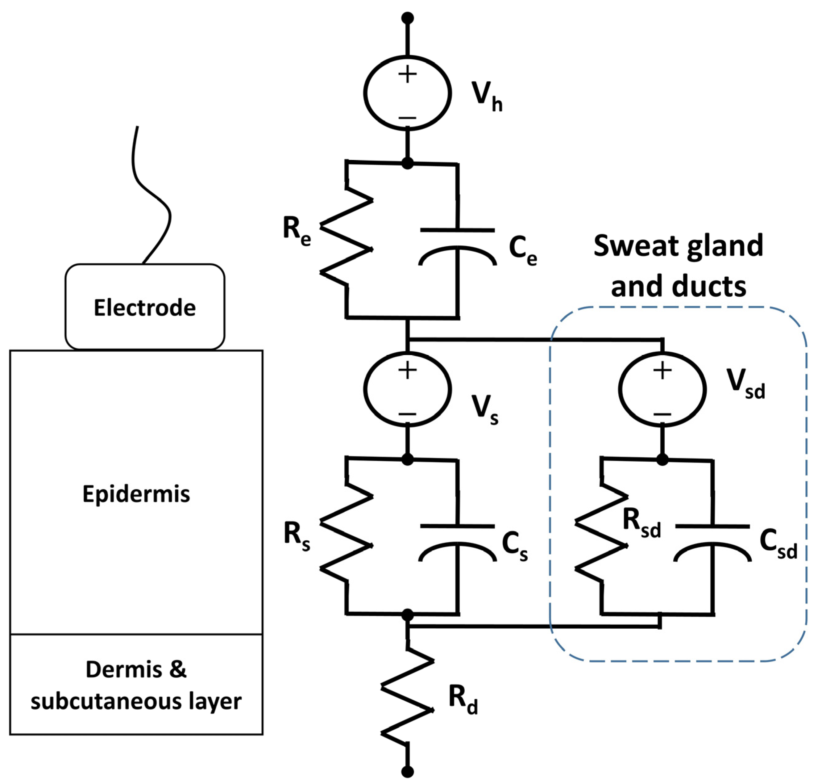
3.5. Electrodermal Activity

3.5.1. Phenomenological Background

Electrodermal activity (EDA) is a measure of the skin’s electrical conductivity. Eccrine sweat glands, found throughout most skin tissue, secrete a dilute electrolytic solution composed of primarily water and sodium chloride. The presence of these conducting solutes on the skin surface gives dermal conductivity, which varies with sweat gland activity. Electrodermal responses (EDRs) are defined as changes in EDA of over 0.05 μS within five seconds and are useful indicators of sweat gland activation [87].
Sweat glands are innervated by the sympathetic branch of the autonomic nervous system and fill with electrolytic fluid upon cholinergic stimulation. The subsequent secretion of these conducting solutes changes the skin’s overall electrical conductance. Thus, the skin’s resistive properties reflect the level and extent of an individual’s sympathetic activity [31]. Homeostatic and thermoregulatory processes produce longer-term fluctuations in EDA, establishing a baseline upon which short-term sympathetically activated EDRs are superimposed.

3.5.2. Current Practice

Similar to bioimpedance, silver-silver chloride (Ag/AgCl) electrodes are commonly used to measure EDA. EDA measurement uses Ohm’s Law (I = V/R) to calculate the electrical resistance at the skin surface. Figure 4 is a model of the electrode-skin interface for electrodermal measurements [88]. Vsd is a potential between the lumen of the sweat duct and the dermis and subcutaneous layers of skin. The parallel Rsd-Csd represents the resistance and capacitance of the wall of the sweat gland and duct. The remaining components of the model are the same as those in Figure 2.
Figure 4. Electrical model of electrode-skin interface for electrodermal measurements.
Electronics 04 00173 g004
The most well-established method of EDA measurement entails the exosomatic detection of current (I) when a constant voltage (V) is applied between two electrodes [89]. Less commonly, a constant current will be applied while voltage is measured to quantify the resistance (R) between the two electrodes. Ideally, electrodes are placed in regions of high sweat gland density where the greatest relative changes in EDA will take place. Typically the palms of the hands or the soles of the feet are the preferred locations [90]. EDRs can be interpreted as measures of physiological and psychological arousal, but they can be indicative of both negative and positive stimuli [89]. In clinical rehabilitation, electrodermal measurements may serve as a complementary access pathway to a motor-based switch [91] or brain-computer interface [92], an indicator of autonomic nervous system response to anxiety in children with autism spectrum disorders [93], and a quantitative measure of motor imagery abilities in individuals with spinal cord injuries [94], among other possibilities.

3.5.3. Textile Innovations

Despite the aforementioned applications in clinical rehabilitation, measurement of electrodermal activity is challenging outside of a controlled environment; typical sensors (wired electrodes on the fingertips) preclude bimanual activity, are susceptible to motion artefact and are sensitive to changes in ambient humidity and temperature. Textile sensing may circumvent some of these limitations and offer long-term, continuous electrodermal measurement.
With the goal of detecting drowsiness, Lee et al. [95] proposed a glove and armband system with textile-based EDA sensors in the fingers and pulse wave sensors in the wrist. Although the authors observed the expected increase in skin impedance and decrease in the phasic components of the electrodermal response in fatigue conditions, the ability of the system to automatically distinguish between states was not demonstrated quantitatively. Conversely, Lanata et al. [28] were able to distinguish between 5 arousal states with high accuracies (>88) using a glove system containing textile sensors at the index and middle fingertips. Sensors were a blended knit, composed of 80% polyester yarn and 20% stainless steel yarn, and were used simultaneously with standard Ag/AgCl electrodes while subjects observed pictures from the international affective picture system (IAPS), designed to elicit emotional arousal. Correlation between textile and standard electrodes was high (Spearman correlation > 0.95) for both tonic and phasic components of skin conductance.
Textile sensing researchers have also explored the ability to reliably measure EDA from locations other than the palm or fingers. Specifically, Fletcher et al. [20] investigated the use of electrodes made of conductive knit fabric (silver-plated nylon blend) to measure EDA from sensors worn on the wrist. Along with traditional sensors to measure photoplethysmogram, temperature, and body motion, the purpose of the low cost, low power “iCalm” device was to identify and measure autonomic arousal in individuals with autism spectrum disorder (ASD), who may be unable to recognize or communicate symptoms of anxiety. The textile-based measurements exhibited a similar phasic response to that observed with the widely used FlexComp system (thoughttechnology.com), although the magnitude of the response was generally attenuated. Textile measurements also exhibited a large baseline drift, which the authors attributed to an insufficient pre-measurement time for skin-electrode interface stabilization. Consequently, they recommended a minimum wait of 15 min before measurement in future studies but did not present the longitudinal data on which this temporal threshold was based.

3.5.4. Merits and Limitations

To date, long-term unobtrusive measurement of EDA has been inconvenient at best, due to the primary sources of reliable response being the palm or fingertips. Recent research has focused on obtaining reliable measurement from other locations (such as the wrists as discussed above [20] and the toes and feet [90]) to improve wearability, but researchers have not yet been able to demonstrate the ability of textile-based devices to gather and analyze EDA signals with sufficient accuracy and repeatability to offer useful feedback to the wearer or remotely to a health professional. Although advances have been made in form factor, cost, and power consumption (e.g., the iCalm device appears smaller, more discreet, and more accurate than the device produced by Lee et al. [95]), quantitative data are needed to establish the clinical relevance of such devices.

3.6. Miscellaneous

Several other papers were found during the review process but fell outside of the above phenomenological categories. Some focused on system [16,47] or garment characteristics [25,40], while one paper examined applications in actuation as opposed to simple monitoring [27] and one explored textile sensing of electroencephalography (EEG) [34].
Angelidis [16] presented a broad overview of a system including electronic textiles and biosensors for vital physical monitoring during physical activity. The conceptual paper gave an overview of an idealized system and identified steps and current barriers to its realization, including signal quality, miniaturization and packaging. Zheng et al. [47] argued that battery life is a determining factor in the usability of wearable technologies and presented a novel battery-scheduling algorithm to extend the operation of multi-battery electronic textile garments.
Schwarz et al. [40] tackled common criticisms of electronic fabrics and yarns by developing yarns with improved elasticity, drape, and mechanical strength. While the authors developed an electrical model to predict the properties of various yarns, an analysis of elasticity and drape was not provided and no recommendations were made about preferred material characteristics.
Specifically targeted at electrical stimulation to alleviate hypertension in older adults, Kim and Cho [27] presented the only paper focused on both monitoring and treatment. A glove-based system was used to sense blood pressure and heart rate in twelve elderly female patients before and after electrical stimulation was delivered through stainless steel thread within the glove. Rings of conductive thread, 20 mm in diameter, were stitched and used to deliver user-controlled electrical stimulation between 1 to 1200 Hz for 15 min. Decreased blood pressure (p < 0.01) was reported following 15 min of glove wear.
Lofhede et al [34] suggest the use of textile electrodes to facilitate long-term recording of EEG in pre-term infants without discomfort or damage to the scalp. The authors investigated two types of textile electrodes, one made from a silver plated elastomer blend, the other knit with nylon, spandex, and conductive silver fibers. When a conductive gel or saline was used as a contact medium, signals from both types of textile electrode were similar to those recorded from standard electrodes at the same locations on the scalp. Although signal quality was deemed to be acceptable in both cases, the five study participants reported no benefits in terms of comfort of textile over standard electrodes. Textile electrodes may prove to be more comfortable in longer term testing, but additional research is necessary to verify the signal quality and diagnostic validity of textile electrodes for EEG.

4. Discussion

In e-textiles research, the ultimate objective is usually full integration, with sensing elements and electronics indistinguishable from everyday clothing. While research and technology has not yet advanced to this point, significant progress has been made toward this goal since the early prototypes of the Wearable Motherboard in 2002 [1]. Primary advances in e-textiles have been in healthcare and monitoring applications. Large multi-lab studies like ProeTEX [96] and MyHeart [97] have led the field in integration. Signal quality has generally been the primary consideration while user comfort and sensor integration have been secondary. Despite significant advances in recent years, circuit and battery integration are still areas in need of additional improvement. As stated in an early review of e-textile technologies by Post et al. [63], integration can only be achieved by departing from the notion of “packaging electronics in hard plastic boxes, however small” [4]. Current research seems to be addressing these limitations with commercially available flexible circuit boards (www.ictboards.com) and batteries (www.solicore.com), although these technologies have been slow to make their way into smart garments, presumably because of the relatively high cost of these flexible components, at the time of writing.
There seem to be other significant gaps between technology advancement and implementation. In general, in e-textile research, there are many studies describing monitoring applications, but few on actuation [14]. Actuation and user feedback through visual effects, sound and even morphological changes are popular in fashion and DIY applications [2]. Although there is much interest in personal information tracking and biofeedback in our current culture, as evidenced by the growing array of wearable technology, this interest has yet to diffuse into the clinical realm. While there is a growing demand for more personal control of health information [16], there are an increasing number of studies where information is sent directly to a coach, clinician, healthcare professional, or simply into a database of other biosignals, without any immediate and direct feedback to the user of the technology. This approach seems to contradict the concepts of patient self-management [98].
In a 2001 review of wearable technology, concerns were raised about “little published data from field trials” [99], a concern that persists to the present day. While research with healthy subjects produces valuable initial data, research with clinical populations is a necessary next step as a number of real-life challenges cannot be simulated or even anticipated in the lab. Ethical concerns of fully integrated textile measurements as well as issues of participant compliance must be addressed before these technologies can move from research into practice. Privacy concerns have also been raised around the storage and transfer of moment-by-moment personal health information, with increasing security measures being implemented [13].
To accelerate future developments, the standardization of interconnections between e-textile elements may be necessary. Upon future mass production, e-textile waste products may also require novel recycling and reuse strategies. While we seek ubiquitous computing, many people remain wary of increasingly invisible technology allowing them to be unknowingly monitored or recorded. However, with mainstream products like Google Glass and Microsoft’s HoloLens, these attitudes may be on the decline.
E-textiles are a comparatively young field with much room for further research and application. However, before these technologies are integrated into clinical rehabilitation practice, there are outstanding technological and ethical hurdles that must be addressed.

5. Recommendations

In light of the above review, we close with the following recommendations for future research.
1
Future research ought to validate textile sensing of a particular physiological or biomechanical phenomenon against its corresponding clinical gold standard. This was a common gap across the reviewed papers.
2
In the spirit of patient self-management, future work may entertain the potential of incorporating textile actuation and hence, immediate sensory feedback to the wearer.
3
While sensing elements have experienced a boon in fabric integration, connecting circuitry and power sources still lag behind in textile assimilation. Truly imperceptible e-textiles will require full system integration.
4
With continued improvements in e-textile signal quality and system integration, it would behoove researchers to initiate testing with clinical populations in ambulatory settings. In particular, future research should consider issues of signal stability over time and across user activities as well as textile sensor integrity with wear and wash.

6. Limitations

Although interesting research has no doubt been documented in conference papers and private or commercial documents, due to the sheer amount of articles published in recent years, we limited the scope of this review to peer-reviewed journal articles. As a consequence, some relevant advancements may have been omitted.

Conclusions

E-textiles constitute an exciting and growing field. Advancements in recent years have brought us closer to the vision of ubiquitous computing, particularly in the fields of remote and ambulatory monitoring in clinical rehabilitation. Many physiological and biomechanical phenomenons can already be measured via textile sensors. Nonetheless, there remain many opportunities for future research such as the integration of power and information processing components, systematic validation against clinical gold standards and sensory feedback via textile actuation.

Conflicts of Interest

The authors declare no conflict of interest.

Author Contributions

A.F. conceived of and conducted the review and critical literature appraisal; T.C. contributed to the writing and editing of various sections; M.S. assisted with the writing of the phenomenological backgrounds of various sections.

References

  1. Park, S.; Mackenzie, K.; Jayaraman, S. The Wearable Motherboard: A Framework for Personalized Mobile Information Processing (PMIP). In Proceedings of the 39th Design Automation Conference, New Orleans, LA, USA, 10–14 June 2002.
  2. Buechley, L.; Eisenberg, M. Fabric PCBs, electronic sequins, and socket buttons: Techniques for e-textile craft. Pers. Ubiquitous Comput. 2009, 13, 133–150. [Google Scholar] [CrossRef]
  3. Meoli, D.; May-Plumlee, T. Interactive electronic textile development: A review of technologies. J. Textile Appar. Technol. Manag. 2002, 2, 1–12. [Google Scholar]
  4. Post, E.R.; Orth, M.; Russo, R.R.; Gershenfeld, N. E-broidery: Design and fabrication of textile-based computing. IBM Syst. J. 2000, 39, 840–860. [Google Scholar] [CrossRef]
  5. Cottet, D.; Grzyb, J.; Kirstein, T.; Tröster, G. Electrical characterization of textile transmission lines. IEEE Trans. Adv. Packag. 2003, 26, 182–190. [Google Scholar] [CrossRef]
  6. Maggie, O. Defining Flexibility and Sewability in Conductive Yarns. In Proceedings of the Materials Research Society (MRS) Symposium, San Francisco, CA, USA, 25–29 April 2011; Volume 736, pp. 37–48.
  7. Bonderover, E.; Wagner, S. A woven inverter circuit for e-textile applications. IEEE Electr. Device Lett. 2004, 25, 295–297. [Google Scholar] [CrossRef]
  8. Lee, J.B.; Subramanian, V. Organic Transistors on Fiber: A First Step towards Electronic Textiles. In Proceedings of the Technical Digest—International Electron Devices Meeting, Washington, WA, USA, 8–10 December 2003.
  9. Lee, J.B.; Subramanian, V. Weave patterned organic transistors on fiber for e-textiles. IEEE Trans. Electr. Devices 2005, 52, 269–275. [Google Scholar] [CrossRef]
  10. Edmison, J.; Jones, M.; Nakad, Z.; Martin, T. Using piezoelectric materials for wearable electronic textiles. In Proceedings of the Sixth International Symposium on Wearable Computers (ISWC), Seattle, WD, USA, 7–10 October 2002; pp. 41–48.
  11. Pacelli, M.; Loriga, G.; Taccini, N.; Paradiso, R.M.D. 3rd IEEE EMBS International Summer School on, B. Sensing fabrics for monitoring physiological and biomechanical variables: E-textile solutions. In Proceedings of the 3rd IEEE/EMBS International Summer School on Medical Devices and Biosensors, Cambridge, MA, USA, 4–6 September 2006.
  12. Pantelopoulos, A.; Bourbakis, N.G. A survey on wearable sensor-based systems for health monitoring and prognosis. IEEE Trans. Syst. Man Cybern. Part C Appl. Rev. 2010, 40, 1–12. [Google Scholar] [CrossRef]
  13. Chan, M.; Esteve, D.; Fourniols, J.Y.; Escriba, C.; Campo, E. Smart wearable systems: Current status and future challenges. Artif. Intell. Med. 2012, 56, 137–156. [Google Scholar] [CrossRef] [PubMed]
  14. Bonato, P. Advances in wearable technology and applications in physical medicine and rehabilitation. J. NeuroEng. Rehabil. 2005, 2. [Google Scholar] [CrossRef] [Green Version]
  15. Adnane, M.; Jiang, Z.; Choi, S.; Jang, H. Detecting specific health-related events using an integrated sensor system for vital sign monitoring. Sensors 2009, 9, 6897–6912. [Google Scholar] [CrossRef] [PubMed]
  16. Angelidis, P.A. Personalised physical exercise regime for chronic patients through a wearable ICT platform. Int. J. Electron. Healthc. 2010, 5, 355–370. [Google Scholar] [CrossRef] [PubMed]
  17. Baek, H.J.; Chung, G.S.; Kim, K.K.; Park, K.S. A smart health monitoring chair for nonintrusive measurement of biological signals. IEEE Trans. Inf. Technol. Biomed. 2012, 16, 150–158. [Google Scholar] [CrossRef] [PubMed]
  18. Bianchi, A.M.; Mendez, M.O.; Cerutti, S. Processing of signals recorded through smart devices: Sleep-quality assessment. IEEE Trans. Inf. Technol. Biomed. 2010, 14, 741–747. [Google Scholar] [CrossRef] [PubMed]
  19. Cho, H.S.; Koo, S.M.; Lee, J.; Cho, H.; Kang, D.H.; Song, H.Y.; Lee, J.W.; Lee, K.H.; Lee, Y.J. Heart monitoring garments using textile electrodes for healthcare applications. J. Med. Syst. 2011, 35, 189–201. [Google Scholar] [CrossRef] [PubMed]
  20. Fletcher, R.R.; Dobson, K.; Goodwin, M.S.; Eydgahi, H.; Wilder-Smith, O.; Fernholz, D.; Kuboyama, Y.; Hedman, E.B.; Poh, M.Z.A.; Picard, R.W.A. ICalm: Wearable sensor and network architecture for wirelessly communicating and logging autonomic activity. IEEE Trans Inf. Technol Biomed. 2010, 14, 215–223. [Google Scholar] [CrossRef] [PubMed]
  21. Gioberto, G.; Dunne, L.E. Overlock-stitched stretch sensors: Characterization and effect of fabric property. J. Textile Appar. Technol. Manag. 2013, 8, 1–14. [Google Scholar]
  22. Giorgino, T.; Tormene, P.; Lorussi, F.; de Rossi, D.; Quaglini, S. Sensor evaluation for wearable strain gauges in neurological rehabilitation. IEEE Trans. Neural Syst. Rehabil. Eng. 2009, 17, 409–415. [Google Scholar] [CrossRef] [PubMed]
  23. Goy, C.B.; Dominguez, J.M.; Gómez López, M.A.; Madrid, R.E.; Herrera, M.C. Electrical characterization of conductive textile materials and its evaluation as electrodes for venous occlusion plethysmography. J. Med. Eng. Technol. 2013, 37, 359–367. [Google Scholar] [CrossRef] [PubMed]
  24. Hannikainen, J.; Vuorela, T.; Vanhala, J. Physiological measurements in smart clothing: A case study of total body water estimation with bioimpedance. Trans. Inst. Meas. Control 2007, 29, 337–354. [Google Scholar] [CrossRef]
  25. Harms, H.; Amft, O.; Troester, G. Estimating posture-recognition performance in sensing garments using geometric wrinkle modeling. IEEE Trans. Inf. Technol. Biomed. 2010, 14, 1436–1445. [Google Scholar] [CrossRef] [PubMed]
  26. Hong, S.; Yang, Y.; Kim, S.; Shin, S.; Lee, I.; Jang, Y.; Kim, K.; Yi, H. Performance study of the wearable one-lead wireless electrocardiographic monitoring system. Telemed. J. E-Health 2009, 15, 166–175. [Google Scholar] [CrossRef] [PubMed]
  27. Kim, R.H.; Cho, G. Effectiveness of the smart healthcare glove system for elderly persons with hypertension. Hum. Factors Ergon. Manuf. 2013, 23, 198–212. [Google Scholar] [CrossRef]
  28. Lanata, A.; Gaetano, V.; Scilingo, E. A novel EDA glove based on textile-integrated electronics for affective computing. Med. Biol. Eng. Comput. 2012, 50, 1163–1172. [Google Scholar] [CrossRef] [PubMed]
  29. Lee, S.; Ahn, Y.; Prabu, A.; Kim, K. Piezoelectric Polymer and Piezocapacitive Nanoweb Based Sensors for Monitoring Vital Signals and Energy Expenditure in Smart Textiles. J. Fiber Bioeng. Inf. 2013, 6, 369–381. [Google Scholar]
  30. Lee, B.W.; Lee, C.; Kim, J.; Lee, M. Optimum conductive fabric sensor sites for evaluating the status of knee joint movements using bio-impedance. Biomed. Eng. Online 2011, 10, 48. [Google Scholar] [CrossRef] [PubMed]
  31. Fowles, D.; Coles, M.; Donchin, E.; Porges, S. The eccrine system and electrodermal activity. In Psychophysiology; Guildford Press: New York, NY, USA, 1985; pp. 51–96. [Google Scholar]
  32. Lee, Y.D.; Chung, W.Y. Wireless sensor network based wearable smart shirt for ubiquitous health and activity monitoring. Sens. Actuators B Chem. 2009, 140, 390–395. [Google Scholar] [CrossRef]
  33. Li, H.; Yang, H.; Li, E.; Liu, Z.; Wei, K. Wearable sensors in intelligent clothing for measuring human body temperature based on optical fiber Bragg grating. Opt. Express 2012, 20, 11740–11752. [Google Scholar] [CrossRef] [PubMed]
  34. Lofhede, J.; Seoane, F.; Thordstein, M. Textile electrodes for EEG recording—A pilot study. Sensors 2012, 12, 16907–16919. [Google Scholar] [CrossRef] [PubMed]
  35. Lopez, G.; Custodio, V.; Ignacio Moreno, J. LOBIN: E-textile and wireless-sensor-network-based platform for healthcare monitoring in future hospital environments. IEEE Trans. Inf. Technol. Biomed. 2010, 14, 1446–1458. [Google Scholar] [CrossRef] [PubMed]
  36. Lorussi, F.; Galatolo, S.; Bartalesi, R. Modeling and characterization of extensible wearable textile-based electrogoniometers. IEEE Sens. J. 2013, 13, 217–228. [Google Scholar] [CrossRef]
  37. Marquez, J.C.; Seoane, F.; Lindecrantz, K. Textrode functional straps for bioimpedance measurements -experimental results for body composition analysis. Eur. J. Clin. Nutr. 2013, 67 (Suppl. 1), S22–S27. [Google Scholar] [CrossRef]
  38. Preece, S.J.; Kenney, L.P.; Major, M.J.; M.J., T.; Lay, E.; Fernandes, B.T. Automatic identification of gait events using an instrumented sock. J. NeuroEng. Rehabil. 2011, 8, 32. [Google Scholar]
  39. Di Rienzo, M.; Rizzo, F.; Meriggi, P.; Castiglioni, P.; Mazzoleni, P.; Parati, G.; Bordoni, B.; Brambilla, G.; Ferratini, M. MagIC system. IEEE Eng. Med. Biol. Mag. 2009, 28, 35–40. [Google Scholar] [CrossRef] [PubMed]
  40. Schwarz, A.; Cuny, L.; Hertleer, C.; Ghekiere, F.; Kazani, I.; de Clercq, G.; de Mey, G.; van Langenhove, L.A. Electrical circuit model of elastic and conductive yarns produced by hollow spindle spinning. Mater. Technol. 2011, 26, 121–127. [Google Scholar] [CrossRef]
  41. Shu, L.; Hua, T.; Wang, Y.; Li, Q.; Feng, D.D.; Tao, X. In-shoe plantar pressure measurement and analysis system based on fabric pressure sensing array. IEEE Trans. Inf. Technol. Biomed. 2010, 14, 767–775. [Google Scholar] [CrossRef] [PubMed]
  42. Song, H.Y.; Lee, J.H.; Kang, D.; Cho, H.; Cho, H.S.; Lee, J.W.; Lee, Y.J. Textile electrodes of jacquard woven fabrics for biosignal measurement. J. Textile Inst. 2010, 101, 758–770. [Google Scholar] [CrossRef]
  43. Tormene, P.; Bartolo, M.; De Nunzio, A.M.; Fecchio, F.; Quaglini, S.; Tassorelli, C.; Sandrini, G. Estimation of human trunk movements by wearable strain sensors and improvement of sensor’s placement on intelligent biomedical clothes. Biomed. Eng. Online 2012, 11, 95. [Google Scholar] [CrossRef] [PubMed]
  44. Vuorela, T.; Seppa, V.P.; Vanhala, J.; Hyttinen, J. Design and implementation of a portable long-term physiological signal recorder. IEEE Trans. Inf. Technol. Biomed. 2010, 14, 718–725. [Google Scholar] [CrossRef] [PubMed]
  45. Yamada, T.; Hayamizu, Y.; Yamamoto, Y.; Yomogida, Y.; Izadi-Najafabadi, A.; Futaba, D.N.; Hata, K. A stretchable carbon nanotube strain sensor for human-motion detection. Nat. Nanotechnol. 2011, 6, 296–301. [Google Scholar] [CrossRef] [PubMed]
  46. Zhang, Z.B.; Shen, Y.H.; Wang, W.D.; Wang, B.Q.; Zheng, J.W. Design and implementation of sensing shirt for ambulatory cardiopulmonary monitoring. J. Med. Biol. Eng. 2011, 31, 207–216. [Google Scholar] [CrossRef]
  47. Zheng, N.; Wu, Z.; Lin, M.; Yang, L.T. Enhancing battery efficiency for pervasive health-monitoring systems based on electronic textiles. IEEE Trans. Inf. Technol. Biomed. 2010, 14, 350–359. [Google Scholar] [CrossRef] [PubMed]
  48. Zysset, C.; Kinkeldei, T.; Münzenrieder, N.; Petti, L.; Salvatore, G.; Tröster, G. Combining electronics on flexible plastic strips with textiles. Textile Res. J. 2013, 83, 1130–1142. [Google Scholar]
  49. Mirvis, D.; Goldberger, A. Electrocardiography. In Braunwald’s Heart Disease, 10th ed.; Saunders Elsevier: Philadelphia, PA, USA, 2015; pp. 114–154. [Google Scholar]
  50. Camm, A.; Malik, M.; Bigger, J.; Breithardt, G.; Cerutti, S.; Cohen, R.; Coumel, P.; Fallen, E.; Kennedy, H.; Kleiger, R.; et al. Heart rate variability: standards of measurement, physiological interpretation and clinical use. Task Force of the European Society of Cardiology and the North American Society of Pacing and Electrophysiology. Circulation 1996, 93, 1043–1065. [Google Scholar]
  51. Neumann, M. Biopotential electrodes. In Clinical Engineering; David, Y., von Maltzahn, W., Neuman, M., Bronzino, J., Eds.; CRC Press: Boca Raton, FL, USA, 2003; pp. 10:1–10:12. [Google Scholar]
  52. Webster, J. Medical Instrumentation: Application and Design, 4th ed.; Wiley: Hoboken, NJ, USA, 2010. [Google Scholar]
  53. Su, L.; Borov, S.; Zrenner, B. 12-lead Holter electrocardiography: Review of the literature and clinical application update. Herzschrittmachertherapie Elektrophysiol. 2013, 24, 92–96. [Google Scholar] [CrossRef]
  54. Acharya, U.R.; Joseph, K.P.; Kannathal, N.; Lim, C.M.; Suri, J.S. Heart rate variability: A review. Med. Biol. Eng. Comput. 2006, 44, 1031–1051. [Google Scholar] [CrossRef] [PubMed]
  55. Appelhans, B.; Luecken, L. Heart rate variability as an index of regulated emotional responding. Rev. General Psychol. 2006, 10, 229–240. [Google Scholar] [CrossRef]
  56. Coyle, M. Ambulatory cardiopulmonary data capture. In Proceedings of the 2nd Annual International IEEE-EMBS Special Topic Conference on Microtechnologies in Medicine & Biology, Madison, WI, USA, 2–4 May 2002; pp. 297–300.
  57. Coosemans, J.; Hermans, B.; Puers, R. Integrating wireless ECG monitoring in textiles. Sens. Actuators A 2006, 130–131, 48–53. [Google Scholar]
  58. Lim, Y.; Kim, K.; Park, K. ECG recording on a bed during sleep without direct skin-contact. IEEE Trans. Biomed. Eng. 2007, 54, 718–725. [Google Scholar] [CrossRef] [PubMed]
  59. Grimnes, S.; Martinsen, O. Bioimpedance Biolectricity Basics, 2nd ed.; Academic: London, UK, 2008. [Google Scholar]
  60. Kyle, U.; Bosaeus, I.; Lorenzo, A.D.; Deurenberg, P.; Elia, M.; Gomez, J.; Heitmann, B.; Kent-Smith, L.; Melchoir, J.; Pirlich, M.; et al. Bioelectrical impedance analysis—Part I: Review of principles and methods. Clin. Nutr. 2004, 23, 1226–1243. [Google Scholar]
  61. Kushner, R. Bioelectrical impedance analysis: A review of principles and applications. J. Am. Coll. Nutr. 1992, 11, 199–209. [Google Scholar] [PubMed]
  62. Togawa, T.; Tamura, T.; Oberg, P.A. Biomedical Transducers and Instruments; CRC Press: Boca Raton, FL, USA, 1997. [Google Scholar]
  63. Cornish, B.; Jacobs, A.; Thomas, B.; Ward, L. Optimizing electrode sites for segmental bioimpedance measurements. Physiol. Meas. 1999, 20, 241–250. [Google Scholar] [CrossRef] [PubMed]
  64. Lescolarde, L.; Yanguas, J.; Lukaski, H.; Alomar, X.; Rosell-Ferrer, J.; Rodas, G. Localized bioimpedance to assess muscle injury. Physiol. Meas. 2013, 34, 237–245. [Google Scholar] [CrossRef] [PubMed]
  65. Kushner, R.F.; Schoeller, D.A. Estimation of total body water by bioelectrical impedance analysis. Am. J. Clin. Nutr. 1986, 44, 417–424. [Google Scholar] [PubMed]
  66. Agarwal, R.; Yadav, R.; Anand, S.; Suri, J.C.; Girija, J. Electrical impedance plethysmography technique in estimating pulmonary function status. J. Med. Eng. Technol. 2007, 31, 1–9. [Google Scholar] [CrossRef] [PubMed]
  67. Azar, R.; al-Moubarak, I.; Barsumau, J.; Smessaert, C.; Vairon, M.X. Assessment and follow-up of nutritional status in hemodialysis patients. Néphrol. Thér. 2009, 5, S317–S322. [Google Scholar] [CrossRef] [PubMed]
  68. Medrano, G.; Eitner, F.; Floege, J.; Leonhardt, S. A novel bioimpedance technique to monitor fluid volume state during hemodialysis treatment. ASAIO J. 2010, 56, 215–220. [Google Scholar] [CrossRef] [PubMed]
  69. Sjedic, E.; Fu, Y.; Pak, A.; Fairley, J.; Chau, T. The effects of rhythmic sensory cues on the temporal dynamics of human gait. PLoS One 2012, 7, e43104. [Google Scholar] [CrossRef] [PubMed] [Green Version]
  70. Masani, K.; Vette, A.; Popovic, M. Controlling balance during quiet standing: proportional and derivative controller generates preceding motor command to body sway position observed in experiments. Gait Posture 2006, 23, 164–172. [Google Scholar] [CrossRef] [PubMed]
  71. Lesperance, A.; Blain, S.; Chau, T. An integrated approach to detecting communicative intent amid hyperkinetic movements in children. Augment. Altern. Commun. 2011, 27, 150–162. [Google Scholar] [CrossRef] [PubMed]
  72. Tong, L.; Song, Q.; Ge, Y.; Liu, M. HMM-based human fall detection and prediction method using tri-axial accelerometer. IEEE Sens. J. 2013, 13, 1848–1856. [Google Scholar]
  73. Gibbs, P.T.; Asada, H.H. Wearable conductive fiber sensors for multi-axis human joint angle measurements. J. NeuroEng. Rehabil. 2005, 2, 18. [Google Scholar] [CrossRef] [PubMed] [Green Version]
  74. Edmison, J.; Jones, M.; Lockhart, T.; Martin, T. An e-textile system for motion analysis. Stud. Health Technol. Inform. 2004, 108, 292–301. [Google Scholar] [PubMed]
  75. Dunne, L.E.; Walsh, P.; Hermann, S.; Smyth, B.; Caulfield, B. Wearable monitoring of seated spinal posture. IEEE Trans. Biomed. Circuits Syst. 2008, 2, 97–105. [Google Scholar] [CrossRef] [PubMed]
  76. Kuang, K.; Cantwell, W.J. An evaluation of a novel plastic optical fibre sensor for axial strain and bend measurements. Meas. Sci. Technol. 2002, 13, 1523. [Google Scholar] [CrossRef]
  77. Lorussi, F.; Galatolo, S.; de Rossi, D.E. Textile-based electrogoniometers for wearable posture and gesture capture systems. IEEE Sens. J. 2009, 9, 1014–1024. [Google Scholar] [CrossRef]
  78. Hall, M.A.; Holmes, G. Benchmarking attribute selection techniques for discrete class data mining. IEEE Trans. Knowl. Data Eng. 2003, 15, 1437–1447. [Google Scholar] [CrossRef]
  79. Gioberto, G.; Dunne, L. Theory and characterization of a top-thread coverstitched stretch sensor. In Proceedings of the IEEE International Conference on Systems, Man and Cybernetics, Seoul, Korea, 14–17 October 2012; pp. 3275–3280.
  80. Sund-Levander, M.; Forsberg, C.; Wahren, L. Normal oral, rectal, tympanic and axillary body temperature in adult men and women: A systematic literature review. Scand. J. Caring Sci. 2002, 16, 122–128. [Google Scholar] [CrossRef] [PubMed]
  81. Charkoudian, N. Skin blood flow in adult human thermoregulation: How it works, when it does not and why. Mayo Clin. Proc. 2003, 78, 603–612. [Google Scholar] [CrossRef] [PubMed]
  82. Oberle, J.; Elam, M.; Karlsson, T.; Wallin, B. Temperature-dependent interaction between vasoconstrictor and vasodilator mechanisms in human skin. Acta Physiol. Scand. 1988, 132, 459–469. [Google Scholar] [CrossRef] [PubMed]
  83. Sund-Levander, M.; Grodzinsky, E. Assessment of body temperature measurement options. Br. J. Nurs. 2013, 22, 944–950. [Google Scholar] [CrossRef]
  84. Nhan, B.; Chau, T. Classifying affective states using thermal infrared imaging of the human face. IEEE Trans. Biomed. Eng. 2010, 57, 979–987. [Google Scholar] [CrossRef] [PubMed]
  85. Allegaert, K.; Casteels, K.; van Gorp, I.; Bogaert, G. Tympanic, infrared skin and temporal artery scan thermometers compared with rectal measurement in children: A real-life assessment. Curr. Ther. Res. 2014, 76, 34–38. [Google Scholar] [CrossRef] [PubMed]
  86. Keefe, F.; Gardner, E. Learned control of skin temperature: Effects of short- and long-term biofeedback training. Behav. Ther. 1979, 10, 202–210. [Google Scholar] [CrossRef]
  87. Blain, S.; Mihailidis, A.; Chau, T. Assessing the potential of electrodermal activity as an alternative access pathway. Med. Eng. Phys. 2008, 30, 498–505. [Google Scholar] [CrossRef] [PubMed]
  88. Boccaletti, C.; Castrica, F.; Fabbri, G.; Santello, M. A non-invasive biopotential electrode for the correct detection of bioelectric currents. In Proceedings of the Sixth IASTED International Conference on Biomedical Engineering, Innsbruck, Austria, 13–15 February 2008; pp. 353–358.
  89. Blain, S.; Chau, T.; Mihailidis, A. Peripheral autonomic signals as access pathways for individuals with severe disabilities: A literature appraisal. Open Rehabil. J. 2008, 1, 27–37. [Google Scholar] [CrossRef]
  90. Kushki, A.; Fairley, J.; Merja, S.; King, G.; Chau, T. Comparison of blood volume pulse and skin conductance responses to mental and affective stimuli at different anatomical sites. Physiol. Meas. 2011, 32, 1529–1539. [Google Scholar] [CrossRef] [PubMed]
  91. Leung, B.; Chau, T. Autonomic responses to correct outcomes and interaction errors during single-switch scanning among children with severe spastic quadriplegic cerebral palsy. J. NeuroEng. Rehabil. 2014, 11, 13. [Google Scholar] [CrossRef] [PubMed]
  92. Falk, T.; Guirgis, M.; Power, S.; Chau, T. Taking NIRS-BCI outside the lab: towards achieving robustness against environmental noise. IEEE Trans. Neural Syst. Rehabil. Eng. 2010, 19, 136–146. [Google Scholar] [CrossRef] [PubMed]
  93. Kushki, A.; Drumm, E.; Mobarak, M.P.; Tanel, N.; Dupuis, A.; Chau, T.; Anagnostou, E. Investigating the autonomic nervous system response to anxiety in children with autism spectrum disorders. PLoS One 2013, 8, e59730. [Google Scholar] [CrossRef] [PubMed]
  94. Grangeon, M.; Charvier, K.; Guillot, A.; Rode, G.; Collet, C. Using sympathetic skin responses in individuals with spinal cord injury as a quantitative evaluation of motor imagery abilities. Phys. Ther. 2012, 92, 831–840. [Google Scholar] [CrossRef] [PubMed]
  95. Lee, Y.; Lee, B.; Lee, M. Wearable sensor glove based on conducting fabric using electrodermal activity and pulse-wave sensors for e-health application. Telemed. J. E-Health 2010, 16, 209–217. [Google Scholar] [CrossRef] [PubMed]
  96. Curone, D.; Secco, E.L.; Tognetti, A.; Loriga, G.; Dudnik, G.; Risatti, M.; Whyte, R.; Bonfiglio, A.; Magenes, G. Smart garments for emergency operators: The ProeTEX project. IEEE Trans. Inf. Technol. Biomed. 2010, 14, 694–701. [Google Scholar] [CrossRef] [PubMed]
  97. Habetha, J. The MyHeart project-fighting cardiovascular diseases by prevention and early diagnosis. In Proceedings of the IEEE EMBS Annual International Conference, New York, NY, USA, 30 August–3 September 2006; pp. 6746–6749.
  98. Shumaker, S.; Ockene, J.; Riekert, K. The Handbook of Health Behavior Change, 3rd ed.; Springer Publishing Company: New York, NY, USA, 2009. [Google Scholar]
  99. Baber, C. Wearable computers: A human factors review. Int. J. Hum. Comput. Interact. 2001, 13, 123–145. [Google Scholar] [CrossRef]

