Communication

Nanosized Particles of Synthetic Silicon Dioxide Delay the Regeneration of Gastric Ulcers Created by N-Methyl-N′-Nitro-N-Nitrosoguanidine and Induce Hyper-Trophic Gastritis-like Symptoms

Department of Pharmaceutical Sciences, Kobe Gakuin University, 1-1-3 Minato Jima, Chuo-ku, Kobe 650-8586, Japan
*
Author to whom correspondence should be addressed.
Sci. Pharm. 2024, 92(2), 20; https://doi.org/10.3390/scipharm92020020
Submission received: 13 February 2024 / Revised: 28 March 2024 / Accepted: 6 April 2024 / Published: 11 April 2024

Abstract

:
Synthetically produced silicon dioxide used as a food additive exhibits nanoparticle size and shape during the early stages of manufacturing. Even when processed into food products, these nanoparticles are detected. Although processing food ingredients into nanoparticles can improve absorption rates or enhance texture, there are concerns about the specific biological effects of nanoparticles. In this study, three types of silica particles, including nanosized particles, were repetitively administered to the stomach using a gastric tube or exposed to a single injection into the submucosal layer of the stomach. Macroscopic and microscopic examinations did not reveal acute toxicity. However, when silica particles were administered to the stomach during the healing and regeneration process of gastric ulcers (induced by injecting the alkylating agent of N-Methyl-N′-Nitro-N-Nitrosoguanidine into the submucosal layer), silica particles with a diameter of 70 nm (SiNPs-70) delayed regeneration more strongly than microsized silica particles with diameters of 300 nm or 1000 nm (SiMPs-300, -1000). Furthermore, fibrosis for tissue regeneration spread throughout the entire mucosa of the stomach, resulting in hypertrophic gastritis-like symptoms. The frequency of this symptom was over 50% with SiNPs-70, 20% with SiMPs-300, and 0% with SiMPs-1000. Although the silica particles used in this study differ from actual samples found in food, the impact of particle size, particularly the effects unique to nanosize, was identified as toxicity in the stomach healing process.

Graphical Abstract

1. Introduction

Synthetic amorphous silica (SAS) is used as a food additive (E551) to maintain the flowability of powdered foods, prevent solidification in paste foods, and enhance transparency in alcohol. SAS exhibits various forms during the manufacturing process, including fumed silica, hydrated silica, precipitated silica, silica gel, and hydrous silica [1]. Scientific viewpoints on silicon dioxide (E551) used as a food additive, issued by the European Food Safety Authority and the U.S. Food and Drug Administration, indicate that even at the maximum estimated exposure level (50 mg/kg body weight per day), it is much lower than the identified No Observed Adverse Effect Level determined through toxicological studies [2]. SAS is generated as nanosized particles (silica nanoparticles—SiNPs) with a primary particle diameter of 1–100 nm, forming microsized aggregates during the production process [3]. A study measuring the amount of SiNPs in 27 foods containing E551 revealed the presence of nanoparticles exceeding 4% in nine items, with a maximum ratio of 33% [4]. Although these SiNPs differ from nanosized SAS found in food, in vitro experiments have shown that SiNPs of the same composition induce cellular toxicity, stimulate the release of inflammatory cytokines, and can pass through barriers, such as the intestinal mucosal barrier, skin barrier, and air–blood barrier [5,6,7]. These findings raise concerns about nanosize-specific toxicity, particularly regarding the potential impact of SAS on the digestive system, where accumulation over time is observed.
Common diseases that affect the stomach and duodenum, which are responsible for the digestion and absorption of food, include peptic ulcers. The estimated lifetime prevalence in the general population of Western countries is 5–10%, constituting an annual incidence rate of 0.1–0.3% [8]. Although Helicobacter pylori infection is a major cause of peptic ulcers, drugs and alcohol act as inhibitory factors in the treatment process [9,10,11]. SiNPs have been found to inhibit cell migration, suggesting worsening ulcers, and induce inflammatory cytokine production in analyses using intestinal and skin epithelial cells [12,13]. Various animal models have been developed to screen antiulcer drugs and elucidate their mechanisms of action, with stress and physical stimuli (clamping the gastric wall with a clamp from the gastric serosa) serving as models for ulcer formation [14,15,16]. The acetic acid ulcer model, which involves the injection of acetic acid into the gastric wall, has been reproduced in various animal species, including mice, rats, cats, and dogs, as an experimental model for creating chronic ulcers [17].
Therefore, in this study, a mouse model of gastric ulcer formation was developed by injecting the alkylating agent N-Methyl-N′-Nitro-N-Nitrosoguanidine (MNNG) into the gastric wall, mimicking the acetic acid ulcer model. The specific effects of SiNPs on the stomach of healthy mice were analyzed, and the impact on the healing process of ulcers was evaluated using macroscopy and microscopy. These studies have the potential to provide information on the toxicity of nanofoods not obtained from traditional toxicological perspectives while offering new insights into the connection between nutrition and healthcare.

2. Materials and Methods

2.1. Silica Particles

Suspension of amorphous SiNPs with a diameter of 70 nm (Sicastar® 43-00-701) and amorphous silica particles with diameters of 300 and 1000 nm (Sicastar® 43-00-302 and 43-00-103, respectively) were purchased from Micromod (Rostock, Germany). These particles were suspended in saline, sonicated for 5 min, and vortexed for 5 min before use. MNNG (Wako Chemical, Osaka, Japan) was dissolved in dimethyl sulfoxide and further diluted with saline. Previous studies have reported the size and zeta potential of these particles in aqueous solution, and their morphology has been observed using transmission electron microscopy [18].

2.2. Animals

Five-week-old specific pathogen-free male ICR mice were purchased from CLEA Japan, Inc. (Tokyo, Japan) and used for analysis at 6 weeks of age. All animals were housed under specific pathogen-free conditions at a room temperature of 22 °C ± 2 °C and humidity of 50 ± 10% with a regular 12 h light (8:00–20:00)/12 h dark (20:00–8:00) cycle and free access to water and pellet food (CE-2 diet: CLEA Japan Inc.). Animal experiments were conducted following the Guidelines for the Care and Use of Laboratory Animals by the Animal Research Committee of Kobe Gakuin University (protocol code A22-35, 01/04/2022).

2.3. MNNG and Silica Particle Injection into the Submucosa of the Stomach

Medetomidine hydrochloride (Meiji Animal Health Co., Ltd., Kumamoto, Japan), midazolam (Nichi-Iko Pharmaceutical Co., Ltd., Toyama, Japan), and butorphanol (Vetorphale, Meiji Animal Health Co., Ltd., Kumamoto, Japan) were intraperitoneally administered to induce anesthesia. Under anesthesia, laparotomy was performed on ICR mice, and either 300 µg of MNNG or 80 µg of each of the three types of silica particles was injected into the submucosa of the stomach using a 28 G injection needle. The stomach was returned to the abdominal cavity, and the endothelium and epithelium were sutured. Atipamezole (Mepatia, Meiji Animal Health Co., Ltd., Kumamoto, Japan) was administered to facilitate recovery, and the mice were kept on a warming pad at 37 °C until they regained consciousness.

2.4. Oral Gavage Administration of Silica Particles

To prepare the silica suspension, 4000 µg of each of three types of silica particles was added to 1500 µL of saline solution, sonicated for 5 min, and vortexed for 5 min. Each mouse was then orally administered 150 µL (400 µg of each of the three types of silica particles) of the silica suspension using a gastric tube. The same amount was orally administered 24 and 48 h after the initial administration.

2.5. Preparation and Observation of the HE- and Azan-Stained Specimens

Medetomidine hydrochloride, midazolam, and butorphanol were intraperitoneally administered to induce anesthesia. The anesthetized mice were euthanized through cervical dislocation, and a thoracotomy was performed to confirm death by observing the heartbeat cessation for >5 min. Then, the stomach was extracted, fixed in 4% paraformaldehyde, and embedded in paraffin. Tissue sections of approximately 1 μm thickness were prepared, followed by deparaffinization in xylene and staining with hematoxylin or Azan. In the case of hematoxylin staining, additional eosin staining was performed. After dehydration and clarification with xylene, the sections were mounted using ENTELLAN new (MERCK) and visualized using a digital camera connected to an optical microscope. The mucosal epithelium that appeared white and thickened was quantified as the incidence of hypertrophic gastritis-like findings (%).

2.6. Experimental Frequency and Statistical Analysis

Mice were used, with three animals per experimental condition conducted three times, resulting in nine animals. Statistical comparisons were made for the incidence of hypertrophic gastritis-like findings obtained from each experiment. Data were analyzed using analysis of variance and Tukey’s honest significant difference test with Kaleida Graph (HULINKS, Tokyo, Japan).

3. Results and Discussion

3.1. Hypertrophic Gastritis-like Lesions form Due to Exposure to Amorphous Nanosilica during the Healing Process of Gastric Ulcers

In the stomach, food undergoes digestion over several hours through the action of protein-digesting enzymes, such as pepsin, and peristaltic movements. The strong acidic environment of gastric acid sterilizes bacteria. Although particulate matter, such as SAS, follows these processes, SAS remains undegraded primarily in the digestive environment of the stomach and intestines, maintaining its particle shape before being absorbed into the body or excreted [19]. Thus, SiNPs within SAS are anticipated to accumulate in the stomach for several hours, potentially causing harmful and inflammatory effects on the gastric mucosal epithelium. To investigate the impact on the stomach, we employed a classic strategy in nanotoxicology and compared the biological effects of nanosized silica particles (SiNPs-70) with those of microsized silica particles (SiMPs-300 and -1000) of the same material and shape. These particles were administered directly to the stomach three times using a gastric tube at 24 h intervals or a single direct injection into the submucosal layer of the stomach, and the stomach tissues were then extracted 24 h later. The stomach was divided into two portions, extending from the greater curvature to the lesser curvature, and the mucosal surface of the excised samples, including the gastric fundus and vestibule, was observed. Notably, no typical signs of epithelial loss indicating acute inflammation were observed in any group that received silica particle administrations (Figure 1A,B). In chronic gastric inflammation, repeated mucosal ulceration and regeneration occur, with some cells undergoing morphological changes toward “precancerous lesions” in the intestinal mucosal epithelium [20]. Proper treatment of gastric inflammation is crucial for maintaining human health, including the reduction in gastric cancer risk, and standard treatment protocols often involve a combination of drug therapy and dietary interventions. Ethanol is a direct factor in inflammation, and the consumption of fatty meats can delay or inhibit regeneration because of increased gastric acid secretion [21,22]. In the case of SiNPs, delayed healing and worsening of gastrointestinal diseases have been suggested. For example, colitis induced by dextran sulfate sodium worsened in mice repeatedly exposed to the oral administration of 10 nm SiNPs, as indicated by worsening disease activity indices (i.e., stool hardness, extent of bleeding, weight, and appearance) [23]. In this study, we modified the acetic acid injection method, which was established and used as a gastric ulcer model, by incorporating a N-nitroso compound model material (MNNG), which is known as one of the causes of gastric cancer. MNNG alkylates the oxygen atom at position 6 of guanine, inducing G:C to A:T base substitution mutations during DNA replication. In previous studies, mice raised with drinking water containing MNNG developed gastric ulcers [24]. Furthermore, gastric ulcers were formed upon injection of MNNG into the submucosal layer, and these ulcers were nearly regenerated and healed within approximately 2–3 days (Figure 1C). After 24 h of MNNG injection, three types of silica particles were directly administered to the stomach using a gastric tube three times at 24 h intervals. In the SiNPs-70 group, severe atrophic morphology of the entire gastric mucosa was observed (Figure 1C). Although the stomach wall is ordinarily transparent, resembling frosted glass, this transparency was compromised in SiNP-70-treated stomachs. When quantifying this loss of transparency as hypertrophic gastrocnemius-like lesions, a significantly increased occurrence rate was observed with smaller particle sizes (Figure 1D). These results demonstrate the impact of silica particles, unobserved in a healthy stomach, during the healing process of ulcers. Moreover, the dramatic increase in the frequency of nanosized particles raises concerns about the excessive intake of nano-shaped foods.

3.2. Amorphous Nanosilica Not Only Delays the Healing of Gastric ulcers but also Induces Fibrosis on the Surface of the Gastric Mucosa

In wound healing and tissue regeneration, the extracellular matrix, which serves as a scaffold for cells, is produced by fibroblasts, leading to the formation of granulation tissue [25,26,27]. Ulcers created using acetic acid resulted in easily discernible granulation tissue of significant size, particularly in the vestibular region where ulcers were formed. Histopathological analysis of hematoxylin and eosin (HE)-stained specimens from ulcers induced by MNNG in this region demonstrated progressing healing and regeneration, notably with increased regeneration and proliferation of chief cells observed in the control group (Figure 2A and Supplementary Figure S1). In contrast, the SiNPs-70 group showed limited advancement in the healing and regeneration of MNNG-induced ulcers, and tissue damage extended to the submucosal layer (Figure 2A). HE-stained specimens from the gastric fundus revealed disrupted fold-like structures in the glandular epithelium and gastric fundic gland layers in the SiNPs-70 group, along with elevations in the mucosal muscular layer and hypertrophy of chief cells (Figure 2A). Azan-stained specimens from the same sites showed minimal collagen fibers stained in dark blue in the gastric fundus and vestibular regions of the control group. However, the SiNPs-70 group exhibited an accumulation of collagen fibers in the glandular epithelium of the gastric fundus and vestibular regions (Figure 2B). Furthermore, in the control group, the ulcerated area in the vestibular region exhibited numerous orange-stained red blood cells, indicating enhanced angiogenesis associated with regeneration. Conversely, these observations lacked in the ulcerated area and its vicinity in the SiNPs-70 group, with minimal inflammatory responses involving neutrophils and macrophages (Figure 2B). Fibrosis at the ulcer formation site is a part of the healing and regeneration process, and factors involved in fibrosis are being studied for their potential use as biomarkers and molecular targets for pre- and post-regeneration diagnostics [28].
The extracellular matrix that constitutes fibrosis undergoes degradation by matrix metalloproteinases as healing and regeneration progress, facilitating the reformation of healthy tissue. Chronic fibrosis is recognized as atrophic gastritis preceding precancerous changes, and studies using mesenchymal stem cells to treat ulcerative colitis have emphasized the improvement of fibrosis as an indicator of effectiveness [29,30,31]. Although this research raises concerns that amorphous nanosilica in stomach acid suppressants induce fibrosis in the ulcer healing process, definitive assertions regarding the equivalent risks posed by foods containing stomach acid suppressants or those generated through nanotechnology are not made. This study is positioned as a hazard investigation demonstrating the potential risks associated with SiNPs, strongly suggesting the need for further understanding and research in nanotoxicology. Simultaneously, it emphasizes the need to carefully consider hypersensitive reactions to foods containing stomach acid suppressants.

4. Conclusions

As individuals age, maintaining a healthy and optimal state of the body becomes challenging; various stomach disorders are expected to occur. Evaluating the stomach under pathological conditions is a valid strategy to examine daily dietary intake safety. We investigated the effects of amorphous silica particles on the stomach and the effect of particle size. We used 6-week-old mice believed to have a healthy stomach and mice of the same age with gastric ulcers induced by chemical substances (Figure 3). While silica particles with a diameter of 70 nm were found to be harmful and inflammatory in both in vitro and in vivo experiments, no effects were observed in healthy stomachs (Figure 1A,B). In contrast, in stomachs with gastric ulcers, smaller diameters of silica particles were associated with delayed healing and excessive fibrosis (Figure 1C,D and Figure 2). These results suggest that nanosilica should not be considered a definitive threat to human health but a potential hazard in certain conditions. In the medical fields utilizing nanotechnology, nanosilica particles are employed as vectors for drug delivery systems, and technologies are being developed to use these particles for the specific and efficient targeting of gastric cancer and inflammatory bowel diseases, as well as for loading various drugs [32,33]. Nanotechnology is also applied to diagnose and detect gastrointestinal diseases, with techniques developed to manipulate various materials (such as carbon, synthetic polymers, polysaccharides, metals, and quantum dots) into nanosized structures [34,35]. Applying these technologies in the food industry is a promising platform for developing safe and functional nanofoods; therefore, safety research results are essential for realizing this potential.

Supplementary Materials

The following supporting information can be downloaded at https://www.mdpi.com/article/10.3390/scipharm92020020/s1, Figure S1: Temporal variations in granulation tissue formation in acetic acid-induced ulcer models: Insights from three distinct experimental schedules.

Author Contributions

Conceptualization, Y.K. and A.O.; methodology, A.I. and A.O.; software, A.I. and A.O.; validation, A.I., Y.K. and A.O.; formal analysis, A.I. and A.O.; investigation, A.I., Y.K. and A.O.; resources, A.I. and A.O.; data curation, A.I., Y.K. and A.O.; writing—original draft preparation, A.I. and A.O.; writing—review and editing, Y.K.; visualization, A.I. and A.O.; supervision, Y.K. and A.O.; project administration, Y.K. and A.O.; funding acquisition, A.O. All authors have read and agreed to the published version of the manuscript.

Funding

This research was funded by JSPS KAKENHI, grant number JP 20K06848. Additional support was provided by the Japan Food Chemical Research Foundation and the Kobe Gakuin Health Science Grant.

Institutional Review Board Statement

Not applicable.

Informed Consent Statement

Not applicable.

Data Availability Statement

The data presented in this study are available upon request from the corresponding author.

Conflicts of Interest

The authors declare no conflicts of interest.

References

  1. Fruijtier-Pölloth, C. The toxicological mode of action and the safety of synthetic amorphous silica—A nanostructured material. Toxicology 2012, 294, 61–79. [Google Scholar] [CrossRef]
  2. EFSA Panel on Food Additives and Nutrient Sources added to Food (ANS); Younes, M.; Aggett, P.; Aguilar, F.; Crebelli, R.; Dusemund, B.; Filipič, M.; Frutos, M.J.; Galtier, P.; Gott, D.; et al. Re-evaluation of silicon dioxide (E 551) as a food additive. EFSA J. 2018, 16, e05088. [Google Scholar] [CrossRef]
  3. Fruijtier-Pölloth, C. The safety of nanostructured synthetic amorphous silica (SAS) as a food additive (E 551). Arch. Toxicol. 2016, 90, 2885–2916. [Google Scholar] [CrossRef] [PubMed]
  4. Dekkers, S.; Krystek, P.; Peters, R.J.B.; Lankveld, D.P.K.; Bokkers, B.G.H.; van Hoeven-Arentzen, P.H.; Bouwmeester, H.; Oomen, A.G. Presence and risks of nanosilica in food products. Nanotoxicology 2011, 5, 393–405. [Google Scholar] [CrossRef] [PubMed]
  5. Murugadoss, S.; Lison, D.; Godderis, L.; Van Den Brule, S.; Mast, J.; Brassinne, F.; Sebaihi, N.; Hoet, P.H. Toxicology of silica nanoparticles an update. Arch. Toxicol. 2017, 91, 2967–3010. [Google Scholar] [CrossRef] [PubMed]
  6. Qi, Y.; Ma, R.; Li, X.; Lv, S.; Liu, X.; Abulikemu, A.; Zhao, X.; Li, Y.; Guo, C.; Sun, Z. Disturbed mitochondrial quality control involved in hepatocytotoxicity induced by silica nanoparticles. Nanoscale 2020, 12, 13034–13045. [Google Scholar] [CrossRef]
  7. De Matteis, V. Exposure to inorganic nanoparticles: Routes of entry, immune response, biodistribution and in vitro/in vivo toxicity evaluation. Toxics 2017, 5, 29. [Google Scholar] [CrossRef] [PubMed]
  8. Sharma, N.; Jha, S. Amorphous nanosilica induced toxicity, inflammation and innate immune responses: A critical review. Toxicology 2020, 441, 152519. [Google Scholar] [CrossRef]
  9. Rosenstock, S.J.; Jørgensen, T. Prevalence and incidence of peptic ulcer disease in a Danish County a prospective cohort study. Gut 1995, 36, 819–824. [Google Scholar] [CrossRef] [PubMed]
  10. Gupta, A.; Shetty, S.; Mutalik, S.; Nandakumar, K.; Mathew, E.M.; Jha, A.; Mishra, B.; Rajpurohit, S.; Ravi, G.; Saha, M.; et al. Treatment of H. pylori infection and gastric ulcer: Need for novel Pharmaceutical formulation. Heliyon 2023, 9, e20406. [Google Scholar] [CrossRef] [PubMed]
  11. Dall, M.; Schaffalitzky de Muckadell, O.B.; Lassen, A.T.; Hallas, J. There is an association between selective serotonin reuptake inhibitor use and uncomplicated peptic ulcers: A populationbased case-control study. Aliment. Pharmacol. Ther. 2010, 32, 1383–1391. [Google Scholar] [CrossRef]
  12. Nguyen, T.N.M.; Sha, S.; Chen, L.-J.; Holleczek, B.; Brenner, H.; Schöttker, B. Strongly increased risk of gastric and duodenal ulcers among new users of low-dose aspirin: Results from two large cohorts with new-user design. Aliment. Pharmacol. Ther. 2022, 56, 251–262. [Google Scholar] [CrossRef]
  13. Zhang, Y.; Hu, L.; Yu, D.; Gao, C. Influence of silica particle internalization on adhesion and migration of human dermal fibroblasts. Biomaterials 2010, 31, 8465–8474. [Google Scholar] [CrossRef]
  14. Cornu, R.; Chrétien, C.; Pellequer, Y.; Martin, H.; Béduneau, A. Small silica nanoparticles transiently modulate the intestinal permeability by actin cytoskeleton disruption in both Caco-2 and Caco-2HT29-MTX models. Arch. Toxicol. 2020, 94, 1191–1202. [Google Scholar] [CrossRef] [PubMed]
  15. Guth, P.H.; Mendick, R. The effect of chronic restraint stress on gastric ulceration in the rat. Gastroenterology 1964, 46, 285–286. [Google Scholar] [CrossRef] [PubMed]
  16. Takagi, K.; Okabe, S.; Saziki, R. A new method for the production of chronic gastric ulcer in rats and the effect of several drugs on its healing. Jpn. J. Pharmacol. 1969, 19, 418–426. [Google Scholar] [CrossRef]
  17. Okabe, S.; Amagase, K. An overview of acetic acid ulcer models—The history and state of the art of peptic ulcer research. Biological and Pharmaceutical Bulletin. Biol. Pharm. Bull. 2005, 28, 1321–1341. [Google Scholar] [CrossRef]
  18. Onodera, A.; Yayama, K.; Tanaka, A.; Morosawa, H.; Furuta, T.; Takeda, N.; Kakiguchi, K.; Yonemura, S.; Yanagihara, I.; Tsutsumi, Y.; et al. Amorphous nanosilica particles evoke vascular relaxation through PI3K/Akt/eNOS signaling. Fundam. Clin. Pharmacol. 2016, 5, 419–428. [Google Scholar] [CrossRef]
  19. Fuentes, C.; Ruiz-Rico, M.; Fuentes, A.; Ruiz, M.J.; Barat, J.M. Degradation of silica particles functionalised with essential oil components under simulated physiological conditions. J. Hazard. Mater. 2020, 399, 123120. [Google Scholar] [CrossRef] [PubMed]
  20. Fujii, Y.; Yoshihashi, K.; Suzuki, H.; Tsutsumi, S.; Mutoh, H.; Maeda, S.; Yamagata, Y.; Seto, Y.; Aburatani, H.; Hatakeyama, M. CDX1 confers intestinal phenotype on gastric epithelial cells via induction of stemness-associated reprogramming factors SALL4 and KLF5. Proc. Natl Acad. Sci. USA 2012, 109, 20584–20589. [Google Scholar] [CrossRef] [PubMed]
  21. Vomero, N.D.; Colpo, E. Nutritional care in peptic ulcer. Arq. Bras. Cir. Dig. 2014, 27, 298–302. [Google Scholar] [CrossRef] [PubMed]
  22. Suzuki, S. Experimental studies on the presumption of the time after food intake from stomach contents. Forensic Sci. Int. 1987, 35, 83–117. [Google Scholar] [CrossRef] [PubMed]
  23. Ogawa, T.; Okumura, R.; Nagano, K.; Minemura, T.; Izumi, M.; Motooka, D.; Nakamura, S.; Iida, T.; Maeda, Y.; Kumanogoh, A.; et al. Oral intake of silica nanoparticles exacerbates intestinal inflammation. Biochem. Biophys. Res. Commun. 2021, 534, 540–546. [Google Scholar] [CrossRef] [PubMed]
  24. Onodera, A.; Kawai, Y.; Kashimura, A.; Ogita, F.; Tsutsumi, Y.; Itoh, N. Suppression of alkylating agent induced cell transformation and gastric ulceration by low-dose alkylating agent pretreatment. Biochem. Biophys. Res. Commun. 2013, 435, 714–719. [Google Scholar] [CrossRef] [PubMed]
  25. Shahin, M.; Konturek, J.W.; Pohle, T.; Schuppan, D.; Herbst, H.; Domschke, W. Remodeling of extracellular matrix in gastric ulceration. Microsc. Res. Tech. 2001, 53, 396–408. [Google Scholar] [CrossRef]
  26. Yamaguchi, H.; Yoshida, N.; Takanashi, M.; Ito, Y.; Fukami, K.; Yanagihara, K.; Yashiro, M.; Sakai, R. Stromal fibroblasts mediate extracellular matrix remodeling and invasion of scirrhous gastric carcinoma cells. PLoS ONE 2014, 9. [Google Scholar] [CrossRef] [PubMed]
  27. Rieder, F.; Brenmoehl, J.; Leeb, S.; Schölmerich, J.; Rogler, G. Wound healing and fibrosis in intestinal disease. Gut 2007, 56, 130–139. [Google Scholar] [CrossRef] [PubMed]
  28. Hermenean, A.; Oatis, D.; Herman, H.; Ciceu, A.; D’Amico, G.; Trotta, M.C. Galectin 1-A key player between tissue repair and fibrosis. Int. J. Mol. Sci. 2022, 23, 5548. [Google Scholar] [CrossRef] [PubMed]
  29. Fox, J.G.; Wang, T.C. Inflammation, atrophy, and gastric cancer. J. Clin. Investig. 2007, 117, 60–69. [Google Scholar] [CrossRef]
  30. Genta, R.M. Gastric atrophy and atrophic gastritis—Nebulous concepts in search of a definition. Aliment. Pharmacol. Ther. 1998, 1 (Suppl. S1), 17–23. [Google Scholar] [CrossRef]
  31. Gómez-Ferrer, M.; Amaro-Prellezo, E.; Dorronsoro, A.; Sánchez-Sánchez, R.; Vicente, Á.; Cosín-Roger, J.; Barrachina, M.D.; Baquero, M.C.; Valencia, J.; Sepúlveda, P. HIF-overexpression and pro-inflammatory priming in human mesenchymal stromal cells improves the healing properties of extracellular vesicles in experimental Crohn’s disease. Int. J. Mol. Sci. 2021, 22, 11269. [Google Scholar] [CrossRef] [PubMed]
  32. Hani, U.; Osmani, R.A.M.; Yasmin, S.; Gowda, B.H.J.; Ather, H.; Ansari, M.Y.; Siddiqua, A.; Ghazwani, M.; Fatease, A.A.; Alamri, A.H.; et al. Novel Drug Delivery Systems as an Emerging Platform for Stomach Cancer Therapy. Pharmaceutics 2022, 14, 1576. [Google Scholar] [CrossRef]
  33. Laroui, H.; Sitaraman, S.V.; Merlin, D. Chapter six—Gastrointestinal Delivery of Anti-inflammatory Nanoparticles. Methods Enzymol. 2012, 509, 101–125. [Google Scholar] [CrossRef] [PubMed]
  34. Laroui, H.; Wilson, D.S.; Dalmasso, G.; Salaita, K.; Murthy, N.; Sitaraman, S.V.; Merlin, D. Nanomedicine in GI. Am. J. Physiol. Gastrointest. Liver Physiol. 2011, 300, G371–G383. [Google Scholar] [CrossRef]
  35. Kanaoujiya, R.; Porwal, D.; Srivastava, S. Applications of nanomaterials for gastrointestinal tumors: A review. Front. Med. Technol. 2022, 4, 997123. [Google Scholar] [CrossRef] [PubMed]
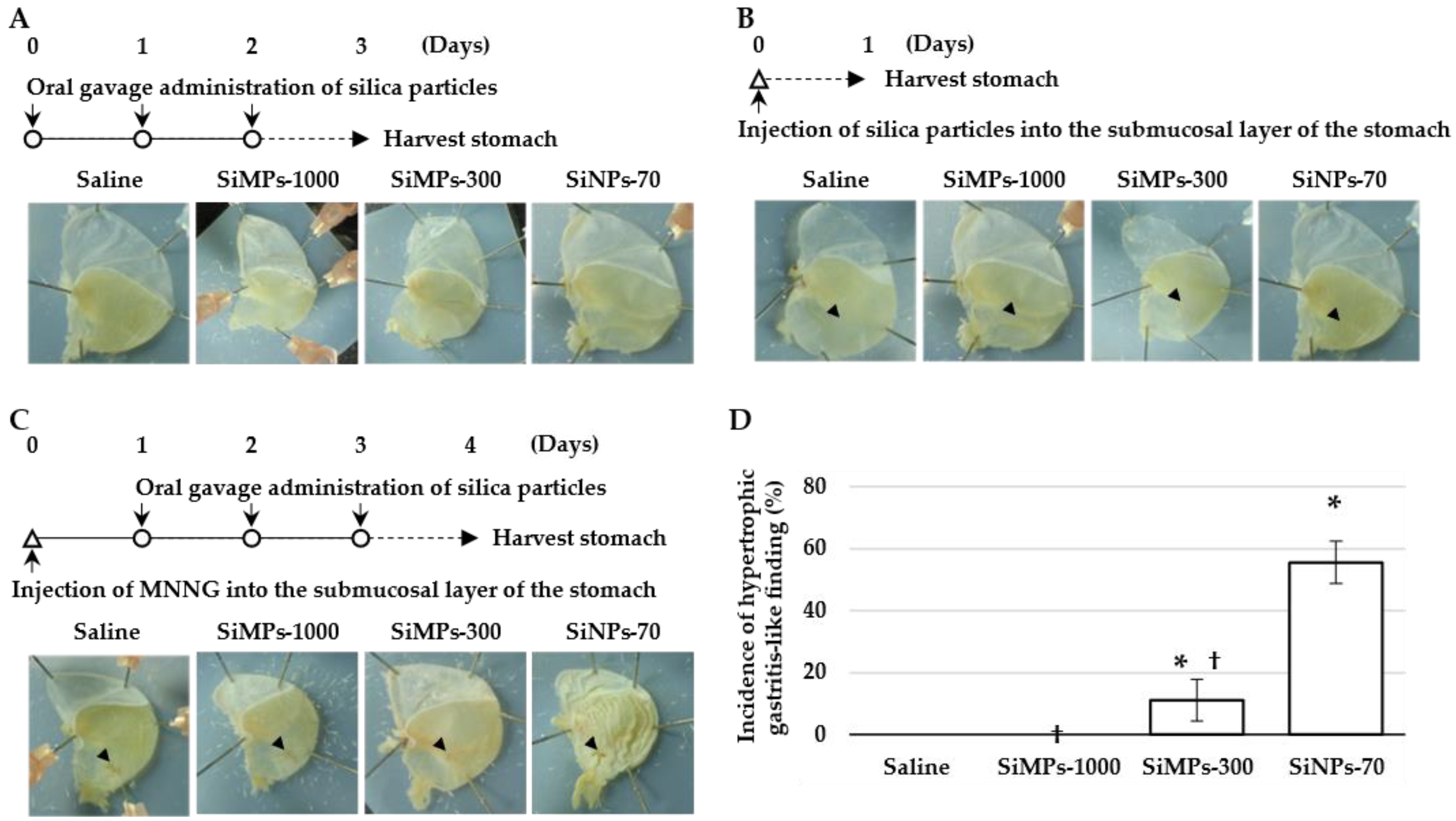
Figure 1. Scheme of silica particles, MNNG exposure, and anatomy of the murine stomach. Three types of silica particles were exposed through oral gavage or injection into the submucosal layer of the stomach in healthy or MNNG-pre-injected mice. (A) Silica particles or vehicle (saline) was administered to healthy mice via oral gavage three times every 24 h. (B) Silica particles or saline was injected into the submucosal layer of the stomach to normal mice once. The arrow indicates the injection site of silica particles. (C) Silica particles or vehicle (saline) was administered via oral gavage to MNNG-pre-injected mice three times every 24 h. For each experiment, 24 h after the last administration of silica particles, the stomach was harvested and then fixed with 4% paraformaldehyde. An incision was made to observe the gastric mucosa, and a digital photograph was then taken. The arrow indicates the injection site of MNNG. (D) The stomachs that lost transparency due to hypertrophy or atrophy as hypertrophic gastrocnemius-like lesions and quantified the frequency of this symptom. * p < 0.0001 vs. saline, † p < 0.0001 vs. SiNPs-70.
Scipharm 92 00020 g001
Figure 2. Histological analysis of the gastric mucosa. Paraffin-embedded blocks were prepared by excising a part of the stomach of saline- and SiNP-70-administered mice shown in Figure 1C. Paraffin-embedded sections were cut out to be observable from the serosa to the mucosal epidermis. (A) HE-stained samples were prepared from the anterior stomach and the MNNG injection site of the glandular stomach. (B) Azan-stained samples were prepared from the anterior stomach and around the MNNG injection site of the glandular stomach. Scalebar = 100 μm.
Scipharm 92 00020 g002
Figure 3. Schematic diagram of the issues and results of this study.
Scipharm 92 00020 g003
Disclaimer/Publisher’s Note: The statements, opinions and data contained in all publications are solely those of the individual author(s) and contributor(s) and not of MDPI and/or the editor(s). MDPI and/or the editor(s) disclaim responsibility for any injury to people or property resulting from any ideas, methods, instructions or products referred to in the content.