Review

Polyphenols, Autophagy and Neurodegenerative Diseases: A Review

by
Vichitra Chandrasekaran
1,2,
Tousif Ahmed Hediyal
1,2,
Nikhilesh Anand
3,
Pavan Heggadadevanakote Kendaganna
2,
Vasavi Rakesh Gorantla
4,
Arehally M. Mahalakshmi
1,2,
Ruchika Kaul Ghanekar
5,
Jian Yang
6,
Meena Kishore Sakharkar
6,* and
Saravana Babu Chidambaram
1,2,*
1
Department of Pharmacology, JSS College of Pharmacy, JSS Academy of Higher Education and Research, Mysuru 570015, India
2
Center for Experimental Pharmacology and Toxicology, Central Animal Facility, JSS Academy of Higher Education and Research, Mysuru 570015, India
3
Department of Pharmacology, College of Medicine, American University of Antigua, Saint John’s P.O. Box W-1451, Antigua and Barbuda
4
Department of Anatomical Science, St. George’s University, West Indies FZ818, Grenada
5
Symbiosis Centre for Research and Innovation (SCRI), Symbiosis International (Deemed University), Pune 412115, India
6
Drug Discovery and Development Research Group, College of Pharmacy and Nutrition, University of Saskatchewan, Saskatoon, SK S7N 5E5, Canada
*
Authors to whom correspondence should be addressed.
Biomolecules 2023, 13(8), 1196; https://doi.org/10.3390/biom13081196
Submission received: 5 July 2023 / Revised: 26 July 2023 / Accepted: 28 July 2023 / Published: 31 July 2023

Abstract

:
Polyphenols are secondary metabolites from plant origin and are shown to possess a wide range of therapeutic benefits. They are also reported as regulators of autophagy, inflammation and neurodegeneration. The autophagy pathway is vital in degrading outdated organelles, proteins and other cellular wastes. The dysregulation of autophagy causes proteinopathies, mitochondrial dysfunction and neuroinflammation thereby contributing to neurodegeneration. Evidence reveals that polyphenols improve autophagy by clearing misfolded proteins in the neurons, suppress neuroinflammation and oxidative stress and also protect from neurodegeneration. This review is an attempt to summarize the mechanism of action of polyphenols in modulating autophagy and their involvement in pathways such as mTOR, AMPK, SIRT-1 and ERK. It is evident that polyphenols cause an increase in the levels of autophagic proteins such as beclin-1, microtubule-associated protein light chain (LC3 I and II), sirtuin 1 (SIRT1), etc. Although it is apparent that polyphenols regulate autophagy, the exact interaction of polyphenols with autophagy markers is not known. These data require further research and will be beneficial in supporting polyphenol supplementation as a potential alternative treatment for regulating autophagy in neurodegenerative diseases.

1. Introduction

Polyphenols are one of the major class of secondary metabolites found in edible and nonedible plants [1]. Polyphenolic compounds are classified based on the number of phenolic rings and functional groups such as phenolic acids (hydroxybenzoic acid and hydroxycinnamic acid), flavonoids (flavanols, flavonols, flavones, flavanones, isoflavones, proanthocyanidins), stilbenes and lignans [2]. The functional classification of polyphenols helps in understanding their bioavailability and health benefits. The main class of plant phenolic compounds are phenolic acids, also called phenolcarboxylic acids or polyphenols. They are divided into two types, hydroxybenzoic acid and hydroxycinnamic acid. Hydroxybenzoic acid has a complex structure and is present in low concentration in red fruits, black radish, onions, tea, etc. Hydroxycinnamic acid (HA) is present in higher concentration in the outer part of ripened fruits which includes caffeic acid, ferulic acid, p-coumaric and sinapic acid [3]. Phenolic acids are potent antioxidants and have several benefits such as being neuroprotective [4], antidiabetic [5], anticancer, anti-inflammatory, etc. [6]. Flavonoids are ubiquitously occurring polyphenolic compounds which are widely distributed in fruits and vegetables and have health benefits such as being antimicrobial, antioxidant, neuroprotective and having an antimalarial activity [7]. The classification of flavonoids is based on their benzopyran structure, the oxidation state of the carbon atom in the heterocycle ring. Flavonoids include flavanols, isoflavones, flavanones, flavonols, proanthocyanidins and flavones [8].
Anthocyanins are a major part of polyphenolic compounds. They are colored pigments that are dissolved in the vacuolar sap of epidermal tissues of plants. They are present in fruits, vegetables and flowers, for example, in grapes, apples, plums, berries, purple cabbages, red hibiscus, red rose, red pineapple sage, red clover, pink blossom, etc. [9]. Stilbene compounds are a larger group of natural-defense polyphenols in plants. They are found in low quantity in the human diet and the most well-known stilbene is resveratrol, which is present in red grapes, blueberries and wine and is reported to have antiaging and antiangiogenic activities [10]. Plant lignans are low-molecular-weight polyphenols that are present in rhizomes, leaves and roots and structurally possess two phenyl propene units. They are found richly in linseed as secoisolariciresinol and in minor quantities as matairesinol. These lignans are metabolized by the intestinal microflora to enterodiol and enterolactone. Lignans possess several pharmacological properties such as antitumor, antiviral and antiasthmatic properties [11].
Polyphenols are studied extensively for their health benefits to humans and animals. Polyphenols possess potent antioxidant and anti-inflammatory properties that help in the treatment of cancers, metabolic and neurodegenerative diseases [12,13], including age-related degenerative diseases [14,15]. Polyphenols provide neuroprotection and neurogenesis by regulating different signaling pathways, mitochondrial functions and antiapoptotic proteins. Polyphenols are capable of scavenging reactive oxygen species (ROS) directly and can thereby help regulate the mitochondrial apoptotic cascade. They exert an anti-inflammatory action by suppressing the expression of proinflammatory transcription factor NF-kB, which leads to apoptosis [16]. Polyphenols have been explored as nutraceuticals and for their usage in treating neurodegenerative diseases, and the majority of them have the ability to penetrate the blood–brain barrier [17]. Moreover, polyphenol supplementation reduces the incidence of psychiatric disorders as they possess an antioxidant activity [18].
Polyphenols such as curcumin and resveratrol produce a neuroprotective effect by activating the protein kinase signaling pathway and also via the upregulation of neurotrophic factors such as BDNF and GDNF [19]. Dysregulation in autophagy leads to neurodegenerative diseases, and evidence has revealed that polyphenol treatment ameliorates autophagy [20,21]. In this review, we summarize the absorption, distribution, metabolism and excretion (ADME), toxicity profile and effects of polyphenols on autophagy and proteinopathies in different neurodegenerative diseases. We used major search engines such as PubMed, SCOPUS, MDPI and Google Scholar to collect the scientific literature. To the best of our literature survey, we did not find many reports on the direct interaction of polyphenols with major proteins that regulate autophagy.

2. Absorption, Distribution, Metabolism and Excretion (ADME) of Polyphenols

In general, polyphenols have poor oral bioavailability due to their high metabolism, the interaction with the intestinal microflora and food matrices. Primarily, they are absorbed in the small intestine and metabolized by deconjugation reaction and deglycosylation. After absorption, they become less complex and undergo phase I (oxidation, reduction and hydrolysis) and phase II (conjugation) biotransformation in the enterocytes. This results in the formation of water-soluble conjugated metabolites (glucuronide, methyl and sulfate derivatives) that are released into the systemic circulation [22]. Polyphenols get excreted by two routes, urinary and biliary. More specifically, the biliary elimination of polyphenols occurs when the large and extensively conjugated metabolite is present, whereas in urine, small conjugates such as monosulfates are excreted [23].
The poor bioavailability of polyphenols is overcome by preparing nanoparticles with food macromolecules [24]. Liu et al. prepared and evaluated a curcumin nanodrug encapsulated in gelatin microspheres in a diabetic wound-healing model. These encapsulated microspheres increased the solubility and improved the bioavailability. Ravichandran et al., on the other hand, compared the kinetic profile of curcumin using conventional and nanoforms using an in vivo model. They found that the concentration of nanocurcumin in plasma and organs was increased in the nanoparticle-treated group (Figure 1) [25,26]. Similarly, Kohli et al. developed natural polysaccharide-based nanoparticles using berberine (BNPs) and found that the bioavailability of BNPs was increased in comparison to the normal free form in in vitro (drug release kinetics), ex vivo (gut permeation study) and in vivo models (Wistar rats) (Figure 2) [27]. Furthermore, the oral bioavailability of resveratrol was reported to be enhanced by preparing polymeric nanoparticles (in vitro and in vivo) [28]. Another interesting polyphenol is Epigallocatechin-3-gallate (EGCG). It is chemically unstable and has low bioavailability. Ramesh and Mandal prepared the solid lipid nanoparticles encapsulating EGCG, which improved the chemical stability and bioavailability [29]. In addition, the structural modification of EGCG by an acetylation process was shown to improve the bioavailability through in vitro and in vivo models [30]. Biasutto et al. added a glucosyl group by linking the succinate group to yield 3,4′,5-tri-(α-d-glucose-3-O-succinyl) resveratrol, which increased the pharmacokinetics when compared with conventional resveratrol in rats [31]. These experiments strongly suggest that novel drug delivery methods could potentially be explored further to improve the bioavailability of polyphenols.

3. Toxicity Profile of Polyphenols

Data reveal that some polyphenols exhibit a potential toxicity at high concentrations. For example, a high concentration of catechins was found to induce DNA damage in mice spleen cells [32], and grape extracts at 75 to 300 µg/mL concentrations promoted mitomycin C induced sister chromatid exchange in human peripheral blood lymphocytes [33]. A comparative toxicity study on quercetin, EGCG and cyanidin 3 glycoside revealed that quercetin caused a blood–brain barrier disruption and neurotoxicity [34]. Green tea polyphenols, when administered to mice at high doses, worsen colitis and colorectal cancer and cause kidney and liver dysfunctions [35]. Moreover, at higher doses, several of the polyphenols have been shown to promote mutagenesis or cause genotoxic and carcinogenic effects. This could potentially be attributed to their chemical structure, and the metabolites that are formed upon degradation. Hence, further research is needed to clearly establish the toxicity profile of polyphenols [36].

4. Autophagy and Neurodegenerative Diseases

Autophagy is a protective mechanism, as it eliminates outdated proteins, organelles and toxic substances and also reprocesses degraded products which is used as a source of energy in anabolic pathways [37]. Autophagy differs between eukaryotic cells and mammalian cells. In the case of eukaryotes, the proteins are degraded by two systems: the ubiquitin–proteasome system and macro-autophagy. In mammals, it is of three types: macro-autophagy, chaperone-mediated autophagy (CMA) and micro-autophagy. Mostly the degradation of misfolded proteins occurs through macro-autophagy in humans. Macro-autophagy executes in three phases known as autophagic flux: autophagosome formation, substrate recognition and autophagosome trafficking and degradation [38,39]. Stimuli such as cellular aging, pH, genetic mutations, oxidative stress resulting from excitotoxicity, native protein misfolding followed by immune activation and ionic strength alter the conformation of proteins [40,41]. The autophagy pathway is first regulated by phosphatidyl inositol 3-kinase (PI3K) along with ULK complex and initiates the formation of phagophore by expressing beclin-1 and AMPK and suppressing mTOR. Further, the formation of phagosomes is completed by two pathways, microtubule-associated protein 1 light chain 3 (LC3) and Atg5-12 [42]. Autophagosomes fuse with lysosomes to form autolysosomes which are responsible for clearing unfolded proteins. The misfolding of specific proteins and their subsequent aggregation cause proteotoxicity or proteinopathies in the brain. Dysregulation in any of the autophagy steps leads to the deposition of unfolded proteins or inclusion bodies in neurons and initiates the progression of neurodegenerative diseases such as Parkinson’s disease (α-synuclein), Alzheimer’s disease (Amyloid-β, neurofibrillary tangle), amyotrophic lateral sclerosis (SOD1, TDP 43) and Huntington’s disease (mutant Htt) [43,44] (Figure 3). Thus, the restoration of the autophagy mechanism is essential for treating neurodegenerative diseases. Several studies demonstrated the neuroprotective effects of polyphenols in neurodegenerative diseases (NDD), and in this review, we summarize the role of polyphenols in regulating autophagy in NDD.

4.1. Alzheimer’s Disease (AD)

Alzheimer’s disease is mainly characterized by the presence of amyloid-β plaques and neurofibrillary tangles in the brain and is responsible for memory loss. In AD, amyloid-β accumulation occurs outside the cell (extracellular), which is derived from an amyloid precursor protein through the sequential action of two proteases β and γ secretases [45]. The oligomers of amyloid-β peptide cause calcium dysregulation, mitochondrial dysfunction and inflammatory reactions. These processes promote the cytotoxicity of Aβ oligomers and the release of cytochrome c, triggering neurodegeneration [46]. In addition, the accumulation of phosphorylated tau proteins within neurons (intracellular) causes an axonal degradation and synaptic dysfunction [47].
Autophagy regulatory proteins such as beclin-1, PARK 2/parkin and nuclear receptor binding factor 2 (NRBF2) are dysregulated in Alzheimer’s disease [38]. The increased expression of microtubule-associated protein light chain-3 (LC3 I and II) and a significant decrease in Lamp-1 protein expression are recorded in the postmortem brain samples of AD patients [48]. In a transgenic mice model, the increased presence of aggregated autophagic vacuoles due to lysosomal dysfunction is reported [49]. Moreover, it was found that the dysregulation of the endosomal autophagic lysosomal pathway in the cortex and hippocampus is responsible for impairing the amyloid precursor protein processing [50].
The administration of polyphenol oleuropein aglycone has been shown to improve autophagic reactions and prevent or delay the occurrence of AD in CRND8 AD transgenic mice by increasing the expression of beclin-1, LC3-I and II, p62 [51] and by regulating the mammalian target of rapamycin (mTOR). Similarly, arctigenin, a polyphenol derived from Arctium Slappa (L.) is reported to inhibit the production of Aβ by suppressing β-site amyloid precursor protein cleavage enzyme 1 expression. This polyphenol also promotes the clearance of Aβ by enhancing autophagy through the AKT/mTOR signaling inhibition and the activation of the AMPK/Raptor pathway in an AD transgenic mouse model [52]. Additionally, resveratrol activates adenosine monophosphate activated protein kinase (AMPK) by increasing intracellular Ca2+ levels and inhibits mTOR, thereby regulating autophagy and the clearance of Aβ [53].
Lychee seed polyphenol (rutin, quercetin, etc.) inhibits the Aβ (1–42) induced activation of the NLR family pyrin domain containing 3 (NLRP3) inflammasome by acting through LRP1/AMPK-mediated autophagy in a transgenic mouse model [54]. An experiment conducted using resveratrol showed that resveratrol provided an anti-inflammatory effect against the Aβ-triggered microglial activation through TLR4/NF-jB/STAT cascade [55], both in in vitro and in vivo models.
Gintonin, a glycolipoprotein fraction isolated from ginseng, reported to decrease Aβ formation via nonamyloidogenic proteins in SHSY5Y cell lines. Gintonin then facilitated sAβPP release by increasing the [Ca2+] load and the activation of PI3K and PKC enzymes. Additionally, gintonin decreased brain neuropathy and memory impairment in wild-type Tg mice via the activation of the lysophosphatidic acid (LPA) receptor [56]. Parallelly, in a study using hippocampal neuronal cells, genistein was shown to protect the neurons from Aβ25-35-induced damage by acting through the estrogen-receptor-mediated pathway and antioxidant properties [57].
Evidence from the above studies suggests that polyphenols regulate autophagy through the AMPK–mTOR pathway by increasing the expression of beclin-1, LC3-I and LC3-II or through the LPA and estrogen receptor pathway in AD. Moreover, they protect from neuroinflammation by acting through the NLRP3 and TLR4/NF-jB/STAT cascade and thereby helps in the clearance of Aβ.

4.2. Parkinson’s Disease (PD)

The accumulation of α-synuclein (SNCA) in the form of Lewy bodies in the brain due to the overexpression of transcriptional and post-transcriptional mechanisms and a decreased degradation of α-synuclein through proteasomal and lysosomal dysfunction are the major pathological hallmark of PD [58]. Deubiquitinated SNCA is degraded by autophagy, and monoubiquitinated SNCA is removed by the proteasomal system [59]. The native SNCA protein is degraded by the CMA by proteases with the help of the HSC70 chaperone protein in the lysosomal membrane [60]. Apart from the degradation of SNCA, the autophagy pathway is also involved in mitochondrial turnover; therefore, the dysregulation of mitochondrial dynamics also results in Parkinson’s disease. Parkin, on the other hand, was found to facilitate mitophagy when impaired mitochondria undergo a macro-autophagy process [61,62].
In a study performed in A53T mutant and wild-type Drosophila models, isorhynchophylline induced mTOR-independent and beclin-1-dependent autophagy and stimulated the clearance of α-synuclein [63]. Similarly, curcumin was reported to reduce the accumulation of A53T α-synuclein by downregulating mTOR/p70S6k in SHSY5Y cells. Jiang et al. showed that curcumin produced neuroprotective effects in PD by facilitating the inhibition of inflammation, oxidative stress and α-synuclein aggregation through the mTOR-dependent autophagic pathway [64]. Resveratrol prevents rotenone-induced cell death by inducing heme oxygenase-1 expression and facilitating autophagy induction by promoting LC3-II and p62 levels [65]. Additionally, resveratrol was shown to enhance autophagic α-synuclein degradation by stimulating AMPK or via the activation of sirtuin-1 and LC-3 deacetylation in a 1-methyl-4-phenyl-1,2,3,6-tetrahydropyridine (MPTP) mouse model of PD [66]. In the MPTP model, gintonin, a glycolipoprotein, was reported to reduce α-synuclein accumulation in the substantia nigra and striatum of mice by regulating the Nrf2/HO-1 pathway [67]. Cell damage induced by overexpression of A53T α-synuclein was also reported to be reduced by caffeic acid by the activation of JNK/Bcl-2-mediated autophagy in SHSY-5Y cell lines and in transgenic mice models of PD [68]. These data indicate that a dysregulation in the autophagic pathway is improved by polyphenols through mTOR, AMPK, SIRT1, Ho-1 and JNK/Bcl-2 signaling, and polyphenols also increase the clearance of α synuclein and thereby help in the mitochondrial turnover in PD.

4.3. Huntington’s Disease (HD)

Huntington’s disease is an autosomal-dominant disease caused by the mutation in the HTT protein, and these mutated proteins form perinuclear cytoplasmic aggregates and intracellular inclusions which are normally removed through the autophagy process. The expanded polyglutamine tract (poly Q) present in the Huntington protein causes the protein to aggregate. The aggregated mutant protein is cytotoxic to specific neuronal population in the striatum and cortex and causes neurotoxicity [69]. In Huntington’s disease, the PIP3 binding protein linked with FYVE protein (Alfy/Wdfy3) is the adaptor protein required for the degradation of mutant HTT [70]. The dysregulation of macro-autophagy leads to HD pathogenesis, especially the ability of autophagic vacuoles to recognize cytosolic cargos [71]. In HD patients, the accumulation of mutant HTT recruits cytosolic beclin-1 resulting in the impairment of beclin-1 complex-mediated autophagy leading to neurotoxicity [72].
Neferine, a bisbenzylisoquinoline alkaloid from Nelumbo nucifera, is shown to alleviate HTT mutant proteins by inducing autophagy via an AMPK or mTOR-dependent manner in the PC-12 HD model [73]. Similarly, in a study carried out using an HEK293 cell model of HD, a phenolic extract from Murtilla berries was reported to reduce polyglutamine peptide aggregation and increase the expression levels of proteins p-62 and LC3-II [74]. Additionally, in an N171-82Q transgenic mice HD model, berberine was found to reduce mutant HTT by facilitating autophagy by increasing the conversion of LC3-I to LC3-II. Interestingly, these animals also show improved motor functions and prolonged survival [75].
Maher et al. observed that fisetin and resveratrol reduced the mutant HTT levels via the activation of extracellular signal regulated kinase (ERK) [76] in PC-12 cells, Drosophila (expressing mutant Httex1), and mice (R6/2). Moreover, resveratrol was shown protective against dopaminergic toxicity in Huntington’s by restoring ATG4, which is responsible for the formation of LC3-II using SHSY5Y cells [77].
Thus, it is clear that polyphenols have the ability to improve beclin-1-mediated autophagy in HD and potentially regulate autophagy proteins such as p62 and LC3 through the ERK, AMPK or mTOR pathways.

4.4. Amyotrophic Lateral Sclerosis (ALS)

ALS is a motor neuron disease characterized by a selective loss of the upper and lower neurons of the brain and spinal cord. The accumulation of superoxide dismutase-1 (SOD1) and transactive response (TAR) DNA-binding protein 43 (TDP-43) proteins due to a dysregulation in the autophagy process has been observed in patients with ALS and also in animal models [78]. Postmortem brain and spinal cord samples from ALS subjects have shown the presence of hyperphosphorylated, ubiquitinated fragments of TDP-43 protein [79]. In a clinical study, spinal cord motor neurons have shown increased levels of endoplasmic reticulum stress (ER-stress) and Er-stress-associated proteins [80]. Also in ALS, the aggregation of other proteins such as SOD-1, FUS and pNFH has been reported [81]. In a transgenic mice model of ALS, a small heat-shock protein B8 was found to reduce the aggregation of mutant SOD1 by increasing the solubility and clearance of mutant SOD-1 through an enhancement of autophagy without affecting the wild-type SOD 1 turnover [82].
In an in vitro model of ALS, resveratrol protects mutant-SOD-1-mediated toxicity by upregulating the expression of sirtuin-1 (SIRT-1) and motor neuron degeneration [83]. Several pieces of evidence show that resveratrol neuroprotection and the upregulation of LC3-II and beclin-1 by activating SIRT-1 and AMPK [84,85] improve ALS symptoms. A similar study using kaempferol from Brazilian green propolis produced protective effects in mutant SOD1 cell line models by increasing AMPK and inhibiting mTOR autophagy processes [86]. Dimethoxy curcumin, an analogue of curcumin, showed a protective effects in a cell-line model expressing TDP-43 mutant, by restoring mitochondrial damage, increasing the electron transfer chain complex, improving the transmembrane potential and upregulating uncoupling protein 2 (UCP2) [87]. The administration of fisetin to SOD1G93A mice and SOD1G85R Drosophila melanogaster increases the survival in mice by activating the extracellular signal-regulated kinase (ERK) pathway and decreases mutant SOD1 [88]. From the above data, it is evident that polyphenols interact with SIRT1, AMPK and ERK to facility autophagy and produce neuroprotection in ALS.

4.5. Multiple Sclerosis (MS)

Multiple sclerosis is an autoimmune disorder that involves neuronal loss, demyelination and chronic neuroinflammation [89]. Other than the clearance of proteins, autophagy plays an important role in both the innate and adaptive response. Autophagy plays a critical role in the regulating proinflammatory responses via interacting with NFκB. A blockade of autophagy increases the proinflammatory milieu [90]. Therefore, autophagy dysregulation prolongs the inflammatory response, which further results in autoimmune disease such as MS [91]. Blood samples obtained from MS patients revealed that numerous ATG genes were involved in the autophagy process [92]. The cerebrospinal fluid of MS patients showed upregulated levels of Atg5 and Parkin when compared with healthy subjects [93]. In chronic MS patients, an ultrastructural examination revealed the presence of synaptic vesicles containing autophagosomes in the dentate nucleus indicating the participative role of the autophagy process in MS pathogenesis [94].
From the pharmacological point of view, it is interesting to note that the administration of curcumin upregulated the protein expression of LC3-II and beclin-1 via the AKT/mTOR pathway, in turn shown to improve the motor function in a mouse model of experimental autoimmune encephalomyelitis (EAE). Further, curcumin downregulates the expression of inflammatory factors such as TNFα, IL-17, etc. [95]. Similarly, treatment with phloretin, a dihydrogen chalcone flavonoid, was shown to suppress neuroinflammation through AMPK-dependent autophagy in an EAE mouse model [96].
Matrine, a quinolizine alkaloid, was shown to upregulate the levels of beclin-1, LC3 and glutathione peroxidase and thereby to enhance mitochondrial autophagy in EAE [97]. Acteoside, a phenylpropanoid glycoside, obtained from Rehmanniae radix, was shown to suppress peroxynitrite (ONOO−) induced excessive mitophagy and to attenuate EAE [98]. These pathological and pharmacological pieces of evidence clearly reveal the role of polyphenols in autophagy regulation in MS.
The modulation of autophagy by polyphenols in neurodegenerative diseases (AD, PD, HD, ALS) has been summarized above and is diagrammatically shown in (Figure 4).

5. Limitations

Polyphenols as natural compounds are present in dietary foods and have many beneficial effects in the management or treatment of chronic diseases. Evidence from clinical and preclinical data suggests that polyphenols regulate autophagy. The exact interaction of polyphenols with autophagy protein markers still needs to be studied. This requires further research and will be beneficial in supporting polyphenol supplementation as a potential adjuvant in the management of neurodegenerative diseases. Moreover, some polyphenols possess some toxic effects and a low bioavailability; therefore, more preclinical and clinical studies are needed.

6. Conclusions

In this review, we summarized the role of polyphenols in regulating autophagy in various neurodegenerative diseases such as AD, PD, HD and ALS. Evidence from preclinical and clinical studies has revealed that polyphenols have the ability to regulate autophagy by modulating key autophagy proteins such as beclin-1, LC3 I and II and p62 and also act via the mTOR, AMPK, SIRT-1 and ERK pathways. Furthermore, evidence also shows that improving autophagy processes by polyphenols suppresses neuroinflammation and oxidative stress and improves the motor functions and memory in neurodegenerative diseases. However, more studies are warranted to support the use of polyphenols as supplements in modulating autophagy and proteinopathies and details on their interaction with autophagy proteins.

Author Contributions

V.C., literature collection, and manuscript drafting; T.A.H., N.A., P.H.K., V.R.G., J.Y. and R.K.G., manuscript editing; S.B.C., A.M.M. and M.K.S., conceptualization, manuscript editing, and finalization of manuscript. All authors have read and agreed to the published version of the manuscript.

Funding

This work was funded partly by the Department of Science and Technology, “Promotion of university research and scientific excellence”, Govt of India (SR/PURSE/2021/81-(C)) and partly by an Agricultural Development Fund (ADF) grant from the Provincial Government of Saskatchewan, Canada (422905).

Institutional Review Board Statement

Not applicable.

Informed Consent Statement

Not applicable.

Data Availability Statement

The data that support the findings of this study are available in standard research databases such as PubMed, Science Direct or Google Scholar, and/or on public domains that can be searched with either keywords or DOI numbers.

Acknowledgments

The authors thank their respective institutions for providing the facilities during the manuscript preparation.

Conflicts of Interest

The authors declare no conflict of interest.

Abbreviations

The following abbreviations are used in this manuscript:
Amyloid beta
ADAlzheimer’s disease
AMPKAdenosine monophosphate activated protein kinase
ALSAmyotrophic lateral sclerosis
EGCGEpigallocatechin-3 gallate
EAEEncephalomyelitis
HDHuntington’s disease
LC3 1 and 2Microtubule-associated protein light chain
mTORMammalian target of rapamycin
PDParkinson’s disease
SNCASynuclein alpha
SOD-1Superoxide dismutase-1
SIRT-1Sirtuin-1
TDP-43TAR DNA-binding protein-43

References

  1. Tresserra-Rimbau, A.; Lamuela-Raventos, R.M.; Moreno, J.J. Polyphenols, Food and Pharma. Current Knowledge and Directions for Future Research. Biochem. Pharmacol. 2018, 156, 186–195. [Google Scholar] [CrossRef] [PubMed]
  2. Prabhu, S.; Molath, A.; Choksi, H.; Kumar, S.; Mehra, R. Classifications of Polyphenols and Their Potential Application in Human Health and Diseases. Int. J. Physiol. Nutr. Phys. Educ. 2021, 6, 293–301. [Google Scholar] [CrossRef]
  3. Abbas, M.; Saeed, F.; Anjum, F.M.; Afzaal, M.; Tufail, T.; Bashir, M.S.; Ishtiaq, A.; Hussain, S.; Suleria, H.A.R. Natural Polyphenols: An Overview. Int. J. Food Prop. 2017, 20, 1689–1699. [Google Scholar] [CrossRef] [Green Version]
  4. Nirmal, J.; Babu, C.S.; Harisudhan, T.; Ramanathan, M. Evaluation of Behavioural and Antioxidant Activity of Cytisus Scoparius Link in Rats Exposed to Chronic Unpredictable Mild Stress. BMC Complement. Altern. Med. 2008, 8, 15. [Google Scholar] [CrossRef] [PubMed] [Green Version]
  5. Saravana Babu, C.; Sathiya, S.; Anbarasi, C.; Prathyusha, N.; Ramakrishnan, G.; Kalaivani, P.; Jyothi Priya, R.; Selvarajan Kesavanarayanan, K.; Verammal Mahadevan, M.; Thanikachalam, S. Polyphenols in Madhumega Chooranam, a Siddha Medicine, Ameliorates Carbohydrate Metabolism and Oxidative Stress in Type II Diabetic Rats. J. Ethnopharmacol. 2012, 142, 331–336. [Google Scholar] [CrossRef]
  6. Kumar, N.; Goel, N. Phenolic Acids: Natural Versatile Molecules with Promising Therapeutic Applications. Biotechnol. Rep. 2019, 24, e00370. [Google Scholar] [CrossRef]
  7. Ullah, A.; Munir, S.; Badshah, S.L.; Khan, N.; Ghani, L.; Poulson, B.G.; Emwas, A.-H.; Jaremko, M. Important Flavonoids and Their Role as a Therapeutic Agent. Molecules 2020, 25, 5243. [Google Scholar] [CrossRef]
  8. Ekalu, A.; Habila, J.D. Flavonoids: Isolation, Characterization, and Health Benefits. Beni-Suef Univ. J. Basic. Appl. Sci. 2020, 9, 45. [Google Scholar] [CrossRef]
  9. Mazza, G.; Miniati, E. (Eds.) Anthocyanins in Fruits, Vegetables, and Grains; CRC Press: Boca Raton, FL, USA, 2017; ISBN 978-1-351-06970-0. [Google Scholar]
  10. Kasiotis, K.M.; Pratsinis, H.; Kletsas, D.; Haroutounian, S.A. Resveratrol and Related Stilbenes: Their Anti-Aging and Anti-Angiogenic Properties. Food Chem. Toxicol. 2013, 61, 112–120. [Google Scholar] [CrossRef]
  11. Cui, Q.; Du, R.; Liu, M.; Rong, L. Lignans and Their Derivatives from Plants as Antivirals. Molecules 2020, 25, 183. [Google Scholar] [CrossRef] [PubMed] [Green Version]
  12. Cory, H.; Passarelli, S.; Szeto, J.; Tamez, M.; Mattei, J. The Role of Polyphenols in Human Health and Food Systems: A Mini-Review. Front. Nutr. 2018, 5, 87. [Google Scholar] [CrossRef] [PubMed] [Green Version]
  13. Pérez-Jiménez, J.; Neveu, V.; Vos, F.; Scalbert, A. Identification of the 100 Richest Dietary Sources of Polyphenols: An Application of the Phenol-Explorer Database. Eur. J. Clin. Nutr. 2010, 64, S112–S120. [Google Scholar] [CrossRef] [PubMed] [Green Version]
  14. Bensalem, J.; Dal-Pan, A.; Gillard, E.; Calon, F.; Pallet, V. Protective Effects of Berry Polyphenols against Age-Related Cognitive Impairment. Nutr. Aging 2015, 3, 89–106. [Google Scholar] [CrossRef] [Green Version]
  15. Rudrapal, M.; Khairnar, S.J.; Khan, J.; Dukhyil, A.B.; Ansari, M.A.; Alomary, M.N.; Alshabrmi, F.M.; Palai, S.; Deb, P.K.; Devi, R. Dietary Polyphenols and Their Role in Oxidative Stress-Induced Human Diseases: Insights into Protective Effects, Antioxidant Potentials and Mechanism(s) of Action. Front. Pharmacol. 2022, 13, 283. [Google Scholar] [CrossRef]
  16. Islam, F.; Islam, M.M.; Khan Meem, A.F.; Nafady, M.H.; Islam, M.R.; Akter, A.; Mitra, S.; Alhumaydhi, F.A.; Emran, T.B.; Khusro, A.; et al. Multifaceted Role of Polyphenols in the Treatment and Management of Neurodegenerative Diseases. Chemosphere 2022, 307, 136020. [Google Scholar] [CrossRef]
  17. Makkar, R.; Behl, T.; Bungau, S.; Zengin, G.; Mehta, V.; Kumar, A.; Uddin, M.S.; Ashraf, G.M.; Abdel-Daim, M.M.; Arora, S.; et al. Nutraceuticals in Neurological Disorders. Int. J. Mol. Sci. 2020, 21, 4424. [Google Scholar] [CrossRef] [PubMed]
  18. Trebatická, J.; Ďuračková, Z. Psychiatric Disorders and Polyphenols: Can They Be Helpful in Therapy? Oxid. Med. Cell Longev. 2015, 2015, 248529. [Google Scholar] [CrossRef] [PubMed] [Green Version]
  19. Di Meo, F.; Valentino, A.; Petillo, O.; Peluso, G.; Filosa, S.; Crispi, S. Bioactive Polyphenols and Neuromodulation: Molecular Mechanisms in Neurodegeneration. Int. J. Mol. Sci. 2020, 21, 2564. [Google Scholar] [CrossRef] [Green Version]
  20. García-Aguilar, A.; Palomino, O.; Benito, M.; Guillén, C. Dietary Polyphenols in Metabolic and Neurodegenerative Diseases: Molecular Targets in Autophagy and Biological Effects. Antioxidants 2021, 10, 142. [Google Scholar] [CrossRef]
  21. Nabavi, S.F.; Sureda, A.; Dehpour, A.R.; Shirooie, S.; Silva, A.S.; Devi, K.P.; Ahmed, T.; Ishaq, N.; Hashim, R.; Sobarzo-Sánchez, E.; et al. Regulation of Autophagy by Polyphenols: Paving the Road for Treatment of Neurodegeneration. Biotechnol. Adv. 2018, 36, 1768–1778. [Google Scholar] [CrossRef]
  22. Di Lorenzo, C.; Colombo, F.; Biella, S.; Stockley, C.; Restani, P. Polyphenols and Human Health: The Role of Bioavailability. Nutrients 2021, 13, 273. [Google Scholar] [CrossRef] [PubMed]
  23. Crespy, V.; Morand, C.; Besson, C.; Cotelle, N.; Vézin, H.; Demigné, C.; Rémésy, C. The Splanchnic Metabolism of Flavonoids Highly Differed According to the Nature of the Compound. Am. J. Physiol. Gastrointest. Liver Physiol. 2003, 284, G980–G988. [Google Scholar] [CrossRef] [PubMed] [Green Version]
  24. Hu, B.; Liu, X.; Zhang, C.; Zeng, X. Food Macromolecule Based Nanodelivery Systems for Enhancing the Bioavailability of Polyphenols. J. Food Drug Anal. 2017, 25, 3–15. [Google Scholar] [CrossRef]
  25. Liu, J.; Chen, Z.; Wang, J.; Li, R.; Li, T.; Chang, M.; Yan, F.; Wang, Y. Encapsulation of Curcumin Nanoparticles with MMP9-Responsive and Thermos-Sensitive Hydrogel Improves Diabetic Wound Healing. ACS Appl. Mater. Interfaces 2018, 10, 16315–16326. [Google Scholar] [CrossRef] [PubMed]
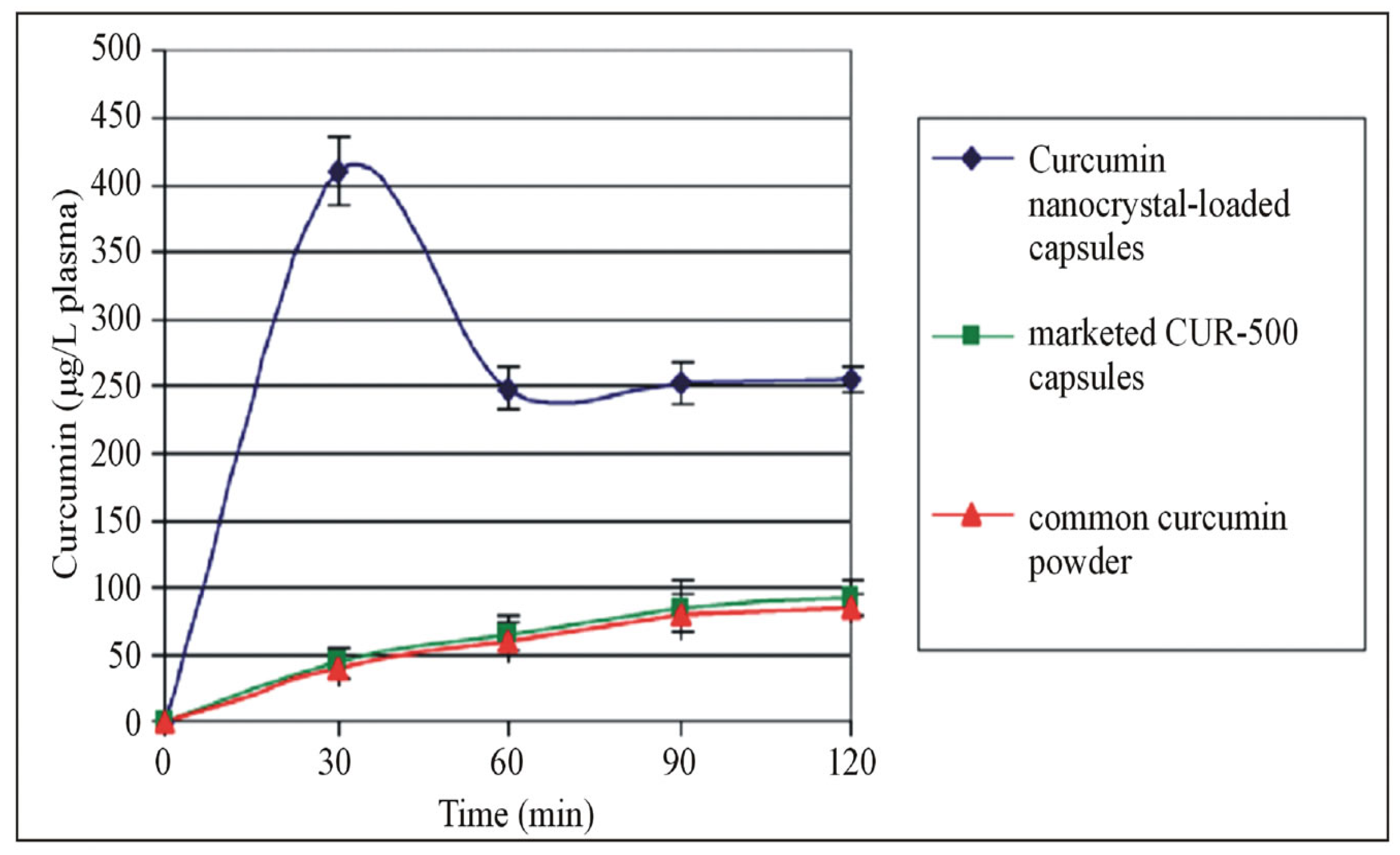
  26. Ravichandran, R. Pharmacokinetic Study of Nanoparticulate Curcumin: Oral Formulation for Enhanced Bioavailability. J. Biomater. Nanobiotechnol. 2013, 2013, 35329. [Google Scholar] [CrossRef]
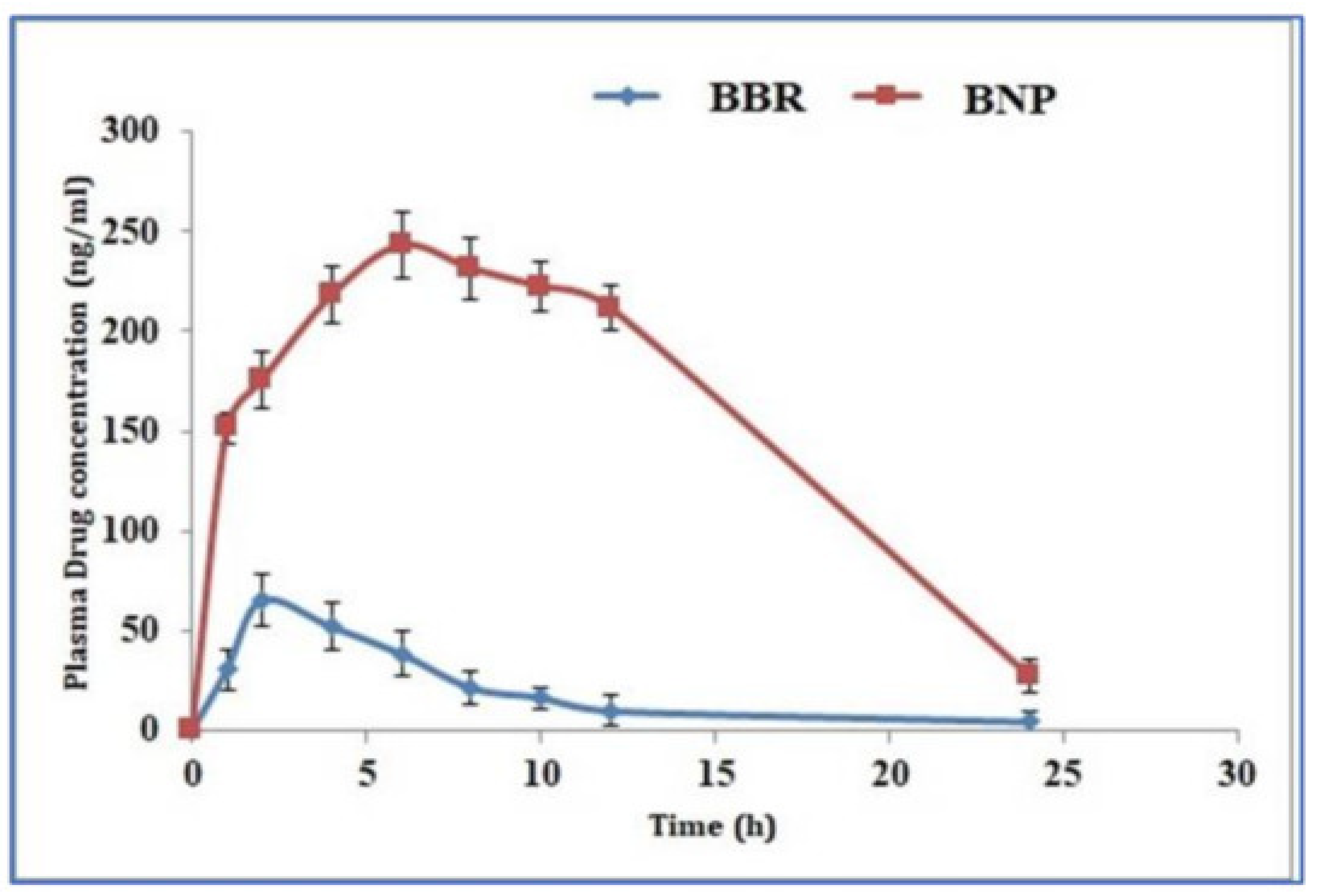
  27. Kohli, K.; Mujtaba, A.; Malik, R.; Amin, S.; Alam, M.S.; Ali, A.; Barkat, M.A.; Ansari, M.J. Development of Natural Polysaccharide–Based Nanoparticles of Berberine to Enhance Oral Bioavailability: Formulation, Optimization, Ex Vivo, and In Vivo Assessment. Polymers 2021, 13, 3833. [Google Scholar] [CrossRef]
  28. Hasija, R.; Chaurasia, S.; Gupta, S. Assessment of Polymeric Nanoparticles to Enhance Oral Bioavailability and Antioxidant Activity of Resveratrol. Indian. J. Pharm. Sci. 2021, 83, 1114–1128. [Google Scholar] [CrossRef]
  29. Ramesh, N.; Mandal, A.K.A. Pharmacokinetic, Toxicokinetic, and Bioavailability Studies of Epigallocatechin-3-Gallate Loaded Solid Lipid Nanoparticle in Rat Model. Drug Dev. Ind. Pharm. 2019, 45, 1506–1514. [Google Scholar] [CrossRef]
  30. Lambert, J.D.; Sang, S.; Hong, J.; Kwon, S.-J.; Lee, M.-J.; Ho, C.-T.; Yang, C.S. Peracetylation as a Means of Enhancing In Vitro Bioactivity and Bioavailability of Epigallocatechin-3-Gallate. Drug Metab. Dispos. 2006, 34, 2111–2116. [Google Scholar] [CrossRef] [Green Version]
  31. Biasutto, L.; Marotta, E.; Bradaschia, A.; Fallica, M.; Mattarei, A.; Garbisa, S.; Zoratti, M.; Paradisi, C. Soluble Polyphenols: Synthesis and Bioavailability of 3,4′,5-Tri(α-d-Glucose-3-O-Succinyl) Resveratrol. Bioorg. Med. Chem. Lett. 2009, 19, 6721–6724. [Google Scholar] [CrossRef]
  32. Fan, P.; Lou, H. Effects of Polyphenols from Grape Seeds on Oxidative Damage to Cellular DNA. Mol. Cell. Biochem. 2004, 267, 67–74. [Google Scholar] [CrossRef]
  33. Li, A.-N.; Li, S.; Zhang, Y.-J.; Xu, X.-R.; Chen, Y.-M.; Li, H.-B. Resources and Biological Activities of Natural Polyphenols. Nutrients 2014, 6, 6020–6047. [Google Scholar] [CrossRef] [Green Version]
  34. Silva, R.F.M.; Pogačnik, L. Polyphenols from Food and Natural Products: Neuroprotection and Safety. Antioxidants 2020, 9, 61. [Google Scholar] [CrossRef] [Green Version]
  35. Murakami, A. Dose-Dependent Functionality and Toxicity of Green Tea Polyphenols in Experimental Rodents. Arch. Biochem. Biophys. 2014, 557, 3–10. [Google Scholar] [CrossRef] [Green Version]
  36. Duda-Chodak, A.; Tarko, T. Possible Side Effects of Polyphenols and Their Interactions with Medicines. Molecules 2023, 28, 2536. [Google Scholar] [CrossRef]
  37. Jung, S.; Jeong, H.; Yu, S.-W. Autophagy as a Decisive Process for Cell Death. Exp. Mol. Med. 2020, 52, 921–930. [Google Scholar] [CrossRef]
  38. Cordero, J.G.; García-Escudero, R.; Avila, J.; Gargini, R.; García-Escudero, V. Benefit of Oleuropein Aglycone for Alzheimer’s Disease by Promoting Autophagy. Oxidative Med. Cell. Longev. 2018, 2018, e5010741. [Google Scholar] [CrossRef] [Green Version]
  39. Parzych, K.R.; Klionsky, D.J. An Overview of Autophagy: Morphology, Mechanism, and Regulation. Antioxid. Redox Signal 2014, 20, 460–473. [Google Scholar] [CrossRef] [Green Version]
  40. Tyedmers, J.; Mogk, A.; Bukau, B. Cellular Strategies for Controlling Protein Aggregation. Nat. Rev. Mol. Cell Biol. 2010, 11, 777–788. [Google Scholar] [CrossRef]
  41. Höhn, A.; Tramutola, A.; Cascella, R. Proteostasis Failure in Neurodegenerative Diseases: Focus on Oxidative Stress. Oxidative Med. Cell. Longev. 2020, 2020, e5497046. [Google Scholar] [CrossRef] [Green Version]
  42. Fujikake, N.; Shin, M.; Shimizu, S. Association Between Autophagy and Neurodegenerative Diseases. Front. Neurosci. 2018, 12, 255. [Google Scholar] [CrossRef] [PubMed] [Green Version]
  43. Chidambaram, S.B.; Essa, M.M.; Rathipriya, A.G.; Bishir, M.; Ray, B.; Mahalakshmi, A.M.; Tousif, A.H.; Sakharkar, M.K.; Kashyap, R.S.; Friedland, R.P.; et al. Gut Dysbiosis, Defective Autophagy and Altered Immune Responses in Neurodegenerative Diseases: Tales of a Vicious Cycle. Pharmacol. Ther. 2022, 231, 107988. [Google Scholar] [CrossRef]
  44. Chung, C.G.; Lee, H.; Lee, S.B. Mechanisms of Protein Toxicity in Neurodegenerative Diseases. Cell. Mol. Life Sci. 2018, 75, 3159–3180. [Google Scholar] [CrossRef] [PubMed] [Green Version]
  45. Chen, G.; Xu, T.; Yan, Y.; Zhou, Y.; Jiang, Y.; Melcher, K.; Xu, H.E. Amyloid Beta: Structure, Biology and Structure-Based Therapeutic Development. Acta Pharmacol. Sin. 2017, 38, 1205–1235. [Google Scholar] [CrossRef] [PubMed] [Green Version]
  46. Jiang, S.; Li, Y.; Zhang, X.; Bu, G.; Xu, H.; Zhang, Y. Trafficking Regulation of Proteins in Alzheimer’s Disease. Mol. Neurodegener. 2014, 9, 6. [Google Scholar] [CrossRef] [Green Version]
  47. Mondragón-Rodríguez, S.; Perry, G.; Zhu, X.; Moreira, P.I.; Acevedo-Aquino, M.C.; Williams, S. Phosphorylation of Tau Protein as the Link between Oxidative Stress, Mitochondrial Dysfunction, and Connectivity Failure: Implications for Alzheimer’s Disease. Oxid. Med. Cell. Longev. 2013, 2013, 940603. [Google Scholar] [CrossRef] [Green Version]
  48. Long, Z.; Chen, J.; Zhao, Y.; Zhou, W.; Yao, Q.; Wang, Y.; He, G. Dynamic Changes of Autophagic Flux Induced by Abeta in the Brain of Postmortem Alzheimer’s Disease Patients, Animal Models and Cell Models. Aging 2020, 12, 10912–10930. [Google Scholar] [CrossRef]
  49. Lai, A.Y.; McLaurin, J. Inhibition of Amyloid-Beta Peptide Aggregation Rescues the Autophagic Deficits in the TgCRND8 Mouse Model of Alzheimer Disease. Biochim. Biophys. Acta 2012, 1822, 1629–1637. [Google Scholar] [CrossRef] [Green Version]
  50. Ba, L.; Chen, X.-H.; Chen, Y.-L.; Nie, Q.; Li, Z.-J.; Ding, F.-F.; Zhang, M. Distinct Rab7-Related Endosomal–Autophagic–Lysosomal Dysregulation Observed in Cortex and Hippocampus in APPswe/PSEN1dE9 Mouse Model of Alzheimer’s Disease. Chin. Med. J. 2017, 130, 2941–2950. [Google Scholar] [CrossRef]
  51. Grossi, C.; Rigacci, S.; Ambrosini, S.; Dami, T.E.; Luccarini, I.; Traini, C.; Failli, P.; Berti, A.; Casamenti, F.; Stefani, M. The Polyphenol Oleuropein Aglycone Protects TgCRND8 Mice against Aß Plaque Pathology. PLoS ONE 2013, 8, e71702. [Google Scholar] [CrossRef] [Green Version]
  52. Zhu, Z.; Yan, J.; Jiang, W.; Yao, X.; Chen, J.; Chen, L.; Li, C.; Hu, L.; Jiang, H.; Shen, X. Arctigenin Effectively Ameliorates Memory Impairment in Alzheimer’s Disease Model Mice Targeting Both β-Amyloid Production and Clearance. J. Neurosci. 2013, 33, 13138–13149. [Google Scholar] [CrossRef] [Green Version]
  53. Kou, X.; Chen, N. Resveratrol as a Natural Autophagy Regulator for Prevention and Treatment of Alzheimer’s Disease. Nutrients 2017, 9, 927. [Google Scholar] [CrossRef] [Green Version]
  54. Qiu, W.-Q.; Pan, R.; Tang, Y.; Zhou, X.-G.; Wu, J.-M.; Yu, L.; Law, B.Y.-K.; Ai, W.; Yu, C.-L.; Qin, D.-L.; et al. Lychee Seed Polyphenol Inhibits Aβ-Induced Activation of NLRP3 Inflammasome via the LRP1/AMPK Mediated Autophagy Induction. Biomed. Pharmacother. 2020, 130, 110575. [Google Scholar] [CrossRef] [PubMed]
  55. Capiralla, H.; Vingtdeux, V.; Zhao, H.; Sankowski, R.; Al-Abed, Y.; Davies, P.; Marambaud, P. Resveratrol Mitigates Lipopolysaccharide- and Aβ-Mediated Microglial Inflammation by Inhibiting the TLR4/NF-ΚB/STAT Signaling Cascade. J. Neurochem. 2012, 120, 461–472. [Google Scholar] [CrossRef] [PubMed] [Green Version]
  56. Hwang, S.H.; Shin, E.-J.; Shin, T.-J.; Lee, B.-H.; Choi, S.-H.; Kang, J.; Kim, H.-J.; Kwon, S.-H.; Jang, C.-G.; Lee, J.-H.; et al. Gintonin, a Ginseng-Derived Lysophosphatidic Acid Receptor Ligand, Attenuates Alzheimer’s Disease-Related Neuropathies: Involvement of Non-Amyloidogenic Processing. J. Alzheimers Dis. 2012, 31, 207–223. [Google Scholar] [CrossRef]
  57. Zeng, H.; Chen, Q.; Zhao, B. Genistein Ameliorates Beta-Amyloid Peptide (25–35)-Induced Hippocampal Neuronal Apoptosis. Free Radic. Biol. Med. 2004, 36, 180–188. [Google Scholar] [CrossRef]
  58. Jowaed, A.; Schmitt, I.; Kaut, O.; Wüllner, U. Methylation Regulates Alpha-Synuclein Expression and Is Decreased in Parkinson’s Disease Patients’ Brains. J. Neurosci. 2010, 30, 6355–6359. [Google Scholar] [CrossRef] [PubMed] [Green Version]
  59. Pan, P.-Y.; Yue, Z. Genetic Causes of Parkinson’s Disease and Their Links to Autophagy Regulation. Park. Relat. Disord. 2014, 20, S154–S157. [Google Scholar] [CrossRef] [PubMed]
  60. Lynch-Day, M.A.; Mao, K.; Wang, K.; Zhao, M.; Klionsky, D.J. The Role of Autophagy in Parkinson’s Disease. Cold Spring Harb. Perspect. Med. 2012, 2, a009357. [Google Scholar] [CrossRef] [Green Version]
  61. Cheung, Z.H.; Ip, N.Y. The Emerging Role of Autophagy in Parkinson’s Disease. Mol. Brain 2009, 2, 29. [Google Scholar] [CrossRef] [Green Version]
  62. Ray, B.; Bhat, A.; Mahalakshmi, A.M.; Tuladhar, S.; Bishir, M.; Mohan, S.K.; Veeraraghavan, V.P.; Chandra, R.; Essa, M.M.; Chidambaram, S.B.; et al. Mitochondrial and Organellar Crosstalk in Parkinson’s Disease. ASN Neuro 2021, 13, 17590914211028364. [Google Scholar] [CrossRef]
  63. Lu, J.-H.; Tan, J.-Q.; Durairajan, S.S.K.; Liu, L.-F.; Zhang, Z.-H.; Ma, L.; Shen, H.-M.; Chan, H.Y.E.; Li, M. Isorhynchophylline, a Natural Alkaloid, Promotes the Degradation of Alpha-Synuclein in Neuronal Cells via Inducing Autophagy. Autophagy 2012, 8, 98–108. [Google Scholar] [CrossRef] [PubMed] [Green Version]
  64. Jiang, T.-F.; Zhang, Y.-J.; Zhou, H.-Y.; Wang, H.-M.; Tian, L.-P.; Liu, J.; Ding, J.-Q.; Chen, S.-D. Curcumin Ameliorates the Neurodegenerative Pathology in A53T α-Synuclein Cell Model of Parkinson’s Disease through the Downregulation of MTOR/P70S6K Signaling and the Recovery of Macroautophagy. J. Neuroimmune Pharmacol. 2013, 8, 356–369. [Google Scholar] [CrossRef]
  65. Lin, T.-K.; Chen, S.-D.; Chuang, Y.-C.; Lin, H.-Y.; Huang, C.-R.; Chuang, J.-H.; Wang, P.-W.; Huang, S.-T.; Tiao, M.-M.; Chen, J.-B.; et al. Resveratrol Partially Prevents Rotenone-Induced Neurotoxicity in Dopaminergic SH-SY5Y Cells through Induction of Heme Oxygenase-1 Dependent Autophagy. Int. J. Mol. Sci. 2014, 15, 1625–1646. [Google Scholar] [CrossRef] [PubMed]
  66. Guo, Y.-J.; Dong, S.-Y.; Cui, X.-X.; Feng, Y.; Liu, T.; Yin, M.; Kuo, S.-H.; Tan, E.-K.; Zhao, W.-J.; Wu, Y.-C. Resveratrol Alleviates MPTP-Induced Motor Impairments and Pathological Changes by Autophagic Degradation of α-Synuclein via SIRT1-Deacetylated LC3. Mol. Nutr. Food Res. 2016, 60, 2161–2175. [Google Scholar] [CrossRef] [PubMed]
  67. Jo, M.G.; Ikram, M.; Jo, M.H.; Yoo, L.; Chung, K.C.; Nah, S.-Y.; Hwang, H.; Rhim, H.; Kim, M.O. Gintonin Mitigates MPTP-Induced Loss of Nigrostriatal Dopaminergic Neurons and Accumulation of α-Synuclein via the Nrf2/HO-1 Pathway. Mol. Neurobiol. 2019, 56, 39–55. [Google Scholar] [CrossRef]
  68. Zhang, Y.; Wu, Q.; Zhang, L.; Wang, Q.; Yang, Z.; Liu, J.; Feng, L. Caffeic Acid Reduces A53T α-Synuclein by Activating JNK/Bcl-2-Mediated Autophagy In Vitro and Improves Behaviour and Protects Dopaminergic Neurons in a Mouse Model of Parkinson’s Disease. Pharmacol. Res. 2019, 150, 104538. [Google Scholar] [CrossRef]
  69. Fatoba, O.; Ohtake, Y.; Itokazu, T.; Yamashita, T. Immunotherapies in Huntington’s Disease and α-Synucleinopathies. Front. Immunol. 2020, 11, 337. [Google Scholar] [CrossRef]
  70. Fox, L.M.; Kim, K.; Johnson, C.W.; Chen, S.; Croce, K.R.; Victor, M.B.; Eenjes, E.; Bosco, J.R.; Randolph, L.K.; Dragatsis, I.; et al. Huntington’s Disease Pathogenesis Is Modified In Vivo by Alfy/Wdfy3 and Selective Macroautophagy. Neuron 2020, 105, 813–821.e6. [Google Scholar] [CrossRef]
  71. Nah, J.; Yuan, J.; Jung, Y.-K. Autophagy in Neurodegenerative Diseases: From Mechanism to Therapeutic Approach. Mol. Cells 2015, 38, 381–389. [Google Scholar] [CrossRef] [Green Version]
  72. Shibata, M.; Lu, T.; Furuya, T.; Degterev, A.; Mizushima, N.; Yoshimori, T.; MacDonald, M.; Yankner, B.; Yuan, J. Regulation of Intracellular Accumulation of Mutant Huntingtin by Beclin 1. J. Biol. Chem. 2006, 281, 14474–14485. [Google Scholar] [CrossRef] [PubMed] [Green Version]
  73. Wong, V.K.W.; Wu, A.G.; Wang, J.R.; Liu, L.; Law, B.Y.-K. Neferine Attenuates the Protein Level and Toxicity of Mutant Huntingtin in PC-12 Cells via Induction of Autophagy. Molecules 2015, 20, 3496–3514. [Google Scholar] [CrossRef] [PubMed] [Green Version]
  74. Pérez-Arancibia, R.; Ordoñez, J.L.; Rivas, A.; Pihán, P.; Sagredo, A.; Ahumada, U.; Barriga, A.; Seguel, I.; Cárdenas, C.; Vidal, R.L.; et al. A Phenolic-Rich Extract from Ugni Molinae Berries Reduces Abnormal Protein Aggregation in a Cellular Model of Huntington’s Disease. PLoS ONE 2021, 16, e0254834. [Google Scholar] [CrossRef]
  75. Jiang, W.; Wei, W.; Gaertig, M.A.; Li, S.; Li, X.-J. Therapeutic Effect of Berberine on Huntington’s Disease Transgenic Mouse Model. PLoS ONE 2015, 10, e0134142. [Google Scholar] [CrossRef]
  76. Maher, P.; Dargusch, R.; Bodai, L.; Gerard, P.E.; Purcell, J.M.; Marsh, J.L. ERK Activation by the Polyphenols Fisetin and Resveratrol Provides Neuroprotection in Multiple Models of Huntington’s Disease. Hum. Mol. Genet. 2011, 20, 261–270. [Google Scholar] [CrossRef] [Green Version]
  77. Vidoni, C.; Secomandi, E.; Castiglioni, A.; Melone, M.A.B.; Isidoro, C. Resveratrol Protects Neuronal-like Cells Expressing Mutant Huntingtin from Dopamine Toxicity by Rescuing ATG4-Mediated Autophagosome Formation. Neurochem. Int. 2018, 117, 174–187. [Google Scholar] [CrossRef]
  78. Pasinelli, P.; Brown, R.H. Molecular Biology of Amyotrophic Lateral Sclerosis: Insights from Genetics. Nat. Rev. Neurosci. 2006, 7, 710–723. [Google Scholar] [CrossRef] [PubMed]
  79. Arai, T.; Hasegawa, M.; Akiyama, H.; Ikeda, K.; Nonaka, T.; Mori, H.; Mann, D.; Tsuchiya, K.; Yoshida, M.; Hashizume, Y.; et al. TDP-43 Is a Component of Ubiquitin-Positive Tau-Negative Inclusions in Frontotemporal Lobar Degeneration and Amyotrophic Lateral Sclerosis. Biochem. Biophys. Res. Commun. 2006, 351, 602–611. [Google Scholar] [CrossRef] [PubMed]
  80. Kanekura, K.; Suzuki, H.; Aiso, S.; Matsuoka, M. ER Stress and Unfolded Protein Response in Amyotrophic Lateral Sclerosis. Mol. Neurobiol. 2009, 39, 81–89. [Google Scholar] [CrossRef] [PubMed]
  81. Brenner, D.; Weishaupt, J.H. Update on Amyotrophic Lateral Sclerosis Genetics. Curr. Opin. Neurol. 2019, 32, 735–739. [Google Scholar] [CrossRef]
  82. Crippa, V.; Sau, D.; Rusmini, P.; Boncoraglio, A.; Onesto, E.; Bolzoni, E.; Galbiati, M.; Fontana, E.; Marino, M.; Carra, S.; et al. The Small Heat Shock Protein B8 (HspB8) Promotes Autophagic Removal of Misfolded Proteins Involved in Amyotrophic Lateral Sclerosis (ALS). Hum. Mol. Genet. 2010, 19, 3440–3456. [Google Scholar] [CrossRef] [Green Version]
  83. Wang, J.; Zhang, Y.; Tang, L.; Zhang, N.; Fan, D. Protective Effects of Resveratrol through the Up-Regulation of SIRT1 Expression in the Mutant HSOD1-G93A-Bearing Motor Neuron-like Cell Culture Model of Amyotrophic Lateral Sclerosis. Neurosci. Lett. 2011, 503, 250–255. [Google Scholar] [CrossRef]
  84. Han, S.; Choi, J.-R.; Soon Shin, K.; Kang, S.J. Resveratrol Upregulated Heat Shock Proteins and Extended the Survival of G93A-SOD1 Mice. Brain Res. 2012, 1483, 112–117. [Google Scholar] [CrossRef]
  85. Mancuso, R.; del Valle, J.; Modol, L.; Martinez, A.; Granado-Serrano, A.B.; Ramirez-Núñez, O.; Pallás, M.; Portero-Otin, M.; Osta, R.; Navarro, X. Resveratrol Improves Motoneuron Function and Extends Survival in SOD1G93A ALS Mice. Neurotherapeutics 2014, 11, 419–432. [Google Scholar] [CrossRef] [PubMed] [Green Version]
  86. Ueda, T.; Inden, M.; Shirai, K.; Sekine, S.-I.; Masaki, Y.; Kurita, H.; Ichihara, K.; Inuzuka, T.; Hozumi, I. The Effects of Brazilian Green Propolis That Contains Flavonols against Mutant Copper-Zinc Superoxide Dismutase-Mediated Toxicity. Sci. Rep. 2017, 7, 2882. [Google Scholar] [CrossRef] [PubMed]
  87. Lu, J.; Duan, W.; Guo, Y.; Jiang, H.; Li, Z.; Huang, J.; Hong, K.; Li, C. Mitochondrial Dysfunction in Human TDP-43 Transfected NSC34 Cell Lines and the Protective Effect of Dimethoxy Curcumin. Brain Res. Bull. 2012, 89, 185–190. [Google Scholar] [CrossRef] [PubMed]
  88. Wang, T.H.; Wang, S.Y.; Wang, X.D.; Jiang, H.Q.; Yang, Y.Q.; Wang, Y.; Cheng, J.L.; Zhang, C.T.; Liang, W.W.; Feng, H.L. Fisetin Exerts Antioxidant and Neuroprotective Effects in Multiple Mutant HSOD1 Models of Amyotrophic Lateral Sclerosis by Activating ERK. Neuroscience 2018, 379, 152–166. [Google Scholar] [CrossRef]
  89. Dobson, R.; Giovannoni, G. Multiple Sclerosis—A Review. Eur. J. Neurol. 2019, 26, 27–40. [Google Scholar] [CrossRef] [Green Version]
  90. Interplay between ER Stress and Autophagy: A Possible Mechanism in Multiple Sclerosis Pathology. Exp. Mol. Pathol. 2019, 108, 183–190. [CrossRef] [PubMed]
  91. Misrielal, C.; Mauthe, M.; Reggiori, F.; Eggen, B.J.L. Autophagy in Multiple Sclerosis: Two Sides of the Same Coin. Front. Cell Neurosci. 2020, 14, 603710. [Google Scholar] [CrossRef]
  92. Igci, M.; Baysan, M.; Yigiter, R.; Ulasli, M.; Geyik, S.; Bayraktar, R.; Bozgeyik, İ.; Bozgeyik, E.; Bayram, A.; Cakmak, E.A. Gene Expression Profiles of Autophagy-Related Genes in Multiple Sclerosis. Gene 2016, 588, 38–46. [Google Scholar] [CrossRef] [PubMed]
  93. Albert, M.; Barrantes-Freer, A.; Lohrberg, M.; Antel, J.P.; Prineas, J.W.; Palkovits, M.; Wolff, J.R.; Brück, W.; Stadelmann, C. Synaptic Pathology in the Cerebellar Dentate Nucleus in Chronic Multiple Sclerosis. Brain. Pathol. 2017, 27, 737–747. [Google Scholar] [CrossRef] [PubMed] [Green Version]
  94. Patergnani, S.; Castellazzi, M.; Bonora, M.; Marchi, S.; Casetta, I.; Pugliatti, M.; Giorgi, C.; Granieri, E.; Pinton, P. Autophagy and Mitophagy Elements Are Increased in Body Fluids of Multiple Sclerosis-Affected Individuals. J. Neurol. Neurosurg. Psychiatry 2018, 89, 439–441. [Google Scholar] [CrossRef]
  95. Boyao, Y.; Mengjiao, S.; Caicai, B.; Xiaoling, L.; Zhenxing, L.; Manxia, W. Dynamic Expression of Autophagy-Related Factors in Autoimmune Encephalomyelitis and Exploration of Curcumin Therapy. J. Neuroimmunol. 2019, 337, 577067. [Google Scholar] [CrossRef] [PubMed]
  96. Dierckx, T.; Haidar, M.; Grajchen, E.; Wouters, E.; Vanherle, S.; Loix, M.; Boeykens, A.; Bylemans, D.; Hardonnière, K.; Kerdine-Römer, S.; et al. Phloretin Suppresses Neuroinflammation by Autophagy-Mediated Nrf2 Activation in Macrophages. J. Neuroinflamm. 2021, 18, 148. [Google Scholar] [CrossRef] [PubMed]
  97. Wang, M.-R.; Zhang, X.-J.; Liu, H.-C.; Ma, W.-D.; Zhang, M.-L.; Zhang, Y.; Li, X.; Dou, M.-M.; Jing, Y.-L.; Chu, Y.-J.; et al. Matrine Protects Oligodendrocytes by Inhibiting Their Apoptosis and Enhancing Mitochondrial Autophagy. Brain Res. Bull. 2019, 153, 30–38. [Google Scholar] [CrossRef]
  98. Li, W.; Deng, R.; Jing, X.; Chen, J.; Yang, D.; Shen, J. Acteoside Ameliorates Experimental Autoimmune Encephalomyelitis through Inhibiting Peroxynitrite-Mediated Mitophagy Activation. Free Radic. Biol. Med. 2020, 146, 79–91. [Google Scholar] [CrossRef] [PubMed]
Figure 1. Pharmacokinetic profile of curcumin. Comparison of the pharmacokinetic profile of curcumin, nanocurcumin and marketed curcumin capsules. The figure is reused as per the journal’s copyright permission [26].
Biomolecules 13 01196 g001
Figure 2. Pharmacokinetic profile of berberine. Comparison of pharmacokinetic profile of berberine and berberine nanoparticles. The figure is reused as per the journal’s copyright permission [27].
Biomolecules 13 01196 g002
Figure 3. Autophagy dysregulation in different neurodegenerative diseases. Different stages of autophagy are affected in neurodegenerative diseases. Parkinson’s disease (PD)—the cargo initiation step, Alzheimer’s disease (AD)—initiation of autophagosome, Huntington’s disease (HD)—formation of autophagosome, amyotrophic lateral sclerosis—autolysosome formation is inhibited. These ultimately lead to cognitive impairment.
Biomolecules 13 01196 g003
Figure 4. Regulation of autophagy by polyphenol supplementation in neurodegenerative diseases. Polyphenol supplementation regulates autophagy and aids in the clearance of misfolded proteins by entering cytoplasm. Polyphenols inhibit the PI3K/AKT pathway and thereby activate the AMPK and SIRT1 pathways and inhibit the mTOR pathway. Furthermore, they activate the ULK1/2 complex, which initiates the autophagy process by increasing the levels of beclin1 and LC3 proteins.
Biomolecules 13 01196 g004
Disclaimer/Publisher’s Note: The statements, opinions and data contained in all publications are solely those of the individual author(s) and contributor(s) and not of MDPI and/or the editor(s). MDPI and/or the editor(s) disclaim responsibility for any injury to people or property resulting from any ideas, methods, instructions or products referred to in the content.

