Article

Transcription Factor and Protein Regulatory Network of PmACRE1 in Pinus massoniana Response to Pine Wilt Nematode Infection

1
Jinshan College, Fujian Agriculture and Forestry University, Fuzhou 350002, China
2
Key Laboratory of Integrated Pest Management in Ecological Forests (Fujian Province University), Fujian Agriculture and Forestry University, Fuzhou 350002, China
3
Forestry College, Fujian Agriculture and Forestry University, Fuzhou 350002, China
4
Fujian Academy of Forestry, Fuzhou 350000, China
*
Authors to whom correspondence should be addressed.
Plants 2024, 13(19), 2672; https://doi.org/10.3390/plants13192672
Submission received: 5 August 2024 / Revised: 17 September 2024 / Accepted: 20 September 2024 / Published: 24 September 2024
(This article belongs to the Special Issue Molecular Biology and Bioinformatics of Forest Trees)

Abstract

:
Pine wilt disease, caused by Bursaphelenchus xylophilus, is a highly destructive and contagious forest affliction. Often termed the “cancer” of pine trees, it severely impacts the growth of Masson pine (Pinus massoniana). Previous studies have demonstrated that ectopic expression of the PmACRE1 gene from P. massoniana in Arabidopsis thaliana notably enhances resistance to pine wilt nematode infection. To further elucidate the transcriptional regulation and protein interactions of the PmACRE1 in P. massoniana in response to pine wilt nematode infection, we cloned a 1984 bp promoter fragment of the PmACRE1 gene, a transient expression vector was constructed by fusing this promoter with the reporter GFP gene, which successfully activated the GFP expression. DNA pull-down assays identified PmMYB8 as a trans-acting factor regulating PmACRE1 gene expression. Subsequently, we found that the PmACRE1 protein interacts with several proteins, including the ATP synthase CF1 α subunit, ATP synthase CF1 β subunit, extracellular calcium-sensing receptor (PmCAS), caffeoyl-CoA 3-O-methyltransferase (PmCCoAOMT), glutathione peroxidase, NAD+-dependent glyceraldehyde-3-phosphate dehydrogenase, phosphoglycerate kinase 1, cinnamyl alcohol dehydrogenase, auxin response factor 16, and dehydrin 1 protein. Bimolecular fluorescence complementation (BiFC) assays confirmed the interactions between PmACRE1 and PmCCoAOMT, as well as PmCAS proteins in vitro. These findings provide preliminary insights into the regulatory role of PmACRE1 in P. massoniana’s defense against pine wilt nematode infection.

1. Introduction

Pine wilt disease is a devastating forest epidemic in pine trees, which causes the yellowing and wilting of needles, stunted growth, premature needle drop, and the dieback of branches. The ultimate damage can lead to the death of the tree. Since its entry into Nanjing city in 1982, the disease has rapidly spread across multiple provinces, posing a serious threat to the ecological security of forests in China and causing significant economic losses [1]. Enhancing the disease resistance of Masson pine (Pinus massoniana) through effective technologies could mitigate the lethal effects of pine wood nematode (Bursaphelenchus xylophilus). Disease resistance in plants is closely related to the expression and regulation of resistance genes. Utilizing modern molecular techniques to uncover mechanisms of resistance in P. massoniana and identifying genes that regulate resistance capacity can expedite the development of resistant pine germplasm. This is crucial for the sustainable control of pine wilt disease [2,3,4].
Plant disease resistance is closely associated with the expression levels of disease-related genes. Numerous studies have demonstrated that different resistant varieties of P. massoniana show significant variations in the expression levels of genes involved in secondary metabolic pathways and antioxidant systems when infected by B. xylophilus. These genes potentially regulate P. massoniana resistance to the nematode [5,6,7,8]. Liu et al. found that enhancing the expression of terpene synthesis-related genes, such as PmGPPS1, PmTPS4, and PmTPS21, improves the plant defense against nematode infection [9,10], thereby confirming the efficacy of disease-resistance genes. Identifying key genes that regulate disease defense and employing molecular genetics to stably enhance their expression in P. massoniana could improve its resistance to pine wilt disease.
Our previous research identified differential gene expression in P. massoniana needles following B. xylophilus infection, with significant enrichment in pathways including phenylalanine metabolism, secondary metabolite biosynthesis, and plant hormone signal transduction [11,12]. Notably, the expression of the leucine-rich repeat (LRR) containing pathogen-specific recognition gene, Avr9/Cf-9 rapidly elicited (ACRE1), was markedly upregulated within a day post-infection. Additionally, genes associated with hypersensitive response pathways, such as plant–pathogen interactions, plant hormone signal transduction, and glutathione metabolism, exhibited altered expression patterns. With escalating nematode infection intensity, the expression of the PmACRE1 gene in P. massoniana was suppressed, indicating a potential correlation between this gene and the tree’s resistance to nematode infection [12,13].
Upon pathogen infection, incompatible plant–pathogen interactions typically trigger a hypersensitive response (HR), leading to rapid local cell death mediated by the production of reactive oxygen species (ROS) and programmed cell death, which inhibits pathogen growth and enhances plant resistance [14]. Resistant pine varieties can activate the expression of ROS-related genes through HR, promoting the production of ROS to inhibit pine wood nematode infection [15]. ACRE genes, which are key players in mediating plant hypersensitive responses, have been extensively studied [16,17,18]. Durrant et al. first reported these genes using cDNA-AFLP technology to study tobacco defense responses mediated by Avr9 and cf-9, identifying 13 cDNA clones encoding ACRE genes induced by Avr9 in tobacco leaves. These ACRE proteins share homology with ethylene response elements, calcium-binding proteins, 13-lipoxygenase, and cyclo-H2 zinc finger proteins, making them readily inducible [19]. Ni et al. cloned a RING-H2 zinc finger protein gene (StRFP1) from potato, homologous to the ACRE132 gene in tobacco. The overexpression of StRFP1 in potato enhanced resistance to late blight, while suppression of StRFP1 expression increased the susceptibility of transgenic potato plants to the disease [20]. De Vega et al. demonstrated that chitosan treatment induced the expression of two ACRE genes, ACRE75 and ACRE180, in tomato early during pathogen infection, promoting callose deposition and jasmonate accumulation, thereby enhancing resistance to gray mold (Botrytis cinerea) [21]. These findings indicate that the expression intensity of ACRE genes mediates plant responses to pathogens and significantly influences their ability to resist pathogen invasion.
In conifers, the only reported ACRE gene is from Scots pine (Pinus sylvestris), where PsACRE regulates resistance to root rot [22]. The PmACRE1 gene in P. massoniana encodes a 207 amino acid protein, differing from the ACRE protein in Scots pine by a single amino acid. It contains a leucine-rich repeat (LRR) motif, highlighting its conservation among pines. The LRR motif mediates protein–protein interactions, providing high-affinity domains for effective protein interaction sites, and is critical for the specific recognition of pathogen elicitors [22,23,24,25]. Previous studies have genetically transformed PmACRE1 into Arabidopsis, demonstrating that transgenic Arabidopsis plants exhibit enhanced defense against pine wood nematode infection, reduced disease incidence, and increased levels of flavonoids, alkaloids, and triterpenoids in their leaves. Co-immunoprecipitation (Co-IP) studies revealed that the ACRE1 protein interacts with proteins involved in secondary metabolism and detoxification pathways in Arabidopsis, indicating that PmACRE1 plays a role in defense against nematode infection and enhances plant resistance [26].
To further elucidate the regulatory mechanisms and protein interactions of PmACRE1 in P. massoniana, we cloned the flanking promoter region of PmACRE1 from the P. massoniana genome and analyzed its cis-acting elements. A transient expression vector was constructed by fusing the promoter with the GFP gene to investigate promoter activity. DNA pull-down assays were performed to capture promoter-binding proteins, followed by mass spectrometry (MS) to identify and analyze associated transcription factors. Co-IP was employed to capture interacting proteins of PmACRE1, and bimolecular fluorescence complementation (BiFC) was used to validate these protein–protein interactions. These methodologies aim to enhance our understanding of the transcription factors regulating PmACRE1 and the associated protein networks, providing theoretical insights into the transcriptional regulation of PmACRE1 in response to pine wood nematode infection and paving the way for breeding disease-resistant P. massoniana varieties.

2. Materials and Methods

2.1. Plant Materials

The plant materials included 3-year-old P. massoniana grown in a greenhouse and Nicotiana benthamiana cultivated in a controlled climate chamber at 25 °C with a 16 h/8 h light/dark cycle, using peatmoss with vermiculite as a culture medium. The plant gene expression vectors p3301-ΔActin::GFP, p2300-35S::YFPN, and p2300-35S::YFPC were provided by the Key Laboratory of Crop Ecology and Molecular Physiology, Fujian Agriculture and Forestry University [27].

2.2. Cloning and Analysis of the PmACRE1 Gene Promoter in P. massoniana

Genomic DNA was extracted from P. massoniana needles using the CTAB method [28]. The promoter region upstream of the PmACRE1 gene’s initiation codon (ATG) was identified from the P. massoniana genome database (currently unreleased). Using genomic DNA as a template, a 1984 bp fragment upstream of the PmACRE1 gene coding sequence (CDS) was amplified with primers PmACRE1-PRO-F (5′-GCTCATGGGTCTACTAACGGTTT-3′) and PmACRE1-PRO-R (5′-TTTTTTTGGGTTTTTTCTTCTGATC-3′) via TransTaq HiFi DNA Polymerase (TransGen Biotech Co., Ltd., Beijing, China). The purified amplified fragment was sequenced and bioinformatics analysis of the PmACRE1 promoter sequence was performed using the PlantCARE online tool (http://bioinformatics.psb.ugent.be/webtools/plantcare/html/, accessed on 26 July 2024).

2.3. Detection of PmACRE1 Gene Promoter Activity

To assess the activity of the PmACRE1 gene promoter, specific cloning primers incorporating sequences from the recombinant vector and Spe I and Sac I restriction sites were utilized: FGFP-PmACRE1-PRO-F (5′-atgattacgaattcGAGCTCGCTCATGGGTCTACTAACGG-3′; SpeI site underlined) and FGFP-PmACRE1-PRO-R (5′-tgtagtccatACTAGTTTTTTTTGGGTTTTTTCTTCTGATC-3′; SacI site underlined). A 1984 bp promoter sequence upstream of the PmACRE1 gene was amplified using HiFi DNA Polymerase (TransGen Biotech Co., Ltd., Beijing, China). The PCR product was purified with the TIANquick Midi Purification Kit (TIANGEN Biotech (Beijing) Co., Ltd., Beijing, China). This purified promoter DNA was seamlessly cloned into a Spe I and Sac I linearized p3301-ΔActin::GFP vector using the pEASY-Basic Seamless Cloning and Assembly Kit (TransGen Biotech Co., Ltd., Beijing, China), resulting in the construction of the recombinant expression vector p3301-PmACRE1pro::GFP. This vector was transiently transformed into N. benthamiana, and the GFP expression driven by the PmACRE1 promoter was analyzed using a Laser Scanning Confocal Microscope (Leica TCS SP8, Leica Microsystems, Wetzlar, Germany).

2.4. Isolation of Proteins Binding to the PmACRE1 Gene Promoter

Biotin was conjugated to the 5′ ends of the promoter amplification primers. The amplified PmACRE1 gene promoter DNA was then purified. Natural soluble proteins were extracted from the P. massoniana needles, using the protein extraction method outlined by Fang et al. [28]. These proteins were incubated with the biotin-labeled PmACRE1 gene promoter. The Dynabeads kilobaseBINDER Kit (Thermo Fisher Scientific Inc., Waltham, MA, USA) was employed to bind the biotin-labeled promoter–protein complexes to magnetic beads. Using a magnetic rack, the beads were isolated to retrieve the promoter-bound proteins. These proteins were subsequently separated by SDS-PAGE and compared with a negative control (incubated without the biotin-labeled promoter). Protein bands of interest were excised, digested with trypsin, and identified by Liquid Chromatography Mass Spectrometry/Mass Spectrometry (LC-MS/MS). The resulting mass spectrometry data were analyzed with Proteome Discoverer 2.5 software and searched against the UniProt protein database to identify the proteins and their functions, with a focus on transcription factors and DNA-binding proteins.

2.5. Subcellular Localization of Proteins Binding to the PmACRE1 Gene Promoter

Total RNA was extracted from P. massoniana needles using the RNAprep Pure Plant Plus Kit (Polysaccharides & Polyphenolics-rich, TIANGEN Biotech (Beijing) Co., Ltd., Beijing, China). The extracted RNA was reverse transcribed into cDNA with the EasyScript One-Step RT-PCR SuperMix kit (TransGen Biotech Co., Ltd., Beijing, China). The coding sequence (CDS) of the PmMYB8 gene was used as a template for amplification, employing the following primers: FGFP-PmMYB8-F: 5′-gacaagacgcgtCCCGGGATGGGGCGCCACTCGTGC-3′ and FGFP-PmMYB8-R: 5′-aggtggaggtccCCCGGGAATTTGGTCCAGAACTG CAGC-3′ (Sma I site underlined). The amplified PmMYB8 gene product was purified using the TIANquick Midi Purification Kit (TIANGEN Biotech (Beijing) Co., Ltd., Beijing, China). The purified PmMYB8 gene fragment was then homologously recombined with the SmaI-linearized p3301-ΔActin::GFP vector using the pEASY-Basic Seamless Cloning and Assembly Kit (TransGen Biotech Co., Ltd., Beijing, China), resulting in the construction of the recombinant expression vector p3301-PmACRE1PRO::PmMYB8-GFP. This vector was transiently transformed into N. benthamiana to investigate the subcellular localization of the PmMYB8 protein.

2.6. Inoculation of Pine Wood Nematode to P. massoniana Seedlings and Proteins Extraction

P. massoniana seedlings were infected with pine wood nematode using the artificial bark inoculation method [29,30]. First, the base of the current year’s pruned branches was wiped with 70% ethanol. A sterile scalpel was used to make a slanted cut approximately 1 cm long, deep enough to reach the xylem. A sterile cotton ball was immediately placed in the wound. Using a sterile pipette tip, 0.2 mL of pine wood nematode suspension (500 nematodes) was inoculated onto the cotton ball. The wound was then gently wrapped and secured with sealing film. Sterile water was added every 2 h to maintain moisture. The seedlings were incubated in a greenhouse at 28 °C. Samples of P. massoniana needles were collected on the 3rd and 5th days post-infection for the extraction of natural soluble proteins [27]. The extracted protein solution was stored at −80 °C.

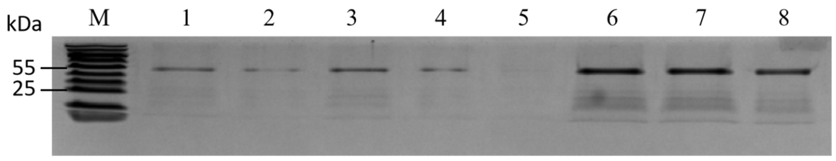
2.7. Co-IP for Isolation of PmACRE1 Interacting Proteins

Interacting proteins of PmACRE1 were isolated using the BeaverBeads Protein A/G Immunoprecipitation Kit (Beaver Biosciences Inc., Suzhou, China). BeaverBeads Protein A/G magnetic beads were first incubated with a specific antibody against PmACRE1 at 26 °C for 60 min to allow antibody binding. The antibody–bead complex was then incubated overnight at 4 °C with natural soluble proteins extracted from P. massoniana needles collected 3- and 5-days post-pine wood nematode infection. This enabled the isolation of PmACRE1 along with its interacting proteins. The eluted protein complexes were subjected to SDS-PAGE and stained with Coomassie Brilliant Blue. Protein bands were excised, digested, and analyzed by HPLC-MS/MS. The data were then analyzed using Proteome Discoverer software and searched against the UniProt database to identify protein information and functionality.

2.8. KEGG Enrichment Analysis for PmACRE1-Interacting Proteins

The FASTA file containing the P. massoniana gene database was obtained from the NCBI database. Gene IDs were extracted and converted using the TBtools-II software. The background set was annotated through the KEGG online website. Subsequently, R language (R-4.3.2), R studio (2021.09.0+351), and necessary compilers were downloaded and installed. The clusterProfiler package 4.0 was loaded into the environment, and the appropriate code was executed to conduct KEGG enrichment analysis. This process culminated in the generation of visualizations to elucidate the functional pathways associated with the PmACRE1-interacting proteins.

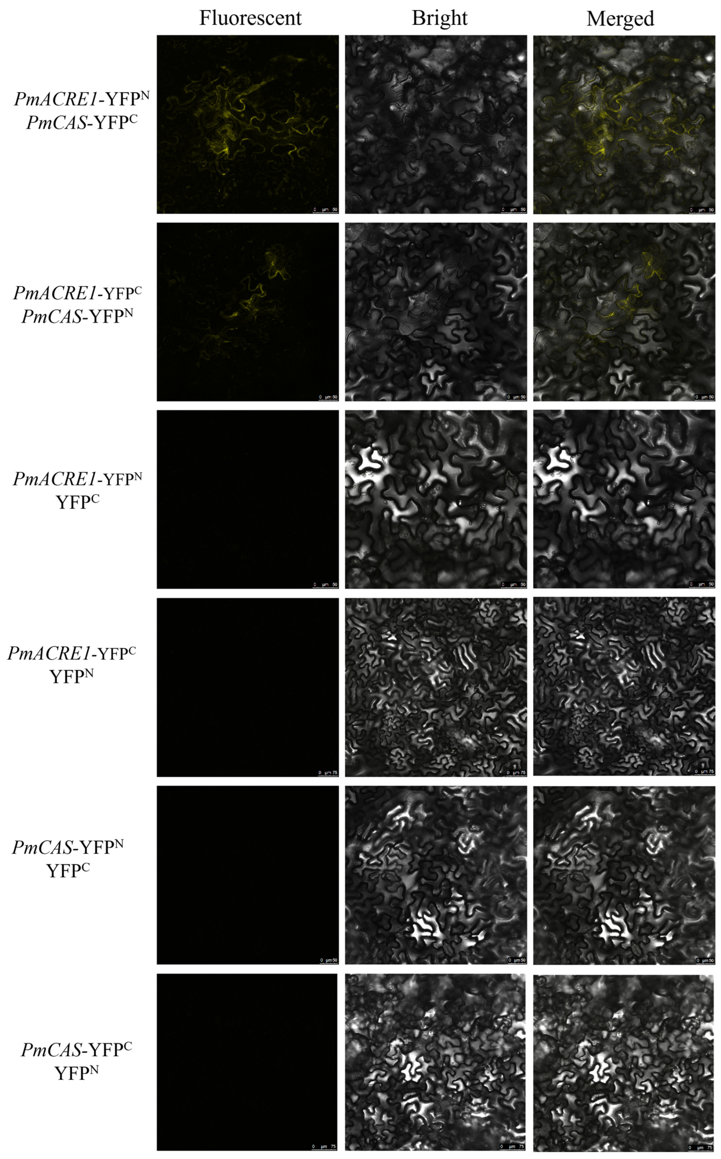
2.9. BiFC Assay for Validating Interactions among PmCCoAOMT, PmCAS, and PmACRE1

Specific amplification primers were designed for PmCCoAOMT, PmCAS, and PmACRE1, incorporating the Spe I site (underlined). The sequences for PmCCoAOMT-2YNC-F and -R were 5′-atggcgcgccACTAGTATGGCAAGCACAGATGTTGCTG-3′ and 5′-cacctcctccACTAGTATAGACACGCCT GCAAAGGGT-3′, respectively. Similarly, primers for PmCAS-2YNC-F and -R were 5′-atggcgcgccACTAGTATGGCTAGCAAGGCAGTTGGC-3′ and 5′-cacctcctccACTAGTATCATCCAAACCACTAGCAAGAA-3′, and for PmACRE1-2YNC-F and -R were 5′-atggcgcgccACTAGTATGGAGGTCCATTCTACTGT AAAT-3′ and 5′-cacctcctccACTAGTGGCTGTACCCCTTTCAAGCATCT-3′. Using cDNA from P. massoniana needles as a template, the genes PmCCoAOMT, PmCAS, and PmACRE1 were amplified, purified, and sequenced. These genes were then individually cloned into the YFP-tagged vectors p2300-2YN and p2300-2YC, which had been linearized with the restriction endonuclease SpeI. The resulting recombinant expression vectors were utilized in a tobacco transient expression system mediated by Agrobacterium. This system allowed for the co-expression of PmACRE1 with either PmCCoAOMT or PmCAS. The interactions between PmACRE1 and PmCCoAOMT, as well as between PmACRE1 and PmCAS, were visualized and confirmed using a Laser Scanning Confocal Microscope (Leica TCS SP8, Leica Microsystems, Wetzlar, Germany).

3. Results

3.1. PmACRE1 Gene Promoter and Its Cis-Elements

In this study, a 1984 bp DNA fragment upstream of the PmACRE1 gene open reading frame was isolated. Sequencing and alignment confirmed its location upstream of the PmACRE1 gene initiation codon. Analysis using the PlantCARE online tool identified 38 cis-acting elements within the PmACRE1 promoter sequence (see Supplementary Figure S1). The promoter contains binding sites for transcription factors such as MYB, MYC, and Myb (MBS), along with elements involved in specific transcriptional regulatory processes (e.g., AE-box, TCA-element, TCCC-motif, and TGA-element), gene transcription activation (CCAAT-box), stress response (STRE), ethylene responsiveness (ERE), gibberellin responsiveness (P-box), abscisic acid responsiveness (ABRE, ABRE3a, ABRE4), low-temperature responsiveness (LTR), light responsiveness (G-Box), anaerobic responsiveness (ARE), and cis-acting element for WRKY (W box) related to plant immune and pathogen infection responses, etc. Additionally, elements related to anthocyanin synthesis regulation (chs-CMA1a) and circadian rhythm regulation (circadian) were identified (see Table 1). Several response elements of unknown function, such as the AAGAA-motif, Unnamed_1, Unnamed_2, and Unnamed_4, were also identified (see Table 1), suggesting that this promoter is regulated by a variety of factors, including light, temperature, plant hormones, pathogens, and stress conditions.

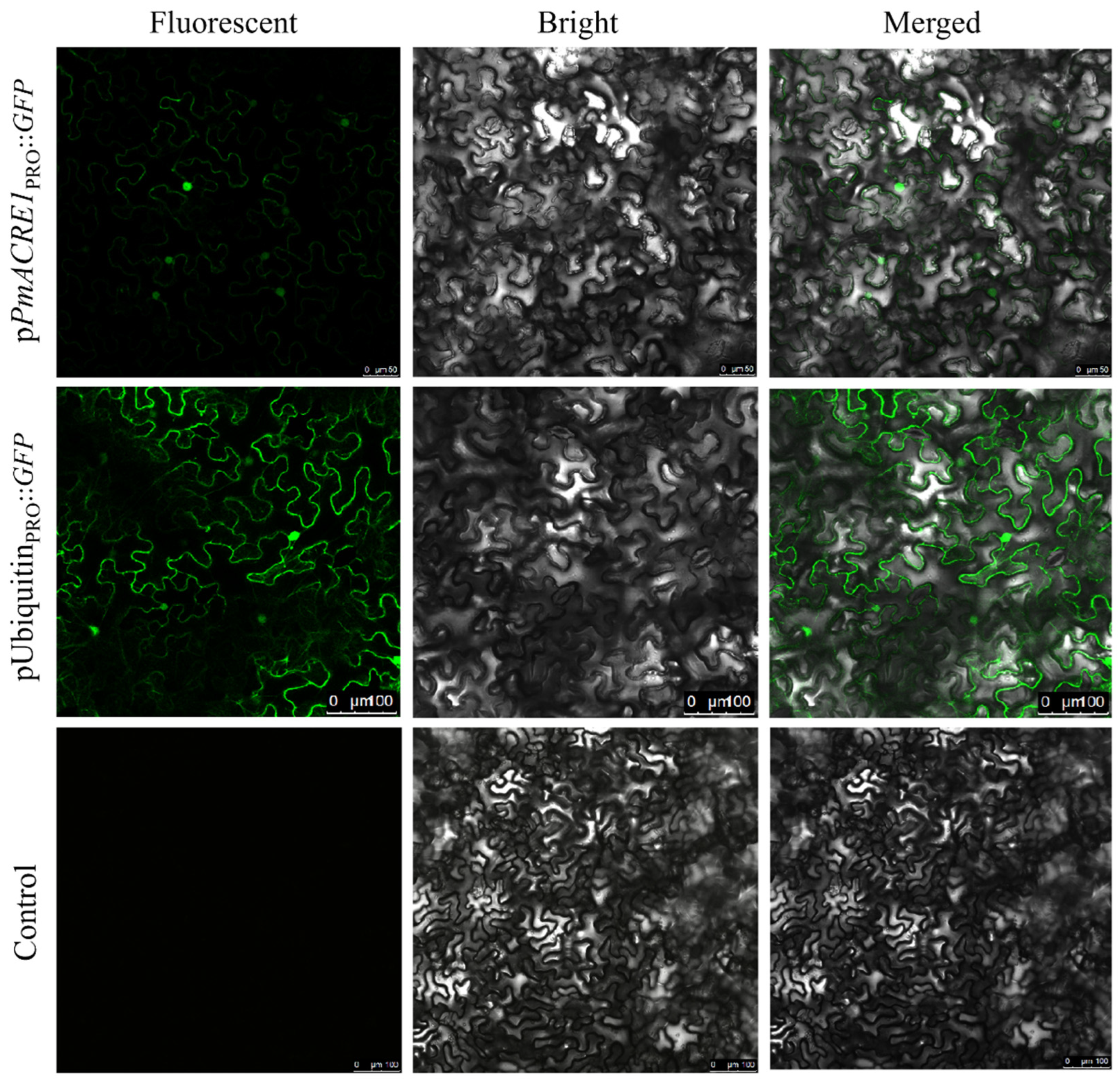
3.2. Activity of the PmACRE1 Gene Promoter

Using an Agrobacterium-mediated transient transformation system in Nicotiana benthamiana leaves, the PmACRE1PRO::GFP recombinant vector was transiently expressed. Tobacco leaves inoculated with an empty GFP vector served as controls. As shown in Figure 1, the PmACRE1PRO::GFP recombinant protein was expressed in both the cell membrane and the nucleus of the tobacco leaves. This observation confirms that the PmACRE1 promoter is active and capable of driving the expression of the reporter gene GFP.

3.3. DNA-Binding Proteins of the PmACRE1 Gene Promoter

Identification of DNA-binding proteins associated with the PmACRE1 gene promoter was achieved using DNA pull-down assays from natural soluble proteins extracted from P. massoniana needles (see Supplementary Figure S2). Mass spectrometry analysis revealed a total of 284 binding proteins (see Supplementary Table S1). Key proteins interacting with the promoter DNA segment included the PmMYB8 transcription factor, ribosomal protein, photosystem II protein K (plastid), propene double bond reductase, glutathione S-transferase, type I chlorophyll a/b binding protein, and ATP synthetase CF1β subunit (chloroplast), among others (see Table 2). Notably, PmMYB8 is a recognized transcription factor that potentially regulates PmACRE1 gene expression.

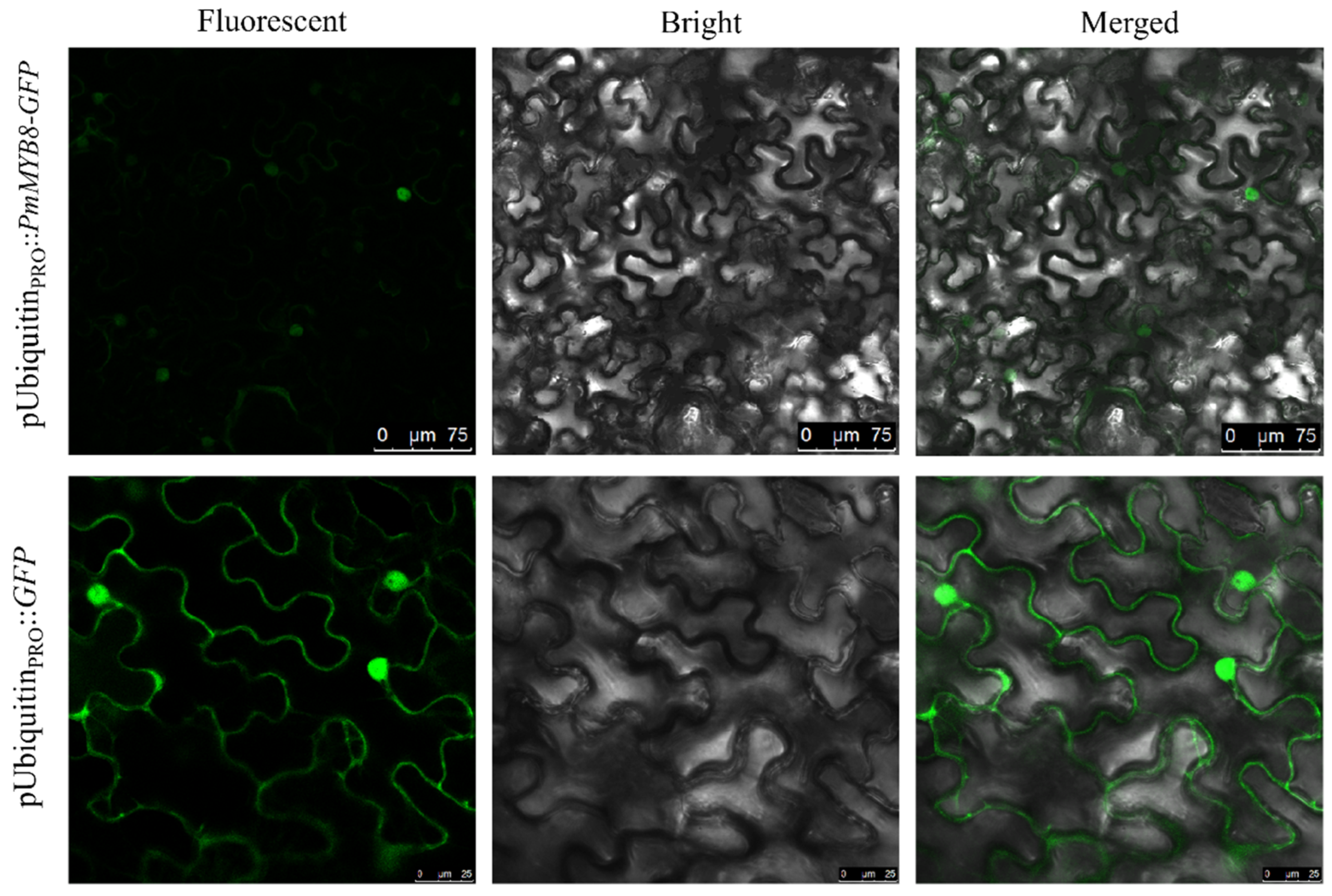
3.4. Subcellular Localization of the PmMYB8 Transcription Factor

The PmMYB8 gene was cloned to construct a recombinant expression vector fused with GFP for transient transformation in N. benthamiana to investigate the subcellular localization of PmMYB8. The results showed green fluorescence localized in the nucleus, whereas the control empty GFP vector exhibited expression both on the cell membrane and in the nucleus (see Figure 2). These findings indicate that the PmMYB8-GFP fusion protein is specifically localized in the nucleus, consistent with the characteristics of transcription factors.

3.5. Proteins Interacting with PmACRE1

The pine wood nematode can colonize pine needles and continue to reproduce, and it can also spread to pine twigs through these needles [31]. To further investigate the regulatory network of PmACRE1 protein in Pine wood Nematode-infected P. massoniana, specific antibodies against PmACRE1 were prepared. Interacting proteins of PmACRE1 were isolated from the natural soluble proteins of P. massoniana needles (see Supplementary Figure S3) using Protein A+G Magnetic Beads-based Co-IP. The PmACRE1 protein and its potential interacting proteins were separated by SDS-PAGE (see Figure 3). Coomassie Brilliant Blue staining revealed that in addition to the PmACRE1 protein, several potential interacting proteins were detected, indicating a complex regulatory network involving PmACRE1 in response to pinewood nematode infection.
To elucidate the types and functions of PmACRE1-interacting proteins, mass spectrometry was performed, identifying 42 proteins detailed in Table 3. These include PmACRE1, ATP synthase CF1 alpha and beta subunits, extracellular calcium-sensing receptor (PmCAS), caffeoyl-CoA 3-O-methyltransferase (PmCCoAOMT), glutathione peroxidase (GSH-Px), NAD+-dependent glyceraldehyde-3-phosphate dehydrogenase, auxin response factor 16 (ARF16), phosphoglycerate kinase 1, cinnamyl alcohol dehydrogenase (PmCAD), and dehydrin 1 protein, among others. These findings suggest that PmACRE1 may play a role in defense against pinewood nematode disease by interacting with these proteins. Notably, PmCCoAOMT, PmCAD, PmCAS, and NAD+-dependent glyceraldehyde-3-phosphate dehydrogenase are involved in secondary metabolic pathways in plants, indicating that PmACRE1 may regulate these pathways through its interactions.

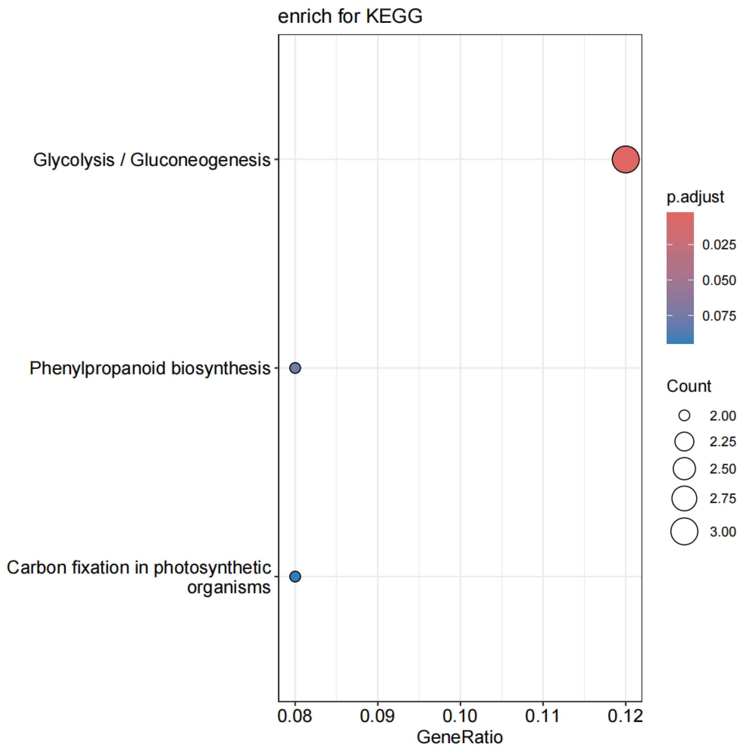
3.6. KEGG Enrichment Analysis of PmACRE1 Interacting Proteins in P. massoniana

Pathway enrichment analysis of PmACRE1-interacting proteins was conducted using the KEGG database. The results indicated that these proteins are enriched in pathways such as Glycolysis/Gluconeogenesis, Phenylpropanoid biosynthesis, and Carbon fixation in photosynthetic organisms (see Figure 4).

3.7. BiFC Validation of Interactions between PmACRE1 and PmCCoAOMT, PmCAS

The interactions between PmACRE1 and proteins involved in plant disease resistance-related secondary metabolic pathways, specifically PmCCoAOMT and PmCAS, were validated using Bimolecular Fluorescence Complementation (BiFC) technology. The coding sequence (CDS) of the CCoAOMT gene was cloned and fused with the N-terminal and C-terminal domains of Yellow Fluorescent Protein (YFP), resulting in the construction of PmCCoAOMT-YFPN and PmCCoAOMT-YFPC recombinant expression vectors. Similarly, PmACRE1-YFPN and PmACRE1-YFPC vectors were also constructed. These vectors were transiently expressed in N. benthamiana leaves, along with a negative control group consisting of YFPN and YFPC without the target genes. Confocal microscopy revealed yellow fluorescent signals in N. benthamiana leaves co-expressing PmACRE1-YFPN with PmCCoAOMT-YFPC, PmACRE1-YFPC with PmCCoAOMT-YFPN, as well as PmACRE1-YFPN with PmCAS-YFPC, and PmACRE1-YFPC with PmCAS-YFPN (see Figure 5 and Figure 6). No fluorescent signals were observed in the negative control group. These results confirm the interactions between PmACRE1 and PmCCoAOMT, as well as PmACRE1 and PmCAS.

4. Discussion

Pine wood nematode infection triggers notable gene expression changes in P. massoniana, including genes involved in flavonoid biosynthesis, plant hormone signal transformation, amino sugar and nucleoside sugar metabolism, and MAPK signaling pathways [32]. Similarly, in Japanese red pine, defense response genes, secondary metabolism genes, transcriptional regulation genes, pathogenesis-related proteins, pinene synthases, and metallothioneins were upregulated following nematode inoculation [33]. Early-stage resistance responses, such as oxidative stress, lignin synthesis, ethylene production, and post-transcriptional mRNA regulation, were activated in the pine trees post-infection [34]. Thus, identifying and validating key genes for their role in disease resistance is crucial. A QTL analysis using a genetic linkage map and phenotypic data from a PWN inoculation test identified the PWD1 locus as a major resistance QTL on Pinus consensus LG03, conferring pine wood nematode resistance dominantly [35]. Transcriptomic analysis indicates that ROS and phenylpropanoid metabolism, particularly lignin synthesis, are crucial for pine resistance to PWN. This is evidenced by the upregulation of genes in phenylpropanoid and lignin synthesis pathways, with cinnamoyl-CoA reductase genes being upregulated in PWN-resistant P. thunbergii and downregulated in PWN-susceptible P. thunbergii. Additionally, lignin content is consistently higher in resistant P. thunbergii compared to susceptible ones [36].
Our previous studies showed that the PmACRE1 (Avr9/Cf-9 rapidly elicited) gene is downregulated during pathogen infection in P. massoniana, indicating its possible role in disease resistance [12]. Transgenic Arabidopsis plants expressing PmACRE1 exhibited enhanced defense against pine wood nematodes. The PmACRE1 protein interacts with proteins involved in secondary metabolism, detoxification, stress response, and primary metabolism, regulating the synthesis of secondary metabolites, promoting flavonoid and phenolic compound production, and increasing APX enzyme activity, thereby enhancing plant resistance [26].
Gene expression regulation is a highly complex process. Transcription factors (TFs) bind to cis-regulatory elements in promoter regions, acting as trans-acting factors to regulate downstream stress-responsive genes. This regulation enables TFs to induce molecular, physiological, and biochemical adjustments in plants, facilitating defense responses to various stresses [37]. Kuang et al. (2020) used WGCNA and cis-motif enrichment to construct a TF regulatory network for ethylene-mediated banana ripening, identifying 25 TFs involved in regulating ripening-related genes with mutual interactions [38]. Zhu et al. (2023) employed DAP-seq to identify the binding sequence of an AP2/ERF TF and its target gene ZmEREB57 in maize [39]. Xie et al. (2023) combined DNA pull-down with LC/MS to identify 31 TFs interacting with CCRM-1 in wheat, showing that TaB3-2A1 binding to CCRM-1 regulates starch content, nitrogen homeostasis, and traits like heading date, plant height, and grain weight [40]. In this study, we cloned a 1984 bp fragment of the PmACRE1 gene promoter from P. massoniana and identified the transcription factor PmMYB8, which regulates PmACRE1 expression.
MYB transcription factors are pivotal regulatory elements in plants, influencing growth, development, metabolism, and environmental responses. Based on conserved amino acids in their DNA-binding domains, MYB transcription factors are categorized into 1R-MYB/MYB-related, R2R3-MYB, 3R-MYB, and 4R-MYB [41]. The R2R3-MYB subfamily, the largest within the MYB family, regulates secondary metabolism, hormone signaling pathways, and defense mechanisms. Craven et al. (2013) identified AC elements in the promoters of genes encoding phenylalanine aminotransferase (PAT), phenylalanine ammonia-lyase (PAL), and glutamine synthetase (GS1b), all involved in R2R3-MYB-mediated transcriptional activation. The R2R3-MYB transcription factor MYB8 regulates the expression of PAT, PAL, and GS1b, thus participating in phenylalanine metabolism [42]. Pandey et al. (2012) reported that ectopic expression of AtMYB12 in tobacco increased the expression of key enzyme genes in the phenylpropanoid pathway, enhancing flavonoid biosynthesis [43]. Yu et al. (2023) demonstrated that transgenic foxtail millet overexpressing SiMYB16 exhibited higher flavonoid and lignin contents and enhanced fatty acid synthase activity under salt stress, suggesting SiMYB16 positively regulates salt tolerance by modulating lignin and suberin biosynthesis [44]. Liu et al. (2024) identified PbrMYB4 as a key gene related to stone cell content in pear fruit through eQTL and gene co-expression network analysis. Genetic transformation confirmed that PbrMYB4 promotes lignin synthesis in pear fruit, callus, and Arabidopsis stems by directly activating 4CL1 expression through binding to the AC-I cis-element [45].
A cell-type-specific protein–protein interaction mediates cellular responses. In Arabidopsis, the interaction between the SM protein SEC1A and the Exocyst subunit SEC6 involves mutual regulation during pollen tube growth [46]. The pepper JAZ protein CaJAZ1-03 negatively regulates drought and ABA signaling, with its stability controlled by the RING-type E3 ubiquitin ligase CaASRF1 [47]. This study identified that the PmACRE1 protein interacts with various proteins enriched in metabolic pathways, such as glycolysis/gluconeogenesis, phenylpropanoid biosynthesis, and carbon fixation. Key interacting proteins include caffeoyl-CoA O-methyltransferase (CCoAOMT), extracellular calcium sensing receptor (CAS), glutathione peroxidase (GSH-Px), and auxin response factor (ARF16). CCoAOMT plays a pivotal role in lignin biosynthesis, a crucial component of plant cell walls that provides structural stability and support [48]. Lignin is essential for secondary metabolic regulation, influencing plant growth, development, and responses to environmental stress [49]. The CCoAOMT role in lignin synthesis underscores its importance in these processes.
Wagner et al. (2011) isolated a cDNA clone encoding the lignin-related enzyme CCoAOMT from a radiata pine differentiating xylem cDNA library [50]. Zhang et al. (2015) cloned the CCoAOMT gene from tea and analyzed its expression in different tea cultivars and leaf maturation stages, confirming its role in catalyzing EGCG methylation [51]. In Arabidopsis, CCoAOMT T-DNA insertion mutants showed reduced lignin content in stems, with lower G-lignin content and a higher S-lignin/G-lignin ratio compared to wild-type plants [52]. Similarly, silencing CCoAOMT in transgenic flax reduced lignin content and altered S-lignin/G-lignin ratios, accompanied by changes in xylem tissue structure [53].
Our study confirmed the interaction between PmACRE1 and PmCCoAOMT, implying that PmACRE1 may influence lignin synthesis via the phenylpropanoid biosynthesis pathway. Additionally, we identified the interaction between PmACRE1 and PmCAS, a protein located on thylakoid membranes. CAS senses extracellular calcium ions and activates signaling pathways critical for stress responses. Jia et al. found that the expression of three CAS homologs and chloroplast genes in wheat is suppressed following fungal infection or fusaric acid application, suggesting that these genes may reduce fusaric acid sensitivity in wheat [54]. Nomura et al. showed that the CAS signaling pathway in chloroplasts affects PTI and ETI in Arabidopsis treated with flg22. RNA-Seq results indicated that CAS regulates the expression of PAMP-induced defense genes and suppresses chloroplast gene expression, possibly via retrograde signaling mediated by singlet oxygen (1O2), highlighting chloroplast-mediated transcriptional reprogramming in plant immune responses [55,56]. Therefore, ACRE1 may interact with CAS to utilize calcium signaling, triggering defense responses in P. massoniana.
Additionally, glutathione peroxidase (GSH-Px) and auxin response factors (ARFs) also participate in the plant’s response to stress. Under stress conditions, GSH-Px enhances plant survival and adaptability by eliminating harmful peroxides and participating in signal transduction processes [57,58]. Auxins such as indole-3-acetic acid (IAA), which are produced in new buds, shoots, and canopy foliage, and can be transported polarialy down the stem, may be sufficient to trigger cambium cell differentiation [59,60]. ARFs are transcription factors that interact with the auxin/indole-3-acetic acid repressors (Aux/IAAs). Upon auxin binding, it is released from these repressors, as auxin induces the degradation of Aux/IAAs, thereby activating the transcription of ARF-targeted genes [61,62]. Additionally, IAA content was highest in the needle leaves surrounding the apical buds [63]. Multiple studies have demonstrated that ARF proteins are essential in enhancing plant stress tolerance [64,65,66].
In summary, this study has identified the transcriptional regulators of the disease-resistance gene PmACRE1 in P. massoniana and elucidated the protein regulatory network involving PmACRE1. The PmACRE1 protein likely interacts with PmCCoAOMT, PmCAS, GSH-Px, ARF16, and other proteins, influencing metabolic pathways such as glycolysis/gluconeogenesis, phenylpropanoid biosynthesis, and carbon fixation. Through these interactions, ACRE1 may regulate P. massoniana resistance to pine wood nematode infestation. This comprehensive understanding of the molecular mechanisms by which PmACRE1 enhances resistance provides a theoretical foundation for leveraging these genes to improve pine tree disease resistance.

Supplementary Materials

The following supporting information can be downloaded at: https://www.mdpi.com/article/10.3390/plants13192672/s1, Table S1: The binding proteins of the PmACRE1 gene promoter in Pinus massoniana; Figure S1: Regulatory Cis-elements identify in the PmACRE1 gene Promoter Sequence; Figure S2: Natural soluble proteins extracted from the stem and needle leaves of Pinus massoniana inoculated with pine wood nematode; Figure S3: Natural soluble proteins extracted from the needle leaves of Pinus massoniana inoculated with pine wood nematode.

Author Contributions

W.X. and F.Z. designed the experiment. X.L., Y.W., J.Z. and Z.L. performed most of the experiments. X.L., Y.H. and W.X. analyzed the data and wrote the manuscript. W.X. and F.Z. edited the manuscript. All authors have read and agreed to the published version of the manuscript.

Funding

This research was supported by the Natural Science Foundation of Fujian Province (2024J01368), the Fujian Province Forestry Science and Technology Project (2024FKJ11), the National Key R & D Program of China (2021YFD1400900), the Scientific Research Foundation for Young Teachers of Jinshan College, Fujian Agriculture and Forestry University (kx211005), the Research Foundation of Education Department of Fujian Province (JAT210660), the National Undergraduate Innovation Training Program (202314046001), the Basic scientific research projects of Fujian Provincial Science and Technology Department provincial public welfare scientific research institutes (2022R1010003), and the Forestry Science and Technology Project of Fujian Province (Minlinwen [2021] 35).

Data Availability Statement

All data generated during this study are included in this published article, and the raw data used or analyzed during the current study are available from the corresponding author on reasonable request.

Conflicts of Interest

The authors declare that they have no competing interests.

References

  1. Wang, X.Z.; Cao, Y.F.; Wang, L.F.; Piao, C.G.; Li, C.L. Current status of pine wilt disease and its control status. J. Environ. Entomol. 2018, 40, 256–267. [Google Scholar]
  2. Li, X.; Liu, X.T.; Wei, J.T.; Li, Y.; Tigabu, M.; Zhao, X.Y. Genetic improvement of Pinus koraiensis in China: Current Situation and future prospects. Forests 2020, 11, 148. [Google Scholar] [CrossRef]
  3. Suontama, M.; Li, Y.; Low, C.B.; Dungey, H.S. Genetic improvement of resistance to cyclaneusma needle cast in Pinus radiata. Can. J. Forest Res. 2019, 49, 128–133. [Google Scholar] [CrossRef]
  4. Grattapaglia, D.; Silva-Junior, O.B.; Resende, R.T.; Cappa, E.P.; Müller, B.S.F.; Tan, B.; Isik, F.; Ratcliffe, B.; El-Kassaby, Y.A. Quantitative genetics and genomics converge to accelerate forest tree breeding. Front. Plant Sci. 2018, 9, 1693. [Google Scholar] [CrossRef] [PubMed]
  5. Modesto, I.; Sterck, L.; Arbona, V.; Gómez-Cadenas, A.; Carrasquinho, I.; Van de Peer, Y.; Miguel, C.M. Insights into the mechanisms implicated in Pinus pinaster resistance to pinewood nematode. Front. Plant Sci. 2021, 12, 690857. [Google Scholar] [CrossRef]
  6. Liu, Q.; Wei, Y.; Xu, L.; Hao, Y.; Chen, X.; Zhou, Z. Transcriptomic profiling reveals differentially expressed genes associated with pine wood nematode resistance in masson pine (Pinus massoniana Lamb.). Sci. Rep. 2017, 7, 4693. [Google Scholar] [CrossRef]
  7. Zheng, H.Y.; Xu, M.; Xu, F.Y.; Ye, J. A comparative proteomics analysis of Pinus massoniana inoculated with Bursaphelenchus xylophilus. Pak. J. Bot. 2015, 47, 1271–1280. [Google Scholar]
  8. Xu, L.; Liu, Z.Y.; Zhang, K.; Lu, Q.; Liang, J.; Zhang, X.Y. Characterization of the Pinus massoniana transcriptional response to Bursaphelenchus xylophilus infection using suppression subtractive hybridization. Int. J. Mol. Sci. 2013, 14, 11356–11375. [Google Scholar] [CrossRef]
  9. Liu, B.; Liu, Q.; Zhou, Z.; Yin, H.; Xie, Y. Overexpression of geranyl diphosphate synthase (PmGPPS1) boosts monoterpene and diterpene production involved in the response to pine wood nematode invasion. Tree Physiol. 2022, 42, 411–424. [Google Scholar] [CrossRef]
  10. Liu, B.; Liu, Q.; Zhou, Z.; Yin, H.; Xie, Y.; Wei, Y. Two terpene synthases in resistant Pinus massoniana contribute to defence against Bursaphelenchus xylophilus. Plant Cell Environ. 2021, 41, 257–274. [Google Scholar] [CrossRef]
  11. Xie, W.F.; Huang, A.Z.; Li, H.M.; Feng, L.Z.; Zhang, F.P.; Guo, W.X. Identification and comparative analysis of microRNAs in Pinus massoniana infected by Bursaphelenchus xylophilus. Plant Growth Regul. 2017, 83, 223–232. [Google Scholar] [CrossRef]
  12. Xie, W.F.; Liang, G.H.; Huang, A.Z.; Zhang, F.P.; Guo, W.X. Comparative study on the mRNA expression of Pinus massoniana infected by Bursaphelenchus xylophilus. J. For. Res. 2020, 31, 75–86. [Google Scholar] [CrossRef]
  13. Xie, W.F.; Liang, G.H. Expression correlation between miRNA and mRNA from needle leaves of Pinus massoniana with Bursaphelenchus xylophilus Infestation. Forest Res. 2018, 31, 7–14. [Google Scholar]
  14. Dangl, J.L.; Jones, J.D.G. Plant pathogens and integrated defence responses to infection. Nature 2001, 411, 826–833. [Google Scholar] [CrossRef] [PubMed]
  15. Xie, Y.; Liu, B.; Gao, K.; Zhao, Y.; Li, W.; Deng, L.; Zhou, Z.; Liu, Q. Comprehensive analysis and functional verification of the Pinus massoniana NBS-LRR gene family involved in the resistance to Bursaphelenchus xylophilus. Int. J. Mol. Sci. 2023, 24, 1812. [Google Scholar] [CrossRef] [PubMed]
  16. Hong, W.; Xu, Y.P.; Zheng, Z.; Cao, J.S.; Cai, X.Z. Comparative transcript profiling by cDNA-AFLP reveals similar patterns of Avr4/Cf-4- and Avr9/Cf-9-dependent defence gene expression. Mol. Plant Pathol. 2007, 8, 515–527. [Google Scholar] [CrossRef]
  17. González-Lamothe, R.; Tsitsigiannis, D.I.; Ludwig, A.A.; Panicot, M.; Shirasu, K.; Jones, J.D. The U-box protein CMPG1 is required for efficient activation of defense mechanisms triggered by multiple resistance genes in tobacco and tomato. Plant Cell. 2006, 18, 1067–1083. [Google Scholar] [CrossRef]
  18. Rowland, O.; Ludwig, A.A.; Merrick, C.; Baillieul, F.; Tracy, F.; Durrant, W.E.; Fitz-Laylin, L.; Nekrasov, V.; Yoshioka, H.; Jonathan, J.D.G. Functional analysis of Avr9/Cf-9 rapidly elicited genes identifies a protein kinase, ACIK1, that is essential for full Cf-9-dependent disease resistance in tomato. Plant Cell. 2005, 17, 295–310. [Google Scholar] [CrossRef]
  19. Durrant, W.E.; Rowland, O.; Piedras, P.; Hammond-Kosack, K.E.; Jones, J.D. cDNA-AFLP reveals a striking overlap in race-specific resistance and wound response gene expression profiles. Plant Cell. 2000, 12, 963–977. [Google Scholar] [CrossRef]
  20. Ni, X.; Tian, Z.; Liu, J.; Song, B.; Xie, C. Cloning and molecular characterization of the potato RING finger protein gene StRFP1 and its function in potato broad-spectrum resistance against Phytophthora infestans. J. Plant Physiol. 2010, 167, 488–496. [Google Scholar] [CrossRef]
  21. de Vega, D.; Holden, N.; Hedley, P.E.; Morris, J.; Luna, E.; Newton, A. Chitosan primes plant defence mechanisms against Botrytis cinerea, including expression of Avr9/Cf-9 rapidly elicited genes. Plant Cell Environ. 2021, 44, 290–303. [Google Scholar] [CrossRef] [PubMed]
  22. Li, G.; Asiegbu, F.O. Induction of Pinus sylvestris PsACRE, a homology of Avr9/Cf-9; rapidly elicited defense-related gene following infection with root rot fungus Heterobasidion annosum. Plant Sci. 2004, 167, 535–540. [Google Scholar] [CrossRef]
  23. Nicaise, V.; Roux, M.; Zipfel, C. Recent advances in PAMP-triggered immunity against bacteria: Pattern recognition receptors watch over and raise the alarm. Plant Physiol. 2009, 150, 1638–1647. [Google Scholar] [CrossRef] [PubMed]
  24. de Vega, D.; Newton, A.C.; Sadanandom, A. Post-translational modifications in priming the plant immune system: Ripe for exploitation? FEBS Letters 2018, 592, 1929–1936. [Google Scholar] [CrossRef] [PubMed]
  25. Schwessinger, B.; Zipfel, C. News from the frontline: Recent insights into PAMP-triggered immunity in plants. Curr. Opin. Plant Biol. 2008, 11, 389–395. [Google Scholar] [CrossRef]
  26. Xie, W.F.; Xu, X.M.; Qiu, W.J.; Lai, X.L.; Liu, M.X.; Zhang, F.P. Expression of PmACRE1 in Arabidopsis thaliana enables host defence against Bursaphelenchus xylophilus infection. BMC Plant Biol. 2022, 22, 541. [Google Scholar] [CrossRef]
  27. Fang, C.X.; Yang, L.K.; Chen, W.X.; Li, L.L.; Zhang, P.L.; Li, Y.Z.; He, H.B.; Lin, W.X. MYB57 transcriptionally regulates MAPK11 to interact with PAL2;3 and modulate rice allelopathy. J. Exp. Bot. 2020, 71, 2127–2141. [Google Scholar] [CrossRef]
  28. Yi, S.; Jin, W.T.; Yuan, Y.N.; Fang, Y.H. An optimized CTAB method for genomic DNA extraction from freshly-picked Pinnae of Fern, Adiantum capillus-veneris L. Bio-Protocol 2018, 8, e2906. [Google Scholar] [CrossRef]
  29. Zhang, Z.Y.; Zhang, K.Y.; Lin, M.S.; Luo, H.W.; Lin, F.Y. Pathogenicity determination of Bursaphelenchus xylophilus isolates to Pine thunbergii. J.Nanjing Agric. Univ. 2002, 25, 43–46. [Google Scholar]
  30. Zhu, L.H.; Zhang, X.Y.; Xia, X.R.; Wan, Y.; Dai, S.J.; Ye, J.R. Pathogenicity of aseptic Bursaphelenchus xylophilus on Pinus massoniana. Sci. Silvae Sin. 2020, 56, 63–69. [Google Scholar]
  31. Wang, Y.; Jin, M.; Wang, Y.; Yang, Y.; Yu, A. Infestation of pine seedlings (Pinus thunbergii Parl.) with pine wood nematode, Bursaphelenchus xylophilus (Nematoda: Aphelenchoididae) through needle leaves. J. Asia-Pac. Entomol. 2024, 27, 102252. [Google Scholar] [CrossRef]
  32. An, Y.; Li, Y.; Ma, L.; Li, D.; Zhang, W.; Feng, Y.; Liu, Z.; Wang, X.; Wen, X.; Zhang, X. Transcriptomic response of Pinus massoniana to infection stress from the pine wood nematode Bursaphelenchus xylophilus. Stress. Biol. 2023, 3, 50. [Google Scholar] [CrossRef]
  33. Shin, H.; Lee, H.; Woo, K.S.; Noh, E.W.; Koo, Y.B.; Lee, K.J. Identification of genes upregulated by pine wood nematode inoculation in Japanese red pine. Tree Physiol. 2009, 29, 411–421. [Google Scholar] [CrossRef]
  34. dos Santos, C.S.S.; de Vasconcelos, M.W. Identification of genes differentially expressed in Pinus pinaster and Pinus pinea after infection with the pine wood nematode. Eur. J. Plant Pathol. 2012, 132, 407–418. [Google Scholar] [CrossRef]
  35. Hirao, T.; Matsunaga, K.; Hirakawa, H.; Shirasawa, K.; Isoda, K.; Mishima, K.; Tamura, M.; Watanabe, A. Construction of genetic linkage map and identification of a novel major locus for resistance to pine wood nematode in Japanese black pine (Pinus thunbergii). BMC Plant Biol. 2019, 19, 424. [Google Scholar] [CrossRef]
  36. Wang, X.Y.; Wu, X.Q.; Wen, T.Y.; Feng, Y.Q.; Zhang, Y. Transcriptomic analysis reveals differentially expressed genes associated with pine wood nematode resistance in resistant Pinus thunbergii. Tree Physiol. 2023, 7, 995–1008. [Google Scholar] [CrossRef]
  37. Wang, Y.; Xu, W.; Chen, Z.X.; Han, B.; Haque, M.E.; Liu, A.Z. Gene structure, expression pattern and interaction of Nuclear Factor-Y family in castor bean (Ricinus communis). Planta 2018, 247, 559–572. [Google Scholar] [CrossRef]
  38. Kuang, J.F.; Wu, C.J.; Guo, Y.F.; Walther, D.; Shan, W.; Chen, J.Y.; Chen, L.; Lu, W.J. Deciphering transcriptional regulators of banana fruit ripening by regulatory network analysis. Plant Biotech. J. 2020, 19, 477–489. [Google Scholar] [CrossRef]
  39. Zhu, J.; Wei, X.; Yin, C.; Zhou, H.; Yan, J.; He, W.; Yan, J.; Li, H. ZmEREB57 regulates OPDA synthesis and enhances salt stress tolerance through two distinct signalling pathways in Zea mays. Plant Cell Environ. 2023, 46, 2867–2883. [Google Scholar] [CrossRef]
  40. Xie, L.; Liu, S.; Zhang, Y.; Tian, W.; Xu, D.; Li, J.; Luo, X.; Li, L.; Bian, Y.; Li, F.; et al. Efficient proteome-wide identification of transcription factors targeting Glu-1: A case study for functional validation of TaB3-2A1 in wheat. Plant Biotech. J. 2023, 21, 1952–1965. [Google Scholar] [CrossRef]
  41. Dubos, C.; Stracke, R.; Grotewold, E.; Weisshaar, B.; Martin, C.; Lepiniec, L. MYB transcription factors in Arabidopsis. Trends Plant Sci. 2010, 15, 573–581. [Google Scholar] [CrossRef]
  42. Craven-Bartle, B.; Pascual, M.B.; Cánovas, F.M.; Avila, C.A. Myb transcription factor regulates genes of the phenylalanine pathway in maritime pine. Plant J. 2013, 74, 755–766. [Google Scholar] [CrossRef]
  43. Pandey, A.; Misra, P.; Chandrashekar, K.; Trivedi, P.K. Development of AtMYB12-expressing transgenic tobacco callus culture for production of rutin with biopesticidal potential. Plant Cell Rep. 2012, 31, 1867–1876. [Google Scholar] [CrossRef]
  44. Yu, Y.; Guo, D.D.; Min, D.H.; Cao, T.; Ning, L.; Jiang, Q.Y.; Sun, X.J.; Zhang, H.; Tang, W.S.; Gao, S.Q.; et al. Foxtail millet MYB-like transcription factor SiMYB16 confers salt tolerance in transgenic rice by regulating phenylpropane pathway. Plant Physiol. Biochem. 2023, 195, 310–321. [Google Scholar] [CrossRef]
  45. Liu, D.L.; Xue, Y.S.; Wang, R.Z.; Song, B.B.; Xue, C.; Shan, Y.F.; Xue, Z.L.; Wu, J. PbrMYB4, a R2R3-MYB protein, regulates pear stone cell lignification through activation of lignin biosynthesis genes. Hortic. Plant J. 2024; in press. [Google Scholar] [CrossRef]
  46. Fan, T.; Fan, Y.; Yang, Y.; Qian, D.; Niu, Y.; An, L.; Xiang, Y. SEC1A and SEC6 synergistically regulate pollen tube polar growth. J. Integr. Plant Biol. 2023, 65, 1717–1733. [Google Scholar] [CrossRef]
  47. Koh, H.; Joo, H.; Lim, C.W.; Lee, S.C. Roles of the pepper JAZ protein CaJAZ1-03 and its interacting partner RING-type E3 ligase CaASRF1 in regulating ABA signaling and drought responses. Plant Cell Environ. 2023, 46, 3242–3257. [Google Scholar] [CrossRef]
  48. Wang, H.M.; Yu, Y.C.; Fu, C.X.; Zhou, G.K.; Gao, H.H. Progress of a key enzyme-caffeoyl-CoA 3-O-methyltransferase in lignin biosynthesis. Genom. Appl. Biol. 2014, 33, 458–466. [Google Scholar]
  49. Tak, H.; Negi, S.; Ganapathi, T.R. Overexpression of MusaMYB31, a R2R3 type MYB transcription factor gene indicate its role as a negative regulator of lignin biosynthesis in banana. PLoS ONE 2017, 12, e172695. [Google Scholar] [CrossRef]
  50. Wagner, A.; Tobimatsu, Y.; Phillips, L.; Flint, H.; Torr, K.; Donaldson, L.; Pears, L.; Ralph, J. CCoAOMT suppression modifies lignin composition in Pinus radiata. Plant J. 2011, 67, 119–129. [Google Scholar] [CrossRef]
  51. Zhang, Y.; Lv, H.P.; Ma, C.Y.; Guo, L.; Tan, J.F.; Peng, Q.H.; Lin, Z. Cloning of a caffeoyl-coenzyme A O-methyltransferase from Camellia sinensis and analysis of its catalytic activity. J. Zhejiang Univ. Sci. B 2015, 16, 103–112. [Google Scholar] [CrossRef]
  52. Do, C.T.; Pollet, B.; Thévenin, J.; Sibout, R.; Denoue, D.; Barrière, Y.; Lapierre, C.; Jouanin, L. Both caffeoyl Coenzyme A 3-O-methyltransferase 1 and caffeic acid O-methyltransferase 1 are involved in redundant functions for lignin, flavonoids and sinapoyl malate biosynthesis in Arabidopsis. Planta 2007, 226, 1117–1129. [Google Scholar] [CrossRef]
  53. Day, A.; Neutelings, G.; Nolin, F.; Grec, S.; Habrant, A.; Crônier, D.; Maher, B.; Rolando, C.; David, H.; Chabbert, B.; et al. Caffeoyl coenzyme A O-methyltransferase down-regulation is associated with modifications in lignin and cell-wall architecture in flax secondary xylem. Plant Physiol. Biochem. 2009, 47, 9–19. [Google Scholar] [CrossRef]
  54. Jia, L.J.; Tang, H.Y.; Wang, W.Q.; Yuan, T.L.; Wei, W.Q.; Pang, B.; Gong, X.M.; Wang, S.F.; Li, Y.J.; Zhang, D.; et al. A linear nonribosomal octapeptide from Fusarium graminearum facilitates cell-to-cell invasion of wheat. Nat. Commun. 2019, 10, 1–20. [Google Scholar] [CrossRef]
  55. Nomura, H.; Komori, T.; Uemura, S.; Kanda, Y.; Shimotani, K.; Nakai, K.; Furuichi, T.; Takebayashi, K.; Sugimoto, T.; Sano, S.; et al. Chloroplast-mediated activation of plant immune signalling in Arabidopsis. Nat. Commun. 2012, 3, 926. [Google Scholar] [CrossRef]
  56. Nomura, H.; Komori, T.; Kobori, M.; Nakahira, Y.; Shiina, T. Evidence for chloroplast control of external Ca2+-induced cytosolic Ca2+ transients and stomatal closure. Plant J. 2008, 53, 988–998. [Google Scholar] [CrossRef]
  57. Madhu; Sharma, A.; Kaur, A.; Upadhyay, S.K. Glutathione peroxidases in plants: Innumerable role in abiotic stress tolerance and plant development. J. Plant Growth Regul. 2023, 42, 598–613. [Google Scholar] [CrossRef]
  58. Bela, K.; Bangash, S.A.K.; Riyazuddin; Csiszár, J. Plant Glutathione Peroxidases: Antioxidant Enzymes in Plant Stress Responses and Tolerance. In Glutathione in Plant Growth, Development, and Stress Tolerance; Hossain, M., Mostofa, M., Diaz-Vivancos, P., Burritt, D., Fujita, M., Tran, L.S., Eds.; Springer: Cham, Switzerland, 2017; pp. 113–126. [Google Scholar]
  59. Fukuda, H. Signals that control plant vascular cell differentiation. Nat. Rev. Mol. Cell Biol. 2004, 5, 379–391. [Google Scholar] [CrossRef]
  60. Aloni, R. Role of hormones in controlling vascular differentiation and the mechanism of lateral root initiation. Planta 2013, 238, 819–830. [Google Scholar] [CrossRef]
  61. Li, Y.H.; Han, S.Q.; Qi, Y.H. Advances in structure and function of auxin response factor in plants. J. Integr. Plant Biol. 2022, 65, 617–632. [Google Scholar] [CrossRef]
  62. Cancé, C.; Martin-Arevalillo, R.; Boubekeur, K.; Dumas, R. Auxin response factors are keys to the many auxin doors. New Phytol. 2022, 235, 402–419. [Google Scholar] [CrossRef]
  63. Chen, H.; Tan, J.; Liang, X.; Tang, S.; Jia, J.; Yang, Z. Molecular mechanism of lateral bud differentiation of Pinus massoniana based on high-throughput sequencing. Sci. Rep. 2021, 11, 9033. [Google Scholar] [CrossRef] [PubMed]
  64. Ha, S.; Tran, L.S. Understanding plant responses to phosphorus starvation for improvement of plant tolerance to phosphorus deficiency by biotechnological approaches. Crit. Rev. Biotechnol. 2014, 34, 16–30. [Google Scholar] [CrossRef] [PubMed]
  65. Sekhwal, M.K.; Swami, A.K.; Sharma, V.; Sarin, R. Identification of drought-induced transcription factors in Sorghum bicolor using GO term semantic similarity. Cell Mol. Biol. Lett. 2015, 20, 1–23. [Google Scholar] [CrossRef]
  66. Zhao, F.Y.; Hu, F.; Zhang, S.Y.; Wang, K.; Zhang, C.R.; Liu, T. MAPKs regulate root growth by influencing auxin signaling and cell cycle-related gene expression in cadmium-stressed rice. Environ. Sci. Pollut. Res. Int. 2013, 20, 5449–5460. [Google Scholar] [CrossRef]
Figure 1. Transient expression of PmACRE1 gene promoter-driven GFP expression in N. benthamiana leaves.
Plants 13 02672 g001
Figure 2. Subcellular localization of PmMYB8 transcription factor in N. benthamiana leaves.
Plants 13 02672 g002
Figure 3. Detection of Proteins Interacting with PmACRE1. M, Protein ladder; Lanes 1–8, Proteins binding on the PmACRE1 gene promoter.
Plants 13 02672 g003
Figure 4. KEGG enrichment analysis of PmACRE1-interacting proteins in P. massoniana.
Plants 13 02672 g004
Figure 5. BiFC Validation of Protein Interaction between PmACRE1 and PmCCoAOMT.
Plants 13 02672 g005
Figure 6. BiFC Validation of Protein Interaction between PmACRE1 and PmCAS.
Plants 13 02672 g006
Table 1. Regulatory cis-elements identified in the PmACRE1 gene promoter sequence.
Cis-Regulatory ElementCore SequenceTotalsFunction
AAGAA-motifgGTAAAGAAA/GAAAGAA3Unknown
ABRECACGTG/CACGTG3cis-acting element involved in the abscisic acid responsiveness
ABRE3aTACGTG1cis-acting element involved in abiotic stress and signaling pathway
ABRE4CACGTA1cis-acting element involved in abiotic stress and signaling pathway
AE-boxAGAAACTT1part of a module for light response
AREAAACCA5cis-acting regulatory element essential for the anaerobic induction
AT~TATA-boxTATATAAA3involved in the formation of a transcription initiation complex
CAAT-boxCAAAT/CAAT39common cis-acting element in promoter and enhancer regions
CCAAT-boxCAACGG1MYBHv1 binding site
EREATTTTAAA1cis-acting element involved in the response to ethylene
G-BoxCACGTG2cis-acting regulatory element involved in light responsiveness
G-boxTAACACGTAG/GCCACGTGGA/TACGTG/CACGTG4cis-acting regulatory element involved in light responsiveness
GT1-motifGGTTAA1light responsive element
LTRCCGAAA1cis-acting element involved in low-temperature responsiveness
MBSCAACTG2MYB binding site involved in drought-inducibility
MREAACCTAA1MYB binding site involved in light responsiveness
MYBCAACAG/CAACCA/TAACCA6MYB binding site
MYB recognition siteCCGTTG1MYB binding site
MYB-like sequenceTAACCA3MYB binding site
MYCCAATTG/CATTTG2basic helix-loop-helix (bHLH) binding motifs
MybCAACTG2MYB binding site
Myb-binding siteCAACAG2MYB binding site
O2-siteGATGATGTGG1cis-acting regulatory element involved in zein metabolism regulation
P-boxCCTTTTG1gibberellin-responsive element
STREAGGGG2involved in peroxisome biogenesis, function, and regulation
TATATATAAAAT2involved in the formation of a transcription initiation complex
TATA-boxTATATAA45core promoter element around −30 of transcription start
TCA-elementCCATCTTTTT2cis-acting element involved in salicylic acid responsiveness
TCCC-motifTCTCCCT1part of a light responsive element
Unnamed_1CGTGG2Unknown
Unnamed_2AACCTAACCT1Unknown
Unnamed_4CTCC6Unknown
W boxTTGACC1a core sequence acts as a binding site for WRKY TFs
WUN-motifAAATTACT/TTATTACAT2wound-responsive element
chs-CMA1aTTACTTAA1part of a light responsive element
circadianCAAAGATATC1cis-acting regulatory element involved in circadian control
Table 2. Identification of binding proteins of the PmACRE1 gene promoter in Pinus massoniana.
AccessionDescriptionSum
PEP
Score
PeptidesUnique
Peptides
AIZ74346.1phosphoglycerate kinase 1 [Pinus massoniana]109.7862626
AHL24663.1ribulose-1,5-bisphosphate carboxylase/oxygenase activase large isoform [Pinus massoniana]116.5572323
ULQ63856.1ATP synthase CF1 beta subunit (chloroplast) [Cuscuta japonica]47.272122
AIZ74323.1actin related protein 1 [Pinus massoniana]28.621111
QEP51812.1elongation factor [Pinus massoniana]35.434101
AIZ74328.1translation elongation factor 1-alpha [Pinus massoniana]33.002101
AFA51418.1extracellular calcium sensing receptor [Pinus massoniana]28.981010
AGC13142.1DHAR class glutathione S-transferase [Pinus tabuliformis]23.51799
ADV40957.1caffeoyl-CoAO-methyltransferase [Pinus radiata]17.37877
AIZ74331.1alpha-tubulin [Pinus massoniana]14.75176
AGT98543.1glutathione peroxidase 2 [Pinus tabuliformis]13.1164
AIZ74330.1cyclophilin [Pinus massoniana]14.91355
QSD59059.1heat shock 90 kDa protein [Pinus sylvestris]10.74555
CAA41404.1Type 1 chlorophyll a /b-binding protein [Pinus sylvestris]7.83522
ACJ70336.1putative ribosomal protein S10, partial [Pinus sylvestris]4.34821
AGC13149.1phi class glutathione S-transferase [Pinus tabuliformis]3.94522
AHA90706.1aquaporin [Pinus massoniana]3.00322
CBM40481.1MYB8 transcription factor [Pinus pinaster]0.75211
ACL14200.1putative ribosomal protein L34, partial [Pinus sylvestris]0.74911
AXQ01589.1photosystem II protein K (plastid) [Pinus pinea]0.71411
YP_008082259.1ribosomal protein S12 (chloroplast) [Pinus massoniana]0.67411
Table 3. Identification of PmACRE1 interacting proteins in P. massoniana.
AccessionDescriptionSum
PEP
Score
PeptidesUnique
Peptides
AHL24663.1ribulose-1,5-bisphosphate carboxylase/oxygenase activase large isoform [Pinus massoniana]68.192323
WCL24039.1ribulose-1,5-bisphosphate carboxylase/oxygenase large subunit (chloroplast) [Pinus massoniana]30.1741313
QEP51812.1elongation factor [Pinus massoniana]18.67282
ASU09148.1disease resistance protein [Pinus massoniana]21.06577
AIZ74328.1translation elongation factor 1-alpha [Pinus massoniana]16.68871
WCL23812.1cytochrome f (chloroplast) [Pinus massoniana]14.92677
AFA51418.1extracellular calcium sensing receptor [Pinus massoniana]13.155176
AIZ74323.1actin related protein 1 [Pinus massoniana]12.51866
WCL24807.1photosystem II 44 kDa protein (chloroplast) [Pinus massoniana]8.53766
WCL24370.1photosytem I subunit VII (chloroplast) 8.04555
WCL24671.1photosystem II protein D1 (chloroplast) [Pinus massoniana]6.06444
WCL25227.1photosystem II 47 kDa protein (chloroplast) [Pinus massoniana]5.58444
AIZ74332.1beta-tubulin [Pinus massoniana]4.89444
AIZ74331.1alpha-tubulin [Pinus massoniana]6.45133
WCL23846.1photosystem II protein D2 (chloroplast) [Pinus massoniana]4.79933
ACY66805.1chlorophyll a/b-binding protein [Pinus massoniana]4.25333
AHJ86267.1glutathione peroxidase [Pinus massoniana]5.426142
AIZ74335.1polyubiquitin 3, partial [Pinus massoniana]2.89422
WCL24189.1ATP synthase CF1 epsilon subunit (chloroplast) [Pinus massoniana]2.56222
WCL23911.1ribosomal protein L2 (chloroplast) [Pinus massoniana]4.18811
AIZ74341.1isocitrate dehydrogenase [Pinus massoniana]3.58111
WCL38145.1photosystem I P700 chlorophyll a apoprotein A1 (chloroplast) [Pinus massoniana]2.01611
ACV88654.1cyclophilin [Pinus massoniana]1.31911
WCL24138.1photosystem I P700 chlorophyll a apoprotein A2 (chloroplast) [Pinus massoniana]1.11711
WCL25009.1cytochrome b6 (chloroplast) [Pinus massoniana]1.0611
WCL25872.1ATP-dependent Clp protease proteolytic subunit (chloroplast) [Pinus massoniana]0.86311
AMR43653.1purple acid phosphatase 1 [Pinus massoniana]0.86111
WCL25530.1ribosomal protein S11 (chloroplast) [Pinus massoniana]0.86111
AIF75959.1putative phosphofructokinase, partial [Pinus massoniana]0.84211
AVP71779.1auxin response factor 16 [Pinus massoniana]0.76331
UFA45708.1bHLH10 [Pinus massoniana]0.68411
WCL23782.1Ycf2 (chloroplast) [Pinus massoniana]0.67711
AHL67654.1caffeoyl-CoA 3-O-methyltransferase [Pinus massoniana]0.63551
AIF75747.1dehydrin 1 protein, partial [Pinus massoniana]0.62151
WCL25253.1hypothetical chloroplast RF68 (chloroplast) [Pinus massoniana]0.59511
UIB01906.12-C-methyl-D-erythritol 4-phosphate cytidylyltransferase [Pinus massoniana]0.58411
Disclaimer/Publisher’s Note: The statements, opinions and data contained in all publications are solely those of the individual author(s) and contributor(s) and not of MDPI and/or the editor(s). MDPI and/or the editor(s) disclaim responsibility for any injury to people or property resulting from any ideas, methods, instructions or products referred to in the content.

