Review

Nanocomposite Materials based on Metal Nanoparticles for the Electrochemical Sensing of Neurotransmitters

1
Department of Electrochemistry and Corrosion, Institute of Physical Chemistry “Ilie Murgulescu” of the Romanian Academy, 202 Splaiul Independentei, 022328 Bucharest, Romania
2
Department of Analytical Chemistry and Environmental Engineering, Faculty of Chemical Engineering and Biotechnologies, University “Politehnica” of Bucharest, 1-7 Polizu Gheorghe, 011061 Bucharest, Romania
*
Authors to whom correspondence should be addressed.
Chemosensors 2023, 11(3), 179; https://doi.org/10.3390/chemosensors11030179
Submission received: 31 January 2023 / Revised: 28 February 2023 / Accepted: 4 March 2023 / Published: 7 March 2023

Abstract

:
Neurotransmitters (NTs) are known as endogenous chemical messengers with important roles in the normal functioning of central and peripheral nervous systems. Abnormal levels of certain NTs, such as dopamine, serotonin and epinephrine, have been linked with several neurodegenerative diseases (such as Alzheimer’s disease, Parkinson’s disease and Huntington’s disease). To date, various strategies have been employed for the quantitative determination of NTs, and nanocomposite materials based on conducting polymers and metal nanoparticles constitute a cornerstone for the development of electrochemical sensors with low costs, stability, fast response rates and high selectivity and sensitivity. The preparation and analytical applications of nanocomposite materials based on metal nanoparticles in the electrochemical sensing of neurotransmitters are discussed in this paper. Recent developments in the electrochemical sensing of neurotransmitters are also discussed with emphasis on the benefits brought by metal nanoparticles in improving the sensitivity of the analytical measurements. The electrochemical synthesis methods for the in situ generation of metal nanoparticles within conducting polymer layers are reviewed. The analytical applications of the nanocomposite-sensing materials towards the detection of neurotransmitters such as dopamine, epinephrine and serotonin are discussed in terms of detection and quantification limits, linear response range, sensitivity and selectivity.

1. Introduction

Neurotransmitters (NTs) are known as chemical messengers involved in the transmission of nerve impulses between two nerve cells (neurons), thus providing important regulatory pathways in the central and peripheral nervous system: cardiac rhythm, muscles movements, emotions (feelings of sadness, fear, stress, happiness and excitement), learning capacity, sleeping behavior and appetite [1,2]. The nervous system is a very complex network that transmits messages (signals) between the brain and various organs in the body, controlling and regulating important processes through neurons, the fundamental cells of the brain. Hence, it is known as the body command or control center.
Billions of neurons communicate with each other by forming a neuronal network, thus transmitting the information in the form of electrical signals or action potentials over long distances through the body [3,4]. In 1921, Otto Loewi discovered an inhibitory substance, ‘vagusstoff’, currently known as the first neurotransmitter discovered, acetylcholine [5]. He also demonstrated that the transmission of NTs at the synapses takes place through mainly chemical and not electrical mechanisms as previously believed. For this excellent contribution to the scientific world, Otto Loewi received the Nobel Prize in Medicine in 1936 [5]. Since 1921, more than 200 NTs have been discovered, but researchers expect to find more biomolecules that show neuroactivity, with the advancements in the neuroscience field giving rise to a novel perspective and understanding of the nervous system [6].
Neuroactive biomolecules can be classified as NTs if specific criteria are met: (a) they are produced and released by the same neuron and kept at the presynaptic terminal; (b) their action on the postsynaptic neuron provides a particular behavior; (c) their exogenous administration must produce the same effect; and (d) their action on the postsynaptic cell can be stopped by a specific process [7].
According to the literature, NTs can be classified based on their chemical structure (e.g., catecholamines, amino acids, indoleamines, neuropeptides and soluble gases) and function (excitatory and inhibitory) [6,7,8]. Epinephrine (EPI), norepinephrine (NE) and dopamine (DA) are known as catecholamines owing to the catechol group present in their chemical structure. Indoleamines comprise serotonin (SER) and histamine (His), along with catecholamines constitute the monoamine NTs. Notable amino acids include glycine (Gly), glutamate (Glu), γ-aminobutyric acid (GABA), aspartate (Asp) and D-serine (Ser) [6,7,8]. Multiple neuropeptides are classified as NTs due to the criteria previously mentioned: opioid peptides, such as enkephalins and dynorphins; and hypothalamic hormones, such as oxytocin and vasopressin, neurotensin and angiotensin. Carbon monoxide (CO), hydrogen sulfide (H2S) and nitric oxide (NO) are part of the gasotransmitter family, synthesized in the nerve cells [9]. In Figure 1 are depicted the chemical structures of NTs responsible for the most encountered neurological disorders in the modern era.
Abnormal concentrations of DA, SER and EPI in the body and dysregulations of the neuronal functions have been linked to Alzheimer’s disease, Parkinson’s disease, depression, schizophrenia, cardiovascular diseases, Huntington’s disease, pheochromocytoma and other life-threatening illnesses [6,8,10,11,12]. A Global Burden of Diseases, Injuries and Risk Factors Study (GBD) from 2016 revealed that neurological disorders are considered the first cause of disability-adjusted life-years (DALYs), a measure for neurological diseases burden, and the second cause of deaths globally [13]. The variation in the concentration of these chemical substances must be analyzed in bodily fluids (urine, blood plasma and cerebrospinal fluid) with high accuracy for a proper medical diagnosis [8,9]. Consequently, electrochemical sensors based on nanocomposite materials have come to the fore for the quantitative analysis of NTs by reason of their cost-effectiveness, reliability, real-time monitoring with fast sampling times and device miniaturization for point-of-care applications [14]. The intercalation of nanosized materials brings novel electric and catalytic properties as well as self-assembly capability, improved sensitivity, selectivity, surface-to-volume ratio and reduced response times [14,15].
According to the literature, the most employed electrodes for NT detection are glassy carbon electrodes (GCEs), carbon screen-printed electrodes (SPEs), carbon paste electrodes (CPEs), diamond electrodes, carbon fiber electrodes (CFEs), pyrolytic carbon electrodes and Au, Ag and Pt electrodes. However, it is worthy to note that carbon microelectrodes and nanoelectrodes have become the most popular tools for performing in vivo assays [14,15,16]. The major challenges associated with the complex composition of biological fluids, ultra-low levels of NTs, low sample volumes and diminished sensing properties can be overcome with the deposition of various electrode modifiers, such as polymers (conducting polymers and molecularly imprinted polymers), metal NPs, metal oxide NPs, metal–organic frameworks (MOFs) and carbon nanomaterials (carbon nanotubes, carbon dots and graphene) [15,17].
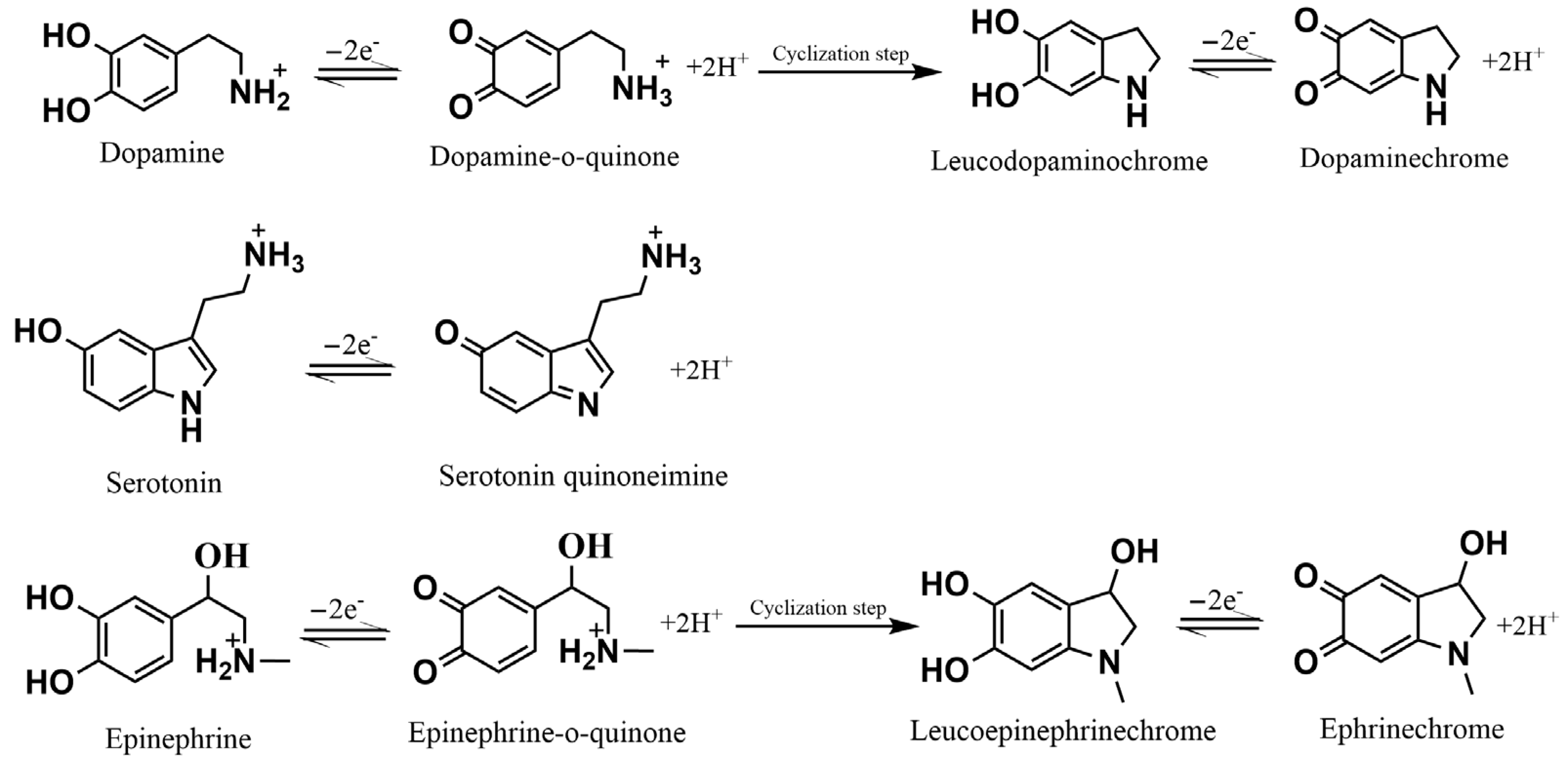
The general oxidation mechanisms of DA, SER and EPI usually proposed in the literature are presented in Figure 2.
This review aims to provide an overview of the recent developments registered in the last decade on the use of electrochemical sensors based on conducting polymers and metal nanoparticles for neurotransmitter detection. The preparation of metal nanoparticles by electrochemical methods directly onto polymeric coatings is mainly addressed as a versatile way for the development of novel electrochemical sensors. A large number of literature reports and research papers was analyzed from this point of view and we are aware that, due to the limited space, not all reported results could be included in the current review. In order to provide a self-consistent approach and content of the scientific discussion of the topic, a short description of the conducting polymers and metal nanoparticles design and properties is firstly introduced, followed by the critical analysis of the proposed electrochemical sensors for neurotransmitter detection. A Conclusion Section summarizing the main achievements and results of the topic, including possible future perspectives, is also included.

2. Electrochemical Sensors

To date, researchers have created multiple NT detection methodologies for in vitro and in vivo analyses to properly understand the nervous system and the association of chemical messengers with altered brain functions: analytical methods (i.e., high-performance liquid chromatography, fast-scan cyclic voltammetry, differential pulse voltammetry, capillary electrophoresis and magnetic resonance spectrometry), optical sensing methods (i.e., fluorescence imaging, colorimetry and chemiluminescence), microdialysis method and positron emission tomography [6,7,8,9]. In vivo applications require the use of high-performance tools due to the presence of very-low levels of NTs in the central nervous system (nM and pM) [6,8]. Real-time concentration measuring is another key factor concerning in vivo detection and the main suitable techniques are high-performance liquid chromatography coupled with microdialysis [18], positron emission tomography [8], magnetic resonance spectrometry [8], electroencephalography [18] and magnetic resonance imaging [18]. In the medical field, a rapid and accurate diagnostic is essential for the access to an optimal treatment, with point-of-care testing meeting all the pre-requisite criteria. These outlined techniques implicate increased costs, qualified personnel and a long time period during the measurements [6]. Thus, electrochemical transduction techniques are currently considered as a good alternative on account of their low costs, device miniaturization, ease of operation, selectivity and sensitivity for biologically active compound determination [6,8,9,11,12,18,19].
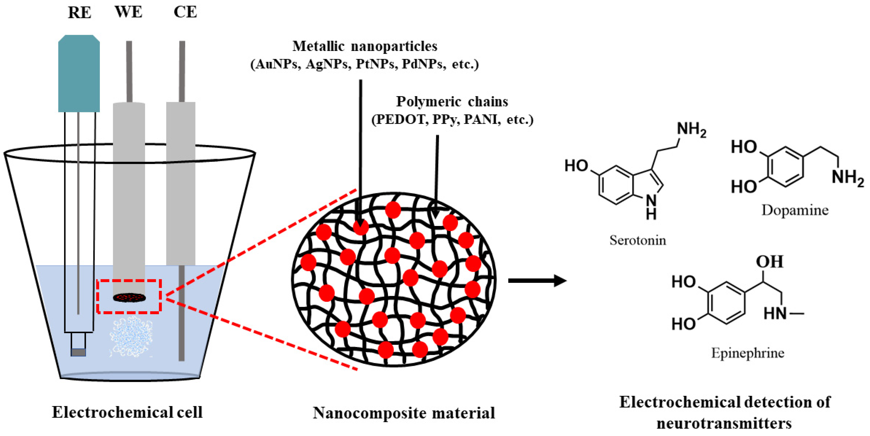
Electrochemical sensors are widely known as analytical devices capable of converting chemical signals into electrical output signals through a transducer, an important constitutive element. The receptor or the sensing element is another integrated part of an electrochemical sensor, providing a proper interaction with the analyte in the form of a redox chemical reaction. This component is responsible for the selectivity towards the analyte of interest in the presence of other interfering species, hence the selected sensing material strongly influences the performance of the sensor [20,21]. In recent decades, nanocomposite materials based on conducting polymers have shown their superiority in terms of enhanced electrocatalytic activity, charge transfer capability, selectivity, sensitivity and reduced fouling of adsorbed species [6,20]. Figure 3 highlights the specific design of nanocomposite-materials-based electrochemical sensors for the electrochemical sensing of the most important NTs in clinical diagnosis.

2.1. Conducting Polymers

Research studies of intrinsically conducting polymers (CPs) started in the 1970s [22] and have been expanded tremendously in the last decade owing to the outstanding properties of these materials, such as electrical conductivity, chemical and mechanical stability, as well as the important applications in various fields, such as batteries, supercapacitors, electrochromic devices, solar cells and sensors [23]. Amongst CPs, polypyrrole (PPy), polyaniline (PANI), polythiophene (PTh) and poly(3,4-ethylenedioxythiophene) (PEDOT) are the most investigated. These CPs have been applied in electrode surface modification for the development of electrochemical sensors through various methodologies that include (i) direct electrochemical polymerization of the corresponding monomers and (ii) chemical polymerization followed by casting of the formed CPs onto electrode surfaces. The modulation of the electrochemical and electrocatalytic properties of CPs deposited as thin layers onto electrode surfaces can be easily achieved in electrolyte solutions during the electrosynthesis or post-synthesis step and ensures the enhancement of the selectivity and sensitivity of the analytical measurements. In this context, the electrical conductivity of CPs can be easily controlled by doping with various dopants during the electrochemical polymerization process. Moreover, the CPs display very good stability during electrochemical cycling between the oxidized (conducting) and the neutral (insulating) forms [22,23]. The deposition of CPs as sensing layers onto electrode surfaces is usually performed by electrochemical and/or chemical polymerization that consists in the formation of a polymeric backbone carrying positive charges that are compensated by the inclusion of anionic species from the polymerization solution. Consequently, the doping ions that could be of inorganic or organic nature markedly influence the final properties of the deposited CP layers, including their structure, morphology and electrical conductivity. In this sense, various surfactants, such as sodium dodecyl sulfate (SDS) and sodium polystyrene sulfonate (PSS), have been investigated as dopants during CP electrodeposition aiming to enhance specific properties, such as the electrical conductivity, electrocatalytic activity, chemical and mechanical stability of the formed polymeric layers [24,25,26]. Alongside surfactants, metallic nanoparticles (MeNPs) have been extensively investigated as dopants in order to enhance mainly the sensitivity of the sensing elements based on CPs. These novel CP-NPs nanocomposite materials have provided peculiar properties such as increased electrochemical-active surface area, electrocatalytic activity towards target analytes and optical properties that could be exploited in the design of novel electrochemical sensors and biosensors. The inclusion of MeNPs into CP layers can be achieved by (i) drop casting of pre-synthesized NPs on top of polymeric layers or (ii) in situ electrochemical deposition of the NPs from solutions containing the appropriate metallic precursors via electrochemical procedures. The first approach benefits from the homogenous size distribution of NPs obtained by the chemical route but suffers from the difficulty in achieving a final homogeneous dispersion of the NPs onto the CP layers. The second approach ensures a straightforward control of the size and distribution of the NPs within the polymeric layers. This review focuses on the procedures for the in situ electrodeposition of MeNPs within the CP matrix by electrochemical methods such as potentiostatic, galvanostatic and potentiodynamic approaches. Among these methodologies, the novel procedures based on the use of alternate voltages and currents are also discussed.

2.2. Noble Metal NPs

Metal nanoparticles have been extensively studied in the last decade due to their outstanding physicochemical properties and they have found applications in a wide range of industrial applications, such as therapeutics and diagnostics, drug delivery systems, energy storage and (bio)sensors technology [27]. Amongst the noble metal nanoparticles (MeNPs), platinum (PtNPs), gold (AuNPs) and silver nanoparticles (AgNPs) have attracted a great deal of interest in the development of selective and sensitive composite materials for (bio)sensor technology thanks to their electrochemical, optical and catalytic properties. MeNPs represent outstanding building blocks in the synthesis of nanostructured composite materials with tunable physicochemical properties. The incorporation of MeNPs within the CP matrix ensures the preparation of a large scale of composite materials displaying novel properties such as increased catalytic activity, enhanced electrochromic properties and chemical stability compared to the pristine components. Thanks to the reduced size and high surface-to-volume ratio, the MeNPs provide enhanced electron transfer capability and catalytic activity of the corresponding sensing composite materials for applications in sensor technology [28].

3. Electrochemical Synthesis Methods of the Composite Material CPs-NPs

3.1. Conventional Methods

The use of nanocomposite materials based on metal nanoparticles in electrochemical sensor technology has attracted special attention in recent years due to the electrochemical, catalytic and optical properties of metal nanoparticles [29]. The in situ synthesis of metal nanoparticles by means of chemical and electrochemical methods had as its main objective the improvement of the analytical performance of the electrochemical sensors. Recent studies have allowed researchers to obtain new information on the influence of nanoparticles on their catalytic, electrochemical and optical properties. Although there is quite a large number of studies and research papers in the field of composite materials based on metal nanoparticles, there is still a special interest in eliminating some disadvantages, such as the agglomeration of metal nanoparticles, reduction in catalytic activity and low chemical stability. In this respect, different methods for the synthesis of nanocomposite materials have been developed both chemically in solutions and by means of electrochemical procedures. It should be noted that the control of the size of the synthesized metal nanoparticles is an extremely difficult major goal to achieve for large-scale applications. The elaboration of new methods for the synthesis of nanocomposite materials allowed the improvement of their physico-chemical properties as well as of the analytical performances of the electrochemical sensors, namely, sensitivity, selectivity, linear response range, detection limit, response time, repeatability and reproducibility.
Electrochemical methods of synthesis of metal nanoparticles involve the use of a metal precursor and its electrochemical reduction by means of potentiostatic procedures, at a modulated [30] or constant potential [31,32,33], on the surface of the electrodes. Another alternative electrochemical synthetic route consists in the galvanostatic control of the electroreduction of the metal precursor under constant current polarization or current pulses. Thus, nanocomposite materials based on metal nanoparticles can be efficiently synthesized on the surface of Pt, Au, semiconductors and carbon or screen-printed electrodes. These electrochemical methods allow a rigorous control of the size, composition and morphology of the nanoparticles. The electric charge used in the in situ synthesis of metal nanoparticles allows an efficient control of the electrochemical process and the estimation of the surface coverage of the electrodeposited metal nanoparticles. The shape and dimensions of the metal nanoparticles depend on the electrochemical parameters, the composition of the deposition solution and the use of surfactants to prevent agglomeration. A disadvantage of these methods is the reduced control of the distribution of nanoparticles on the surface of the electrode.
The use of CPs as immobilization matrix for MeNPs turned out to be a very promising platform in the development of sensitive and selective electrochemical sensors. The CP matrix provides a suitable microenvironment, biocompatibility and stability for the subsequent MeNP immobilization. For instance, PPy decorated with AuNPs has been developed as a sensitive electrochemical platform for serotonin detection [34]. An electrochemical sensor was prepared by a two-step method: in the first step, the polymerization of pyrrole monomer is achieved by multipulse amperometry, followed in the second step by the electrodeposition of AuNPs by scanning the electrode potential from −0.2 to +1.2 V vs. Ag/AgCl in an aqueous solution containing the HAuCl4 precursor for a fixed number of potential cycles. The morphological characterization of the PPy–AuNPs composite material revealed a maximum size of AuNPs of 48 (±5.5) nm. The electrochemical procedure provided a composite PPy–AuNPs coating with an increased electrochemically active surface area and catalytic effect towards serotonin oxidation. A preconcentration step of serotonin at the electrochemical sensor surface resulted in increased sensitivity and selectivity over interfering species such as dopamine and noradrenalin. A low detection limit of 33.22 nM was obtained. The sensor was successfully applied in serotonin detection in serum samples.
The synergy between the CP immobilization matrix and the MeNPs has been exploited in the improvement of the selectivity of the proposed electrochemical sensors for neurotransmitters. The resolution of the analytical signals between various neurotransmitters is a challenging task for the developed electrochemical sensors. On the other hand, there could be a significant interference due to the presence of ascorbic acid and uric acid in real samples. In this sense, various CPs-MeNP-based composite materials were investigated with the aim of improving the selectivity of the analytical measurements. A PANI–AuNPs composite material was developed for the selective detection of dopamine [35]. The in situ electrodeposition of AuNPs onto the PANI layer was achieved by linear sweep voltammetry (LSV) providing a better loading of AuNPs compared to the cyclic voltammetry method (CV). An average size of 65 nm for the AuNPs was observed. The PANI–AuNPs composite material displayed enhanced sensitivity and selectivity in dopamine detection in the presence of ascorbic acid and uric acid with peak potential separations of 108 and 346 mV, respectively. The detection of dopamine occurs at a potential of 0.168 V at a neutral pH. A low detection limit of 16 μM was reported. The overall improved analytical performance was ascribed to the synergic effects of PANI and the in situ electrodeposited AuNPs. The PANI was investigated as a reliable and stable matrix for neurotransmitter electroanalysis in connection with AuNPs towards the development of fast, simple, sensitive and selective electrochemical sensors. The use of screen-printed electrodes as substrates for sensors development ensured the fabrication of single-use sensors [36]. This approach allows the design of disposable sensors for point-of-care testing devices as well as to potential commercially available single-use sensors for the management of chronic diseases. The in situ electrodeposition of Au, Pd and PtNPs onto a PANI coating was successfully achieved in an aqueous solution and the obtained composite materials displayed good electroactivity in alkaline media [37].
Amongst CPs, PEDOT revealed outstanding capability in the incorporation of MeNPs and ensuring the fabrication of sensitive electrochemical platforms. The incorporation of reduced graphene oxide and AgNPs within the PEDOT matrix ensured the preparation of a sensitive electrochemical sensor for serotonin detection [38]. The nanocomposite material displayed increased electron transfer capability and overall improved analytical performance with a low detection limit of 0.1 nM. The proposed sensor ensured the serotonin detection in the presence of some interfering species, such as ascorbic acid, uric acid and tyrosine. The inclusion of carbon-based nanomaterials within the CP matrix alongside MeNPs provided a versatile route for increasing the electron transfer rate and the selectivity and the sensitivity of the analytical measurements. Thus, an electrochemical sensor based on reduced graphene oxide and AuNPs was designed for the selective detection of dopamine [39]. The sensor displayed an increased electroactive surface area and a linear response range of 0.1–100 μM, with a low detection limit value of 0.098 μM.
These results demonstrate the feasibility of the CPs–MeNPs composite materials in the development of fast, reliable, sensitive and selective sensors for neurotransmitter detection. Despite the processability and easy of fabrication of MeNPs by electrochemical methods, there are still important issues to be addressed, such as the polydispersity distribution of MeNPs over the CP matrix, the precise size control of the final metal nanoparticles and the reproducibility from one synthesis to another. However, promising results are encouraging the progress of research in the field of the in situ electrodeposition of MeNPs onto currently available CPs as well as new CPs for electrochemical-sensing applications.

3.2. Innovative Methods

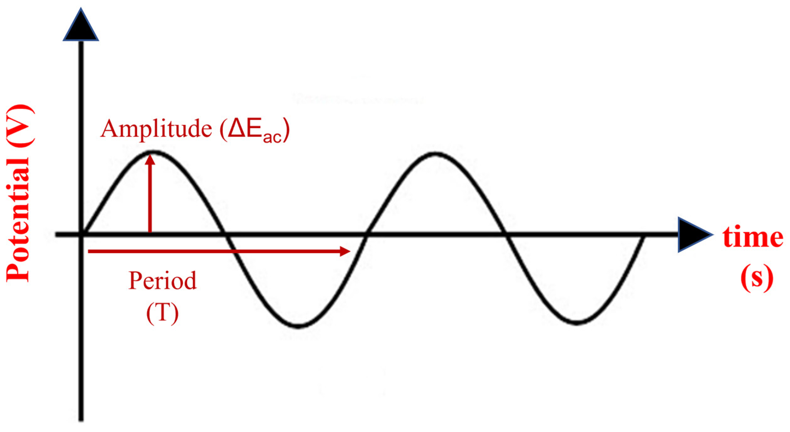
The in situ electrochemical deposition of MeNPs by means of potentiostatic, potentiodynamic and galvanostatic methods represents a straightforward and feasible approach in the design of MeNP-based composite materials for sensing applications. In addition to the electrochemical methods listed above, novel electrochemical preparation procedures have been recently proposed by our research group. The novel procedures consist in the use of a sin wave excitation signal of a selected frequency and amplitude that was superimposed on a constant potential/current. The novel preparation procedures were implemented under potentiostatic or galvanostatic control. In the potentiostatic control, a sinusoidal voltage was superimposed over a constant potential. This approach is referred to as sinusoidal voltage (SV) procedure. The schematic representation of the SV excitation signal is shown in Figure 4, where the frequency of the sinusoidal wave is designated as the reciprocal of the period T and the amplitude is marked as ΔEac. The frequency of the sinusoidal voltage could be scanned in the range from 10 kHz to 0.01 Hz, while the amplitude can be changed between 50 and 350 mV. This SV procedure was applied successfully in the electrodeposition of biocomposite materials based on PEDOT and tyrosinase onto gold microdisk electrodes and their arrays for dopamine electroanalysis [40,41,42,43,44,45]. The incorporation of the enzyme within the PEDOT matrix was achieved thanks to the electrostatic attraction between the polarons on the polymer backbone and the electrical charge of the enzyme at a given pH of the electrodeposition solution. For the sake of comparison, the PEDOT matrix alone was prepared by both potentiostatic and SV procedures [46]. In addition, the PEDOT–tyrosinase biocomposite material was also prepared by potentiostatic and potentiodynamic methods in order to elucidate the benefits brought by the novel SV procedure. The SV procedure ensured a higher porosity and roughness of the PEDOT matrix and an enhanced amount of the immobilized enzymes compared to the classical methods, i.e., the potentiostatic and potentiodynamic approaches. Thanks to the enhanced analytical performance of the biosensors obtained by the SV procedure, the novel procedure was further improved by using sinusoidal voltages of a narrow frequency range or a selected frequency value. It was demonstrated that SV signals of frequencies higher than 1 kHz provided a PEDOT matrix with properties such as those of the potentiostatic or galvanostatic methods, while the SV signals of low frequencies in the range of a tens of millihertz ensured the increased roughness of the PEDOT layer. The selection of the constant potential was optimized according to the electrochemical behavior of the corresponding monomer EDOT during the electrochemical process, and a value of +0.60 V vs. Ag/AgCl/KCl (3 M) ensured a separation between the contribution of the SV excitation signal and the constant potential. In this way, the clear benefit of the SV signal was demonstrated.
The SV procedure was also successfully applied in the in situ electrodeposition of Pt and AuNPs within the PEDOT matrix. The use of SV signals of the selected frequency of 50 mHz and a modulable amplitude provided homogenous distribution and good size control of the electrodeposited metallic nanoparticles. The in situ electrodeposition of Pt and AuNPs was achieved in an aqueous solution from the corresponding metallic precursors within a narrow potential window designed according to the nature of the metallic nanoparticles. The experimental parameters of the SV procedure, namely, the frequency and amplitude of the SV signal, the value of the constant potential and the electrodeposition time, were optimized in order to obtain electrochemical sensors with an improved overall analytical performance. In a first attempt, the PtNPs were prepared onto the PEDOT coating by using a SV signal of a wide frequency range [47]. The PtNPs electrodeposited by means of the SV procedure showed improved catalytic activity in an acidic solution. The morphology of the PtNPs was investigated by means of scanning electron microscopy and atomic force microscopy, proving the successful electrodeposition of the metallic nanoparticles. The wide frequency range of the SV excitation signal was optimized in order to allow a finer control of the size distribution of the metallic nanoparticles. Moreover, the constant potential was judiciously selected upon the voltammetric characterization of the metallic precursor in an aqueous solution at the polymer-modified electrode. The proper selection of the applied constant potential ensured a clear separation of the contribution of the SV excitation signal in the metallic nanoparticles electro-synthesis. In this way, the advantages of the SV procedure could be identified compared to classical potentiostatic and galvanostatic methods. The use of an SV signal with a fixed frequency of 50 mHz provided the successful in situ synthesis of AuNPs with narrow-size distribution homogeneous dispersion within the polymeric PEDOT matrix [48]. The prepared electrochemical sensor enabled the quantification of caffeic acid in food samples with a good analytical performance. The SV procedure based on the fixed frequency excitation signal was also applied in the synthesis of AgNPs [49]. The AgNP-based electrochemical sensor developed by the novel SV procedure demonstrated an enhanced analytical performance compared to other electrochemical sensors prepared by potentiostatic and galvanostatic methods. The AgNPs–PEDOT-based sensor displayed a high sensitivity of 32.2 µM−1 cm−2, limit of detection of 1.9 µM and the quantification limit of 6.5 µM. These results demonstrate the usefulness and the capability of the SV procedure in the development of reliable, sensitive and robust electrochemical sensors based on electrodes modified by CPs–MeNPs composite materials.
In addition to the SV procedure, another innovative preparation method has been established by using a sinusoidal current of a selected frequency superimposed on a constant current. This approach is referred to as the SC procedure. In this case, the optimization of the experimental parameters, in terms of the amplitude and frequency of the excitation sinusoidal current, the value of the constant current and the final electrodeposition time, is very challenging. While the SV procedure provides a straightforward way to control the final potential during the electrosynthesis of conducting polymer coatings and metallic nanoparticles, the SC procedure is very demanding in the establishment of the optimum parameters. Despite these challenging technical issues, we succeeded in the electrosynthesis of a PEDOT–tyrosinase biocomposite coating by using the SC procedure [50,51]. The applied SC signal provides a final potential value in the range of the required value for the electropolymerization of the EDOT monomer or the electrochemical reduction of the metallic precursors in the case of AgNPs/AuNPs. Despite the quite large amplitude of the SC signal, the resulting potential reach the specific value demonstrating a linear behavior of the electrochemical system. The further development of the SV and SC procedures was recently demonstrated by the in situ preparation of PB nanoparticles within the PEDOT layer [52]. The hybrid material was synthesized in two steps by means of the SV and SC procedures: in the first step, the electrochemical polymerization of the EDOT monomer in the presence of ferricyanide ions was performed by SV procedure, while in the second step, the PEDOT coating doped with ferricyanide ions was immersed in a Fe(III)-containing solution and the SV or SC procedure was applied in order to ensure the in situ formation of PBNPs within the PEDOT layer. This approach based on the use of SV and/or SC procedures represents a novel synthetic route for preparing hybrid inorganic–organic materials. The developed electrochemical sensor displayed a good analytical performance towards potassium ion detection compared to commercially available screen-printed electrodes based on PB-containing ink. In conclusion, the novel SV and SC preparation methods provide reliable, fast, cost-effective and straightforward procedures for the design and synthesis of composite materials with applications in (bio)sensor technology.

4. Electrochemical Sensors for Dopamine, Serotonin and Adrenaline Detection

As aforementioned, from a clinical perspective, electrochemical sensors coated with nanocomposite materials constitute the starting point in the development of point-of-care applications for neurological biomarkers due to an increasing trend of neurological disorders in recent decades. Nonetheless, sensor selectivity still remains a major problem concerning the complex composition of human biological fluids with multiple chemical species possessing similar chemical structures [53]. The modulation of composite material properties by intercalating, in the conductive polymeric matrix, various nanomaterials with beneficial physical and chemical properties can acquire a desired synergistic effect for the proper electrochemical detection of NTs.
The current section provides outstanding data with respect to clinically relevant biomolecules, more explicitly, monoamine NTs, the most encountered disease-associated chemical messengers: DA, SER and EPI. The scientific progress of the developed electrochemical sensors modified with nanocomposite materials based on metal nanoparticles from the past 10 years is highlighted with reference to the limit of detection (LOD), the limit of quantification (LOQ), linear response range, sensitivity, selectivity and the electrochemical method implemented.

4.1. Dopamine

DA is a catecholamine that provides important key roles in the central nervous system, being associated with the reward system, attention, learning behavior [54], sleep cycles [55] and motor control functions [55]. By hydroxylating the L-Tyrosine precursor [56] with tyrosine hydroxylase enzyme, L-tyrosine is converted to L-Dihydroxyphenylalanine (L-DOPA), which can be further transformed into DA by the action of the aromatic L-amino acid decarboxylase (DOPA decarboxylase) [54,55]. Therefore, L-Tyrosine is considered the precursor of DA, and DA is also a precursor for other important catecholamines, such as NE and EPI [54]. Disturbances in DA concentration are linked with Parkinson’s disease, Huntington’s disease, schizophrenia [55], drug addiction and memory problems [8]. Table 1 presents the recent developments in electrochemical sensors for DA detection with respect to limit of detection, linear response range, sensitivity and detection method. More attention has been paid to AuNPs for sensor elaboration due to their excellent properties: enhanced electrical conductivity, enhanced specific surface area provided by their reduced dimensions, biocompatibility, the possibility of shape and size modulation and versatility regarding functionalization with various materials and nanomaterials [57].
Ahmed et al. [58] successfully developed an electrochemical sensor with a gold-decorated porous silicon-poly-3-hexylthiophene (Au@PSi-P3HT) nanocomposite material deposited onto a GCE for DA sensing. The nanocomposite material was synthesized by means of chemical stain etching, sonication and photo-reduction processes. The sensor exhibited increased sensitivity (0.5112 µA µM1), linear response range (1–460 µM), good limit of detection (0.63 µM), good reproducibility, repeatability and stability [58]. In contrast, the same group developed an electrochemical sensor modified only with the porous silicon (PSi) for DA determination and it provided unsatisfactory results compared with the nanocomposite material. The beneficial effects of the P3HT polymeric matrix and Au nanoparticles (Au NPs) are clearly seen with regard to the excellent electrocatalytic activity and sensor selectivity, thus providing an enhanced charge transfer capability at the electrode/electrolyte interface [71].
Polymelamine (PM) is considered a very interesting conducting polymer with applications in analytical chemistry. It contains amine groups that provide a good interaction with Au NPs, at the same time preventing their aggregation [72]. Harsini et al. [59] modified a CPE with a composite material consisting of polymelamine (PM) and AuNPs for DA sensing. The polymerization of melamine and the deposition of AuNPs were carried out through electrochemical routes. The good stability and distribution of metallic nanoparticles in the polymeric matrix as well as the excellent selectivity for DA in the presence of uric acid (UA) and ascorbic acid (AA) were successfully assessed. Furthermore, the sensor response was linear in the concentration range of 0.2–11 µM with a LOD of 0.067 µM [59].
Other studies regarding gold-modified electrochemical sensors were conducted with different composite materials in order to verify the electrochemical response toward DA. Chu et al. [60] prepared a composite material consisting of an electrodeposited layer of polyaniline (PAN)-conducting polymer and a self-assembled AuNPs layer on an polyvinylpyridine (PVP)-modified indium tin oxide electrode (ITO). In this study, Au NPs demonstrated a high effectiveness in improving the overall electric conductivity of the composite material and enhancing the oxidation process of the aniline monomer. Moreover, the obtained electrochemical sensor effectively detected DA in the presence of UA, providing a linear response in the concentration range of 1–100 µM and a reduced LOD (0.91 µM) [60]. The electrochemical behavior of DA was also evaluated with a novel composite material introduced by Khudaish et al. [62], namely, a film of poly (2,4,6-triaminopyrmidine) (PTAP) modified with AuNPs.
According to the literature, tri-composite materials based on Au NPs, conducting polymers and sp2-hybridized carbon nanomaterials [73] have gained popularity for sensing applications. Inagaki et al. [61] employed a liquid–liquid interfacial reaction for the synthesis of a thin film of polythiophene (PT), Au NPs and carbon nanotubes (CNTs). The main advantage of the film is that it can be easily transferable to any substrate, in this case, an ITO/glass-working electrode. CNTs act as stabilizers for the polymer matrix and together with AuNPs can enhance the charge transfer capability and the electroactive surface area of the electrode. It was concluded that a smaller amount of AuNPs generates the optimum response for DA with a linear response in the concentration range of 1–10 µM, a good sensitivity (19.492 µA µM1) and a reduced LOD (0.69 µM) [61]. Pan et al. [63] pointed out that the reduced conductivity of an overoxidized poly (3,4-ethylenedioxythiophene) (OPEDOT) film can be overcome with the intercalation of other conductive materials: Au NPs and electrochemically reduced graphene oxide (ERGO). The obtained electrode, OPEDOT-AuNPs-ERGO/GCE, successfully demonstrated its selectivity for DA in the presence of UA and AA with remarkable results: 1 µM LOD and 4–100 µM linear response range [63].
AgNPs, as well as AuNPs, have been employed in electrochemical-sensing applications by reason of their low costs, simple synthesis reactions, biocompatibility, high specific surface area, shape and size variations and adequate stability [74]. Paulraj et al. [64] modified a GCE with Ag-doped PANI nanocomposites for H2O2 and DA detection. The composite was synthesized by means of a mechano-chemical oxidative polymerization process. A PANI-conducting polymer, which acts as a substrate for AgNPs, prevents their agglomeration and can also interact with the analyte (DA) through hydrogen bonding. Hence, the sensor provided a linear response range for DA of 10–90 µM, a low LOD (1.9 µM) and a great sensitivity (0.102 µA µM1) [64]. Even though AuNPs are implemented much more frequently, copper nanoparticles (CuNPs) [67], palladium nanoclusters [68,70] and PtNPs [69] have also been explored as potentially useful sensing materials.
In addition to the utility of conducting polymers and metallic nanoparticles in developing electrochemical sensors, research in the electrochemical engineering branch also focuses on surfactants or doping ions, such as SDS, PSS and poly (2-acrylamido-2-methyl-1-sulfonate) (PAMPS). Their intercalation in the polymeric backbone alongside metallic nanoparticles causes an enhanced interaction with the analyte, thus providing outstanding sensing properties of the composite material [26]. For instance, Atta et al. [31] prepared an electrochemical sensor with a composite film consisting of Au NPs, PEDOT and SDS-doping ions for DA determination. The experimental results demonstrated a high diffusion of DA in the electrodeposited material and an increased charge transfer capability through the interaction of DA cations with the amino groups (-NH2) of Au and the electrostatic interactions of DA cations with the anionic surfactant [31]. At present, the presence of surfactants is considered as a great benefit concerning the proper interaction with biologically active compounds, the excellent electrochemical properties generated and the synergism created between them and metal nanoparticles in terms of enhanced sensitivity.
Since 2011, two-dimensional materials based on metal carbides, nitrides and carbonitrides (MXene) have demonstrated outstanding properties for electrochemical sensors/biosensors development: increased surface area and conductivity, the presence of variable hydrophilic groups and the possibility of composition variation. Notably, DA has been the center of great interest for analytical applications [75]. Ni et al. [76] proposed a composite material consisting of titanium carbide (Ti3C2), graphitized MWCNTs and ZnO nanospheres. The Ti3C2/G-MWCNTs/ZnO NSP sensor detected DA on a wide linear range of 0.01–30 µM, with a LOD of 3.2 nM. Moreover, the superior anti-interference properties were evaluated in the presence of Gly, oxalic acid, ascorbic acid, uric acid, leucine, alanine and 5-fold glucose [76]. Another recent study by Zheng et al. [77] focused on a novel composite material comprised of Ti3C2 nanosheets and Pd/Pt NPs attached through a DNA template on a GCE for DA detection. The 1:1.1:2.5 mass ratio of Ti3C2, Pd and Pt was responsible for the excellent sensitivity (1.05 mA mM1cm2), large concentration range (0.2–1000 µM) and reduced LOD (30 nM). The developed sensor displayed increased selectivity, sensitivity, accuracy and repeatability [77]. Ankitha et al. [78] implemented a hydrothermal process to create a composite material based on Nb2CTx MXene and MoS2 deposited on a carbon cloth for DA sensing. The author demonstrated that the 12.5% MoS2 composition provided remarkable sensing properties towards DA with a wide concentration range (1 fM–100 µM) and an excellent LOD (0.23 fM) [78]. Composite materials based on MXene are widely explored because they can enhance the overall stability of materials by preventing MXene sheet stacking, the conductive properties and the active surface area [75].

4.2. Serotonin

5-hydroxytryptamine or serotonin is a monoamine NTs that modulates important physiological and neuronal functions: appetite, sleeping behaviors, mood and learning capacity [6]. Most psychiatric disorders (e.g., psychosis, schizophrenia and depression) [6] are influenced by changes in SER concentration values, but a major advantage for the world of psychiatry lies in the discovery of serotonin re-uptake inhibitors, the most prescribed antidepressants [79]. L-tryptophan amino acid is the precursor of SER, the biosynthesis process characterized as a two-step reaction: 5-hydroxytryptophan (5-HTP) is produced from L-tryptophan amino acid with the action of tryptophan hydroxylase enzyme followed by a decarboxylation process through the action of aromatic amino acid decarboxylase. This final step initiates the biosynthesis of SER, also known as the happiness hormone [80].
Taking into account the essential role of serotonin in the organism and the impaired quality of life associated with its imbalance, simple and efficient measurements have been sought. For this aim, the synergism created between metal nanoparticles and conducting polymers in electrochemical sensors can enhance the analytical properties for the investigation of real samples. In order to gain a general perspective of the most recently developed electrochemical sensors for SER sensing, in real and synthetic probes, we summarized the most significant research studies in Table 2.
AuNPs are still considered an optimum choice for sensor development, with a notable number of research papers providing detailed and promising results for NT detection in synthetic and biological samples. Tertiș et al. [34] prepared a novel electrochemical sensor based on a graphite screen-printed electrode (GSPE) modified with PPyNPs and AuNPs for the efficient detection of SER. The AuNPs@PPy/GSPE sensor displayed a LOD of 33.22 nM with a linear response in the concentration range of 0.1–15 µM and a good sensitivity (0.3316 µA µM1), mainly attributed to the presence of AuNPs that enhance the active surface area of the electrode. It was tested in the presence of interfering species (ascorbic acid, paracetamol and acetylsalicylic acid) with promising results and also in human serum samples with good recovery times [34]. A study of Li et al. [82] reported the modification of a GCE with a composite material consisting of over oxidized-polypyrrole (PPyox) and AuNPs for SER and DA electrochemical sensing. The Au nanoclusters were electrochemically synthesized with mean diameters of 80 nm and a homogenous distribution. The sensor efficiently responded towards SER in the concentration range of 7 × 10⁻³–2.2 µM with a LOD of 0.001 µM, in 0.1 M PBS, pH = 7 [82].
An interesting approach is attributed to Sadanandhan et al. [83] and constitutes the electrodeposition of polyaniline nanowires (PANIS) on the surface of a GCE, followed by the electrodeposition of AuNPs on the surface of PANIS/GCE through chronoamperometry (CA). PANIS nanostructures were assembled with a template of anilinium-3-pentadecyl phenyl sulphonic acid by template electrochemical polymerization. The obtained sensor demonstrated its efficiency for SER sensing over a wide concentration range, 0.3–1000 µM, with a LOD of 0.025 µM, comparable with the new research data from the literature. Moreover, it simultaneously detected SER, DA, AA and UA in 0.1 M PBS (pH = 7.4) and human serum samples, demonstrating its potential application for NT sensing [83]. In general, nanowire nanocomposites based on conducting polymers and metal nanoparticles are seen as high-performance materials useful for the development of sensors, supercapacitors, light emitting diodes and batteries [90].
Complex sensing materials with multilayered nanocomposites are desirable for NT determination because each material exhibits a different chemical or physical property that can further enhance the overall analytical response of the sensor. Chung et al. [81] simultaneously detected SER and DA with an electrochemical sensor composed of bottom and upper substrates. The bottom substrate consisting of AuNPs@rGO was drop-casted on the surface of an SPCE. In the next step, the upper substrate formation, the monomer 2,2:5,2-terthiophene-3-(p-benzoic acid) (TBA), was electropolymerized to pTBA by cycling the potential between 0 and +1.4V (vs. Ag/AgCl) in a solution of 0.1 M PBS, pH = 7.4. Moreover, in order to form a catalytic material, a palladium complex (Pd (C2H4N2S2)2) was chosen for covalent bonding with the conducting polymer, pTBA, in the upper substrate. The best current response was ascribed to a concentration of 1 mg/mL AuNPs@rGO and the optimum thickness of the pTBA layer was achieved during three cycles of electropolymerization. The AuNPs@rGO/pTBA-Pd (C2H4N2S2)2/SPCEs sensor is useful for SER detection in a wide concentration range, 0.02–200 µM, and it has a reduced LOD of 2.5 nM. Additionally, the SER concentration was also evaluated from cancerous cells (MCF7) and non-cancerous cells (MCF10A) [81]. Xue et al. [84] proposed a novel material with a double-layered membrane of rGO/PANI nanomaterials and molecularly imprinted polymers (MIPs) modified with AuNPs for SER sensing in a 0.1 M PBS (pH = 7.5) solution and human serum probes. The first layer, the rGO/PANI nanomaterial, was obtained via electrostatic adsorption and electrodeposited on the surface of a GCE via cyclic voltammetry by cycling the potential between −0.2 and 0.8 V. The second layer, AuNPs@MIPs, was prepared at a constant potential of 0.9 V, in an electrolyte solution containing SER, AuNPs, p-aminothiophenol (p-ATP) and PBS solution. The presence of the rGO/PANI nanocomposite with a diameter of 93 nm imparted a high specific surface area, good stability and conductivity, and the presence of AuNPs@MIPs enhanced the sensor conductivity and thus the overall sensitivity. Notably, the rGO/PANI/AuNPs@MIP sensor detected SER in 0.1 M PBS (pH = 7.5) on a wide concentration range, 0.2–10 µM, with a lower LOD of 11.7 nM [84].
Several studies expressed the beneficial effects of ternary nanocomposites for SER-sensing applications. For instance, Selvarajan et al. [85] implemented the ultrasonic irradiation technique to generate a uniform layer of ternary nanocomposites, Cu2O NPs, AgNPs and PPy by drop-casting it onto a GCE. The Ag/Ppy/Cu2O/GCE sensor with a mean nanocomposite size of 20 nm showed higher electrocatalytic properties compared with unmodified GCE, PPy/GCE and Cu2O/Ppy/GCE sensors. Furthermore, linear dependences were acquired between the peak currents and the analyte concentration in the range of 0.01–250 µM with a reduced LOD of 0.0124 µM [85]. Other electrochemical sensors based on ternary nanocomposites from the literature intercalate AgNPs with multi-walled carbon nanotubes (MWCNTs) and a PPy-conducting polymer [86] or with PEDOT-conducting polymer and rGO [38].
Nonetheless, various complex electrochemical sensors based on nanocomposite materials successfully detected SER in real and synthetic probes with high accuracy, selectivity and sensitivity, thus providing a potential application for point-of-care testing in the medical area. The presence of conducting polymers assures an anti-biofouling effect and good selectivity [91], whereas metal nanoparticles enhance the sensitivity [92]. The best electrochemical capacities were established through the optimization of working parameters, such as buffer concentration, pH, temperature and type and concentration of the supporting electrolytes, and the by the use of sensitive electrochemical techniques, such as square wave voltammetry (SWV) and DPV [93].

4.3. Epinephrine/Adrenaline

EPI or adrenalin is a catecholamine NT produced in adrenal glands (outside the nervous system) and axon terminals (inside the nervous system). For this reason, it is considered both a hormone and a NT [19,94]. Its effects are mostly encountered during fight-or-flight responses, a condition caused by a very stressful situation where the organism produces several effects: elevated blood glucose levels, tachycardia, mydriasis and blood transport to vital areas needed for survival, such as muscles, lungs and heart [95,96]. EPI is biosynthesized from the same precursor as DA, L-tyrosine, by the action of phenylethanolamine-N-methyltransferase on NE [54]. NE or noradrenalin is also a catecholamine, the precursor of EPI, and is mainly associated with stressful situations [94]. It provides similar effects as EPI in the organism and its reduced levels in the brain can cause depressive episodes [95].
The immediate identification of EPI in human biological samples is of primordial importance due to an increase in neurological disorders in recent decades. For this aim, simple and reliable techniques are required to obtain a proper diagnosis, to avoid unpleasant complications or just to improve life quality. Electrochemical sensors based on nanocomposite materials can check all these criteria, with miniaturized sensors being an excellent option in physiological monitoring. Table 3 summarizes the main research studies of nanocomposite-materials-based electrochemical sensors for EPI detection in real and synthetic probes with high accuracy, stability, reproducibility, selectivity and sensitivity.
Liu et al. [97] reported the electrochemical detection of EPI with an electrochemical sensor consisting of MIP and AuNPs. Firstly, AuNPs were electrodeposited on the surface of a GCE from a solution of 4 mM HAuCl4 by applying a constant potential of −0.2 V for 500 s. Subsequently, the MIP/AuNP-sensing material was achieved by immersing previously formed AuNPs/GCE in a solution of 0.1 M PBS (pH = 6), the monomer, 3-thiophene boronic acid (3-TBA) and EPI, followed by the removal of EPI with 0.05 M HCl. Several parameters were optimized for a proper electrochemical response: pH = 6 for the electropolymerization solution, the effective mole ratio 3-TBA: EPI was 4:1, 20 electropolymerization cycles and 50 mV/s scan rate. In these optimum working conditions, the MIP/AuNPs/GCE sensor demonstrated its superiority in terms of a wide linear range (0.09–100 µM), LOD (0.07 µM), good reproducibility, stability and selectivity in the presence of interfering species (AA, UA and DA) [97].
The following studies describe several designs of electrochemical sensors based on PPy-conducting polymers and AuNPs for EPI sensing, the most extensively used materials. PPy has a good electrical conductivity, stability, ease of fabrication and surface modification, large surface area and low charge-transfer resistance [108,109]. However, the intercalation of AuNPs with a multitude of shapes and sizes assures an improvement of surface-to-volume ratio, electrocatalytic properties and redox behavior for multiplex assays [57]. Ghanbari et al. [98] proposed a chemical modification of a GCE with a nanocomposite material consisting of rGO, zinc oxide nano-sheets (ZnO) and AuNPs for EPI sensing in pharmaceutical and real samples. Scanning electron microscopy (SEM) images revealed a uniform distribution of AuNPs and PPy nanofibers with mean diameters of 100 nm on the surface of rGO sheets and ZnO nano-sheets with 1 µm diameter. The Au/ZnO/PPy/RGO/GCE sensor simultaneously detected AA, EPI and UA, providing high oxidation currents in 0.1 M PBS (pH = 7). Regarding EPI, a 0.06 µM LOD was obtained with a linear current increase in the concentration range of 0.6–500 µM [98]. Lu et al. [101] modified a gold electrode (AuE) with multilayers of single-walled carbon nanotubes (SWCNTs)/AuNPs and PPy NPs through the layer-by-layer method, a thin-film fabrication technique. The ternary nanocomposite effectively separated the oxidation peak potentials of UA and AA interferents from EPI. As a result, the sensor is useful for the selective determination of EPI in 0.05 M PBS (pH = 7), human plasma, serum and urine [101]. Another similar study of Mao et al. [99] described the use of poly (ionic liquids) that act as binding agents between AuNPs with 13–16 nm diameter and PPy nanotubes (PPyNTs), drop-casted on the surface of a GCE. In optimum conditions (0.05 M PBS with pH = 7.4, 20 °C), the Au/PILs/PPyNTs/GCE sensor response towards EPI was linear in the concentration range of 0.035–0.960 nM, with a LOD of 298.9 nM and a good sensitivity of 42.7799 µA/mM. Furthermore, the CA method was implemented to investigate the interference behavior in the presence of EPI, glucose, D-fructose, sucrose, citric acid, UA, AA and DA. The results showed the anti-interferent capability of the sensor as well as its good stability and sensitivity [99].
Alternatively, Yousif et al. [106] proposed novel textile-based sensors comprising rGO/Ag nanocomposites prepared by the electron-beam irradiation technique, followed by the pad-dry-cure method to impregnate the nanomaterials in cotton and polyester textiles. CV, LSV, DPV and SWV methods were implemented to evaluate the electrochemical behavior of rGO/AgNP/cotton and rGO/AgNP/polyester sensors. From the LSV results, it was concluded that the rGO/AgNP/cotton sensor possessed the highest current density associated with an increased selectivity compared with the rGO/AgNP/polyester sensor. This occurs as a consequence of the enhanced structural porosity of the cotton fabric. The highest sensitivities were achieved by using SWV and DPV methods. The prepared rGO/AgNP/cotton and rGO/AgNP/polyester sensors exhibited wide linear ranges in the concentration ranges of 0.5–40 µM and 1–30 µM as well as reduced LODs, 9.73 nM and 3.05 nM, respectively [106]. In terms of electrode material, boron-doped diamond (BDD), a p-type semiconductor, has also been explored for EPI sensing owing to its specific properties: chemical and mechanical stability, low background currents and high potential window [14,110].
The present section successfully described the most notable research studies based on various chemically or electrochemically developed nanocomposite materials with electrocatalytic effects for EPI determination. Most researchers used DPV and SWV voltammetric detection methods, instead of CV and LSV, due to their low capacitive current [93] and improved selectivity [111]. However, the real-time monitoring of NTs in the brain area (in vivo) requires the employment of fast-scan cyclic voltammetry (FSCV), an electrochemical technique that measures NT release on a millisecond time scale per scan [112,113]. Moreover, it should be noted that the electrode material is crucial for biological measurements. Usually, carbon-based electrodes are desirable for NT sensing due to their biocompatibility, conductivity, good chemical and spatial resolution [114,115]. Another essential parameter that must be considered while designing implantable sensors for NT monitoring is the size of the electrode. FSCV conducted on conventional GCEs produces large background currents and can also damage the neuronal tissue. For this aim, carbon microelectrodes and nanoelectrodes are sought. Nonetheless, for the long-term monitoring of NTs, the selectivity, sensibility and antifouling capability must be improved and the large background charging currents removed [113,114]. In optimum working conditions, the electrochemical sensors displayed good sensitivity, LOD, stability and reproducibility coupled with low costs, ease of operation and the possibility of device miniaturization for the manufacturing of portable devices.

5. Conclusions and Future Perspectives

The monitoring of neurotransmitters remains a challenging task for analytical chemists and clinicians due to the great importance of neurotransmitters level in the management of chronic diseases, such as Parkinson’s and Alzheimer’s diseases. The development of novel analytical tools based on electrodes modified with nanocomposite materials remains very active in the last decade. Amongst the most investigated analytical tools for neurotransmitters determination, electrochemical sensors based on CPs-MeNP-sensing materials demonstrated improved sensitivity and selectivity. The ease of fabrication, low-cost materials, enhanced electrochemical and electrocatalytic properties and the possibility of functionalization of MeNP-bearing sensing materials represent the current technological achievement in the above-discussed research topic. The developed electrochemical sensors have outstanding capabilities in the monitoring of neurotransmitters with the very-low detection limits and linear response range that are required for real sample analysis. The complex composition of biological samples containing several major interfering species such as ascorbic acid, uric acid and tyrosine has been successfully addressed by using CPs–MeNP-based sensing elements. The selectivity of the analytical measurements is a challenging task in real sample analysis and the proposed electrochemical sensors demonstrated very good anti-interference capability for dopamine, serotonin and epinephrine detection in both synthetic and complex matrices. In addition to the selectivity factor, the very-low level of neurotransmitters in biological fluids represents another difficult issue in the development of novel electrochemical sensors. Thanks to the outstanding properties of MeNPs, mainly Au, Pt and AgNPs, the sensitivity of the measurements has been greatly enhanced, with the achievement of very-low detection limits in the range of nM and pM range being possible.
The use of various electrode substrates, ranging from conventional electrodes such as glassy carbon, metal or semiconductors to screen-printed electrodes, has paved the way to the development of single-use, disposable sensors with a great potential for commercialization. The versatility of the preparation methods for CPs–MeNPs ensured their successful immobilization over the wide range of substrates mentioned above, but also including flexible, textile-based substrates for patch sensor design. The electrochemical in situ reduction of metal precursors over polymeric coatings showed great potential for electrochemical sensor development by a finer control of the size distribution and polydispersity of the metal nanoparticles. Novel electrochemical preparation methods based on sinusoidal voltages and currents for CPs–MeNP synthesis have also been developed. The synergic effects of the conducting polymers and the metal nanoparticles resulted in an overall increased analytical performance. In addition, the processability, the good mechanical and chemical stability of the prepared CPs–MeNP-sensing materials as well as of new materials based on graphdiyne quantum dots enhanced their analytical applications [116,117,118]. All these efforts were devoted to the fabrication of fast, sensitive, selective, reliable and versatile electrochemical sensors for the real-time monitoring of neurotransmitters. The increasing impact of neurological diseases prognosed for the coming years underpins the research on the topic of electrochemical sensors for neurotransmitter detection. Despite the great achievements reported in the last decade, there are still challenging issues to be addressed such as the improved reproducibility of the CPs–MeNP synthesis from one batch to another, improved selectivity and sensitivity, enhanced antifouling properties of the sensing composite materials, miniaturization and fast integration with smart electronics for the real-time monitoring and collection of analytical data. Additionally, the design of novel conducting polymers will provide a suitable and stable microenvironment matrix for metal nanoparticle in situ electrodeposition in the search for novel electrochemical sensors. Finally, the development of implantable electrochemical sensors for the real-time monitoring of neurotransmitters and their integration within internet-of-things devices and applications constitute the most challenging tasks to be achieved in the near future.

Author Contributions

Conceptualization, S.-A.L., C.L. and S.L.; writing—original draft preparation, S.-A.L. and S.L.; writing—review and editing, S.-A.L., C.L. and S.L.; supervision, S.L.; project administration, S.L.; funding acquisition, S.L. All authors have read and agreed to the published version of the manuscript.

Funding

The authors thank the Ministry of Research, Innovation and Digitization, CCCDI—UEFISCDI, for the grant PN-III-P2-2.1-PED-2021-3693 (607PED/27.06.2022), within the PNCDI III program.

Institutional Review Board Statement

Not applicable.

Informed Consent Statement

Not applicable.

Data Availability Statement

Not applicable.

Acknowledgments

This work was supported by a grant of the Ministry of Research, Innovation and Digitization, CCCDI—UEFISCDI, project number PN-III-P2-2.1-PED-2021-3693, within PNCDI III.

Conflicts of Interest

The authors declare no conflict of interest.

References

  1. Moon, J.M.; Thapliyal, N.; Hussain, K.K.; Goyal, R.N.; Shim, Y.B. Conducting polymer-based electrochemical biosensors for neurotransmitters: A review. Biosens. Bioelectron. 2018, 102, 540–552. [Google Scholar] [CrossRef] [PubMed]
  2. Tavakolian-Ardakani, Z.; Hosu, O.; Cristea, C.; Mazloum-Ardakani, M.; Marrazza, G. Latest trends in electrochemical sensors for neurotransmitters: A review. Sensors 2019, 19, 2037. [Google Scholar] [CrossRef] [PubMed] [Green Version]
  3. Laughlin, S.B.; Sejnowski, T.J. Communication in neuronal networks. Science 2003, 301, 1870–1874. [Google Scholar] [CrossRef] [Green Version]
  4. Herculano-Houzel, S. The human brain in numbers: A linearly scaled-up primate brain. Front. Hum. Neurosci. 2009, 3, 1–11. [Google Scholar] [CrossRef] [PubMed] [Green Version]
  5. McCoy, A.N.; Tan, S.Y. Otto Loewi (1873–1961): Dreamer and Nobel laureate. Singapore Med. J. 2014, 55, 3–4. [Google Scholar] [CrossRef] [Green Version]
  6. Banerjee, S.; McCracken, S.; Faruk Hossain, M.; Slaughter, G. Electrochemical Detection of Neurotransmitters. Biosensors 2020, 10, 101. [Google Scholar] [CrossRef]
  7. Niyonambaza, S.D.; Kumar, P.; Xing, P.; Mathault, J.; De Koninck, P.; Boisselier, E.; Boukadoum, M.; Miled, A. A Review of neurotransmitters sensing methods for neuro-engineering research. Appl. Sci. 2019, 9, 4719. [Google Scholar] [CrossRef] [Green Version]
  8. Su, Y.; Bian, S.; Sawan, M. Real-time: In vivo detection techniques for neurotransmitters: A review. Analyst 2020, 145, 6193–6210. [Google Scholar] [CrossRef]
  9. Perry, M.; Li, Q.; Kennedy, R.T. Review of recent advances in analytical techniques for the determination of neurotransmitters. Anal. Chim. Acta 2010, 653, 1–22. [Google Scholar] [CrossRef] [Green Version]
  10. Baluta, S.; Zając, D.; Szyszka, A.; Malecha, K.; Cabaj, J. Enzymatic platforms for sensitive neurotransmitter detection. Sensors 2020, 20, 423. [Google Scholar] [CrossRef] [Green Version]
  11. Suppiramaniam, V.; Bloemer, J.; Reed, M.; Bhattacharya, S. Neurotransmitter Receptors. Compr. Toxicol. Third Ed. 2018, 6–15, 174–201. [Google Scholar] [CrossRef]
  12. Marc, D.T.; Ailts, J.W.; Campeau, D.C.A.; Bull, M.J.; Olson, K.L. Neurotransmitters excreted in the urine as biomarkers of nervous system activity: Validity and clinical applicability. Neurosci. Biobehav. Rev. 2011, 35, 635–644. [Google Scholar] [CrossRef]
  13. Feigin, V.L.; Nichols, E.; Alam, T.; Bannick, M.S.; Beghi, E.; Blake, N.; Culpepper, W.J.; Dorsey, E.R.; Elbaz, A.; Ellenbogen, R.G.; et al. Global, regional, and national burden of neurological disorders, 1990–2016: A systematic analysis for the Global Burden of Disease Study 2016. Lancet Neurol. 2019, 18, 459–480. [Google Scholar] [CrossRef] [Green Version]
  14. Sanghavi, B.J.; Wolfbeis, O.S.; Hirsch, T.; Swami, N.S. Nanomaterial-based electrochemical sensing of neurological drugs and neurotransmitters. Microchim. Acta 2015, 182, 1–41. [Google Scholar] [CrossRef] [Green Version]
  15. Rajarathinam, T.; Kang, M.; Hong, S.; Chang, S. Nanocomposite-Based Electrochemical Sensors for Neurotransmitters Detection in Neurodegenerative Diseases. Chemosensors 2023, 11, 103. [Google Scholar] [CrossRef]
  16. Kaur, H.; Siwal, S.S.; Saini, R.V.; Singh, N.; Thakur, V.K. Significance of an Electrochemical Sensor and Nanocomposites: Toward the Electrocatalytic Detection of Neurotransmitters and Their Importance within the Physiological System. ACS Nanosci. Au 2022, 3, 1–27. [Google Scholar] [CrossRef]
  17. Lakard, S.; Pavel, I.A.; Lakard, B. Electrochemical biosensing of dopamine neurotransmitter: A review. Biosensors 2021, 11, 179. [Google Scholar] [CrossRef]
  18. Hugo, V.; Castro, C.; Lucía, C.; Valenzuela, L.; Carlos, J.; Sánchez, S.; Peña, K.P.; Pérez, S.J.L.; Ibarra, J.O.; Villagrán, A.M. An Update of the Classical and Novel Methods Used for Measuring Fast Neurotransmitters during Normal and Brain Altered Function. Curr. Neuropharmacol. 2014, 12, 490–508. [Google Scholar] [CrossRef] [Green Version]
  19. Labib, M.; Sargent, E.H.; Kelley, S.O. Electrochemical Methods for the Analysis of Clinically Relevant Biomolecules. Chem. Rev. 2016, 116, 9001–9090. [Google Scholar] [CrossRef]
  20. Hulanicki, A.; Stanislav, G.; Folke, I. Chemical sensors: Definitions and classification. Pure Appl. Chem. 1991, 63, 1247–1250. [Google Scholar] [CrossRef]
  21. Baranwal, J.; Barse, B.; Gatto, G.; Broncova, G.; Kumar, A. Electrochemical Sensors and Their Applications: A Review. Chemosensors 2022, 10, 363. [Google Scholar] [CrossRef]
  22. Diaz, A.F.; Castillo, J.I.; Logan, J.A.; Lee, W.-Y. Electrochemistry of conducting polypyrrole films. J. Electroanal. Chem. 1981, 129, 115–132. [Google Scholar] [CrossRef]
  23. Inzelt, G. Conducting polymers. A new era in electrochemistry. In Monographs in Electrochemistry, 2nd ed.; Scholz, F., Ed.; Springer: Berlin/Heidelberg, Germany, 2012; ISBN 978-3-642-27620-0. [Google Scholar]
  24. Lyutov, V.; Efimov, I.; Bund, A.; Tsakova, V. Electrochemical polymerization of 3,4-ethylenedioxythiophene in the presence of dodecylsulfate and polysulfonic anions—An acoustic impedance study. Electrochim. Acta 2014, 122, 21–27. [Google Scholar] [CrossRef]
  25. Lyutov, V.; Gruia, V.; Efimov, I.; Bund, A.; Tsakova, V. An acoustic impedance study of PEDOT layers obtained in aqueous solution. Electrochim. Acta 2016, 190, 285–293. [Google Scholar] [CrossRef]
  26. Tsakova, V.; Seeber, R. Conducting polymers in electrochemical sensing: Factors influencing the electroanalytical signal. Anal. Bioanal. Chem. 2016, 408, 7231–7241. [Google Scholar] [CrossRef]
  27. Darabdhara, G.; Das, M.R.; Singh, S.P.; Rengan, A.K.; Szunerits, S.; Boukherroub, R. Ag and Au nanoparticles/reduced graphene oxide composite materials: Synthesis and application in diagnostics and therapeutics. Adv. Colloid Interface Sci. 2019, 271, 101991. [Google Scholar] [CrossRef]
  28. Qiao, Z.; Zhang, J.; Hai, X.; Yan, Y.; Song, W.; Bi, S. Recent advances in templated synthesis of metal nanoclusters and their applications in biosensing, bioimaging and theranostics. Biosens. Bioelectron. 2021, 176, 112898. [Google Scholar] [CrossRef]
  29. Zhao, X.; Zhao, H.; Yan, L.; Li, N.; Shi, J.; Jiang, C. Recent Developments in Detection Using Noble Metal Nanoparticles. Crit. Rev. Anal. Chem. 2020, 50, 97–110. [Google Scholar] [CrossRef]
  30. Espino-López, I.E.; Romero-Romo, M.; de Oca-Yemha, M.G.M.; Morales-Gil, P.; Ramírez-Silva, M.T.; Mostany, J.; Palomar-Pardavé, M. Palladium Nanoparticles Electrodeposition onto Glassy Carbon from a Deep Eutectic Solvent at 298 K and Their Catalytic Performance toward Formic Acid Oxidation. J. Electrochem. Soc. 2019, 166, D3205–D3211. [Google Scholar] [CrossRef]
  31. Atta, N.F.; Galal, A.; El-Ads, E.H. Gold nanoparticles-coated poly(3,4-ethylene-dioxythiophene) for the selective determination of sub-nano concentrations of dopamine in presence of sodium dodecyl sulfate. Electrochim. Acta 2012, 69, 102–111. [Google Scholar] [CrossRef]
  32. Tian, J.; Peng, D.; Wu, X.; Li, W.; Deng, H.; Liu, S. Electrodeposition of Ag nanoparticles on conductive polyaniline/cellulose aerogels with increased synergistic effect for energy storage. Carbohydr. Polym. 2017, 156, 19–25. [Google Scholar] [CrossRef]
  33. Agrisuelas, J.; González-Sánchez, M.I.; Valero, E. Hydrogen peroxide sensor based on in situ grown Pt nanoparticles from waste screen-printed electrodes. Sens. Actuators B Chem. 2017, 249, 499–505. [Google Scholar] [CrossRef]
  34. Tertiș, M.; Cernat, A.; Lacatiș, D.; Florea, A.; Bogdan, D.; Suciu, M.; Săndulescu, R.; Cristea, C. Highly selective electrochemical detection of serotonin on polypyrrole and gold nanoparticles-based 3D architecture. Electrochem. Commun. 2017, 75, 43–47. [Google Scholar] [CrossRef]
  35. Mahalakshmi, S.; Sridevi, V. In Situ Electrodeposited Gold Nanoparticles on Polyaniline-Modified Electrode Surface for the Detection of Dopamine in Presence of Ascorbic Acid and Uric Acid. Electrocatalysis 2021, 12, 415–435. [Google Scholar] [CrossRef]
  36. Selvolini, G.; Lazzarini, C.; Marrazza, G. Electrochemical nanocomposite single-use sensor for dopamine detection. Sensors 2019, 19, 3097. [Google Scholar] [CrossRef] [Green Version]
  37. Hatchett, D.W.; Quy, T.; Goodwin, N.; Millick, N.M. In-Situ Reduction of Au, Pd, and Pt Metal Precursors in Polyaniline: Electrochemistry of Variable Metal Content Polymer/Metal Composites in Alkaline Solution. Electrochim. Acta 2017, 251, 699–709. [Google Scholar] [CrossRef]
  38. Sadanandhan, N.K.; Cheriyathuchenaaramvalli, M.; Devaki, S.J.; Ravindranatha Menon, A.R. PEDOT-reduced graphene oxide-silver hybrid nanocomposite modified transducer for the detection of serotonin. J. Electroanal. Chem. 2017, 794, 244–253. [Google Scholar] [CrossRef]
  39. Park, D.J.; Choi, J.H.; Lee, W.J.; Um, S.H.; Oh, B.K. Selective electrochemical detection of dopamine using reduced graphene oxide sheets-gold nanoparticles modified electrode. J. Nanosci. Nanotechnol. 2017, 17, 8012–8018. [Google Scholar] [CrossRef]
  40. Lupu, S.; Lete, C.; Balaure, P.C.; Del Campo, F.J.; Muñoz, F.X.; Lakard, B.; Hihn, J.Y. In Situ electrodeposition of biocomposite materials by sinusoidal voltages on microelectrodes array for tyrosinase based amperometric biosensor development. Sens. Actuators B Chem. 2013, 181, 136–143. [Google Scholar] [CrossRef]
  41. Lupu, S.; Lete, C.; Balaure, P.C.; Caval, D.I.; Mihailciuc, C.; Lakard, B.; Hihn, J.Y.; del Campo, F.J. Development of amperometric biosensors based on nanostructured tyrosinase-conducting polymer composite electrodes. Sensors 2013, 13, 6759–6774. [Google Scholar] [CrossRef] [Green Version]
  42. Lete, C.; Berger, D.; Matei, C.; Lupu, S. Electrochemical and microgravimetric studies of poly[3,4-ethylenedioxythiophene]-tyrosinase biocomposite material electrodeposited onto gold electrodes by a sinusoidal voltages method. J. Solid State Electrochem. 2016, 20, 3043–3051. [Google Scholar] [CrossRef]
  43. Lupu, S.; Lete, C.; JavierdelCampo, F. Dopamine Electroanalysis Using Electrochemical Biosensors Prepared by a Sinusoidal Voltages Method. Electroanalysis 2015, 27, 1649–1659. [Google Scholar] [CrossRef]
  44. Lete, C.; Lupu, S.; Lakard, B.; Hihn, J.Y.; Del Campo, F.J. Multi-analyte determination of dopamine and catechol at single-walled carbon nanotubes—Conducting polymer—Tyrosinase based electrochemical biosensors. J. Electroanal. Chem. 2015, 744, 53–61. [Google Scholar] [CrossRef]
  45. Lete, C.; Lakard, B.; Hihn, J.Y.; del Campo, F.J.; Lupu, S. Use of sinusoidal voltages with fixed frequency in the preparation of tyrosinase based electrochemical biosensors for dopamine electroanalysis. Sens. Actuators B Chem. 2017, 240, 801–809. [Google Scholar] [CrossRef]
  46. Lupu, S.; Del Campo, F.J.; Muñoz, F.X. Sinusoidal voltage electrodeposition and characterization of conducting polymers on gold microelectrode arrays. J. Electroanal. Chem. 2012, 687, 71–78. [Google Scholar] [CrossRef]
  47. Lupu, S.; Lakard, B.; Hihn, J.Y.; Dejeu, J. Novel in situ electrochemical deposition of platinum nanoparticles by sinusoidal voltages on conducting polymer films. Synth. Met. 2012, 162, 193–198. [Google Scholar] [CrossRef] [Green Version]
  48. Bottari, D.; Pigani, L.; Zanardi, C.; Terzi, F.; Paturca, S.V.; Grigorescu, S.D.; Matei, C.; Lete, C.; Lupu, S. Electrochemical sensing of caffeic acid using gold nanoparticles embedded in poly(3,4-ethylenedioxythiophene) layer by sinusoidal voltage procedure. Chemosensors 2019, 7, 65. [Google Scholar] [CrossRef] [Green Version]
  49. García-Guzmán, J.J.; López-Iglesias, D.; Cubillana-Aguilera, L.; Bellido-Milla, D.; Palacios-Santander, J.M.; Marin, M.; Grigorescu, S.D.; Lete, C.; Lupu, S. Silver nanostructures-poly(3,4-ethylenedioxythiophene) sensing material prepared by sinusoidal voltage procedure for detection of antioxidants. Electrochim. Acta 2021, 393, 139082. [Google Scholar] [CrossRef]
  50. García-Guzmán, J.J.; López-Iglesias, D.; Cubillana-Aguilera, L.; Lete, C.; Lupu, S.; Palacios-Santander, J.M.; Bellido-Milla, D. Assessment of the polyphenol indices and antioxidant capacity for beers and wines using a tyrosinase-based biosensor prepared by sinusoidal current method. Sensors 2019, 19, 66. [Google Scholar] [CrossRef] [Green Version]
  51. García Guzmán, J.J.; Aguilera, L.C.; Milla, D.B.; Rodríguez, I.N.; Lete, C.; Palacios Santander, J.M.; Lupu, S. Development of Sonogel-Carbon based biosensors using sinusoidal voltages and currents methods. Sens. Actuators B Chem. 2018, 255, 1525–1535. [Google Scholar] [CrossRef]
  52. Leau, S.A.; Lete, C.; Marin, M.; Javier, F.; Diaconu, I.; Lupu, S. Electrochemical sensors based on antimony tin oxide-Prussian blue screen-printed electrode and PEDOT-Prussian blue for potassium ion detection. J. Solid State Electrochem. 2023. [Google Scholar] [CrossRef]
  53. Yoon, H. Current trends in sensors based on conducting polymer nanomaterials. Nanomaterials 2013, 3, 524–549. [Google Scholar] [CrossRef] [Green Version]
  54. Juárez Olguín, H.; Calderón Guzmán, D.; Hernández García, E.; Barragán Mejía, G. The role of dopamine and its dysfunction as a consequence of oxidative stress. Oxid. Med. Cell. Longev. 2016, 2016, 9730467. [Google Scholar] [CrossRef] [Green Version]
  55. Klein, M.O.; Battagello, D.S.; Cardoso, A.R.; Hauser, D.N.; Bittencourt, J.C.; Correa, R.G. Dopamine: Functions, Signaling, and Association with Neurological Diseases. Cell. Mol. Neurobiol. 2019, 39, 31–59. [Google Scholar] [CrossRef]
  56. Daubner, S.C.; Le, T.; Wang, S. Tyrosine hydroxylase and regulation of dopamine synthesis. Arch. Biochem. Biophys. 2011, 508, 1–12. [Google Scholar] [CrossRef] [Green Version]
  57. Saha, K.; Agasti, S.S.; Kim, C.; Li, X.; Rotello, V.M. Gold nanoparticles in chemical and biological sensing. Chem. Rev. 2012, 112, 2739–2779. [Google Scholar] [CrossRef] [Green Version]
  58. Ahmed, J.; Faisal, M.; Alsareii, S.A.; Jalalah, M.; Harraz, F.A. A novel gold-decorated porous silicon-poly(3-hexylthiophene) ternary nanocomposite as a highly sensitive and selective non-enzymatic dopamine electrochemical sensor. J. Alloys Compd. 2022, 931, 167403. [Google Scholar] [CrossRef]
  59. Harsini, M.; Widyaningrum, B.A.; Fitriany, E.; Ayuparamita, D.R.; Farida, A.N.; Farida, A.; Kurniawan, F.; Wibawasakti, S.C. Electrochemical synthesis of Polymelamine/gold nanoparticle modified carbon paste electrode as voltammetric sensor of dopamine. Chinese J. Anal. Chem. 2022, 50, 100052. [Google Scholar] [CrossRef]
  60. Chu, W.; Zhou, Q.; Li, S.; Zhao, W.; Li, N.; Zheng, J. Oxidation and sensing of ascorbic acid and dopamine on self-Assembled gold nanoparticles incorporated within polyaniline film. Appl. Surf. Sci. 2015, 353, 425–432. [Google Scholar] [CrossRef]
  61. Inagaki, C.S.; Oliveira, M.M.; Bergamini, M.F.; Marcolino-Junior, L.H.; Zarbin, A.J.G. Facile synthesis and dopamine sensing application of three component nanocomposite thin films based on polythiophene, gold nanoparticles and carbon nanotubes. J. Electroanal. Chem. 2019, 840, 208–217. [Google Scholar] [CrossRef]
  62. Khudaish, E.A.; Al-Nofli, F.; Rather, J.A.; Al-Hinaai, M.; Laxman, K.; Kyaw, H.H.; Al-Harthy, S. Sensitive and selective dopamine sensor based on novel conjugated polymer decorated with gold nanoparticles. J. Electroanal. Chem. 2016, 761, 80–88. [Google Scholar] [CrossRef]
  63. Pan, J.; Liu, M.; Li, D.; Zheng, H.; Zhang, D. Overoxidized poly(3,4-ethylenedioxythiophene)–gold nanoparticles–graphene-modified electrode for the simultaneous detection of dopamine and uric acid in the presence of ascorbic acid. J. Pharm. Anal. 2021, 11, 699–708. [Google Scholar] [CrossRef] [PubMed]
  64. Paulraj, P.; Umar, A.; Rajendran, K.; Manikandan, A.; Kumar, R.; Manikandan, E.; Pandian, K.; Mahnashi, M.H.; Alsaiari, M.A.; Ibrahim, A.A.; et al. Solid-state synthesis of Ag-doped PANI nanocomposites for their end-use as an electrochemical sensor for hydrogen peroxide and dopamine. Electrochim. Acta 2020, 363, 137158. [Google Scholar] [CrossRef]
  65. Xu, G.; Liang, S.; Zhang, M.; Fan, J.; Feng, J.; Yu, X. Studies on the electrochemical and dopamine sensing properties of AgNP-modified carboxylated cellulose nanocrystal-doped poly(3,4-ethylenedioxythiophene). Ionics Kiel 2017, 23, 3211–3218. [Google Scholar] [CrossRef]
  66. Pandian, P.; Kalimuthu, R.; Arumugam, S.; Kannaiyan, P. Solid phase mechanochemical synthesis of Poly(o-anisidine) protected Silver nanoparticles for electrochemical dopamine sensor. Mater. Today Commun. 2021, 26, 102191. [Google Scholar] [CrossRef]
  67. Ulubay, Ş.; Dursun, Z. Cu nanoparticles incorporated polypyrrole modified GCE for sensitive simultaneous determination of dopamine and uric acid. Talanta 2010, 80, 1461–1466. [Google Scholar] [CrossRef]
  68. Atta, N.F.; El-Kady, M.F.; Galal, A. Palladium nanoclusters-coated polyfuran as a novel sensor for catecholamine neurotransmitters and paracetamol. Sens. Actuators B Chem. 2009, 141, 566–574. [Google Scholar] [CrossRef]
  69. Atta, N.F.; El-Kady, M.F. Novel poly(3-methylthiophene)/Pd, Pt nanoparticle sensor: Synthesis, characterization and its application to the simultaneous analysis of dopamine and ascorbic acid in biological fluids. Sens. Actuators B Chem. 2010, 145, 299–310. [Google Scholar] [CrossRef]
  70. Atta, N.F.; El-Kady, M.F.; Galal, A. Simultaneous determination of catecholamines, uric acid and ascorbic acid at physiological levels using poly(N-methylpyrrole)/Pd-nanoclusters sensor. Anal. Biochem. 2010, 400, 78–88. [Google Scholar] [CrossRef]
  71. Ahmed, J.; Faisal, M.; Harraz, F.A.; Jalalah, M.; Alsareii, S.A. Development of an amperometric biosensor for dopamine using novel mesoporous silicon nanoparticles fabricated via a facile stain etching approach. Phys. E Low-Dimens. Syst. Nanostruct. 2022, 135, 114952. [Google Scholar] [CrossRef]
  72. Amidi, S.; Ardakani, Y.H.; Amiri-Aref, M.; Ranjbari, E.; Sepehri, Z.; Bagheri, H. Sensitive electrochemical determination of rifampicin using gold nanoparticles/poly-melamine nanocomposite. RSC Adv. 2017, 7, 40111–40118. [Google Scholar] [CrossRef] [Green Version]
  73. Cao, Q.; Puthongkham, P.; Venton, B.J. Review: New insights into optimizing chemical and 3D surface structures of carbon electrodes for neurotransmitter detection. Anal. Methods 2019, 11, 247–261. [Google Scholar] [CrossRef] [PubMed] [Green Version]
  74. Wani, I.A. Review—Recent Advances in Biogenic Silver Nanoparticles & NanoComposite Based Plasmonic-Colorimetric and Electrochemical Sensors. ECS J. Solid State Sci. Technol. 2021, 10, 047003. [Google Scholar] [CrossRef]
  75. Ankitha, M.; Arjun, A.M.; Shabana, N.; Rasheed, P.A. A Mini Review on Recent Advances in MXene Based Electrochemical Wearable Sensing Devices. Biomed. Mater. Devices 2022. [Google Scholar] [CrossRef]
  76. Ni, M.; Chen, J.; Wang, C.; Wang, Y.; Huang, L.; Xiong, W.; Zhao, P.; Xie, Y.; Fei, J. A high-sensitive dopamine electrochemical sensor based on multilayer Ti3C2 MXene, graphitized multi-walled carbon nanotubes and ZnO nanospheres. Microchem. J. 2022, 178, 107410. [Google Scholar] [CrossRef]
  77. Zheng, J.; Wang, B.; Ding, A.; Weng, B.; Chen, J. Synthesis of MXene/DNA/Pd/Pt nanocomposite for sensitive detection of dopamine. J. Electroanal. Chem. 2018, 816, 189–194. [Google Scholar] [CrossRef]
  78. Ankitha, M.; Shabana, N.; Mohan Arjun, A.; Muhsin, P.; Abdul Rasheed, P. Ultrasensitive electrochemical detection of dopamine from human serum samples by Nb2CTx-MoS2 hetero structures. Microchem. J. 2023, 187, 108424. [Google Scholar] [CrossRef]
  79. Kondziella, D. The Top 5 Neurotransmitters from a Clinical Neurologist’s Perspective. Neurochem. Res. 2017, 42, 1767–1771. [Google Scholar] [CrossRef]
  80. Lv, J.; Liu, F. The role of serotonin beyond the central nervous system during embryogenesis. Front. Cell. Neurosci. 2017, 11, 1–7. [Google Scholar] [CrossRef]
  81. Chung, S.; Akhtar, M.H.; Benboudiaf, A.; Park, D.S.; Shim, Y.B. A Sensor for Serotonin and Dopamine Detection in Cancer Cells Line Based on the Conducting Polymer−Pd Complex Composite. Electroanalysis 2020, 32, 520–527. [Google Scholar] [CrossRef]
  82. Li, J.; Lin, X. Simultaneous determination of dopamine and serotonin on gold nanocluster/overoxidized-polypyrrole composite modified glassy carbon electrode. Sens. Actuators B Chem. 2007, 124, 486–493. [Google Scholar] [CrossRef]
  83. Sadanandhan, N.K.; Devaki, S.J. Gold nanoparticle patterned on PANI nanowire modified transducer for the simultaneous determination of neurotransmitters in presence of ascorbic acid and uric acid. J. Appl. Polym. Sci. 2017, 134, 1–9. [Google Scholar] [CrossRef]
  84. Xue, C.; Wang, X.; Zhu, W.; Han, Q.; Zhu, C.; Hong, J.; Zhou, X.; Jiang, H. Electrochemical serotonin sensing interface based on double-layered membrane of reduced graphene oxide/polyaniline nanocomposites and molecularly imprinted polymers embedded with gold nanoparticles. Sens. Actuators B Chem. 2014, 196, 57–63. [Google Scholar] [CrossRef]
  85. Selvarajan, S.; Suganthi, A.; Rajarajan, M. A novel highly selective and sensitive detection of serotonin based on Ag/polypyrrole/Cu2O nanocomposite modified glassy carbon electrode. Ultrason. Sonochem. 2018, 44, 319–330. [Google Scholar] [CrossRef]
  86. Cesarino, I.; Galesco, H.V.; Machado, S.A.S. Determination of serotonin on platinum electrode modified with carbon nanotubes/polypyrrole/silver nanoparticles nanohybrid. Mater. Sci. Eng. C 2014, 40, 49–54. [Google Scholar] [CrossRef]
  87. Ghanbari, K.; Bonyadi, S. An electrochemical sensor based on Pt nanoparticles decorated over-oxidized polypyrrole/reduced graphene oxide nanocomposite for simultaneous determination of two neurotransmitters dopamine and 5-Hydroxy tryptamine in the presence of ascorbic acid. Int. J. Polym. Anal. Charact. 2020, 25, 105–125. [Google Scholar] [CrossRef]
  88. Khan, M.Z.H.; Liu, X.; Tang, Y.; Zhu, J.; Hu, W.; Liu, X. A glassy carbon electrode modified with a composite consisting of gold nanoparticle, reduced graphene oxide and poly(L-arginine) for simultaneous voltammetric determination of dopamine, serotonin and L-tryptophan. Microchim. Acta 2018, 185, 3–12. [Google Scholar] [CrossRef] [PubMed]
  89. Ashraf, G.; Asif, M.; Aziz, A.; Iftikhar, T.; Liu, H. Rice-Spikelet-like Copper Oxide Decorated with Platinum Stranded in the CNT Network for Electrochemical in Vitro Detection of Serotonin. ACS Appl. Mater. Interfaces 2021, 13, 6023–6033. [Google Scholar] [CrossRef] [PubMed]
  90. Kausar, A. Polymeric nanocomposites reinforced with nanowires: Opening doors to future applications. J. Plast. Film Sheeting 2019, 35, 65–98. [Google Scholar] [CrossRef]
  91. Soloducho, J.; Cabaj, J. Conducting Polymers in Sensor Design. In Conducting Polymers; Faris, Y., Ed.; IntechOpen: London, UK, 2016. [Google Scholar]
  92. Malathi, S.; Pakrudheen, I.; Kalkura, S.N.; Webster, T.J.; Balasubramanian, S. Disposable biosensors based on metal nanoparticles. Sens. Int. 2022, 3, 100169. [Google Scholar] [CrossRef] [PubMed]
  93. Deroco, P.B.; Giarola, J.d.F.; Wachholz Júnior, D.; Arantes Lorga, G.; Tatsuo Kubota, L. Paper-Based Electrochemical Sensing Devices, 1st ed.; Elsevier B.V.: Amsterdam, The Netherlands, 2020; Volume 89. [Google Scholar]
  94. Ellis, L.; Farrington, D.P.; Hoskin, A.W. Handbook of Crime Correlates, 2nd ed.; Ellis, L., Farrington, D.P., Hoskin, A.W., Eds.; Academic Press: San Diego, CA, USA, 2019; ISBN 9780128044179. [Google Scholar]
  95. Teleanu, R.I.; Niculescu, A.G.; Roza, E.; Vladâcenco, O.; Grumezescu, A.M.; Teleanu, D.M. Neurotransmitters—Key Factors in Neurological and Neurodegenerative Disorders of the Central Nervous System. Int. J. Mol. Sci. 2022, 23, 5954. [Google Scholar] [CrossRef] [PubMed]
  96. Kozlowska, K.; Walker, P.; McLean, L.; Carrive, P. Fear and the Defence Cascade: Clinical Implications and Management; Harvard Review of Psychiatry; Harvard Medical School: Boston, MA, USA, 2015; ISBN 9780128033579. [Google Scholar]
  97. Liu, F.; Kan, X. Conductive imprinted electrochemical sensor for epinephrine sensitive detection and double recognition. J. Electroanal. Chem. 2019, 836, 182–189. [Google Scholar] [CrossRef]
  98. Ghanbari, K.; Hajian, A. Electrochemical characterization of Au/ZnO/PPy/RGO nanocomposite and its application for simultaneous determination of ascorbic acid, epinephrine, and uric acid. J. Electroanal. Chem. 2017, 801, 466–479. [Google Scholar] [CrossRef]
  99. Mao, H.; Zhang, H.; Jiang, W.; Liang, J.; Sun, Y.; Zhang, Y.; Wu, Q.; Zhang, G.; Song, X.M. Poly(ionic liquid) functionalized polypyrrole nanotubes supported gold nanoparticles: An efficient electrochemical sensor to detect epinephrine. Mater. Sci. Eng. C 2017, 75, 495–502. [Google Scholar] [CrossRef]
  100. Zou, L.; Li, Y.; Cao, S.; Ye, B. Gold nanoparticles/polyaniline Langmuir-Blodgett Film modified glassy carbon electrode as voltammetric sensor for detection of epinephrine and uric acid. Talanta 2013, 117, 333–337. [Google Scholar] [CrossRef] [PubMed]
  101. Lu, X.; Li, Y.; Du, J.; Zhou, X.; Xue, Z.; Liu, X.; Wang, Z. A novel nanocomposites sensor for epinephrine detection in the presence of uric acids and ascorbic acids. Electrochim. Acta 2011, 56, 7261–7266. [Google Scholar] [CrossRef]
  102. Li, J.; Lin, X.Q. Electrodeposition of gold nanoclusters on overoxidized polypyrrole film modified glassy carbon electrode and its application for the simultaneous determination of epinephrine and uric acid under coexistence of ascorbic acid. Anal. Chim. Acta 2007, 596, 222–230. [Google Scholar] [CrossRef]
  103. Dorraji, P.S.; Jalali, F. Novel sensitive electrochemical sensor for simultaneous determination of epinephrine and uric acid by using a nanocomposite of MWCNTs-chitosan and gold nanoparticles attached to thioglycolic acid. Sens. Actuators B Chem. 2014, 200, 251–258. [Google Scholar] [CrossRef]
  104. Zhan, S.; Xu, C.; Chen, J.; Xiao, Q.; Zhou, Z.; Xing, Z.; Gu, C.; Yin, Z.; Liu, H. A novel epinephrine biosensor based on gold nanoparticles coordinated polydopamine-functionalized acupuncture needle microelectrode. Electrochim. Acta 2022, 437, 141468. [Google Scholar] [CrossRef]
  105. Fouad, D.M.; El-Said, W.A. Selective Electrochemical Detection of Epinephrine Using Gold Nanoporous Film. J. Nanomater. 2016, 2016, 6194230. [Google Scholar] [CrossRef] [Green Version]
  106. Yousif, N.M.; Attia, R.M.; Balboul, M.R. Adrenaline biosensors based on r Go/Ag nanocomposites functionalized textiles using advanced electron beam irradiation technique. J. Organomet. Chem. 2022, 972, 122392. [Google Scholar] [CrossRef]
  107. Thanh, T.D.; Balamurugan, J.; Tuan, N.T.; Jeong, H.; Lee, S.H.; Kim, N.H.; Lee, J.H. Enhanced electrocatalytic performance of an ultrafine AuPt nanoalloy framework embedded in graphene towards epinephrine sensing. Biosens. Bioelectron. 2017, 89, 750–757. [Google Scholar] [CrossRef]
  108. Jain, R.; Jadon, N.; Pawaiya, A. Polypyrrole based next generation electrochemical sensors and biosensors: A review. TrAC-Trends Anal. Chem. 2017, 97, 363–373. [Google Scholar] [CrossRef]
  109. Kaur, G.; Adhikari, R.; Cass, P.; Bown, M.; Gunatillake, P. Electrically conductive polymers and composites for biomedical applications. RSC Adv. 2015, 5, 37553–37567. [Google Scholar] [CrossRef]
  110. Marcu, M.; Spataru, T.; Calderon-Moreno, J.M.; Osiceanu, P.; Preda, L.; Spataru, N. Anodic Voltammetry of Epinephrine at Graphene-Modified Conductive Diamond Electrodes and Its Analytical Application. J. Electrochem. Soc. 2018, 165, B523–B529. [Google Scholar] [CrossRef]
  111. Scott, K. Electrochemical Principles and Characterization of Bioelectrochemical Systems; Elsevier Ltd.: Amsterdam, The Netherlands, 2016; ISBN 9781782423966. [Google Scholar]
  112. Borgus, J.R.; Wang, Y.; Discenza, D.J.; Venton, B.J. Spontaneous Adenosine and Dopamine Cotransmission in the Caudate-Putamen Is Regulated by Adenosine Receptors. ACS Chem. Neurosci. 2021, 12, 4371–4379. [Google Scholar] [CrossRef]
  113. Venton, B.J.; Cao, Q. Fundamentals of fast-scan cyclic voltammetry for dopamine detection. Analyst 2020, 145, 1158–1168. [Google Scholar] [CrossRef]
  114. Cao, Q.; Shin, M.; Lavrik, N.V.; Venton, B.J. 3D-Printed Carbon Nanoelectrodes for In Vivo Neurotransmitter Sensing. Nano Lett. 2020, 20, 6831–6836. [Google Scholar] [CrossRef]
  115. Hao, S.W.; Jin, Q.H. Association between the +104T/C polymorphism in the 5′UTR of GDF5 and susceptibility to knee osteoarthritis: A meta-analysis. Mol. Med. Rep. 2013, 7, 485–488. [Google Scholar] [CrossRef] [Green Version]
  116. Berni, A.; Lahcen, A.A.; Salama, K.N.; Amine, A. 3D-porous laser-scribed graphene decorated with overoxidized polypyrrole as an electrochemical sensing platform for dopamine. J. Electroanal. Chem. 2022, 919, 116529. [Google Scholar] [CrossRef]
  117. Shabana, N.; Arjun, A.M.; Ankitha, M.; Mohandas, S.A.; Gangadharan, P.; Rasheed, P.A. A flexible and sensitive electrochemical sensing platform based on dimethyl sulfoxide modified carbon cloth: Towards the detection of dopamine and carvedilol. Anal. Methods 2023, 15, 685–692. [Google Scholar] [CrossRef]
  118. Bai, Q.; Luo, H.; Yi, X.; Shi, S.; Wang, L.; Liu, M.; Du, F.; Yang, Z.; Sui, N. Nitrogen-Doped Graphdiyne Quantum-dots as an Optical-Electrochemical sensor for sensitive detection of dopamine. Microchem. J. 2022, 179, 107521. [Google Scholar] [CrossRef]
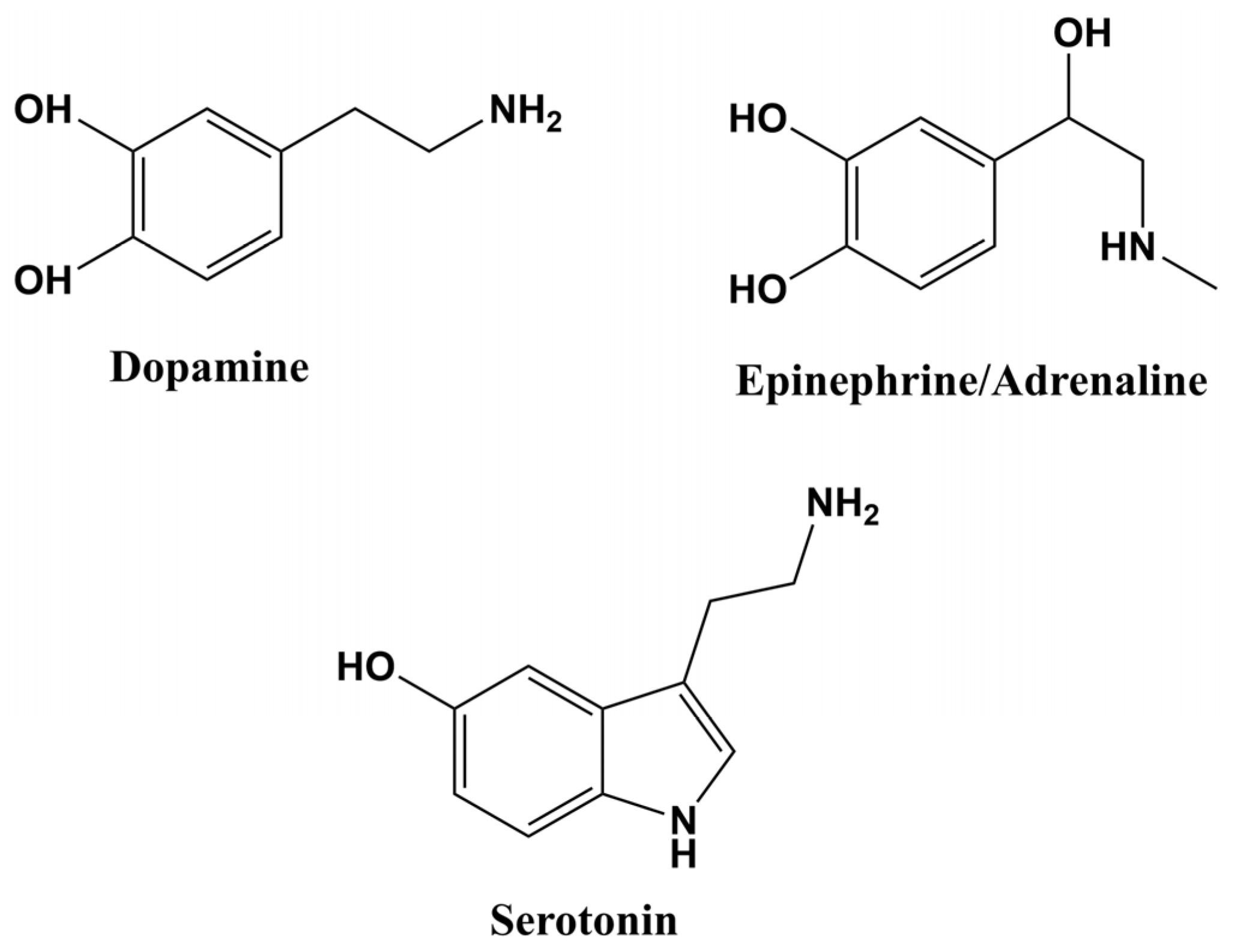
Figure 1. The chemical structures of DA, EPI and SER.
Chemosensors 11 00179 g001
Figure 2. The general oxidation mechanisms of DA, SER and EPI.
Chemosensors 11 00179 g002
Figure 3. Schematic illustration of the specific design of electrochemical sensors based on nanocomposite materials implemented for NT sensing.
Chemosensors 11 00179 g003
Figure 4. Schematic representation of the SV excitation signal.
Chemosensors 11 00179 g004
Table 1. Electrochemical sensors for DA detection from recent studies.
ElectrodeDetection MethodLOD
(µM)
Linear
Response Range
(µM)
Sensitivity
(µA µM−1)
Sample
Matrix
Refs.
Au@PSi-P3HT/GCECA6.3 × 10−11–4600.51120.1 M PBS
(pH = 7)
[58]
AuNPs/PM/CPEDPV6.7 × 10−22 × 10−1–11-0.1 M PBS
(pH = 6)
[59]
AuNPs/PAN/ITOCA9.1 × 10−11–1020.09280.1 M H2SO4
(pH = 1)
[60]
PT/Au/CNT/ITO/glassDPV6.9 × 10−11–1019.4921 mM
acetate buffer
(pH = 4)
[61]
Au/PEDOT-Aunano…SDSLSV3.9 × 10−45 × 10−1–200.03810.1 M PBS
(pH = 7.4)
[31]
AuNPs/PTAP/GCEDPV1.7 × 10−215 × 10−2–15 × 10−16.5800.1 M PBS
(pH = 7.2)
[62]
OPEDOT/AuNPs/ERGO/GCESWV14–102-10 mM PBS
(pH = 7.4)
[63]
Ag/PANI/GCECA1.910–900.1020.1 M PBS
(pH = 6)
[64]
PEDOT/AgNPs/CNCC/GCECA1.7 × 10−25 × 10−2–782-0.2 M PBS
(pH = 7.4)
[65]
POA@Ag/GCECA8.3 × 10−15–45-0.1 M PBS
(pH = 6)
[66]
Cu/PPy/GCEDPV8.5 × 10−410−3–10−1-0.1 M PBS
(pH = 7)
[67]
Pt/PF (BE)/Pd (CV)DPV4.8 × 10−25 × 10−1–1020.4780.1 M H2SO4[68]
Pt/PMT/PdnanoDPV9 × 10−35 × 10−2–11.370.1 M PBS
(pH = 7.4)
[69]
Pt/PMPy/PdnanoDPV1.2 × 10−210−1–100.710.1 M PBS
(pH = 7.4)
[70]
PSi-P3HT—porous silicon-poly-3-hexylthiophene; GCE—glassy carbon electrode; AuNPs—gold nanoparticles; PM—polymelamine; CPE—carbon paste electrode; PAN/PANI—polyaniline; ITO—indium tin oxide; PT—polythiophene; CNT—carbon nanotubes; PEDOT—poly(3,4-ethylene-dioxythiophene); Aunano—gold nanoparticles; SDS—sodium dodecyl sulfate; PTAP—poly(2,4,6-triaminopyrmidine); OPEDOT—overoxidized poly(3,4-ethylene-dioxythiophene); ERGO—electrochemically reduced graphene oxide; Ag—silver; CNCC—carboxylated cellulose nanocrystals; POA—poly(o-anisidine); Cu—copper; PPy—polypyrrole; Pt—platinum electrode; PF (BE)—polyfuran film prepared by bulk electrolysis; Pd (CV)—palladium nanoclusters electrodeposited through cyclic voltammetry; PMT—poly(3-methylthiophene); Pdnano—palladium nanoparticles; PMPy—poly(N-methylpyrrole); CA—chronoamperometry; DPV—differential pulse voltammetry; LSV—linear sweep voltammetry; SWV—square wave voltammetry; PBS—phosphate-buffered saline.
Table 2. Electrochemical sensors for SER detection from recent studies.
ElectrodeDetection MethodLOD
(µM)
Linear
Response Range
(µM)
Sensitivity
(µA µM−1)
Sample
Matrix
Refs.
AuNPs@PPy/GSPESWV33.22 × 10−310−1–150.33160.02 M PBS
(pH = 7.4)
[34]
AuNPs@rGO/pTBA-Pd (C2H4N2S2)2/NFSWV2.5 × 10−32 × 10−2–2 × 10²-0.1 M PBS
(pH = 7.4)
[81]
nano-Au/PPyox/GCEDPV1 × 10−37 × 10−3–22 × 10−1-0.1 M PBS
(pH = 7)
[82]
PANIS/Au/GCEDPV25 × 10−33 × 10−1–103-0.1 M PBS
(pH = 7.4)
[83]
rGO/PANI/AuNPs@MIPsDPV11.7 × 10−32 × 10−1–10-0.1 M PBS
(pH = 7.5)
[84]
Ag/PPy/Cu2O/GCEDPV124 × 10−410−2–250-0.1 M PBS
(pH = 7.2)
[85]
Pt/MWCNT/PPy/AgNPsDPV15 × 10−25 × 10−1–5-0.2 M PBS
(pH = 8)
[86]
PEDOTNTs/rGO/AgNPs/GCEDPV1 × 10−410−3–5 × 10−214.3040.1 M PBS
(pH = 8)
[38]
PtNPs/OPPy/rGO/GCEDPV106 × 10−310–470-0.1 M PBS
(pH = 7)
[87]
P-Arg/ErGO/AuNP/GCEDPV30 × 10−310−2–5 × 10−15.970.1 M PBS
(pH = 7)
[88]
CNTs-Cu2O-CuO@PtCA3 × 10−310–5 × 10²-0.1 M PBS
(pH = 7.4)
[89]
AuNPs—gold nanoparticles; PPy—polypyrrole; GSPE—graphite-based screen-printed electrode; rGO—reduced graphene oxide; pTBA—polyterthiphene; Pd (C2H4N2S2)2—palladium complex; NF—Nafion; nano-Au—gold nanoparticles; PPyox—overoxidized polypyrrole; GCE—glassy carbon electrode; PANIS—polyaniline nanowires; PANI—polyaniline; MIPs—molecularly imprinted polymers; Ag—silver nanoparticles; Cu2O—copper oxide; Pt—platinum electrode; MWCNT—multi—walled carbon nanotubes; PEDOTNTs—poly(3,4-ethylene-dioxythiophene) nanotubes; rGO—reduced graphene oxide; PtNPs—platinum nanoparticles; OPPy—over-ozidized polypyrrole; P-Arg—poly(L-arginine); ErGO—reduced graphene oxide; CNTs—carbon nanotubes; CuO—copper oxide; SWV—square wave voltammetry; DPV—differential pulse voltammetry; CA—chronoamperometry; PBS—phosphate-buffered saline.
Table 3. Electrochemical sensors for EPI detection from recent studies.
ElectrodeDetection MethodLOD
(µM)
Linear
Response Range
(µM)
Sensitivity
(µA µM−1)
Sample
Matrix
Refs.
MIP/AuNPs/GCEDPV7.6 × 10−29 × 10−2–102-0.1 M PBS
(pH = 7)
[97]
Au/ZnO/PPy/RGO/GCEDPV6 × 10−26 × 10−1–5 × 10²-0.1 M PBS
(pH = 7)
[98]
Au/PILs/PPyNTs/GCEDPV298.9 × 10−335–96042.77990.05 M PBS
(pH = 7.4)
[99]
GNPs/Pan-LB/GCESWV8 × 10−24 × 10−1–10-0.2 M PBS
(pH = 6)
[100]
PPy/AuNPs/SWCNTs-AuEDPV2 × 10−34 × 10−3–10−1-0.05 M PBS
(pH = 7)
[101]
Nano-Au/PPyox/GCEDPV3 × 10−23 × 10−1–21-0.1 M PBS
(pH = 7)
[102]
AuNPs/TGA/CS-MWCNTsCA60 × 10−34 × 10−1–112.310.1 M PBS
(pH = 7)
[103]
AuNPs/PDA/ANDPV0.261–103-0.1 M PBS
(pH = 7.4)
[104]
Au nanoporous film/AuECV1950–103-0.01 M PBS
(pH = 7)
[105]
rGO/AgNPs cotton and rGO/AgNPs/polyesterSWV
DPV
9.73 × 10⁻³
3.05 × 10⁻³
0.5–40
1–30
-0.1 M PBS
(pH = 7)
[106]
AuPt@GRCA0.9 × 10−315 × 10−4–96 × 10−116280.1 M PBS
(pH = 7.4)
[107]
MIP—molecularly imprinted polymer; AuNPs—gold nanoparticles; GCE—glassy carbon electrode; Au—gold nanoparticles; ZnO—zinc oxide; PPy—polypyrrole; RGO—reduced graphene oxide; PILs—poly(ionic liquids); PPyNTs—polypyrrole nanotubes; GNPs—gold nanoparticles; PAn-LB—polyaniline Langmuir-Blodgett film; SWCNTs—single-walled carbon nanotubes; AuE—gold electrode; nano-au—gold nanoparticles; PPyox—overoxidized polypyrrole; TGA—thioglycolic acid; CS—chitosan; MWCNTs—multi-walled carbon nanotubes; PDA—polydopamine; AN—acupuncture needle; AuPt—gold and platinum nanoparticles; GR—graphene; DPV—differential pulse voltammetry; SWV—square wave voltammetry; CA—chronoamperometry; CV—cyclic voltammetry; PBS—phosphate-buffered saline.
Disclaimer/Publisher’s Note: The statements, opinions and data contained in all publications are solely those of the individual author(s) and contributor(s) and not of MDPI and/or the editor(s). MDPI and/or the editor(s) disclaim responsibility for any injury to people or property resulting from any ideas, methods, instructions or products referred to in the content.