Article

Bioactivities of Waste Cork and Phloem Fractions of Quercus cerris Bark

by
Ali Umut Șen
1,*,
Daiana Almeida
2,3,
Tayse F. F. da Silveira
2,3,*,
Tânia S. P. Pires
2,3,
Mikel Añibarro-Ortega
2,3,
Filipa Mandim
2,3,
Lillian Barros
2,3,
Isabel C. F. R. Ferreira
2,3,
Helena Pereira
1 and
Ângela Fernandes
2,3
1
Centro de Estudos Florestais, Laboratório Associado TERRA, Instituto Superior de Agronomia, Universidade de Lisboa Tapada da Ajuda, 1349-017 Lisboa, Portugal
2
Centro de Investigação de Montanha (CIMO), Instituto Politécnico de Bragança, Campus de Santa Apolónia, 5300-253 Bragança, Portugal
3
Laboratório Associado para a Sustentabilidade e Tecnologia em Regiões de Montanha (SusTEC), Instituto Politécnico de Bragança, Campus de Santa Apolónia, 5300-253 Bragança, Portugal
*
Authors to whom correspondence should be addressed.
Processes 2024, 12(10), 2081; https://doi.org/10.3390/pr12102081
Submission received: 10 August 2024 / Revised: 17 September 2024 / Accepted: 23 September 2024 / Published: 25 September 2024
(This article belongs to the Special Issue Sustainability Use of Wood/Wood Residues and Other Bioenergy Sources)

Abstract

:
Recently, more and more researchers have begun to consider using waste bark fractions to produce value-added biochemicals and materials, as well as for energy production. Extraction is often the first operation in biomass biorefineries. Here we obtained hydroethanolic extracts from waste cork and phloem fractions of Quercus cerris bark and analyzed them to determine their antioxidant, antimicrobial, and nitric oxide (NO) production inhibition properties and their hepatotoxicity. The antioxidant properties were investigated by ex vivo TBARSs as well as OxHLIA antioxidant assays, the antibacterial properties against Gram-positive and Gram-negative bacteria isolated from food and clinical sources, and antifungal properties against Aspergillus brasiliensis and A. fumigatus. The NO production inhibition activity was assessed in a lipopolysaccharide (LPS)-stimulated murine macrophage (RAW 264.7) cell line, and antiproliferative activities were determined against five different cell lines, including lung (NCI-H460), gastric (AGS), breast (MCF7), and colon (CaCo2) tumor cell lines, as well as a non-tumor cell line (PLP2). The hydroethanolic maceration of waste cork and phloem yielded 4.4% and 2.4% extracts, respectively. Gallic acid glucosides, phenolic acids, and ellagic acid were identified in both extracts. The waste cork and phloem extracts showed antioxidant, antimicrobial, antifungal, and antiproliferative properties but also showed hepatotoxicity in the case of waste cork. Both bark fractions varied in terms of their bioactivity, with waste cork extracts showing, in general, higher bioactivity than phloem extracts

1. Introduction

Biomass is described as biological-derived (of plant or animal origin) material excluding minerals, and fossilized materials (EN 16575:2014) [1]. According to this broad definition, biomass includes wood and forestry wastes, agricultural crops, residues of forest-based and agro-industries, municipal solid waste, algae, microorganisms, marine organisms, etc. Many of these biomasses already undergo valorization processes, particularly regarding their use as energy sources. Biomass contributes to approximately 6% of global energy production, 55% of global renewable energy production, and 2.5% of global electricity production, according to the latest International Energy Agency (IEA) data. These figures will likely increase in the future due to the global shift to renewable energy driven by the depleting fossil resources and the environmental problems associated with their use.
Biorefineries are integrated processes that convert biomass into a range of value-added products and/or into energy through a set of cascaded thermochemical or biological processes [2]. All biorefinery systems seem to share at least two common traits: they facilitate waste reduction and offer sustainability. Thus, by closing material conversion loops, they contribute to the sustainable production of materials, chemicals, and energy from renewable sources under the overall bioeconomy concept [3]. Among the different biomass types that can be used as feedstocks in biorefineries, tree barks are attracting increasing interest [3].
Barks are the outermost components that cover stems and branches, and are defined as all the tissues (comprising the cortex, phloem, epidermis, and periderm) external to the vascular cambium of trees, shrubs, or lianas [4,5]. Tree barks are structurally composed of inner bark (including conducting and non-conducting phloem tissues) and outer bark or rhytidome, consisting of one periderm [6] or, in most cases, of successive periderms separated by phloem layers [5]. Some species have periderms, with a substantial formation of cork cells leading to cork-rich barks [5]. One such case is Quercus cerris (Turkey oak), which develops a cork-rich rhytidome under specific growth conditions [7].
Tree barks are heterogeneous lignocellulosic solid materials at structural and chemical levels, possessing low polysaccharide and high lignin and inorganic (ash) contents, and they often have inorganic impurities [3,8]. Therefore, the production of value-added materials from tree barks is complex and the traditional single-step chemical or biological processes cannot be applied due to low product yields and ash-related operational problems [3]. Therefore, tree barks are often regarded as waste material.
On the other hand, the distinctive properties of barks may be advantageous for a biorefinery route because they are rich sources of valuable chemical products such as lipophilic and phenolic extractives. Extraction is usually the first operation in a multi-step biomass biorefinery, and barks are promising sources for the production of chemical and biochemical extracts due to their high amounts of extractable compounds and their diversity. In subsequent processes, other components can be selectively removed from the previously extracted bark, such as inorganic compounds, lignin depolymerization products [2], as well as carbohydrates derived from the polysaccharide fraction [9,10], therefore closing off the biorefinery system by producing value-added chemicals and materials and achieving full resource valorization.
Different bark components are already processed at a commercial scale. For instance, oak, chestnut, and wattle barks are used at industrial scale for tannin production, eucalypt and birch barks are used for extractives, and cork oak bark is used for cork production [3]. Cork is a unique bark component, given the singularity of its particular set of properties, and cork-rich barks are, therefore, potentially valuable resources [6]. The cork from Quercus suber is one of the most valuable non-wood forest products, and has a well-established and consolidated market value due to its interesting applications, e.g., as cork stoppers for sealing wine bottles, as cork granulates in composites for insulation and surfacing, and as adsorption materials; it can also be used as a source of energy, which can be produced from the cork waste powder, and thus provides an example of a full resource valorization loop [6]. It is therefore understandable that other cork-rich barks have been considered as potential cork providers. This is the case of the cork-rich Quercus cerris bark; a research program was developed to characterize it and explore the different valorization routes, including, as a first step, bark fractionation and separation of a pure cork fraction that comprises approximately 8% of the total bark weight [11]. The cork of Q. cerris has a similar chemical composition to that of Q. suber, but with less suberin and higher lignin and ash content [6]. The composition of lipophilic extractives of both corks is similar, with friedelin triterpenoids acting as the principal compounds [12].
However, the Q. cerris bark fractionation process for obtaining a pure cork fraction generates substantial waste fractions composed of refuse-grade cork (high-density and phloem-containing cork) that are rejected for cork-stopper production, making up approximately 19% of the total bark weight, and also of a phloem fraction which represents up to 73% of the total bark weight [11]. Together, these waste fractions correspond to approximately 90% of the bark weight and must therefore be considered for valorization in a biorefinery route. Previously, it was shown that extracts from these waste cork and phloem fractions exhibit antioxidant and antimicrobial properties that can be detected in in vitro conditions using chemical methods [13,14], but their true bioactive potential was not tested in biological matrices, nor was their phenolic composition determined, although this is closely related to their bioactive properties.
This study aims to address this knowledge gap by conducting the first comparative analysis of the phenolic profile and the bioactive properties of polar extracts from waste cork and phloem from Q. cerris bark. UPLC-MS analysis, antioxidant properties, antimicrobial and antifungal properties, as well as the antiproliferative activity, hepatotoxicity, and nitric oxide production inhibition properties of Q. cerris waste cork and phloem fractions were determined. The results will provide knowledge that will be useful for the valorization of phloem and cork residues from other oak barks by producing bioactive agents for several applications, including skincare agents and health supplements, infection control, food preservatives, and cancer treatment.

2. Materials and Methods

2.1. Standards and Reagents

Trolox (6-hydroxy-2,5,7,8-tetramethylchroman-2-carboxylic acid) and PBS (Phosphate-buffered saline) were purchased from Sigma (St. Louis, MO, USA). Formic and acetic acids were purchased from Prolabo (VWR International, Rosny-sous-Bois, France). Ethyl acetate (99.8%) was purchased from Fisher Scientic (Lisbon, Portugal). Phenolic compound standards were purchased from Extrasynthese (Genay, France). Fetal bovine serum (FBS), L-glutamine, Hank’s balanced salt solution (HBSS), and trypsin–EDTA (ethyl-enediaminetetraacetic acid) were obtained from Hyclone (Logan, UT, USA). Acetic acid, ellipticine, sulforhodamine B (SRB), dimethyl sulfoxide (DMSO, trypan blue, tri-chloroacetic acid (TCA), and tris (tris(hydroxymethyl)amino-methane) were obtained from Sigma Chemical Co. (St. Louis, MO, USA). The cell lines CaCo-2 (Catalogue No. 860102022) and RAW 264.7 (Catalogue No. 91062702) were commercially acquired from the European Collection of Authenticated Cell Cultures—ECACC, in turn NCI-H460 (ACC 737) and MCF-7 (ACC 115) were acquired from the Leibniz Institute DSMZ-German Collection of Microorganisms and Cell Cultures GmbH. Microorganisms, Mueller–Hinton agar (MH), malt agar (MA), as well as positive controls (ampicillin, streptomycin, methicillin, and ketoconazole) were purchased from Frilabo, Porto, Portugal. All other chemicals and solvents were of analytical grade and were purchased from common sources. Water was treated in a Milli-Q water purification system (TGI Pure Water Systems, Greenville, SC, USA).

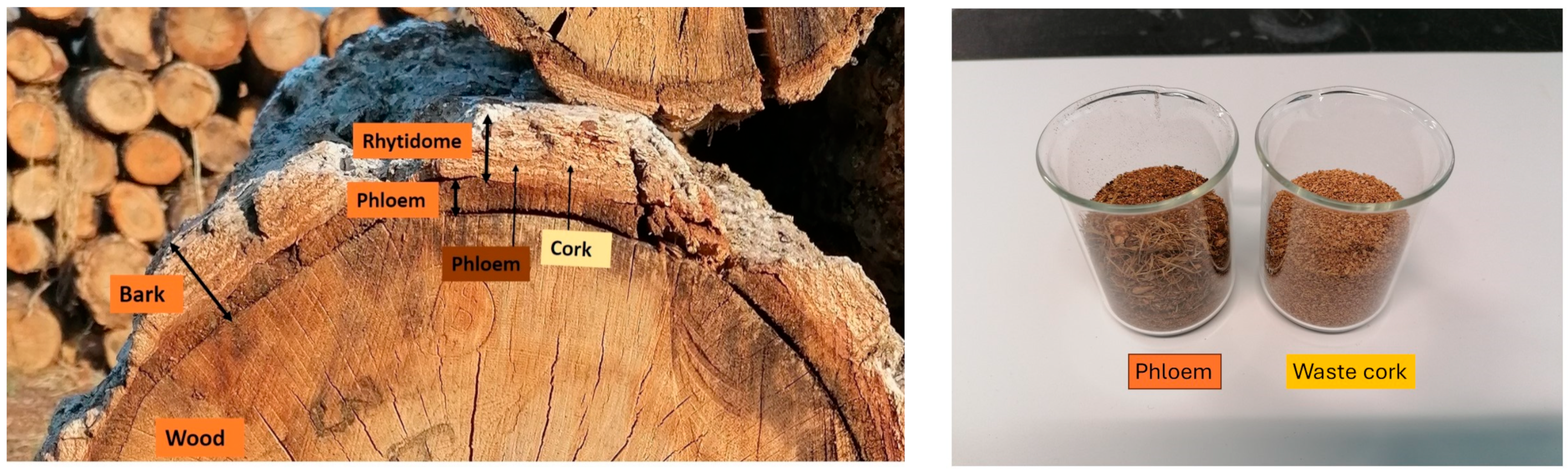
2.2. Preparation of Cork and Phloem Samples

Samples of cork and phloem were obtained from the bark of approximately 100 mature trees of Quercus cerris that were harvested in Kahramanmaras, Turkey (Figure 1) in 2015. The bark was processed as a composite sample by trituration, followed by size and density separation in an industrial-scale two-step separation process, as previously described [11]. The phloem was separated as a high-density fraction in the first separation step, while good-grade (low-density) and low-grade (high-density) cork fractions were separated in the second separation step. The phloem and low-grade (waste) cork samples were re-granulated, sieved into 40–60 mesh (350–420 μm) particle size, and kept in air-tight bags for further experiments.

2.3. Preparation of Hydroethanolic Extracts

The waste cork and phloem specimens were subjected to a two-step mild hydroethanolic maceration extraction by agitating approximately 3 g of sample with a 100 mL mixture of ethanol (EtOH) and water (H2O) (80:20, v/v) at room temperature (25 °C) for 2 h. The biomass–solvent mixture was filtered through filter paper (Whatman No. 4 with 20–25 μm pore size). The filtered extracts were concentrated at 40 °C using a Büchi R-210 rotary evaporator (BUCHI Labortechnik. Flawil, Switzerland) under reduced pressure. The concentrated solutions were frozen and lyophilized using a FreeZone 4.5 lyophilizer (Labconco, Kansas City, MO, USA) [15].

2.4. UHPLC-DAD-ESI-IT-MS Characterization of Phenolic Compounds

The hydroethanolic extracts prepared above were dissolved in a solution of ethanol and water (80:20, v/v) to obtain a final concentration of 10 mg/mL. This solution was filtered using 0.22 μm disposable nylon filter disks and used for the determination of phenolic composition by using an ultra-high-performance liquid chromatography (UHPLC) system that was equipped with a diode array detector (DAD) operating at three different ultraviolet wavelengths (280 nm, 330 nm, and 370 nm). The UHPLC system was connected to an electrospray ionization ion trap mass spectrometry detector (ESI-IT-MS) operating in the negative ion mode. The instruments used were the Dionex Ultimate 3000 LC system and the Linear Ion Trap LTQ XL mass spectrometer (Thermo Scientific in San Jose, CA, USA). The chromatographic method and the mass spectrometry conditions used can be found elsewhere [15].
The identification of phenolic compounds was based on comparisons of retention times, UV-VIS signals, mass spectra of the samples and of available standards (gallic, protocatechuic, caffeic and ellagic acids), as well as previously reported data. The quantification of phenolic compounds was made with calibration curves obtained from standards based on their UV-VIS signals, or from a structurally similar compound when the standard compound was not available, by integrating the peak areas in the chromatograms using the baseline-to-valley information and applying baseline projection. The results of the quantification were expressed as mg/g of the extract.

2.5. Antioxidant Activity

The antioxidant activity of waste cork and phloem extracts was assessed using two different ex vivo antioxidant assays: the thiobarbituric acid-reactive substances (TBARSs) assay and the oxidative hemolysis (OxHLIA) assay.

2.5.1. Thiobarbituric Acid-Reactive Substances (TBARSs) Assay

Prior to the TBARSs assay, the hydroethanolic extracts were first dissolved in water and then subjected to dilutions ranging from 10 to 0.3125 mg/mL. Porcine (Sus scrofa) brain cell homogenates were homogenized in tris-HCl (20 mM, pH 7.4). Afterwards, 200 μL extract samples were mixed with 100 μL brain cell homogenates, 100 μL Ascorbic acid (C6H8O6, 0.1 mM), 100 μL Iron (II) sulfate (FeSO4, 10 μM) and incubated at 37.5 °C for 60 min. Afterwards, 500 μL trichloroacetic acid (C2HCl3O2, 28% w/v) and 380 μL thiobarbituric acid (TBA, C4H4N2O2S, 2% w/v) were added and the mixture was incubated at 80 °C for 20 min. After the incubation, the inhibition of lipid peroxidation in porcine brain cell homogenates was determined by measuring the decrease in thiobarbituric acid-reactive substances (TBARSs) and the color intensity of the malondialdehyde–thiobarbituric acid (MDA–TBA) complex by measuring the absorbance at 532 nm. The lipid peroxidation inhibition ratio was calculated as a percentage using the following formula: (A − B)/A × 100, where A represents the absorbance of the control sample and B corresponds to the absorbance of the extract samples. The results were expressed in terms of effective concentration (EC50) values, which indicate the concentration (in µg/mL) of the sample that provides 50% antioxidant activity and were calculated from a graph showing the percentage of inhibition against the concentration of the extract. Trolox was used as a positive control.

2.5.2. Oxidative Hemolysis (OxHLIA) Assay

The assessment of the oxidative hemolysis inhibition activity of the extracts (OxHLIA antioxidant assay) was performed using red blood cells (RBCs) isolated from healthy sheep. An erythrocyte solution (2.8% v/v; 200 µL) was mixed with either the extract solution (ranging from 0.0938 to 3 mg/mL) or water (used for complete hemolysis). After a pre-incubation period of 10 min at 37 °C with shaking, 2,2′-azobis (2-methylpropionamide) dihydrochloride (AAPH, 200 µL, 160 mM in PBS, radical initiator was added. The optical density was measured at 690 nm at approximately 10 min intervals using a microplate reader (Bio-Tek Instruments, ELX800, Santa Clara, CA, USA) until complete hemolysis occurred. The results were expressed as inhibitory concentration (IC50) values (in µg/mL) at Dt (time point) of 60 min. These IC50 values represent the extract concentration required to inhibit 50% of the erythrocyte population from the hemolytic action. Trolox was also used as a positive control [16].

2.6. Antibacterial Activity

The antibacterial activities of waste cork and phloem extracts were assessed using a microdilution method where the minimum inhibitory concentrations (MICs) and the minimum bactericidal concentrations (MBCs) were determined. These assays were performed applying a serial dilution technique in 96-well microtiter plates (ThermoFisher Scientific, Lisbon, Portugal) [17].
Gram-negative bacteria (Enterobacter cloacae (ATCC 49741), Escherichia coli (ATCC 25922), Pseudomonas aeruginosa (ATCC 9027), Salmonella enterica (ATCC 13076), and Yersinia enterocolitica (ATCC 8610)), as well as Gram-positive bacteria (Bacillus cereus (ATCC 11778), Listeria monocytogenes (ATCC 19111), and Staphylococcus aureus (ATCC 25923)), isolated from food sources, were selected to test the antibacterial activity of the hydroethanolic extracts of waste cork and phloem.
Additionally, Gram-negative bacteria (E. coli, Klebsiella pneumoniae, Morganella morganii, Proteus mirabilis, and P. aeruginosa), as well as Gram-positive bacteria (Enterococcus faecalis, L. monocytogenes, and Methicillin-resistant Staphylococcus aureus (MRSA)) were clinical isolates obtained from patients hospitalized in various departments at the Hospital Center of Trás-os-Montes and Alto Douro (Vila Real, Portugal) to evaluate the antibacterial activity of the hydroethanolic extracts of waste cork and phloem.
The MIC determinations on all bacteria were conducted using the colorimetric assay described by [18]. Firstly, the samples were dissolved in 5% (v/v) dimethyl sulfoxide (DMSO) and 95% of autoclaved distilled water to give a final concentration of 20 mg/mL for the stock solution. Then, 90 μL of this concentration was added to the first well (96-well microplate) in duplicate with 100 μL of Tryptic Soy Broth (TSB). After that, 90 μL of TSB medium was added to the remaining wells. Then, the samples were serially diluted to obtain the concentration ranges (10 to 0.03125 mg/mL). Finally, 10 μL of inoculum (standardized at 1.5 × 10⁶ Colony Forming Unit (CFU)/mL) was added to all the wells to ensure the presence of 1.5 × 10⁵ CFU. Two negative controls were prepared, one with TSB and another one with the extract. Two positive controls were prepared with TSB and each inoculum and another with medium, antibiotics, and bacteria. Ampicillin (10 mg/mL), streptomycin (1 mg/mL), and methicillin (1 mg/mL) were used as positive controls, whereas the negative control was 5% dimethyl sulfoxide (DMSO). The microplates were incubated at 37 °C for 24 h. The MICs of samples were detected following the addition of (40 μL) 0.2 mg/mL p-iodonitrotetrazolium chloride (INT) and incubation at 37 °C for 30 min. MIC was defined as the lowest concentration that inhibits the visible bacterial growth, which is determined by a change in the coloration from yellow to pink that occurs if the microorganisms are viable. To determine the MBC, 10 μL of liquid from each well that showed no change in color was plated on a solid medium, blood agar (7% sheep blood), and incubated at 37 °C for 24 h. The lowest concentration that yielded no growth determined the MBC. The MBC was defined as the lowest concentration required to kill bacteria.

2.7. Antifungal Activity

To determine the antifungal activity of waste cork and phloem, Aspergillus brasiliensis (ATCC 16404) and A. fumigatus (ATCC 204305) were used. The microdilution method was conducted to determine the minimum inhibitory and fungicidal concentrations (MICs, and MFCs, expressed in mg per mL) [19]. The micromycetes were maintained on malt agar and the cultures stored at 4 °C, after which they were placed in new medium and incubated at 25 °C for 72 h. In order to investigate the antifungal activity, the fungal spores were washed from the surface of agar plates with sterile 0.85% saline containing 0.1% Tween 80 (v/v). The spore suspension was adjusted with sterile saline to a concentration of approximately 1.0 × 10⁵ in a final volume of 100 μL per well. Firstly, the samples were dissolved in 5% (v/v) DMSO and 95% of autoclaved distilled water to provide a final concentration of 10 mg/mL for the stock solution. Afterwards, 90 μL of this concentration was added to the first well (96-well microplate) in duplicate with 100 μL of Malt Extract Broth (MEB).
After that, 90 μL of MEB medium was added to the remaining wells. Then, the samples were serially diluted to obtain the concentration ranges (10 to 0.03125 mg/mL). The minimum inhibitory concentration (MIC) determinations were performed by a serial dilution technique using a 96-well microplate. The lowest concentrations without visible growth (at the binocular microscope) were defined as MICs. The MFC was determined by serial subcultivation of 2 μL of the tested compounds dissolved in medium and inoculated for 72 h then added to microplates containing 100 μL of MEB per well and subjected to further incubation for 72 h at 26 °C. The lowest concentration with no visible growth was defined as the MFC, indicating 99.5% death of the original inoculum. Commercial fungicide ketoconazole (1 mg/mL) was used as the positive control, whereas the negative control was 5% DMSO.

2.8. Antiproliferative Activity

The antiproliferative activity of the hydroethanolic extracts was assessed within a concentration range between 400 and 6.25 μg/mL using the sulforhodamine B colorimetric assay against four human tumor cell lines: gastric adenocarcinoma (AGS), colorectal adenocarcinoma (CaCo-2), lung carcinoma (NCI-H460), and breast carcinoma (MCF-7). The cell lines were maintained in accordance with the manufacturer’s recommendations and used only up to a maximum of passage 50, to ensure their viability and quality control [20]. A primary culture obtained from pig liver (PLP2) and established according to the previously described procedure [21] was tested for hepatotoxicity. Ellipticine was applied as a positive control. The results were expressed as GI50 values (μg/mL), which represent the extract concentration accountable for 50% inhibition of cell proliferation.

2.9. Nitric Oxide Production Inhibition

The capacity of the extracts to inhibit the nitric oxide (NO) production (anti-inflammatory activity) within a range of concentrations (from 400 to 6.25 μg/mL) was studied using a lipopolysaccharide (LPS)-stimulated murine macrophage cell line (RAW 264.7). NO production was quantified based on the nitrite concentration utilizing the Griess Reagent System kit [22]. Dexamethasone (50 mM) was used as a positive control, while cells with and without LPS were tested as the negative control. The results were expressed as EC50 values (μg/mL), representing the extract concentration responsible for 50% of NO production inhibition.

2.10. Statistical Analysis

All bioactivity and phenolic composition tests were conducted in triplicate, and the outcomes were presented as the mean values along with the corresponding standard deviations (SD). The results (except for those pertaining to the antibacterial activity) were analyzed using Student’s t-test in order to determine the significant difference between two different samples with a 5% significance level (Version 25, IBM Corp., New York, NY, USA).

3. Results

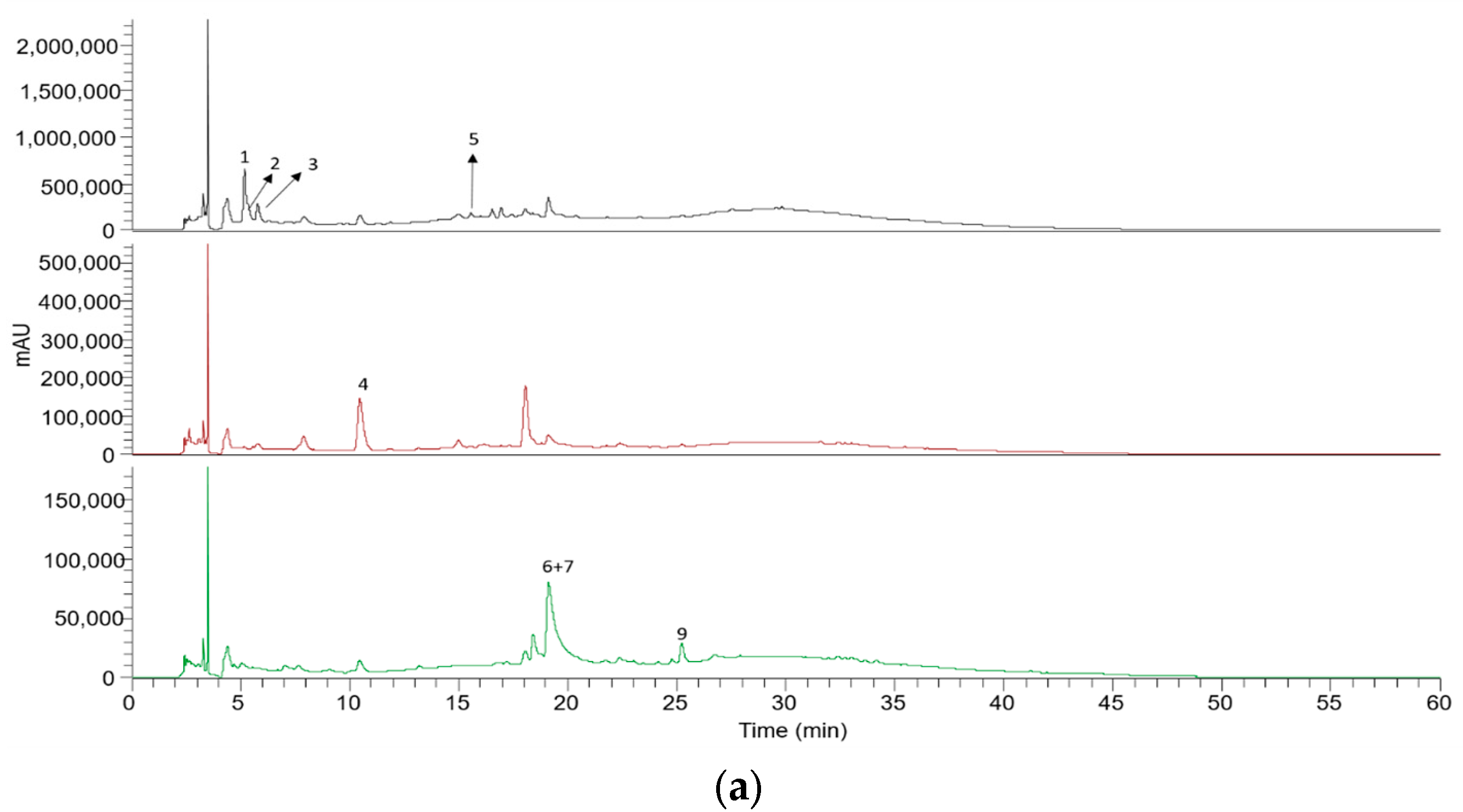
3.1. UHPLC-DAD-ESI-IT-MS Analysis of Phenolic Compounds

The hydroethanolic extraction of waste cork (WCE) and phloem (PE) extracts obtained by maceration resulted in 4.4% and 2.4% extract yields, respectively. The phenolic compounds were tentatively identified in waste cork and phloem based on their chromatographic and UV-Vis characteristics, as well as fragmentation patterns (Figure 2, Table 1). The phenolic profile of the samples was similar and composed principally of phenolic acids and ellagic acid, with gallic acid glucosides being the major phenolic compounds identified (Table 1).
Epicatechin was not detected in waste cork and phloem. This suggests that tannins in waste cork and phloem are predominantly of the hydrolysable tannin type and that condensed tannins are not major components.

3.2. Ex Vivo Antioxidant Activity

Waste cork and phloem showed antioxidant activity as confirmed by the cell-based TBARSs and OxHLIA methods, with TBARSs indicating a lower antioxidant activity (EC50) and OxHLIA a stronger antioxidant activity (IC50) when compared to Trolox (Table 2).

3.3. Antibacterial and Antifungal Activity

The current study presents the first detailed antimicrobial activity profile for waste cork and phloem of Q. cerris bark.
Both waste cork and phloem extracts showed antibacterial activities against Gram-positive and Gram-negative bacteria (Table 3) isolated from food sources and clinical specimens, as well as against methicillin-resistant bacteria (MRSA). However, their antimicrobial activities were much lower than those of the antibiotics used as reference.
Waste cork and phloem extracts also exhibited antifungal activities (Table 4), although they were lower than those of the ketoconazole that we used as a reference.

3.4. Antiproliferative, Hepatotoxicity, and Inhibition of NO Production Activities

Antiproliferative assays measure the ability of extracts to inhibit cell proliferation and are particularly useful for evaluating the potential efficiency of different drugs against tumor cells, while the side effect of the extracts can be assessed by hepatotoxicity assays.
Hydroethanolic extracts of waste cork and phloem from Q. cerris bark exhibit the capacity to interfere with the normal proliferation of several studied cancer cell lines (Table 5), including gastric cancer, lung cancer, colon cancer, and breast cancer cell lines, as well as non-tumor cell lines.
Neither waste cork nor phloem extracts exhibit NO production inhibition properties (Table 5), i.e., the ability to reduce or inhibit inflammation.

4. Discussion

The mild maceration yielded much less extract compared to extractions using harsher conditions such as an extended time and higher temperature and compared with cyclic Soxhlet and soxtec extractions. In fact, the extract yields obtained via Soxhlet extraction of the same waste cork and phloem samples were 2.5 and 1.7 times higher (Table 6) [23]. The extract yields depended on the material and were higher for the waste cork than for phloem (1.8 times higher). Other studies also showed different extract yields for different plant tissues, e.g., in Pseudotsuga menziesii bark, the polar extractives amounted to 23.5% and 27.3% in cork and phloem, respectively [24]. In carrots (Daucus carota L.), extract yields decreased from peel to phloem and xylem [25], and in olive trees extract yields were higher in the bark than in wood [23].
The hydroethanolic extract yield of waste cork was 46.7% higher than that reported for pure cork that was also obtained by maceration (Table 6). The same was reported for extractions using Soxhlet extractions, for which there was a 27.6% difference [12]. Since the extraction of the phloem fraction always results in a lower yield than that of waste cork, this suggests that the phloem tissues that accompany the cork in the waste cork fractions, most probably corresponding to the phloem layers included in the rhytidome, have a higher extract content than that of the bark phloem. This may be caused by an enrichment in secondary metabolites in the phloem layers that accompany the process of traumatic periderm formation that is at the base of rhytidome development.
Although the extract yields of waste cork and phloem were relatively low when this mild hydroethanolic extraction method was used, they are still significant and valuable for biorefineries, especially when purity and selectivity are major concerns. Since maceration extraction is performed at low temperature, the bioactive compounds present in the extracts may be protected from thermal degradation, e.g., the decrease in certain phenolic compounds such as quercetin with increased extraction temperature may be avoided [27,28]. Therefore, maceration should be considered as a special biorefinery process when targeting bioactive bark extracts. On the other hand, an overall higher extract yield as well as a hemicellulose fraction may be obtained from barks by other, more intensive biorefinery processes [9,29,30]. Therefore, both “low-yield and selective” and “high-yield and extensive” biorefinery processes may be applied to different barks, namely to oak barks, which usually contain similar or higher phenolic extractive contents and compositions [3].
Waste cork contained a slightly higher amount of phenolic compounds than phloem (Table 1). The phenolic compounds shown on chromatographic peaks 3, 4, and 6 were identified through comparison with authentic standards. Peaks 1, 2, and 7 yielded deprotonated ions at m/z 477, 345, and 187, respectively, and fragment ions at m/z 169 and 124, indicating the presence of gallic acid derivatives [28]. Additionally, the fragment ions at m/z 183 appearing in both peaks 1 and 2 suggest the presence of methyl gallate structures [31]. The neutral losses of 294 Da (in peak 1) and 162 Da (in peak 2) imply the loss of pentose and hexose moieties. Therefore, peaks 1 and 2 were assigned as methyl gallate–pentosyl–hexoside and methyl gallate hexoside, respectively. Compound 7 had a deprotonated molecule at m/z 491 and base peak in MS2 at m/z 359, denoting the neutral loss of 132 that was attributed to a pentose group, followed by fragment ions (m/z 344, 313) that were previously described for isolariciresinol in conifer woods [32]. Therefore, compound 7 was tentatively identified as isolariciresinol pentoside. Peak 8 ([M − H] at m/z 187) also shows fragment ions at m/z 169 and 125, indicating another gallic acid derivative. The difference between compound 8 and gallic acid’s molecular weight is 18 Da, suggesting that this compound can be a gallic acid monohydrate. This compound has been found in Dendranthema indicum flowers and sorghum wholegrains [33,34]. Peak 5 was another gallic acid derivative that was found in the samples. It showed [M − H] at m/z 787 and the presence of m/z 313, suggesting the presence of galloyl glucose [31], while the successive loss of 152 moieties implies the presence of other galloyl groups in the molecules. Therefore, peak 5 was identified as tetragalloylglucose [31]. Peak 9 exhibited [M − H] at m/z 461 and fragment ions at m/z 315 ([M − H] 146) and 300 ([M − H] 146 − 15), which were attributed to the successive loss of rhamnose and methyl groups linked to an ellagic acid. From these data, peak 8 was assigned as methyl ellagic acid rhamnoside [31].
Caffeic acid and tetragalloyglucose were only detected in waste cork. Caffeic acid exhibits antioxidant activities [35] and inhibits leukotriene biosynthesis [36]. Tetragalloylglucose (4GG) is the almost fully esterified form of glucose and showed bioactive properties including inhibition of HIV integrase, inhibition of HCV protease, anti-cancer activity against S-100 sarcoma, as well as inhibition of HBV antigen [37,38].
Ex vivo antioxidant tests enable a biologically relevant assessment of antioxidant activity compared to in vitro chemical antioxidant tests. Because ex vivo tests involve living cells or cells isolated from living organisms, which better simulate the complex biological environment. In contrast, in vitro chemical antioxidant tests solely apply chemical methods and do not involve living cells, thereby potentially failing to accurately predict how extract compounds will behave in biological systems. The difference between the results of the applied antioxidant tests may arise from the characteristics of the two tests, since the TBARSs method measures the antioxidant activity of extracts against lipid-derived free radicals, while OxHLIA method measures the antioxidant activity of extracts against a broader range of free radicals (both lipid-derived and hydrophilic-derived).
In all cases, the ex vivo antioxidant tests confirmed the antioxidant activity of Q. cerris bark extracts that had been previously detected by chemical methods in in vitro conditions [13,14]. The IC50 antioxidant activity (OxHLIA assay) of phloem was better than that of Trolox, and above that of waste cork, which was similar to the results obtained for Trolox (Table 3). The EC50 TBARSs results showed poor antioxidant activity in relation to Trolox, especially for phloem, indicating that the antioxidant bioactivity of these extracts mainly relates to hydrophilic-derived radicals, as determined by the OxHLIA assay.
A biorefinery approach involving different extractive routes for screening lipophilic and hydrophilic extractives seems to be particularly interesting for cork-rich barks. The results also suggest that other oak barks may be similarly valorized in a biorefinery system, even if only a few contain significant amount of cork. The antioxidant activity of waste cork and phloem hydroethanolic extracts was stronger or comparable, respectively, to that of Juglans regia green husks [39].
The microdilution method was selected to study the antimicrobial properties because it is a widely used method for determining the minimum inhibitory concentration of antibacterial or antifungal extracts. It has previously been reported that Q. suber and Q. cerris cork extracts contain phenolic bioactive compounds and exhibit antimicrobial activity [14,40,41,42,43,44]. The antimicrobial activity of the phenolic extracts of waste cork and phloem is comparable to that of phenolic extracts of Rhizipora racemosa [45].
Waste cork hydroethanolic extracts had a greater inhibitory effect against gastric cancer and lung cancer cell lines with smaller GI50 values, while phloem extracts had a greater inhibitory effect against colon and breast cancer cell lines (Table 5). Phloem extracts do not exhibit hepatotoxicity for the PLP2 cell line, while cork extracts show a certain hepatotoxicity. Interestingly, the anti-inflammatory activity (measured by Inhibition of NO Production) of cork extracts was previously reported for Q. suber cork in Portugal [41] which may indicate the effect of cork species on the anti-inflammatory activity. Our results agree with previous results on the variability in the bioactivity in different plant tissues [46,47].
Thus, the overall results suggest that hydroethanolic extracts of waste cork and phloem from Q. cerris bark from Turkey contain bioactive compounds that appear to be effective against certain tumor cells such as gastric, colon, and breast cell cancer cell lines, but they do not show anti-inflammatory activity. The hepatotoxicity of cork and phloem extracts differs therefore, hepatotoxicity screening of the hydroethanolic extracts for specific compounds should be performed prior to possible drug development.

5. Conclusions

Waste cork and phloem fractions obtained from fractionation of Quercus cerris bark were extracted using a hydroethanolic solvent under mild maceration conditions, and the phenolic components and biologically active properties of the extracts were determined.
The following specific conclusions were drawn in this study:
  • The hydroethanolic maceration of waste cork had higher extract yield than that of phloem.
  • Gallic acid glucosides, phenolic acids, and ellagic acid were identified in waste cork and phloem fractions.
  • The antioxidant activity of waste cork exceeded that of phloem and was comparable to pure cork.
  • Waste cork and phloem extracts may be associated with antiproliferation of gastric, colon, and breast tumor cell lines.
  • Phloem extracts did not exhibit hepatotoxicity.
  • The bioactivities of waste cork and phloem extracts vary considerably, with distinct antioxidant, antimicrobial, and antiproliferative activities.
Further research should explore the phenolic composition and the bioactive potential of extracts from waste fractions of other cork-rich barks.

Author Contributions

Conceptualization, investigation, writing—original draft preparation, A.U.Ș.; formal analysis, D.A., F.M., M.A.-O., T.S.P.P. and T.F.F.d.S.; funding acquisition, supervision, L.B.; research mentor, methodology, writing—review and editing, I.C.F.R.F. and H.P.; writing—review and editing, visualization; supervision, A.U.Ș., H.P. and Â.F. All authors have read and agreed to the published version of the manuscript.

Funding

A.U. Sen acknowledges the research support of FCT (DL 57/2016). The authors are thankful to FCT for providing financial support via national funds to CIMO (UIDB/00690/2020 (DOI: 10.54499/UIDB/00690/2020) and UIDP/00690/2020 (DOI: 10.54499/UIDP/00690/2020)), SusTEC (LA/P/0007/2020 (DOI: 10.54499/LA/P/0007/2020)) and CEF (UIDB/00239/2020 and UIDP/00239/2020 ((DOI: 10.54499/UIDB/00239/2020 and DOI: 10.54499/UIDP/00239/2020)). Furthermore, the contracts of L. Barros and Â. Fernandes (DOI: 10.54499/CEECINST/00016/2018/CP1505/CT0008) were funded by FCT via the institutional scientific employment program.

Data Availability Statement

Data are contained within the article.

Conflicts of Interest

The authors declare no conflicts of interest.

References

  1. Sherwood, J.; Clark, J.H.; Farmer, T.J.; Herrero-Davila, L.; Moity, L. Recirculation: A New Concept to Drive Innovation in Sustainable Product Design for Bio-Based Products. Molecules 2016, 22, 48. [Google Scholar] [CrossRef]
  2. FitzPatrick, M.; Champagne, P.; Cunningham, M.F.; Whitney, R.A. A Biorefinery Processing Perspective: Treatment of Lignocellulosic Materials for the Production of Value-Added Products. Bioresour. Technol. 2010, 101, 8915–8922. [Google Scholar] [CrossRef] [PubMed]
  3. Şen, U.; Esteves, B.; Pereira, H. Pyrolysis and Extraction of Bark in a Biorefineries Context: A Critical Review. Energies 2023, 16, 4848. [Google Scholar] [CrossRef]
  4. Trockenbrodt, M. Survey and Discussion of the Terminology Used in Bark Anatomy. IAWA J. 1990, 11, 141–166. [Google Scholar] [CrossRef]
  5. Angyalossy, V.; Pace, M.R.; Evert, R.F.; Marcati, C.R.; Oskolski, A.A.; Terrazas, T.; Kotina, E.; Lens, F.P.; Mazzoni-Viveiros, S.C.; Angeles, G. IAWA List of Microscopic Bark Features. IAWA J. 2016, 37, 517–615. [Google Scholar] [CrossRef]
  6. Pereira, H. Cork: Biology, Production and Uses; Elsevier: Amsterdam, Netherlands, 2007; ISBN 978-0-444-52967-1. [Google Scholar]
  7. Şen, A.; Miranda, I.; Santos, S.; Graça, J.; Pereira, H. The Chemical Composition of Cork and Phloem in the Rhytidome of Quercus cerris Bark. Ind. Crops Prod. 2010, 31, 417–422. [Google Scholar] [CrossRef]
  8. Giannotas, G.; Kamperidou, V.; Barboutis, I. Tree bark utilization in insulating bio-aggregates: A review. Biofuels Bioprod. Biorefining 2021, 15, 1989–1999. [Google Scholar] [CrossRef]
  9. Le Normand, M.; Edlund, U.; Holmbom, B.; Ek, M. Hot-Water Extraction and Characterization of Spruce Bark Non-Cellulosic Polysaccharides. Nord. Pulp. Pap. Res. J. 2012, 27, 18–23. [Google Scholar] [CrossRef]
  10. Le Normand, M.; Moriana, R.; Ek, M. Isolation and Characterization of Cellulose Nanocrystals from Spruce Bark in a Biorefinery Perspective. Carbohydr. Polym. 2014, 111, 979–987. [Google Scholar] [CrossRef]
  11. Şen, A.; Leite, C.; Lima, L.; Lopes, P.; Pereira, H. Industrial Valorization of Quercus Cerris Bark: Pilot Scale Fractionation. Ind. Crops Prod. 2016, 92, 42–49. [Google Scholar] [CrossRef]
  12. Vieira, P.G.; de Melo, M.M.R.; Şen, A.; Simões, M.M.Q.; Portugal, I.; Pereira, H.; Silva, C.M. Quercus Cerris Extracts Obtained by Distinct Separation Methods and Solvents: Total and Friedelin Extraction Yields, and Chemical Similarity Analysis by Multidimensional Scaling. Sep. Purif. Technol. 2020, 232, 115924. [Google Scholar] [CrossRef]
  13. Şen, A.; Miranda, I.; Esteves, B.; Pereira, H. Chemical Characterization, Bioactive and Fuel Properties of Waste Cork and Phloem Fractions from Quercus Cerris L. Bark. Ind. Crops Prod. 2020, 157, 112909. [Google Scholar] [CrossRef]
  14. Şen, U.; Viegas, C.; Duarte, M.P.; Maurício, E.M.; Nobre, C.; Correia, R.; Pereira, H.; Gonçalves, M. Maceration of Waste Cork in Binary Hydrophilic Solvents for the Production of Functional Extracts. Environments 2023, 10, 142. [Google Scholar] [CrossRef]
  15. Petropoulos, S.A.; Fernandes, Â.; Finimundy, T.C.; Polyzos, N.; Pinela, J.; Ivanov, M.; Soković, M.; Ferreira, I.C.F.R.; Barros, L. The Bioactivities and Chemical Profile of Turnip-Rooted Parsley Germplasm. Horticulturae 2022, 8, 639. [Google Scholar] [CrossRef]
  16. Lockowandt, L.; Pinela, J.; Roriz, C.L.; Pereira, C.; Abreu, R.M.V.; Calhelha, R.C.; Alves, M.J.; Barros, L.; Bredol, M.; Ferreira, I.C.F.R. Chemical Features and Bioactivities of Cornflower (Centaurea Cyanus L.) Capitula: The Blue Flowers and the Unexplored Non-Edible Part. Ind. Crops Prod. 2019, 128, 496–503. [Google Scholar] [CrossRef]
  17. Pires, T.C.S.P.; Dias, M.I.; Barros, L.; Calhelha, R.C.; Alves, M.J.; Oliveira, M.B.P.P.; Santos-Buelga, C.; Ferreira, I.C.F.R. Edible Flowers as Sources of Phenolic Compounds with Bioactive Potential. Food Res. Int. 2018, 105, 580–588. [Google Scholar] [CrossRef]
  18. Pires, T.C.; Dias, M.I.; Barros, L.; Alves, M.J.; Oliveira, M.B.P.; Santos-Buelga, C.; Ferreira, I.C. Antioxidant and antimicrobial properties of dried Portuguese apple variety (Malus domestica Borkh. cv Bravo de Esmolfe). Food Chem. 2018, 240, 701–706. [Google Scholar] [CrossRef]
  19. Heleno, S.A.; Ferreira, I.C.F.R.; Esteves, A.P.; Ćirić, A.; Glamočlija, J.; Martins, A.; Soković, M.; Queiroz, M.J.R.P. Antimicrobial and demelanizing activity of Ganoderma lucidum extract, p-hydroxybenzoic and cinnamic acids and their synthetic acetylated glucuronide methyl esters. Food Chem. Toxicol. 2013, 58, 95–100. [Google Scholar] [CrossRef]
  20. De la Fuente, B.; Pinela, J.; Mandim, F.; Heleno, S.A.; Ferreira, I.C.; Barba, F.J.; Berrada, H.; Caleja, C.; Barros, L. Nutritional and bioactive oils from salmon (Salmo salar) side streams obtained by Soxhlet and optimized microwave-assisted extraction. Food Chem. 2022, 386, 132778. [Google Scholar] [CrossRef]
  21. Mandim, F.; Graça, V.C.; Calhelha, R.C.; Machado, I.L.F.; Ferreira, L.F.V.; Ferreira, I.C.F.R.; Santos, P.F. Synthesis, Photochemical and in Vitro Cytotoxic Evaluation of New Iodinated Aminosquaraines as Potential Sensitizers for Photodynamic Therapy. Molecules 2019, 24, 863. [Google Scholar] [CrossRef]
  22. Taofiq, O.; Calhelha, R.C.; Heleno, S.; Barros, L.; Martins, A.; Santos-Buelga, C.; Queiroz, M.J.R.P.; Ferreira, I.C.F.R. The Contribution of Phenolic Acids to the Anti-Inflammatory Activity of Mushrooms: Screening in Phenolic Extracts, Individual Parent Molecules and Synthesized Glucuronated and Methylated Derivatives. Food Res. Int. 2015, 76, 821–827. [Google Scholar] [CrossRef] [PubMed]
  23. Báidez, A.G.; Gómez, P.; Del Río, J.A.; Ortuño, A. Dysfunctionality of the Xylem in Olea europaea L. Plants Associated with the Infection Process by Verticillium dahliae Kleb. Role of Phenolic Compounds in Plant Defense Mechanism. J. Agric. Food Chem. 2007, 55, 3373–3377. [Google Scholar] [CrossRef]
  24. Ferreira, J.P.A.; Miranda, I.; Gominho, J.; Pereira, H. Chemical Characterization of Cork and Phloem from Douglas Fir Outer Bark. Holzforschung 2016, 70, 475–483. [Google Scholar] [CrossRef]
  25. Zhang, D.; Hamauzu, Y. Phenolic Compounds and Their Antioxidant Properties in Different Tissues of Carrots (Daucus Carota L.). J. Food Agric. Env. 2004, 2, 95–100. [Google Scholar]
  26. Sen, U.; Almeida, D.; da Silveira, T.F.F.; Pires, T.S.P.; Añibarro-Ortega, M.; Mandim, F.; Barros, L.; Ferreira, I.C.F.R.; Pereira, H.; Fernandes, Â. Exploring the Bioactive Properties of Hydroethanolic Cork Extracts of Quercus Cerris and Quercus Suber. Processes 2024, 12, 1579. [Google Scholar] [CrossRef]
  27. Lefebvre, T.; Destandau, E.; Lesellier, E. Selective Extraction of Bioactive Compounds from Plants Using Recent Extraction Techniques: A Review. J. Chromatogr. A 2021, 1635, 461770. [Google Scholar] [CrossRef]
  28. Dobiáš, P.; Pavlíková, P.; Adam, M.; Eisner, A.; Beňová, B.; Ventura, K. Comparison of Pressurised Fluid and Ultrasonic Extraction Methods for Analysis of Plant Antioxidants and Their Antioxidant Capacity. Cent. Eur. J. Chem. 2010, 8, 87–95. [Google Scholar] [CrossRef]
  29. Liu, S.; Lu, H.; Hu, R.; Shupe, A.; Lin, L.; Liang, B. A Sustainable Woody Biomass Biorefinery. Biotechnol. Adv. 2012, 30, 785–810. [Google Scholar] [CrossRef]
  30. Le Normand, M.; Moriana, R.; Ek, M. The Bark Biorefinery: A Side-Stream of the Forest Industry Converted into Nanocomposites with High Oxygen-Barrier Properties. Cellulose 2014, 21, 4583–4594. [Google Scholar] [CrossRef]
  31. Singh, A.; Bajpai, V.; Kumar, S.; Sharma, K.R.; Kumar, B. Profiling of Gallic and Ellagic Acid Derivatives in Different Plant Parts of Terminalia Arjuna by HPLC-ESI-QTOF-MS/MS. Nat. Prod. Commun. 2016, 11, 1934578X1601100227. [Google Scholar] [CrossRef]
  32. Patyra, A.; Dudek, M.K.; Kiss, A.K. LC-DAD–ESI-MS/MS and NMR Analysis of Conifer Wood Specialized Metabolites. Cells 2022, 11, 3332. [Google Scholar] [CrossRef] [PubMed]
  33. Kang, J.; Price, W.E.; Ashton, J.; Tapsell, L.C.; Johnson, S. Identification and Characterization of Phenolic Compounds in Hydromethanolic Extracts of Sorghum Wholegrains by LC-ESI-MSn. Food Chem. 2016, 211, 215–226. [Google Scholar] [CrossRef] [PubMed]
  34. Zhong, L.; Yuan, Z.; Rong, L.; Zhang, Y.; Xiong, G.; Liu, Y.; Li, C. An Optimized Method for Extraction and Characterization of Phenolic Compounds in Dendranthema Indicum Var. Aromat. Flower. Sci. Rep. 2019, 9, 7745. [Google Scholar] [CrossRef] [PubMed]
  35. Gülçin, İ. Antioxidant Activity of Caffeic Acid (3, 4-Dihydroxycinnamic Acid). Toxicology 2006, 217, 213–220. [Google Scholar] [CrossRef]
  36. Yasuko, K.; Tomohiro, N.; Sei-Itsu, M.; Ai-Na, L.; Yasuo, F.; Takashi, T. Caffeic Acid Is a Selective Inhibitor for Leukotriene Biosynthesis. Biochim. Biophys. Acta (BBA)-Lipids Lipid Metab. 1984, 792, 92–97. [Google Scholar] [CrossRef]
  37. Zhang, J.; Li, L.; Kim, S.-H.; Hagerman, A.E.; Lü, J. Anti-Cancer, Anti-Diabetic and Other Pharmacologic and Biological Activities of Penta-Galloyl-Glucose. Pharm. Res. 2009, 26, 2066–2080. [Google Scholar] [CrossRef]
  38. Xiang, Y.; Ju, H.; Li, S.; Zhang, Y.; Yang, C.; Wang, Y. Effects of 1, 2, 4, 6-Tetra-O-Galloyl-β-D-Glucose from P. Emblica on HBsAg and HBeAg Secretion in HepG2. 2.15 Cell Culture. Virol. Sin. 2010, 25, 375–380. [Google Scholar] [CrossRef]
  39. Vieira, V.; Pereira, C.; Abreu, R.M.V.; Calhelha, R.C.; Alves, M.J.; Coutinho, J.A.P.; Ferreira, O.; Barros, L.; Ferreira, I.C.F.R. Hydroethanolic Extract of Juglans Regia L. Green Husks: A Source of Bioactive Phytochemicals. Food Chem. Toxicol. 2020, 137, 111189. [Google Scholar] [CrossRef]
  40. Sánchez-Hernández, E.; González-García, V.; Casanova-Gascón, J.; Barriuso-Vargas, J.J.; Balduque-Gil, J.; Lorenzo-Vidal, B.; Martín-Gil, J.; Martín-Ramos, P. Valorization of Quercus Suber L. Bark A Source Phytochem. Antimicrob. Act. Against Apple Tree Diseases. Plants 2022, 11, 3415. [Google Scholar] [CrossRef]
  41. Carriço, C.; Ribeiro, H.M.; Marto, J. Converting Cork By-Products to Ecofriendly Cork Bioactive Ingredients: Novel Pharmaceutical and Cosmetics Applications. Ind. Crops Prod. 2018, 125, 72–84. [Google Scholar] [CrossRef]
  42. Mota, S.; Pinto, C.; Cravo, S.; Rocha e Silva, J.; Afonso, C.; Sousa Lobo, J.M.; Tiritan, M.E.; Cidade, H.; Almeida, I.F. Quercus Suber: A Promising Sustainable Raw Material for Cosmetic Application. Appl. Sci. 2022, 12, 4604. [Google Scholar] [CrossRef]
  43. Gonçalves, F.; Correia, P.; Silva, S.P.; Almeida-Aguiar, C. Evaluation of Antimicrobial Properties of Cork. FEMS Microbiol. Lett. 2016, 363, fnv231. [Google Scholar] [CrossRef] [PubMed]
  44. Hassikou, R.; Oulladi, H.; Arahou, M. Antifungal Activity of Quercus Suber Extracts on Trichophyton Rubrum and Candida Albicans. Phytothérapie 2014, 12, 206–212. [Google Scholar] [CrossRef]
  45. Ita, B.N.; Eduok, S.I. In Vitro Antioxidant and Antifungal Activities of Rhizophora Racemosa GFW Mey. Stem Bark Extracts. Sci. Afr. 2022, 15, e01091. [Google Scholar] [CrossRef]
  46. Wang, H.; Qiu, C.; Chen, L.; Abbasi, A.M.; Guo, X.; Liu, R.H. Comparative Study of Phenolic Profiles, Antioxidant and Antiproliferative Activities in Different Vegetative Parts of Ramie (Boehmeria nivea L.). Molecules 2019, 24, 1551. [Google Scholar] [CrossRef]
  47. Srinivas, B.K.; Shivamadhu, M.C.; Devegowda, P.S.; Mathew, G.; Tamizhmani, T.; Prabhakaran, S.G.; Jayarama, S. Screening and Evaluation of Lectin and Anti-Cancer Activity from the Phloem Exudate/Sap of the Indian Dietary Ethnomedicinal Plants. Pharmacogn. J. 2019, 11, 570–578. [Google Scholar] [CrossRef]
Figure 1. Bark of mature Quercus cerris trees showing the inner bark (phloem) and the rhytidome-containing phloem tissues (dark brown color), and cork tissues (light brown color) on the left. The ground waste cork and phloem samples used in this study are shown on the right.
Processes 12 02081 g001
Figure 2. Representative chromatograms of (a) WCE at 280, 330 and 370 nm, respectively; (b) PE at 280 and 370 nm. Peak numbers are given in Table 1.
Processes 12 02081 g002aProcesses 12 02081 g002b
Table 1. Chromatographic (retention time) and spectral (UV, MS, and MS/MS) data and suggested assignment of phenolic compounds, and content of phenolic compounds (mg/g extract) in Q. cerris waste cork (WCE) and phloem (PE) hydroethanolic extracts (mean ± SD, n = 3).
PeakRt (min)λ Max (nm)[M − H]MS2MS3Compoundmg/g Extract* Student’s t-Test
p-Value
WCEPE
15.17264477183169, 124Methyl gallate–pentosyl–hexoside4.57 ± 0.013.21 ± 0.01<0.001
25.39-345183169, 124Methyl gallate hexoside0.12 ± 0.03tr-
35.785.78260, 292153-Protocatechuic acid1.62 ± 0.011.35 ± 0.01<0.001
410.45323179--Caffeic acid0.72 ± 0.01--
515.56279787617, 635573, 447, 403, 313, 279, 235Tetragalloylglucose0.49 ± 0.04--
619.08366301--Ellagic acid2.35 ± 0.071.62 ± 0.01<0.001
719.14-491359344, 313Isolariciresinol pentoside2.21 ± 0.011.38 ± 0.12<0.001
821.8-187169, 125-Gallic acid monohydrate-tr-
925.21-461315300Methyl ellagic acid rhamnoside1.34 ± 0.011.33 ± 0.010.001
tr—traces; standard calibration curves used for quantification: gallic acid (y = 131.538x + 292.163, R2 = 0.9969, LOD = 8.05 μg/mL; LOQ = 24.41 μg/mL, peaks 1, 2 and 5), protocatechuic acid (y = 214.168x + 27.102, R2 = 0.9999, LOD = 0.14 μg/mL; LOQ = 0.52 μg/mL, peak 3), caffeic acid (y = 533.585x + 1 × 10⁶, R2 = 0.9909, LOD = 0.78 μg/mL; LOQ = 1.97 μg/mL, peaks 4), and ellagic acid (y = 26.719x – 31.7255, R2 = 0.9986, LOD = 0.41 μg/mL; LOQ = 1.22 μg/mL, peaks 6, 7, and 9); * Mean statistical differences obtained using the t-Student test; [M − H]: Deprotonated molecule; MS2: First stage product ion spectra; MS3: Second stage product ion spectra.
Table 2. Antioxidant activity by lipid peroxidation (TBARSs) and oxidative hemolysis inhibition (OxHLIA) of Q. cerris waste cork (WCE) and phloem (PE) extracts (mean ± SD, n = 3).
TBARSs EC50 (μg/mL)OxHLIA IC50 (μg/mL)
WCE79 ± 421 ± 1
PE660 ± 8013.9 ± 0.8
Trolox 5.4 ± 3.019.7 ± 0.4
* Student’s t-Test p-Value<0.001<0.001
* Mean statistical differences obtained by t-Student test.
Table 3. Antibacterial activity of hydroethanolic extracts of Q. cerris waste cork (WCE) and phloem (PE) (mg/mL) against Gram-negative and Gram-positive bacteria isolated from food sources and clinical specimens.
Antibacterial ActivityWCEPEStreptomycin *Methicillin *Ampicillin *
Food IsolatesMICMBCMICMBCMICMBCMICMBCMICMBC
Gram-negative bacteria
E. cloacae5>105>100.0070.007n.t.n.t0.150.15
E. coli5>105>100.010.01n.t.n.t.0.150.15
P. aeruginosa10>1010>100.060.06n.t.n.t.0.630.63
S. enterica5>105>100.0070.007n.t.n.t.0.150.15
Y. enterocolitica2.5>102.5>100.0070.007n.t.n.t.0.150.15
Gram-positive bacteria
B. cereus10>1010>100.0070.007n.t.n.t.n.t.n.t.
L. monocytogenes10>1010>100.0070.007n.t.n.t.0.150.15
S. aureus2.5>105>100.0070.0070.0070.0070.150.15
Clinical isolates
Gram-negative bacteria
E. coli2.5>102.5>10<0.15<0.15<0.0078<0.0078n.t.n.t.
K. pneumoniae5>105>1010>10<0.0078<0.0078n.t.n.t.
M. morganii5>105>10>10>10<0.0078<0.0078n.t.n.t.
P. mirabilis5>105>10<015<0.15<0.0078<0.0078n.t.n.t.
P. aeruginosa10>1010>10>10>100.51n.t.n.t.
Gram-positive bacteria
E. faecalis5>105>10<0.15<0.15n.t.n.t.<0.0078<0.0078
L. monocytogenes10>105>10<0.15<0.15<0.0078<0.0078n.t.n.t.
MRSA5>105>10<0.15<0.15n.t.n.t.0.250.5
* Positive controls; n.t.: not tested; MRSA: methicillin-resistant Staphylococcus aureus; MIC: minimum inhibitory concentration; MBC: minimum bactericidal concentration.
Table 4. Antifungal activities of hydroethanolic extracts of Q. cerris waste cork (WCE) and phloem (PE) (mg/mL).
WCEPEKetoconazole *
MICMFCMICMFCMICMFC
A. brasiliensis5>105>100.060.125
A. fumigatus10>105>100.51
* Positive control; MIC: minimum inhibition concentration; MFC: minimum fungicidal concentration.
Table 5. Antiproliferative, hepatotoxicity, and inhibition of NO production activities of hydroethanolic extracts of Q. cerris waste cork (WCE) and phloem (PE) (mean ± SD, n = 3).
WCEPEPositive ControlStudent’s t-Test p-Value
Antiproliferative Activity (GI50 μg/mL) a
against Tumor Cell Lines
Ellipticine
AGS173 ± 6230 ± 81.23 ± 0.03<0.001
CaCO2238 ± 10167 ± 101.21 ± 0.02<0.001
NCI-H460247 ± 19>4001.03 ± 0.09-
MCF-7230 ± 10138 ± 171.02 ± 0.02<0.001
Hepatotoxicity (GI50 µg/mL) a
PLP2226 ± 15>4001.4 ± 0.1-
Inhibition of NO production (EC50 μg/mL) b Dexamethasone
RAW 264.7>400>4006.3 ± 0.4-
a GI50: extract concentration required for 50% inhibition of cell proliferation); b EC50: extract concentration required to achieve 50% inhibition of NO production.
Table 6. Extraction yields (%) of Q. cerris waste cork and phloem and comparison with yields reported in the literature.
SampleExtraction Time (h)Extraction
Temperature (°C)
Extraction MethodExtraction
Agent
Yield (%)Reference
Waste cork225MacerationEtOH/Water
(80/20, v/v)
4.4This work
Phloem225Maceration2.4This work
Pure cork225Maceration3.0[26]
Waste cork18 *75–100 **SoxhletEtOH (100%)
followed by H2O (100%)
7.4[12]
Phloem18 *75–100 **Soxhlet5.9[12]
Pure cork1.5 *75–100 **Soxtec5.8[7]
* With each solvent; ** 75 °C for EtOH extraction and 100 °C for H2O extraction.
Disclaimer/Publisher’s Note: The statements, opinions and data contained in all publications are solely those of the individual author(s) and contributor(s) and not of MDPI and/or the editor(s). MDPI and/or the editor(s) disclaim responsibility for any injury to people or property resulting from any ideas, methods, instructions or products referred to in the content.

