
Photothermal Effect of 970 nm Diode Laser Irradiation on Enterococcus faecalis Biofilms in Single-Rooted Teeth Ex Vivo


Abstract
:1. Introduction
2. Materials and Methods
2.1. Teeth Selection
2.2. Specimen Preparation
2.3. Bacteria
2.4. Groups
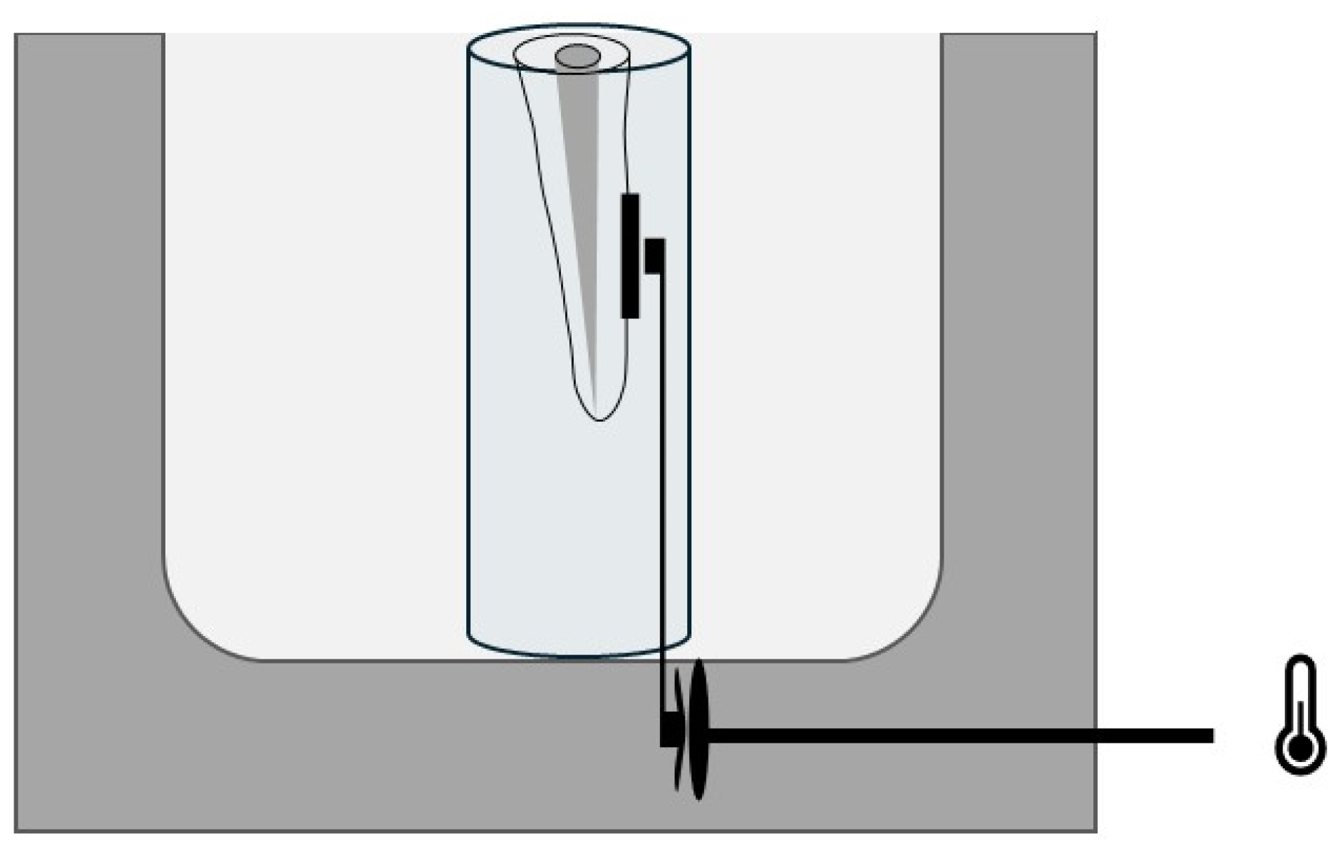
2.5. Temperature
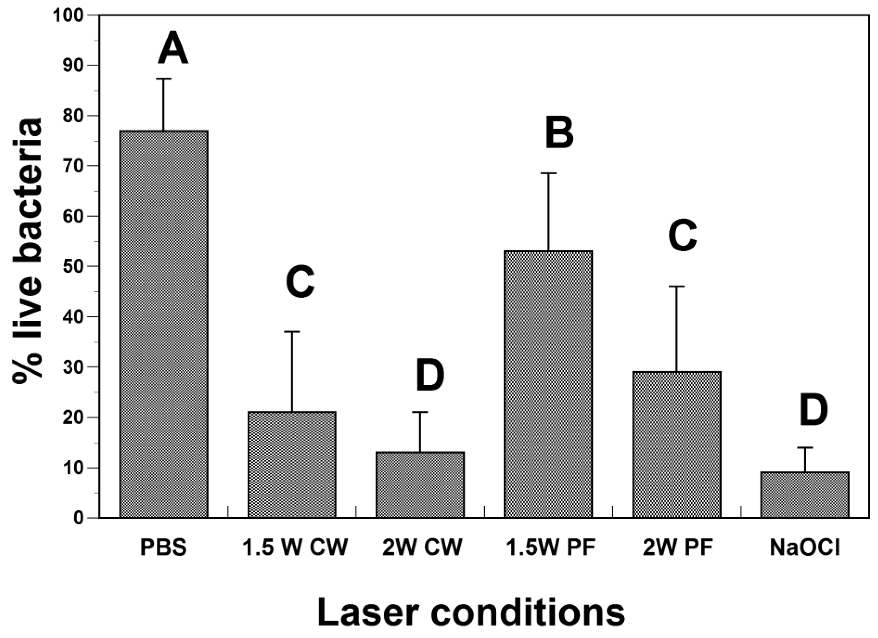
3. Results
4. Discussion
5. Conclusions
Author Contributions
Funding
Institutional Review Board Statement
Informed Consent Statement
Data Availability Statement
Conflicts of Interest
References
- Kakehashi, S.; Stanley, H.R.; Fitzgerald, R.J. The Effects of Surgical Exposures of Dental Pulps in Germ-Free and Conventional Laboratory Rats. Oral. Surg. Oral. Med. Oral. Pathol. 1965, 20, 340–349. [Google Scholar] [CrossRef]
- Sundqvist, G. Bacteriological Studies of Necrotic Dental Pulps. Ph.D. Thesis, University of Umea, Umea, Sweeden, 1976. [Google Scholar]
- Schilder, H. Cleaning and shaping the root canal. Dent. Clin. N. Am. 1974, 18, 269–296. [Google Scholar] [CrossRef] [PubMed]
- Siqueira, J.F., Jr.; Rocas, I.N.; Santos, S.R.; Lima, K.C.; Magalhaes, F.A.; de Uzeda, M. Efficacy of instrumentation techniques and irrigation regimens in reducing the bacterial population within root canals. J. Endod. 2002, 28, 181–184. [Google Scholar] [CrossRef] [PubMed]
- Haapasalo, M.; Shen, Y.; Wang, Z.; Gao, Y. Irrigation in endodontics. Br. Dent. J. 2014, 216, 299–303. [Google Scholar] [CrossRef]
- Zehnder, M. Root canal irrigants. J. Endod. 2006, 32, 389–398. [Google Scholar] [CrossRef] [PubMed]
- Pashley, E.L.; Birdsong, N.L.; Bowman, K.; Pashley, D.H. Cytotoxic effects of NaOCl on vital tissue. J. Endod. 1985, 11, 525–528. [Google Scholar] [CrossRef] [PubMed]
- Wikstrom, A.; Romani Vestman, N.; Rakhimova, O.; Lazaro Gimeno, D.; Tsilingaridis, G.; Brundin, M. Microbiological assessment of success and failure in pulp revitalization: A randomized clinical trial using calcium hydroxide and chlorhexidine gluconate in traumatized immature necrotic teeth. J. Oral. Microbiol. 2024, 16, 2343518. [Google Scholar] [CrossRef]
- Zandi, H.; Petronijevic, N.; Mdala, I.; Kristoffersen, A.K.; Enersen, M.; Rocas, I.N.; Siqueira, J.F., Jr.; Orstavik, D. Outcome of Endodontic Retreatment Using 2 Root Canal Irrigants and Influence of Infection on Healing as Determined by a Molecular Method: A Randomized Clinical Trial. J. Endod. 2019, 45, 1089–1098.e5. [Google Scholar] [CrossRef]
- Peters, L.B.; Wesselink, P.R. Periapical healing of endodontically treated teeth in one and two visits obturated in the presence or absence of detectable microorganisms. Int. Endod. J. 2002, 35, 660–667. [Google Scholar] [CrossRef]
- Rodrigues, R.C.V.; Zandi, H.; Kristoffersen, A.K.; Enersen, M.; Mdala, I.; Orstavik, D.; Rocas, I.N.; Siqueira, J.F., Jr. Influence of the Apical Preparation Size and the Irrigant Type on Bacterial Reduction in Root Canal-treated Teeth with Apical Periodontitis. J. Endod. 2017, 43, 1058–1063. [Google Scholar] [CrossRef]
- Vianna, M.E.; Horz, H.P.; Conrads, G.; Zaia, A.A.; Souza-Filho, F.J.; Gomes, B.P. Effect of root canal procedures on endotoxins and endodontic pathogens. Oral. Microbiol. Immunol. 2007, 22, 411–418. [Google Scholar] [CrossRef] [PubMed]
- Peters, O.A.; Schonenberger, K.; Laib, A. Effects of four Ni-Ti preparation techniques on root canal geometry assessed by micro computed tomography. Int. Endod. J. 2001, 34, 221–230. [Google Scholar] [CrossRef] [PubMed]
- Kouchi, Y.; Ninomiya, J.; Yasuda, H.; Fukui, K.; Moriyama, T.; Okamoto, H. Location of Streptococcus mutans in the dentinal tubules of open infected root canals. J. Dent. Res. 1980, 59, 2038–2046. [Google Scholar] [CrossRef] [PubMed]
- Berutti, E.; Marini, R.; Angeretti, A. Penetration ability of different irrigants into dentinal tubules. J. Endod. 1997, 23, 725–727. [Google Scholar] [CrossRef] [PubMed]
- Gulabivala, K.; Patel, B.; Evans, G.; Ng, Y. Effects of mechanical and chemical procedures on root canal surfaces. Endod. Top. 2005, 10, 103–122. [Google Scholar] [CrossRef]
- Prada, I.; Mico-Munoz, P.; Giner-Lluesma, T.; Mico-Martinez, P.; Collado-Castellano, N.; Manzano-Saiz, A. Influence of microbiology on endodontic failure. Literature review. Med. Oral. Patol. Oral. Cir. Bucal 2019, 24, e364–e372. [Google Scholar] [CrossRef]
- Alghamdi, F.; Shakir, M. The Influence of Enterococcus faecalis as a Dental Root Canal Pathogen on Endodontic Treatment: A Systematic Review. Cureus 2020, 12, e7257. [Google Scholar] [CrossRef]
- Estrela, C.; Silva, J.A.; de Alencar, A.H.; Leles, C.R.; Decurcio, D.A. Efficacy of sodium hypochlorite and chlorhexidine against Enterococcus faecalis—A systematic review. J. Appl. Oral. Sci. 2008, 16, 364–368. [Google Scholar] [CrossRef]
- Safavi, K.E.; Spangberg, L.S.; Langeland, K. Root canal dentinal tubule disinfection. J. Endod. 1990, 16, 207–210. [Google Scholar] [CrossRef]
- Meire, M.; de Moor, R.J.G. Principle and antimicrobial efficacy of laser-activated irrigation: A narrative review. Int. Endod. J. 2024, 57, 841–860. [Google Scholar] [CrossRef]
- Olivi, G. Laser use in endodontics: Evolution from direct laser irradiation to laser-activated irrigation. J. Laser Dent. 2013, 21, 58–71. [Google Scholar]
- Zanin, I.C.; Goncalves, R.B.; Junior, A.B.; Hope, C.K.; Pratten, J. Susceptibility of Streptococcus mutans biofilms to photodynamic therapy: An in vitro study. J. Antimicrob. Chemother. 2005, 56, 324–330. [Google Scholar] [CrossRef] [PubMed]
- Komerik, N.; Nakanishi, H.; MacRobert, A.J.; Henderson, B.; Speight, P.; Wilson, M. In vivo killing of Porphyromonas gingivalis by toluidine blue-mediated photosensitization in an animal model. Antimicrob. Agents Chemother. 2003, 47, 932–940. [Google Scholar] [CrossRef] [PubMed]
- Soukos, N.S.; Chen, P.S.; Morris, J.T.; Ruggiero, K.; Abernethy, A.D.; Som, S.; Foschi, F.; Doucette, S.; Bammann, L.L.; Fontana, C.R.; et al. Photodynamic therapy for endodontic disinfection. J. Endod. 2006, 32, 979–984. [Google Scholar] [CrossRef]
- Wilson, B.C.; Patterson, M.S. The physics of photodynamic therapy. Phys. Med. Biol. 1986, 31, 327–360. [Google Scholar] [CrossRef]
- Blanken, J.; de Moor, R.J.; Meire, M.; Verdaasdonk, R. Laser induced explosive vapor and cavitation resulting in effective irrigation of the root canal. Part 1: A visualization study. Lasers Surg. Med. 2009, 41, 514–519. [Google Scholar] [CrossRef]
- De Groot, S.D.; Verhaagen, B.; Versluis, M.; Wu, M.K.; Wesselink, P.R.; van der Sluis, L.W. Laser-activated irrigation within root canals: Cleaning efficacy and flow visualization. Int. Endod. J. 2009, 42, 1077–1083. [Google Scholar] [CrossRef]
- De Moor, R.J.; Blanken, J.; Meire, M.; Verdaasdonk, R. Laser induced explosive vapor and cavitation resulting in effective irrigation of the root canal. Part 2: Evaluation of the efficacy. Lasers Surg. Med. 2009, 41, 520–523. [Google Scholar] [CrossRef]
- Anton, Y.O.C.I.; Marger, L.; di Bella, E.; Abdelaziz, M.; Feilzer, A.; Krejci, I. Activation of endodontic irrigants using a 9.3 microm CO2 and diode lasers: A laboratory proof of concept model. Am. J. Dent. 2024, 37, 39–46. [Google Scholar]
- Do, Q.L.; Gaudin, A. The Efficiency of the Er: YAG Laser and PhotonInduced Photoacoustic Streaming (PIPS) as an Activation Method in Endodontic Irrigation: A Literature Review. J. Lasers Med. Sci. 2020, 11, 316–334. [Google Scholar] [CrossRef]
- George, R.; Walsh, L.J. Apical extrusion of root canal irrigants when using Er:YAG and Er,Cr:YSGG lasers with optical fibers: An in vitro dye study. J. Endod. 2008, 34, 706–708. [Google Scholar] [CrossRef] [PubMed]
- Helvacioglu Kivanc, B.; Deniz Arisu, H.; Yanar, N.O.; Silah, H.M.; Inam, R.; Gorgul, G. Apical extrusion of sodium hypochlorite activated with two laser systems and ultrasonics: A spectrophotometric analysis. BMC Oral. Health 2015, 15, 71. [Google Scholar] [CrossRef] [PubMed]
- Olivi, G.; DiVito, E.E. Advanced laser-activated irrigation: PIPSTM technique and clinical protocols. In Lasers in Endodontics. Scientific Background and Applications, 1st ed.; Olivi, G., de Moor, R., DiVito, E., Eds.; Springer: Berlin/Heidelberg, Germany, 2016; pp. 219–292. [Google Scholar]
- Schoop, U.; Kluger, W.; Moritz, A.; Nedjelik, N.; Georgopoulos, A.; Sperr, W. Bactericidal effect of different laser systems in the deep layers of dentin. Lasers Surg. Med. 2004, 35, 111–116. [Google Scholar] [CrossRef] [PubMed]
- Beer, F.; Buchmair, A.; Wernisch, J.; Georgopoulos, A.; Moritz, A. Comparison of two diode lasers on bactericidity in root canals—An in vitro study. Lasers Med. Sci. 2012, 27, 361–364. [Google Scholar] [CrossRef]
- Schoop, U.K.; Kluger, W.; Dervisbergovic, S.; Goharkhay, K.; Wenisch, J.; Georgopoulos, A.; Sperr, W.; Moritz, A. Innovative wavelengths in endodontic treatment. Lasers Surg. Med. 2006, 38, 624–630. [Google Scholar] [CrossRef]
- Beltes, C.; Economides, N.; Sakkas, H.; Papadopoulou, C.; Lambrianidis, T. Evaluation of Antimicrobial Photodynamic Therapy Using Indocyanine Green and Near-Infrared Diode Laser Against Enterococcus faecalis in Infected Human Root Canals. Photomed. Laser Surg. 2017, 35, 264–269. [Google Scholar] [CrossRef]
- Pirnat, S.; Lukac, M.; Ihan, A. Study of the direct bactericidal effect of Nd:YAG and diode laser parameters used in endodontics on pigmented and nonpigmented bacteria. Lasers Med. Sci. 2011, 26, 755–761. [Google Scholar] [CrossRef]
- Beltes, C.; Sakkas, H.; Economides, N.; Papadopoulou, C. Antimicrobial photodynamic therapy using Indocyanine green and near-infrared diode laser in reducing Entrerococcus faecalis. Photodiagn. Photodyn. Ther. 2017, 17, 5–8. [Google Scholar] [CrossRef]
- Katalinic, I.; Budimir, A.; Bosnjak, Z.; Jakovljevic, S.; Anic, I. The photo-activated and photo-thermal effect of the 445/970 nm diode laser on the mixed biofilm inside root canals of human teeth in vitro: A pilot study. Photodiagn. Photodyn. Ther. 2019, 26, 277–283. [Google Scholar] [CrossRef]
- Benedicenti, S.; Cassanelli, C.; Signore, A.; Ravera, G.; Angiero, F. Decontamination of root canals with the gallium-aluminum-arsenide laser: An in vitro study. Photomed. Laser Surg. 2008, 26, 367–370. [Google Scholar] [CrossRef]
- Lan, W.H. Temperature elevation on the root surface during Nd:YAG laser irradiation in the root canal. J. Endod. 1999, 25, 155–156. [Google Scholar] [CrossRef] [PubMed]
- Stabholz, A.; Sahar-Helft, S.; Moshonov, J. Lasers in endodontics. Dent. Clin. N. Am. 2004, 48, 809–832. [Google Scholar] [CrossRef] [PubMed]
- Borges, C.C.; Estrela, C.; Lopes, F.C.; Palma-Dibb, R.G.; Pecora, J.D.; De Araujo Estrela, C.R.; Sousa-Neto, M.D. Effect of different diode laser wavelengths on root dentin decontamination infected with Enterococcus faecalis. J. Photochem. Photobiol. B 2017, 176, 1–8. [Google Scholar] [CrossRef] [PubMed]
- Alfredo, E.; Marchesan, M.A.; Sousa-Neto, M.D.; Brugnera-Junior, A.; Silva-Sousa, Y.T. Temperature variation at the external root surface during 980-nm diode laser irradiation in the root canal. J. Dent. 2008, 36, 529–534. [Google Scholar] [CrossRef] [PubMed]
- Anagnostaki, E.; Mylona, V.; Parker, S.; Lynch, E.; Grootveld, M. Systematic Review on the Role of Lasers in Endodontic Therapy: Valuable Adjunct Treatment? Dent. J. 2020, 8, 63. [Google Scholar] [CrossRef]
- Dawasaz, A.A. In Vivo Efficacy of Diode Laser as a Monotherapy in Root Canal Disinfection: A Systematic Review and Meta-Analysis. Photobiomodul. Photomed. Laser Surg. 2022, 40, 59–70. [Google Scholar] [CrossRef]
- Klinke, T.; Klimm, W.; Gutknecht, N. Antibacterial effects of Nd:YAG laser irradiation within root canal dentin. J. Clin. Laser Med. Surg. 1997, 15, 29–31. [Google Scholar] [CrossRef]
- Provoost, C.; Rocca, G.T.; Thibault, A.; Machtou, P.; Bouilllaguet, S. Influence of Needle Design and Irrigant Flow Rate on the Removal of Enterococcus faecalis Biofilms In Vitro. Dent. J. 2022, 10, 59. [Google Scholar] [CrossRef]
- Manoil, D.; Filieri, A.; Gameiro, C.; Lange, N.; Schrenzel, J.; Wataha, J.C.; Bouillaguet, S. Flow cytometric assessment of Streptococcus mutans viability after exposure to blue light-activated curcumin. Photodiagn. Photodyn. Ther. 2014, 11, 372–379. [Google Scholar] [CrossRef]
- Stocks, S.M. Mechanism and use of the commercially available viability stain, BacLight. Cytom.Part A J. Int. Soc. Anal. Cytol. 2004, 61, 189–195. [Google Scholar] [CrossRef]
- Gutknecht, N.; Franzen, R.; Schippers, M.; Lampert, F. Bactericidal effect of a 980-nm diode laser in the root canal wall dentin of bovine teeth. J. Clin. Laser Med. Surg. 2004, 22, 9–13. [Google Scholar] [CrossRef] [PubMed]
- Sarda, R.A.; Shetty, R.M.; Tamrakar, A.; Shetty, S.Y. Antimicrobial efficacy of photodynamic therapy, diode laser, and sodium hypochlorite and their combinations on endodontic pathogens. Photodiagn. Photodyn. Ther. 2019, 28, 265–272. [Google Scholar] [CrossRef] [PubMed]
- Orstavik, D.; Haapasalo, M. Disinfection by endodontic irrigants and dressings of experimentally infected dentinal tubules. Endod. Dent. Traumatol. 1990, 6, 142–149. [Google Scholar] [CrossRef] [PubMed]
- Shen, Y.; Stojicic, S.; Haapasalo, M. Antimicrobial efficacy of chlorhexidine against bacteria in biofilms at different stages of development. J. Endod. 2011, 37, 657–661. [Google Scholar] [CrossRef] [PubMed]
- Giard, J.C.; Hartke, A.; Flahaut, S.; Benachour, A.; Boutibonnes, P.; Auffray, Y. Starvation-induced multiresistance in Enterococcus faecalis JH2-2. Curr. Microbiol. 1996, 32, 264–271. [Google Scholar] [CrossRef] [PubMed]
- Stuart, C.H.; Schwartz, S.A.; Beeson, T.J.; Owatz, C.B. Enterococcus faecalis: Its role in root canal treatment failure and current concepts in retreatment. J. Endod. 2006, 32, 93–98. [Google Scholar] [CrossRef] [PubMed]
- Cimpean, S.I.; Pop-Ciutrila, I.S.; Matei, S.R.; Colosi, I.A.; Costache, C.; Nicula, G.Z.; Badea, I.C.; Colceriu Burtea, L. Effectiveness of Different Final Irrigation Procedures on Enterococcus faecalis Infected Root Canals: An In Vitro Evaluation. Materials 2022, 15, 6688. [Google Scholar] [CrossRef]
- Kurian, A.H.; Sethi, S.; Aneja, K.; Gupta, A.; Virmani, S.; Abraham, D. Elimination of Enterococcus faecalis from root canal system using laser-activated nanoparticles: A systematic review. Lasers Med. Sci. 2023, 38, 81. [Google Scholar] [CrossRef]
- Puleio, F.; Pirri, R.; Tosco, V.; Lizio, A.S.; Tripodi, P.; la Spina, I.; la Fauci, V.; Squeri, R. Assessment of 2′-Fucosyllactose and Lacto-N-Neotetraose Solution as an Irrigant in E. faecalis-Infected Root Canals: An In Vitro Study. Clin. Pract. 2024, 14, 1348–1356. [Google Scholar] [CrossRef] [PubMed]
- da Costa Ribeiro, A.; Nogueira, G.E.C.; Antoniazzi, J.H.; Moritz, A.; Zezell, D.M. Effects of Diode Laser (810 nm) Irradiation on Root Canal Walls: Thermographic and Morphological Studies. J. Endod. 2007, 33, 252–255. [Google Scholar] [CrossRef]
- Gutknecht, N.; Franzen, R.; Meister, J.; Vanweersch, L.; Mir, M. Temperature evolution on human teeth root surface after diode laser assisted endodontic treatment. Lasers Med. Sci. 2005, 20, 99–103. [Google Scholar] [CrossRef] [PubMed]
- Eriksson, A.R.; Albrektsson, T. Temperature threshold levels for heat-induced bone tissue injury: A vital-microscopic study in the rabbit. J. Prosthet. Dent. 1983, 50, 101–107. [Google Scholar] [CrossRef] [PubMed]
- Zou, Z.; Bhandari, J.; Xiao, B.; Liang, X.; Zhang, Y.; Yan, G. Effect of using diode laser on Enterococcus faecalis and its lipoteichoic acid (LTA) in chronic apical periodontitis. Lasers Med. Sci. 2021, 36, 1059–1066. [Google Scholar] [CrossRef] [PubMed]
- Braun, A.; Hagelauer, F.J.; Wenzler, J.; Heimer, M.; Frankenberger, R.; Stein, S. Microcrack Analysis of Dental Hard Tissue After Root Canal Irradiation with a 970-nm Diode Laser. Photomed. Laser Surg. 2018, 36, 621–628. [Google Scholar] [CrossRef]
- Saunders, E.M. In vivo findings associated with heat generation during thermomechanical compaction of gutta-percha. 2. Histological response to temperature elevation on the external surface of the root. Int. Endod. J. 1990, 23, 268–274. [Google Scholar] [CrossRef]
- Faria, M.I.; Sousa-Neto, M.D.; Souza-Gabriel, A.E.; Alfredo, E.; Romeo, U.; Silva-Sousa, Y.T. Effects of 980-nm diode laser on the ultrastructure and fracture resistance of dentine. Lasers Med. Sci. 2013, 28, 275–280. [Google Scholar] [CrossRef]
- Saunders, E.M. In vivo findings associated with heat generation during thermomechanical compaction of gutta-percha. 1. Temperature levels at the external surface of the root. Int. Endod. J. 1990, 23, 263–267. [Google Scholar] [CrossRef]


| Emission Mode | Power | ∆ T °C | p < 0.05 |
|---|---|---|---|
| Continuous mode | 1.5 W | 7.1 ± 2.8 | A,a |
| 2 W | 7.9 ± 3.1 | A | |
| Pulsed mode | 1.5 W | 4 ± 1.6 | B,b |
| 2 W | 5.9 ± 2.3 | B |
Disclaimer/Publisher’s Note: The statements, opinions and data contained in all publications are solely those of the individual author(s) and contributor(s) and not of MDPI and/or the editor(s). MDPI and/or the editor(s) disclaim responsibility for any injury to people or property resulting from any ideas, methods, instructions or products referred to in the content. |
© 2024 by the authors. Licensee MDPI, Basel, Switzerland. This article is an open access article distributed under the terms and conditions of the Creative Commons Attribution (CC BY) license (https://creativecommons.org/licenses/by/4.0/).
Share and Cite
Tanner, S.; Thibault, A.; Leprince, J.G.; Bouillaguet, S. Photothermal Effect of 970 nm Diode Laser Irradiation on Enterococcus faecalis Biofilms in Single-Rooted Teeth Ex Vivo. Dent. J. 2024, 12, 308. https://doi.org/10.3390/dj12100308
Tanner S, Thibault A, Leprince JG, Bouillaguet S. Photothermal Effect of 970 nm Diode Laser Irradiation on Enterococcus faecalis Biofilms in Single-Rooted Teeth Ex Vivo. Dentistry Journal. 2024; 12(10):308. https://doi.org/10.3390/dj12100308
Chicago/Turabian StyleTanner, Soraya, Anna Thibault, Julian Grégoire Leprince, and Serge Bouillaguet. 2024. "Photothermal Effect of 970 nm Diode Laser Irradiation on Enterococcus faecalis Biofilms in Single-Rooted Teeth Ex Vivo" Dentistry Journal 12, no. 10: 308. https://doi.org/10.3390/dj12100308