Review

Bioengineered Anthocyanin-Enriched Tomatoes: A Novel Approach to Colorectal Cancer Prevention

1
Department of Food Nutrition Dietetics and Health, Kansas State University, Manhattan, KS 66506, USA
2
Department of Horticulture and Nature Resources, Kansas State University, Manhattan, KS 66506, USA
3
Department of Grain Science and Industry, Kansas State University, Manhattan, KS 66506, USA
*
Author to whom correspondence should be addressed.
Foods 2024, 13(18), 2991; https://doi.org/10.3390/foods13182991
Submission received: 10 August 2024 / Revised: 11 September 2024 / Accepted: 18 September 2024 / Published: 21 September 2024

Abstract

:
Colorectal cancer (CRC) remains a significant global health challenge, with barriers to effective prevention and treatment including tumor recurrence, chemoresistance, and limited overall survival rates. Anthocyanins, known for their strong anti-cancer properties, have shown promise in preventing and suppressing various cancers, including CRC. However, natural sources of anthocyanins often fail to provide sufficient quantities needed for therapeutic effects. Bioengineered crops, particularly anthocyanin-enriched tomatoes, offer a viable solution to enhance anthocyanin content. Given its large-scale production and consumption, tomatoes present an ideal target for bioengineering efforts aimed at increasing dietary anthocyanin intake. This review provides an overview of anthocyanins and their health benefits, elucidating the mechanisms by which anthocyanins modulate the transcription factors involved in CRC development. It also examines case studies demonstrating the successful bioengineering of tomatoes to boost anthocyanin levels. Furthermore, the review discusses the effects of anthocyanin extracts from bioengineered tomatoes on CRC prevention, highlighting their role in altering metabolic pathways and reducing tumor-related inflammation. Finally, this review addresses the challenges associated with bioengineering tomatoes and proposes future research directions to optimize anthocyanin enrichment in tomatoes.

1. Introduction

CRC has now become a world health issue; it is the third most common malignancy and the second most leading reason for cancer-related deaths [1]. The global burden of CRC has increased substantially, with a 157% rise in incidence from 1990 to 2019, resulting in 2.17 million cases in 2019 [2]. Such elevation is particularly evident in developing regions, such as East Asia and Middle Socio-Demographic Index Regions, where the burden is growing the fastest [3]. In China, CRC poses a severe challenge, with 560,000 new cases and 290,000 deaths in 2020 alone, and projections indicating a 64% increase in incident cases by 2040 [1]. A summary report on CRC has been published by the Surveillance, Epidemiology, and End Results program under the National Cancer Institute. The incidence of CRC in the US was reported at a rate of 36.5 cases per 100,000 individuals annually. Although the rate of people being diagnosed with colon or rectal cancer each year has dropped overall since the mid-1980s in the US, it is still a threatening and raging disease in the world as a whole. All types of cancer have some similar patterns and nature of growth and spread. When a cancer is identified solely within the original site of occurrence, it is categorized as localized (sometimes denoted as stage 1). Conversely, if it has disseminated to another area of the body, it is classified as either regional or distant. In the case of colorectal cancer, the distribution among local stage diagnoses stands at 34.8%, the regional stage at 36%, and the distant stage at 23%. Five years of relative survival rates for localized, regional, and distant CRC were reported as 91.1%, 73.7%, and 15.7%, respectively. The interplay between these genetic predispositions and environmental exposures underscores the multifactorial nature of CRC etiology. Effective screening programs and early diagnosis, along with lifestyle modifications, are essential to mitigate the rising incidence and improve survival rates [4,5].
Cancer treatment has a variety of technologies and methodologies, including surgery, chemotherapy, radiation therapy, and emerging biotechnologies like immunotherapy and targeted therapy [6]. While these treatments and strategies are crucial for managing and potentially curing cancer, they often come with significant side effects and complications [7,8]. On average, 10% of colorectal surgery cases lead to small bowel obstruction (SBO) due to peritoneal adhesions post-surgery. The recurrence of adhesive SBO is linked to a lower survival rate [9]. The late adverse effects of radiotherapy on CRC include bowel obstructions and dysfunction, manifesting through symptoms such as fecal incontinence, lose or solid stools, gas, evacuation difficulties, and sexual dysfunction [10]. In contrast, plant-based compounds exhibit several special outcomes of CRC treatment. For example, plant-based compounds effectively manage chemotherapy-related side effects, delay tumor growth, and modulate multiple molecular pathways linked with multidrug resistance [11,12]. Within plants, one can find bioactive phytochemicals including flavonoids, polyphenols, and catechins, which play a crucial character in reducing tumor cell propagation by obstructing cell cycle checkpoints and fostering apoptosis [11].
Anthocyanins represent water-soluble pigments within the phenolic compound category. Berries, currants, grapes, and specific tropical fruits exhibit a notable abundance of anthocyanins. The presence of high anthocyanin content is characteristic of leafy vegetables, grains, roots, and tubers displaying hues ranging from red to purplish-blue [13]. The potential anti-cancer effects of anthocyanin may emanate from a variety of biological processes, encompassing anti-oxidative stress, anti-inflammatory actions, anti-mutagenesis properties, the stimulation of differentiation, the suppression of proliferation, the arrest of the cell cycle, and the induction of apoptosis, alongside anti-invasion, anti-metastasis, anti-angiogenesis effects, and the sensitization of cancer cells to chemotherapy [14]. Several studies proved the power of anthocyanin to fight against CRC [15,16]. In particular, the consumption of anthocyanin extracts from strawberries, black raspberries, tart cherries, and blackberries with higher doses could significantly decrease tumor proliferation [17]. The higher doses are equal to a higher number of fruits, which may not be affordable for middle- and low-income families, especially from those countries where such fruits need to be imported. While berries are renowned for their high anthocyanin content, several other fruits and vegetables are often less expensive and contain anthocyanin, but in trace amounts [18]. The biosynthesis of anthocyanins among those plants can be possibly manipulated by bioengineering. For instance, the overexpression of MYB genes like VvmybA1 from red grape and Ruby from ‘Moro’ blood orange in Mexican lime has resulted in significant anthocyanin accumulation in leaves, flowers, and fruits, thereby increasing the antioxidant activity of the transgenic plants [19,20]. Additionally, the development of anthocyanin-rich cultivars such as the ‘Sun Black’ tomato in Europe and the ‘Kufri Neelkanth’ purple potato in India demonstrates the successful application of genetic engineering in enhancing anthocyanin content without compromising yield [19].
To increase the affordable supply of anthocyanin to all classes of people around the globe, a frequently consumed, less expensive, and easily cultivatable crop could be subjected to bioengineering. Consequently, the target population could have access and purchase capacity. Tomato (Solanum lycopersicum), second only to potatoes in terms of vegetable consumption in the US, presents a promising opportunity for the consideration of bioengineering to enhance anthocyanin due to its status as one of the most extensively studied crop species at the metabolomic level. Thus, it establishes itself as a primary model for the examination of flavonoids and phenylpropanoids [21,22]. Bioengineered tomato containing high anthocyanin levels was documented in 2008, and US government agencies recently approved bioengineered purple tomatoes for market, a significant advancement for enriching human diets. On top of that, EU legislation started approving bioengineered crops in various sectors, with slow and challenging approval processes [21]. To understand the importance of bioengineered anthocyanin-enriched tomatoes to face CRC, this review focused on accumulating available knowledge and information on the role of bioengineered anthocyanin-enriched tomatoes in preventing CRC and its inflammation. In addition, this review is committed to showcasing the mechanism to prevent CRC through anthocyanin extracts from bioengineered tomatoes as well as other natural sources.

2. Anthocyanins

2.1. Natural Sources of Anthocyanin

Anthocyanins have been historically documented as “colored cell sap” for many centuries. In 1835, a German botanist officially designated this compound [23]. Within the classification of secondary phytochemicals termed flavonoids, anthocyanin is identified as a specific subset [24]. Anthocyanin produces a diverse range of colors including purple, pink, blue, and red in plant organisms, namely flowers, vegetables, and fruits [24]. The modification of anthocyanin’s chemical structure through processes like hydroxylation, methylation, glycosylation, and acylation enhances their color variations and stability. The structure of the major type of anthocyanin is presented in Figure 1. The color of anthocyanins transitions to blue as the number of hydroxyl groups in the B-ring increases, whereas methylation induces a shift toward the red end of the spectrum. The methylation of the B-ring is important for reducing oxidation susceptibility and enhancing anthocyanin stability [24]. Anthocyanins have a unique UV−Vis absorption pattern for their extended chromophore with eight double bonds alone with a positive charge on the O2 ring in an acidic environment. This pattern shows an absorption peak among the visible range of 465 nm and 550 nm. It also gives a peak at another absorption between 270 nm and 280 nm [25]. Naturally, a rich diversity of over 635 distinct anthocyanins has been identified [26]. Among these, six primary anthocyanidins—cyanidin, pelargonidin, delphinidin, petunidin, peonidin, and malvidin—dominate, collectively representing about 90% of all identified anthocyanins [27]. The distribution of these anthocyanidins in natural sources is typically 30% cyanidin, 22% delphinidin, 18% pelargonidin, 7.5% peonidin, 7.5% malvidin, and 5% petunidin [28]. A list of plant sources that contain comparatively higher amounts of anthocyanin is presented in Table 1. Although anthocyanins can be isolated from various plants like sweet potato, hibiscus, corn, cabbage, and carrot, berries are primarily recognized as the main reservoir of anthocyanins and provide about 29.17% of anthocyanin of all sources. Following berries, other significant sources of anthocyanins include grape, sour cherry, black rice, and black carrot. Furthermore, purple maize, pomegranate fruit, saffron, banana, and mao fruit also serve as notable sources of anthocyanins [29].

2.2. Factors That Affect the Yield of Anthocyanin

The yield of anthocyanin, a pigment with significant antioxidant properties, is influenced by several factors, such as the type of solvent used, soil temperature, plant preparation methods, extraction techniques, cofactor selection, and environmental conditions [50].

2.2.1. Extraction Techniques

Likewise, extraction techniques influence anthocyanin yields because different extraction techniques have different principles which alter the extraction yield and quality [51]. For instance, in the study on freeze-dried strawberry puree, two chloroform-based methods yielded the highest anthocyanin content, while an extraction solvent MeOH:H2O: HCl extracted an intermediate amount of anthocyanin, and the pH differential method yielded the lowest anthocyanin content, despite its shorter processing time [52]. In a separate investigation, the utilization of enzyme–microwave-assisted extraction on Hibiscus sabdariffa L. led to a recorded anthocyanin output of 15.37 ± 0.41 mg/g, whereas a microwave-assisted extraction yielded 13.01 ± 0.22 mg/g, ultrasound-assisted extraction yielded 11.73 ± 0.28 mg/g, and enzyme-assisted extraction led to an output of 11.06 ± 0.33 mg/g, correspondingly [53]. Similarly, ultrasound-assisted extraction of Myrciaria cauliflora Berg.’s skin showed that increasing the extraction time improved anthocyanin yield from 200 mg (at 1st min) to 400 mg (at 3rd min) per 100 g DW and antioxidant capacity, making it the most effective method compared to conventional and high-pressure extractions [54]. Optimized ultrasound-assisted extraction demonstrated a yield of 311 ± 5 mg of anthocyanin from 100 g of Hibiscus sabdariffa L. calyces, surpassing the yield obtained through the traditional extraction method [55]. On top of that, another study on Hibiscus rosa-sinensis petals revealed that ultrasound-assisted extraction yielded 179.32 mg/L of anthocyanins, outperforming microwave-assisted extraction at 155.45 mg/L and conventional extraction at 100.88 mg/L [56]. A highly efficient separation methodology has been developed, referred to as counter-current chromatography (CCC). This technique demonstrated extraction purities exceeding 90% for specific anthocyanins, including delphinidin-3-glucoside, cyanidin-3-rutinoside, and delphinidin-3-rutinoside [57]. In another investigation, anthocyanins were extracted from strawberries utilizing high-speed counter-current chromatography, employing a biphasic solvent system composed of tert-butyl methyl ether, n-butanol, acetonitrile, water, and trifluoroacetic acid in a ratio of 2.5:2.0:2.5:5.0:1.0%. The identified anthocyanins included pelargonidin-3-rutinoside, cyanidin-3-glucoside, and pelargonidin-3-glucoside, exhibiting purities of 95.6%, 96.2%, and 99.3%, respectively [58]. These findings collectively highlight that advanced extraction techniques, particularly those involving ultrasound, enzyme–microwave assistance, and current, tend to enhance anthocyanin yields significantly compared to conventional methods, with specific numeric data underscoring the superior efficiency of these modern techniques.

2.2.2. Extraction Solvents

The extraction of anthocyanins found in various plants is significantly influenced by the choice of extraction solvents. It affects both yield and stability. Among the various solvents that were investigated, chloroform–methanol, ethanol, and methanol showed the most significant degree of anthocyanin extraction capacity, approximately 10–12 A/g FW. Solvents like methanol–water, pH differential buffers, and acetone demonstrated a moderate efficiency in extracting anthocyanins, ranging from 6.8 to 8.8 A/g FW. Moreover, a combination of different solvents with water displayed the least effective solvents, with an extraction yield of less than 5 A/g FW of strawberry puree [50]. In contrast, only distilled water demonstrated superior solvency compared to 96% ethanol for the extraction of anthocyanins from Butterfly Pea flowers. The water extraction method, in particular, resulted in the production of anthocyanin, 4841 mg/g Butterfly Pea [59]. Novel techniques such as microwave–ultrasonic-assisted extraction (MUAE) in combination with natural deep eutectic solvents (DESs) have displayed potential in the efficient retrieval of anthocyanins from purple perilla leaves, attaining a maximum output of 619.62 mg/100 g under ideal conditions [60]. Likewise, the utilization of natural deep eutectic solvents (NaDES), specifically formulations involving choline chloride and malic acid, in conjunction with ultrasonication-assisted extraction, has proven to be successful in the retrieval of chokeberry anthocyanins. Moreover, additional enhancements have been noted through the integration of hydroxypropyl-β-cyclodextrin (HP-β-CD) to boost both yield and stability. The extraction efficiency experienced enhancement with HP-β-CD concentrations of up to 3% (w/w) [61]. Additionally, DESs based on choline chloride and xylitol significantly increased the anthocyanin yield from Euterpe edulis Mart. The increase in fruit pulp by 42% compared to methanolic extraction also demonstrated superior antioxidant capacity and slower degradation under heat and light [62].

2.2.3. Extraction pH

The extraction pH showed influences on both the quantity and quality of anthocyanins pulled out from various plant sources. For instance, in the study on extracted anthocyanin from Hibiscus rosa-sinensis, scientists compared the outcome of pH 2 and pH 4, revealing that the highest total anthocyanin (cyanidin-3-glucoside) yields were 8.33 mg/L at pH 2 and 9.56 mg/L at pH 4 [63]. Similarly, in the extraction of anthocyanins from water caltrop hull, the optimal pH was found to be 4.49, which resulted in a 70.3% anthocyanin extraction efficiency [64]. In red radish, a lower pH of 2.5 yielded higher anthocyanin content (98.02 mg/100 g, FW) compared to pH 4.5, which also resulted in a lighter color and higher polymeric color percentage, suggesting that lower pH conditions are more favorable for maintaining anthocyanin stability and color intensity [65]. The type of anthocyanin extracted also varies with pH; for example, in strawberries, pelargonidin was quantified as a major abundance of anthocyanin in methanol–chloroform solvent, while delphinidin dominated in other category solvents, indicating that solvent pH can alter the anthocyanin profile [50]. Additionally, the pH-sensitive film study demonstrated that anthocyanin extracts from Lycium ruthenicum Murr. changed color across a pH range of 2.0 to 10.0, further illustrating the impact of pH on anthocyanin stability and visual properties [66]. Overall, these studies collectively highlight that lower pH conditions generally enhance anthocyanin yield and stability, while higher pH conditions can lead to different anthocyanin profiles and potentially higher extraction efficiencies, depending on the plant source and extraction method used.

2.2.4. Cultivar and Genetic Factors

Genetic diversity and bioengineering have significantly impacted anthocyanin yield in various fruits and vegetables, as evidenced by multiple studies. In blueberries and bilberries, the upregulation of the transcriptional activator MYBA1 in conjunction with VmANS resulted in a threefold elevation in anthocyanin concentration, underscoring the involvement of specific structural genes in anthocyanin biosynthesis [67]. Similarly, in the context of olive fruits, specifically the Carolea and Tondina varieties, research findings indicated that the overall concentration of anthocyanins was greater in the ‘Tondina’ cultivar compared to the ‘Carolea’ cultivar. Inconsistencies were also observed concerning the genetic composition [68]. In the case of tomatoes, the ectopic expression of the MYB-like transcription factor PhAN4 triggered the synthesis of atypical anthocyanins like delphinidin and petunidin derivatives, while augmenting the plant’s capacity to combat oxidative stress, a crucial aspect for applications in modern agriculture [69]. Examination of the gene expression patterns associated with anthocyanin production in violet tomatoes indicated that key regulatory genes like SlANT1 and SlAN1 are pivotal in activating structural genes, thereby facilitating anthocyanin deposition in the fruit skin [70]. In woodland strawberries, altering the expression of the R2R3 MYB activator MYB10 significantly affected anthocyanin concentrations, with overexpression increasing and knockdown decreasing the levels of cyanidin and pelargonidin glucosides [71]. The incorporation of the SmMYB1 gene into an anthocyanin-deficient eggplant variety resulted in elevated levels of anthocyanins in multiple plant parts, notably the fruit pulp, alongside enhanced resistance to frost stress [72]. In horticulture crops, the synchronized expression of the structural genes implicated in anthocyanin synthesis is impacted by external elements like light and temperature, which have an impact on the expression of regulatory genes [73]. The enhancement of regulatory and structural genes in blueberries during the later phases of fruit growth resulted in a notable accumulation of anthocyanin, causing the fruit to exhibit a blue color [74]. Within Asian pears, the transcription factor PyMYB10 belonging to the R2R3-MYB family was recognized as a key player in the regulation of anthocyanin production, displaying a positive association between its expression and the anthocyanin levels in red-skinned varieties [75]. These studies collectively demonstrate that the targeted manipulation of gene expression and bioengineering can effectively enhance anthocyanin yield in fruits and vegetables, providing valuable insights for breeding programs aimed at improving nutritional properties and stress tolerance.

2.3. Health Benefits and Functional Properties of Anthocyanins

Anthocyanin consumption offers a range of specific health benefits, particularly in controlling and preventing chronic diseases.

2.3.1. Circulatory-Related Health Issues

Clinical trials have shown that anthocyanin-rich foods significantly reduce fasting blood glucose by a notable percentage, HbA1c by 2%, total cholesterol by 1%, triglycerides by 0.3%, and low-density lipoprotein by 0.5%, while increasing high-density lipoprotein by 0.3% [76]. Anthocyanins from roselle administered to Wistar rats resulted in a decreased heart rate and blood pressure across varying doses of 50, 100, and 200 mg/kg body weight. The findings indicate that the impact of anthocyanins on rats implies a notable antihypertensive efficacy, potentially through the modulation of elements within the renin–angiotensin–aldosterone system [77]. Anthocyanin treatment at 10 mg/kg, 0.4 μL/h decreased a salt-induced rise in heart rate and blood pressure. Additionally, it hindered sympathetic nerve activation by lowering LF/HF and raising RMSSD and SDNN levels in hypertension induced by high salt [78]. Clinical trials have shown that anthocyanin supplementation significantly reduces body mass index by an average of 0.36 kg/m2, and is particularly effective at doses of 300 mg/day or less over four weeks [79]. Moreover, anthocyanins have demonstrated the ability to lower fasting blood glucose, HbA1c, total cholesterol, triglycerides, and levels of low-density lipoprotein, while simultaneously elevating high-density lipoprotein levels in various clinical and pre-clinical investigations. These findings suggest a beneficial influence on metabolic health indicators linked to obesity [76].

2.3.2. Chronic Liver Disease

Research conducted on 2288 adult participants from the United States revealed that a higher consumption of anthocyanins was found to be significantly linked to a reduced likelihood of Non-Alcoholic Fatty Liver Disease. In an investigation, hepatic fibrosis was induced in a mouse model by CCL4, where anthocyanin treatment with doses of 100 and 200 mg/kg notably reduced hepatic fibrosis, inhibited hepatic stellate cell proliferation, and reversed blocked autophagic flux through the modulation of the circ_0000623/miR-351-5p/TFEB pathway [76]. Black raspberry anthocyanins also showed protective effects against adrenoleukodystrophy by ameliorating serum biochemical parameters, reducing liver damage, and promoting apoptosis in HepG2 cells through the TGF-β and NF-κB pathways [80]. Black raspberry anthocyanins (25, 50, 100, 150, and 200 μg/mL) and cyanidin-3-O-rutinoside (10, 20, 40, 80, and 100 μM) demonstrated cytotoxic effects on t-HSC/Cl-6, HepG2, and Hep3B cells, as well as induced cell death programming in HepG2 cells. These compounds were found to downregulate Bcl-2 protein expression, upregulate Bax levels, and facilitate cytochrome C release, cleaved caspase-3, cleaved caspase-9, and cleaved PARP in HepG2 cells [80]. The findings of the study indicate that the preventive impact of anthocyanin from black raspberry on alcoholic liver disease is mediated through antioxidative and apoptotic pathways.

2.3.3. Immunity Responses

In animal research, anthocyanin-enriched diets have demonstrated the ability to restore cellular immunity parameters, specifically the proportion of T helper cells (CD3+CD4+) and T cytotoxic lymphocytes (CD3+CD8+), which are commonly disturbed in cases of obesity [81]. BALB/c mice with leukemia were provided with a diet containing anthocyanins extracted from purple glutinous indica rice at doses of 0, 20, 50, or 100 mg/kg over three weeks. The supplementation of anthocyanins was observed to stimulate the populations of CD3 (T cell), CD19 (B cell), CD11b (monocyte), and Mac-3 (macrophage) in the leukemia-afflicted mice. Moreover, treatment with AUPGA was found to enhance macrophage phagocytosis while reducing the activity of NK cells [82]. Fluorosis was induced in four-week-old healthy male Wistar rats through the administration of fluoride for 90 days. The experimental intervention involved the administration of anthocyanin extracted from blueberries at a concentration of 100 mg/kg body weight per day. The levels of IgG and IL-1 markers exhibited a significant increase in the groups treated with anthocyanins. These immunoglobulins and cytokines play a crucial role in both the immune response and immune regulation [83]. Therefore, the results indicate the potential of anthocyanins in enhancing immune responses in living organisms.

2.3.4. Cancers

Anthocyanins’ broad antitumor activities include anti-inflammatory, antioxidant, anti-metastasis, and anti-mutagenesis effects and also reverse drug resistance and increasing sensitivity to chemotherapy, making them versatile agents in cancer treatment [84]. The MCF 7 breast cancer cell line was treated with anthocyanins (50 µg/mL) from Cordyline australis (cabbage tree). The study revealed that anthocyanin treatment with 65 ± 2.1% cytotoxicity induced profound activities in caspase-3 (157%), caspase-8 (142%), and caspase-9 (147%). These findings indicated anthocyanin’s potential in promoting apoptosis and inhibiting cancer cell proliferation [85]. Moreover, a human study with 25 CRC patients who received oral bilberry extract (containing 0.5–2.0 g of anthocyanins) for 7 days demonstrated a dose-dependent existence of anthocyanins in circulation and tumor tissues. The concentrations reached around 179 ng/g of tumor tissue at the highest dose. This treatment resulted in a 7% reduction in tumor cell proliferation [15]. Furthermore, dietary factors rich in anthocyanins, such as berries and grapes, have been associated with protective effects against CRC due to their high levels of cancer-fighting phytochemicals [86]. In another animal study, phycocyanin, a pigment–protein complex, demonstrated anti-CRC activities by reducing tumor numbers and inhibiting epithelial cell proliferation, alongside altering the composition of the microbiota inside the gut and affecting the IL-17 signaling pathway [87]. Collectively, these findings underscore the multifaceted role of anthocyanins in cancer prevention, highlighting their potential as complementary therapeutic agents in oncology. Future investigation is necessary to elucidate the precise mechanisms (molecular and cellular levels), optimal dosages, and available sources for their effective use in cancer prevention and treatment.

2.4. Bioaccessibility and Absorption of Anthocyanin

Based on the above discussion, without any doubt, anthocyanin has numerous health benefits and functional properties. Furthermore, to maximize the benefits of anthocyanin to protect our health, the bioaccessibility and bioavailability through the gastrointestinal tract (GIT) of this compound should be kept in mind [88]. This is because the plant-based matrix, for instance, the cell wall and fibrous tissue structure, could affect the absorption of starch, lipids, proteins, and functional phytochemicals [89]. An in vitro gastrointestinal model was used and demonstrated anthocyanin’s bioaccessibility from purple carrots and potatoes sunk at 44.62 and 71.8%, respectively [90]. On top of that, the total phenolic and carotenoid contents and the antioxidant activities of purple tomato were significantly reduced by 37–72%, and degradation seemed to have occurred during the in vitro digestion [91]. Although the evidence showed lower bioaccessibility, anthocyanin could rapidly be absorbed from the stomach and enter the systemic circulation within minutes. Then, they reach maximum concentrations after a few hours but rapidly decline afterwards [92]. Anthocyanins are extensively metabolized before entering systemic circulation since some of their metabolites are generally more concentrated than the original compounds [93,94]. Effective strategies that could enhance the bioavailability of anthocyanin through the oral consumption of raw plants need to be discovered. At this point, increasing the oral consumption of anthocyanin-enriched plant organs could be an easy approach.
On the other hand, anthocyanin could be extracted from anthocyanin-enriched plant organs which could minimize the limitation identified due to the plant-based matrix. However, anthocyanins exhibit sensitivity to pH variations and may degrade substantially within the digestive system, especially during the acidic gastric phase. Such degradation influences their bioavailability to the tissue and the organ’s biological activity [95]. An in vitro study was conducted on the availability of anthocyanin extracted from raspberry in GIT and revealed that only around 5% anthocyanin could be available in serum available material [96]. However, delivering a significant and effective dose of anthocyanin could maximize its potential, where modern methods such as encapsulating could be adopted. Recent studies demonstrated that microencapsulated anthocyanin not only exhibited a superior antioxidant capacity but also enhanced anthocyanin retention compared to nonencapsulated anthocyanins and cyanidin-3-glucoside in both the gastric and intestinal phases [97]. On top of that, a study on microencapsulated (by whey protein/fructo-oligosaccharide) anthocyanins (extracted from black soybean peels) found a higher concentration in circulation. This reflects the enhancement of absorption of anthocyanins by microencapsulation, which might be the presence of encapsulated materials that made more anthocyanin transportation to the absorption system [98]. Overall, ensuring the bioavailability of anthocyanin absorption by GIT and adapting appropriate anthocyanin delivery methods like encapsulation could enhance the health benefits and functional properties of anthocyanins.

3. Effect of Anthocyanin on Modulation of the Signaling Pathways in CRC

CRC is a multifaceted disease influenced by the alteration of several key signaling pathways. Each pathway contributes uniquely as well as collaboratively to the initiation, expansion, and progression of CRC. The NFκB signaling pathway is one of them, and it is significantly involved in CRC through its role in inflammation. The overactivation of this pathway triggers the upregulation of genes such as IL1B, CXCL8, IL1A, and CSF2, which are well known for tumor growth and poor differentiation [99]. Similarly, the Wnt/β-catenin signaling pathway is also found to be hyperactivated in almost all CRC patients. This pathway influences the propagation of cancer stem cells, chemoresistance, epithelial–mesenchymal transition, and metastasis [100,101]. On top of that, this pathway’s interaction with the PI3K/AKT pathway further enhances CRC progression. Such conditions promote cellular migration and proliferation, and resistance to PI3K or AKT inhibitors can be mitigated by targeting Wnt/β-catenin signaling [102,103]. Another signaling pathway known as the JAK/STAT pathway, along with other pathways, is found to be dysregulated in CRC stem cells, contributing to their survival, proliferation, and self-renewal properties, which are critical for disease progression and reappearance [104]. Moreover, the p53 signaling pathway is also often mutated in CRC, plays a key part in cell cycle regulation and apoptosis, and its inactivation leads to unchecked cellular proliferation and tumor growth. Another pathway is known as the PI3K/AKT/mTOR pathway, which is also a critical axis in CRC, by controlling cellular autophagy, metabolism, metastasis, and cell cycle progression. Its activation is related to poor prognosis, and the inhibitors targeting this pathway are being explored, although resistance mechanisms often emerge [105]. The TGFβ/BMPs signaling pathway is involved in CRC through its dual role in tumor suppression and promotion, depending on the context and stage of cancer. The dysregulation of this pathway can lead to enhanced epithelial–mesenchymal transition and metastasis. Collectively, these pathways form a complex network that drives CRC pathogenesis, and understanding their interplay is essential for establishing noble and effective therapeutic strategies. For instance, targeting the Wnt/β-catenin pathway in combination with PI3K/AKT inhibitors has shown the potential to overwhelm drug resistance and improve treatment results [103,106]. Additionally, controlling the NFκB and JAK/STAT pathways could provide new avenues for targeting CRC stem cells and reducing tumor recurrence [99,104]. Therefore, a comprehensive approach that considers the intricate interactions among these pathways is crucial for advancing CRC therapy and improving patient prognosis. Treatment with anthocyanin showed promising protective effects against CRC through altering the above signaling pathways. The effect of anthocyanin on the modulation of the transcription factors that are involved in forming CRC has been presented in Table 2.

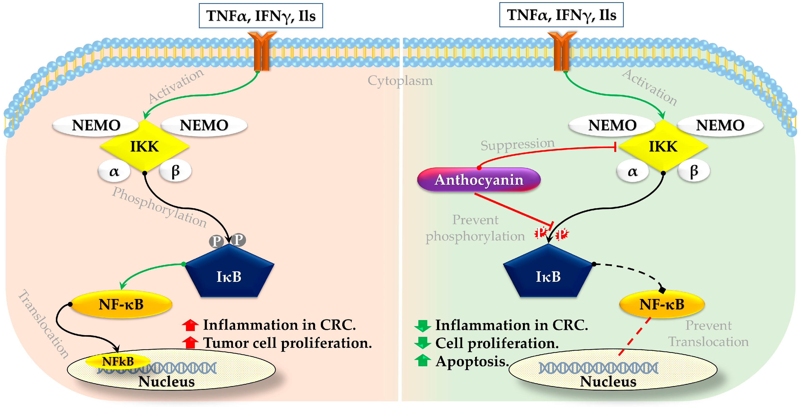
3.1. Anthocyanin on NFκB Signaling Pathway

Studies have found that anthocyanin treatment has promising effects on the NFκB signaling pathway in CRC [86]. The beneficial mechanism involves the suppression of IκB kinase (IKK) activity, preventing phosphorylation and the subsequent degradation of IκB proteins [125,126]. Such inhibition results in holding the NFκB in the cytoplasm, which prevents NFκB’s translocation to the nucleus where it would otherwise activate the transcription of genes involved in cancer inflammation, cell proliferation, and survival [127] (Figure 2). By inhibiting this pathway, anthocyanins help reduce inflammation and tumor growth in CRC cells.
In an investigation, HCT116, HT29, and SW620 cells (human colon cancer cell line) were treated with 10, 25, 50, and 100 µM of anthocyanin (Cyanidin chloride) for 24, 48, and 72 h. The results revealed that treatment with anthocyanin induced apoptosis and significantly impeded cellular proliferation and colony formation in those three colon cancer cells. In particular, anthocyanin suppressed the NFκB signaling pathway while enhancing the activation of the nuclear factor erythroid 2-related factor 2 (Nrf2) pathway in colon cancer cells [126]. Another study observed that HCT116 to anthocyanidin (delphinidin) at a concentration of 30–240 µM for 48 h resulted in decreased cell viability, the initiation of apoptosis, PARP cleavage, the activation of caspases-3, -8, and -9, the upregulation of Bax with a simultaneous downregulation of the Bcl-2 protein, and cell cycle arrest at the G2/M phase. Introducing anthocyanin to HCT116 cells also led to the inhibition of IKKα, phosphorylation and degradation of IκBα, phosphorylation of NFκB/p65 at Ser536, translocation of NFκB/p65 to the nucleus, NFκB/p65 binding to DNA, and induction of NFκB’s transcription. The study concludes that the use of anthocyanin on HCT116 cells can block the NFκB pathway, resulting in apoptosis and arrest at the G2/M phase in CRC [129].

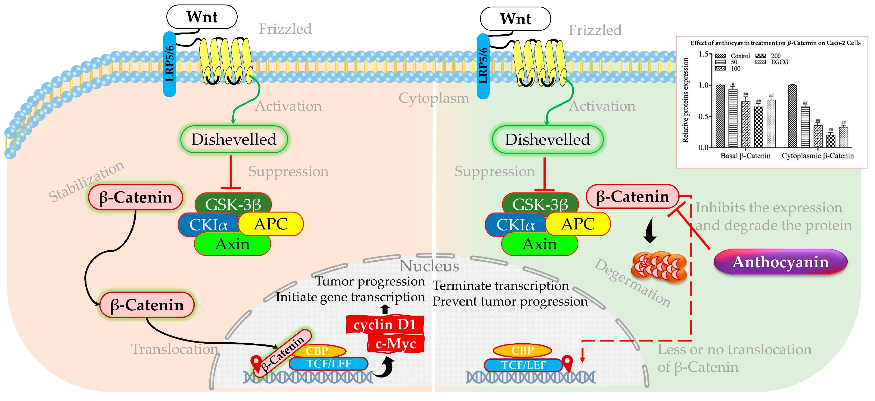
3.2. Anthocyanin on Wnt/β-Catenin Signaling Pathway

The crosstalk between inflammatory signaling and the Wnt/β-catenin pathway leads to the relocation of β-catenin to the cell nucleus, subsequently stimulating the synthesis of cancer-promoting proteins such as cyclin D1 and c-Myc (Figure 3). Moreover, in many human cancers such as CRC, up to 70–80% elevation of c-Myc expression was observed [130]. This process leads to the proliferation of stem cells while impeding their differentiation [131]. The overactivation of Wnt signaling is a common feature in nearly all cases of CRC and significantly contributes to various cancer-related activities such as the spread of cancer stem cells, angiogenesis, epithelial–mesenchymal transition, resistance to chemotherapy, and metastasis [101]. Other transcription factors also could influence the Wnt/β-catenin signaling pathway. A case in point is FOXO3a, which typically functions as a suppressor of tumor growth and has been shown to impede the Wnt/β-catenin pathway in various cancer forms. On top of that, in cervical carcinoma, FOXO3a overexpression inhibits cell invasion and migration by negatively regulating the Wnt/β-catenin pathway, suggesting a comparable mechanism could be at play in CRC [132]. The interaction between FOXO3a and microRNAs also influences the Wnt/β-catenin pathway, as seen in various cancers where microRNAs either synergize with or antagonize FOXO3a to modulate tumor growth and metastasis [133]. Another transcription factor called RUNX3 downregulates c-Myc expression, a key oncogene, through two parallel pathways: directly at the transcriptional level and by attenuating β-catenin/TCFs, downstream of BMPs in CRC cells [134]. RUNX3 also binds to the METTL3 promoter, activating circMETTL3 transcription, which in turn acts as an miR-107 sponge to regulate PER3 signaling, thereby restraining CRC development and metastasis [135].
Anthocyanins have been found to interact with the Wnt/β-catenin signaling pathway, a critical regulator of cell growth and differentiation that is frequently disrupted in colorectal cancer [136]. The mechanism underlying this interaction involves the inhibition of the pathway by anthocyanins, which reduces the levels of β-catenin, thus preventing its cytoplasmic accumulation and subsequent movement into the nucleus (Figure 3) [137]. A study utilizing anthocyanin-enriched purple-fleshed potatoes was conducted to investigate its impact on colon cancer. It was discovered that the anthocyanin extract derived from purple potatoes exhibited a greater suppression of cytoplasmic and nuclear β-catenin levels compared to sulindac in colon cancer stem cells with active p53 and p53 that were attenuated by shRNA. Furthermore, the anthocyanin extract also effectively inhibited downstream targets of the Wnt/β-catenin pathway, namely c-Myc and cyclin D1, in colon cancer stem cells with functional p53 and p53 that were attenuated by shRNA. These findings provide evidence supporting the inhibition of β-catenin nuclear translocation by anthocyanin extract, thereby restraining the growth of colon cancer stem cells [138].
Figure 3. Schematic representation of activated and inhibited Wnt/β-catenin pathway. Hashes indicate significant differences with control group only at the (#) p ≤ 0.05 and (##) p ≤ 0.01 levels, respectively. Adopted and modified from [101,136,139].
Foods 13 02991 g003
HCT116 cells were subjected to treatment with hawthorn proanthocyanidin extract of 200, 250, and 300 μg/mL. The findings indicated a reduction in the mRNA expression of Wnt1, c-Myc, and Slug in the treatment cohort, which was opposite to the control group. Through Western blot analysis, there was a significant decrease in the quantities of c-Myc and Snail protein of HCT116 cells that were exposed to high hawthorn proanthocyanidin extract for 48 h [140]. Moreover, a group of adult male Sprague Dawley rats was subjected to 4 mg N-methylnitrosourea per ml water/mouse to induce CRC, followed by intraperitoneal administration of either 12.5 mg/kg 5-fluorouracil or oral gavage 2.25 to 4.5 g/kg of Punica peel extract. N-MNU increased the mRNA expression of the β-Catenin, K-ras, and C-Myc genes in colon tissues, as compared to the negative control group. Treatment with Punica peel extract effectively decreased the expression levels of the investigated genes (β-Catenin, K-ras, and C-Myc) in a dose-dependent manner compared to the N-MNU group (untreated). The potential mechanism of action for these effects of the Punica peel extract, which include apoptosis promotion, inflammation mitigation, and suppression of tumor cell proliferation in vivo, may involve the inhibition of the Wnt/β-Catenin signaling pathway [141].

3.3. Anthocyanin on JAK/STAT Signaling Pathway

The relationship between CRC and the JAK/STAT signaling pathway is multifaceted and significant in understanding the disease’s progression and treatment resistance. The JAK/STAT pathway maintains a crucial transmembrane signal transduction mechanism activated by various cytokines, growth factors, and other molecules, driving processes such as proliferation, immune response, and malignancy [142]. The dysregulation of this pathway is strongly associated with cancer progression, including CRC, where it contributes to therapy resistance. Specifically, active STAT3 signaling has been shown to mediate resistance to chemoradiotherapy in the CRC cell lines. STAT3, along with both JAK1 and JAK2, play a role in the proliferation, viability, invasion, and migration of CRC cells by controlling the expression of various genes, including Bcl-2, p16ink4a, p21waf1/cip1, p27kip1, E-cadherin, VEGF, and MMPs [143]. Notably, it has been observed that anthocyanins have the potential to inhibit the activation of STAT3 and disrupt the JAK/STAT signaling pathway in CRC [84].
As potential sources of anthocyanin, blueberry and malvidin were found to possess inhibitory effects on the JAK/STAT3 pathway, thereby impeding the proliferation of cancer cells, as confirmed in a study [144]. Pretreatment with anthocyanin at a dosage of 100 mg/kg resulted in a significant reduction in hepatic JAK2/STAT3/P53 signaling activation in vivo [145]. Research focusing on MAPK and STAT-3 signaling revealed that at concentrations from 80 to 100 µM, delphinidin (an anthocyanidin) can effectively hinder the phosphorylation (suppression) of these transcription factors in HCT116 cells. The suppression of STAT-3, p38, and ERK1/2 phosphorylation, combined with the modulation of pro-apoptotic protein expression, is supposed to trigger the anti-cancer properties of delphinidin in CRC [111].

3.4. Anthocyanin on p53 Signaling Pathway

Talking about the p53 signaling pathway is highly significant in CRC due to its control over a wide range of cellular responses like DNA repair, cell cycle arrest, cellular senescence, and programmed cell death. In CRC, the TP53 gene is frequently mutated, with approximately 43–50% of tumors harboring missense mutations that not only impair the tumor-suppressing functions of wild-type p53 but also confer gain-of-function activities that promote cancer progression, stemness, and metastasis [146,147]. Additionally, epigenetic modifications, for example, DNA methylation and histone modifications, further compromise p53 function in CRC, even in cases where TP53 is not mutated, by inactivating downstream genes essential for p53 signaling [148]. One of many KLF4 can suppress p53 expression by directly acting on its promoter. The discovery was made that KLF4 functions to inhibit the apoptotic pathway dependent on p53 through the direct inhibition of TP53 and direct suppression of BAX expression [149]. The loss of Klf4 expression, observed in a subset of CRC cases, suggests its role as an early event in tumor development [150]. KLF4, a Zn finger transcription factor, functions as a tumor suppressor in CRC by upregulating p21WAF1/Cip1 and downregulating cyclin D1, thereby inhibiting cell proliferation and tumorigenesis [151]. Anthocyanin exhibited potent antiproliferative activities by promoting caspase-mediated cell death in a p53-independent manner, particularly in CRC stem cells, through the elevation of proteins mediating mitochondrial apoptosis, such as Bax and cytochrome C [152]. Also, the role of anthocyanins in modulating the KLF4 signaling pathway in CRC is less direct, but can be inferred and proven by several studies [153].
An investigation utilized anthocyanin-enriched purple-fleshed potatoes to elucidate its impact on colon cancer through both in vivo and in vitro studies involving colon cancer stem cells [138]. The anthocyanin extract derived from purple potatoes, when administered at a concentration of 5.0 μg/mL, demonstrated a significant reduction in proliferation rates by 63% and 32% in colon cancer stem cells possessing functional p53 and those with attenuated p53 via shRNA, respectively, as compared to the control group. These findings strongly indicate that the anthocyanin extract derived from purple potatoes effectively hinders the proliferation of colon cancer stem cells irrespective of their p53 status [138]. The same treatment led to an elevation in Cytochrome c levels regardless of the p53 status, suggesting that the activation of apoptosis may occur through the mitochondria-mediated apoptotic pathway [138]. Aronia berries are well known for their noteworthy levels of anthocyanin and proanthocyanidin [154,155]. Exposure to 50 µg monomeric anthocyanin/mL of Aronia extract for 24 h resulted in a 60% inhibition of growth in HT-29 [156]. Moreover, another examination illustrated that Aronia berry extract diminished the viability of CRC cells (SW480, HCT116). The Aronia berry extract impeded not only the proliferation, migration, survival, and invasion of CRC cells but also induced apoptosis in these cells. After the extract, an analysis of the genome-wide transcriptome identified 439 differentially expressed genes in both CRC cells. These genes were subsequently employed for pathway analysis of the p53 signaling pathway [157]. On top of that, investigation on DU-145 tumor xenografts in athymic nude mice revealed that daily oral consumption of anthocyanin (8 mg/kg) for 14 weeks resulted in a notable dose-dependent increase in apoptosis, significant reduction in p53 and Bcl-2 expressions (along with escalated Bax expression), and marked decrease in PSA and AR expressions. These results suggest that in the xenograft model, anthocyanin treatment considerably hinders tumor growth [115]. Moreover, not on CRC, but treatment with Cyanidin-3-O-glucoside (20 μM) for 24 h demonstrated inhibitory effects on the epithelial–mesenchymal transition process within this cellular context, leading to a notable reduction in the migratory and invasive capabilities of MDA-MB-231 and MDA-MB-468 cancer cells through the enhancement of KLF4 expression at the protein level [121]. In general, the data indicate that anthocyanins can regulate the p53 signaling pathway and trigger apoptosis in CRC cells, underscoring their potential as adjunct therapeutic agents in CRC management.

3.5. Anthocyanin on mTOR Signaling Pathway

Research has shown that KLF4 could also influence the mTOR pathway and the p53-dependent cell-cycle pathway in CRC cell line HCT116. The overexpression of Klf4 was found to suppress mTOR pathway activity, leading to decreased levels of phosphorylated mTOR and p70S6K1 (pS371). Conversely, the downregulation of KLF4 resulted in a relative increase in phosphorylated mTOR and p70S6K1 (pS371) in CRC [158].
The treatment of cells with Cy3G at a concentration of 20 μM for 24 h exhibited inhibition of the epithelial–mesenchymal transition process. Furthermore, it significantly reduced the migratory and invasive capabilities of cancer cells by upregulating the expression of KLF4 at the protein level [121]. Additionally, investigations into the effects of anthocyanins on cancer cell survival and the AMPK/mTOR pathway revealed that anthocyanins extracted from Meoru exerted growth inhibitory effects by regulating the mTOR or GSK3β/β-catenin pathway in HT-29 colon and Hep3B cells, respectively [159]. Moreover, a separate study illustrated that anthocyanin from Lycium ruthenicum Murray inhibited cancer cell proliferation, suppressed migration and invasion, induced apoptosis, and caused G2/M phase cell cycle arrest through the activation of the AMPK/mTOR autophagy pathway [160]. These findings provide evidence that anthocyanins have the potential to enhance cancer management by modulating AMPK/mTOR or AMPK/Wnt signaling pathways.

3.6. Anthocyanin on PI3K-AKT Signaling Pathway

The PI3K/Akt/mTOR pathway plays an important role not only in controlling the proliferation and apoptosis of cancer cells but also in promoting normal and tumor angiogenesis [161]. AKT phosphorylation is responsible for driving cancer formation and progression by modulating the transcription factors that belong to the FOXO family, which are essential for suppressing growth and proliferation [162]. Current research suggests that anthocyanins possess the ability to prevent CRC cell proliferation and enhance apoptosis by influencing various signaling pathways, including the PI3K/AKT pathway that interacts with FOXO3a. Particularly, anthocyanins have demonstrated the capability to decrease PI3K protein expression and impede AKT phosphorylation, resulting in the activation of FOXO3a, thereby facilitating apoptosis and impeding tumor growth [163].
A study conducted on Caco-2 human colon carcinoma revealed that the anthocyanin extract, obtained from bilberries and blackcurrant at various concentrations, significantly reduced the proliferation of Caco-2 cells. This extract also triggered apoptosis through the activation of caspase-3 cleavage and increased the expression of cyclin-dependent kinase inhibitor 1 (p21Waf/Cif1) in a manner that depended on the dosage. Moreover, the anthocyanin extract exhibited a dose-dependent elevation in intracellular reactive oxygen species within Caco-2 cells, accompanied by a slight rise in the overall antioxidant status of the cells. These results further confirm the role of anthocyanins in enhancing apoptotic pathways mediated by FOXO [164]. Another study has also confirmed that anthocyanins can downregulate the PI3K/AKT pathway, which is recognized for its negative regulation of FOXO3a, thereby encouraging apoptosis and hindering tumor growth in human colon cancer HT29 cells [163]. Overall, the modulation of different co-factors in the PI3K/AKT signaling pathway by anthocyanins represents a promising strategy for CRC treatment, leveraging their natural bioactive properties to inhibit cancer cell growth and promote apoptosis through multiple molecular mechanisms.

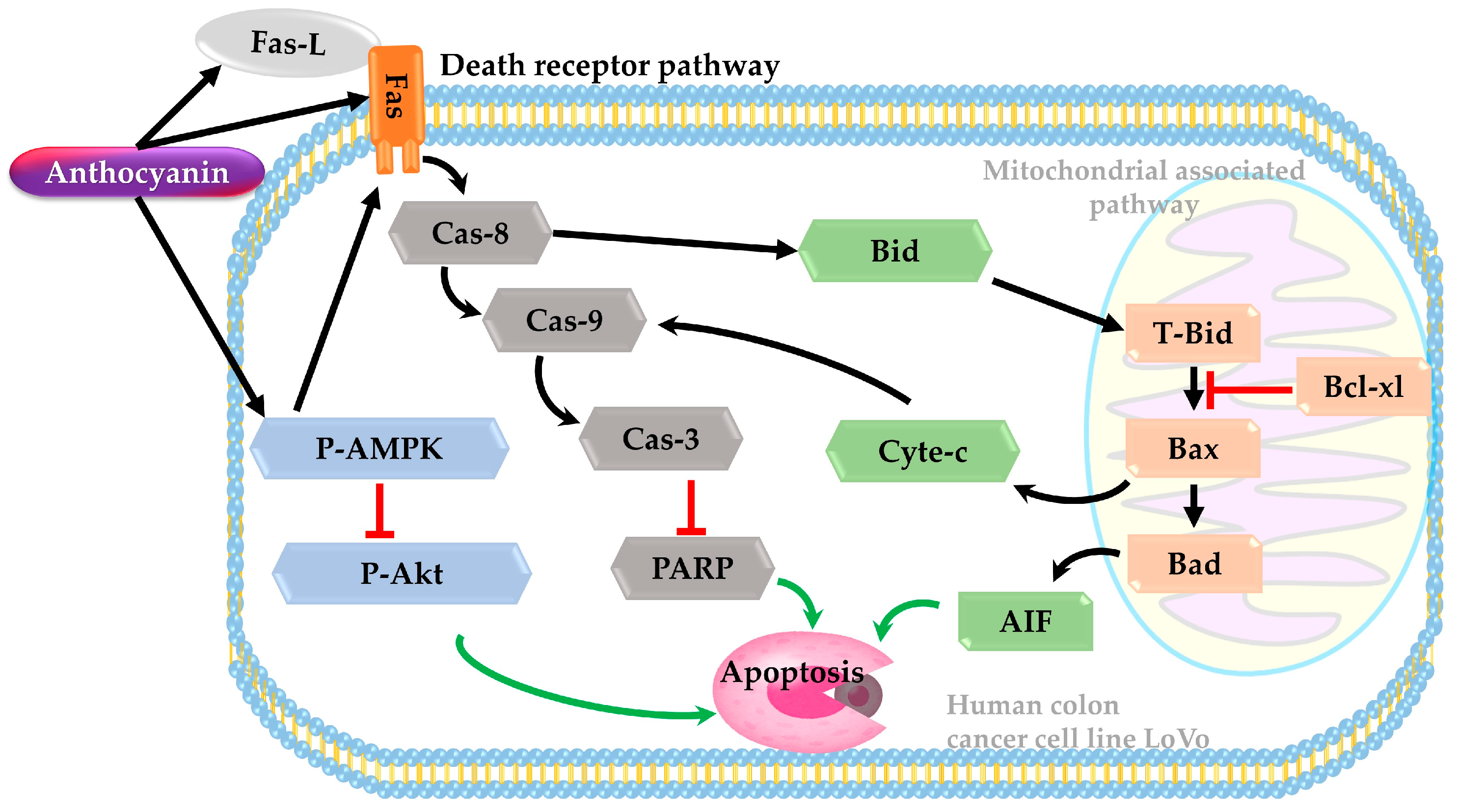
3.7. Death Receptor Pathway

The death receptor pathway is a process that involves cell surface receptors that send apoptotic signals. Apoptosis, a well-orchestrated cellular demise mechanism crucial for preventing cancer progression, can be circumvented by malignant cells. AMP-activated protein kinase (AMPK) plays a pivotal role in maintaining cellular energy balance. The activation of AMPK can modulate various effector molecules involved in the control and progression of malignancies. AMPK activation can hinder the metabolic expansion of tumors by regulating energy levels, enforcing metabolic checkpoints, and restraining cell proliferation.
A study was carried out on the human colon cancer cell line LoVo using anthocyanin extract from Hibiscus flowers. The cells were treated with different concentrations of anthocyanin (1, 2, and 3 mg/mL) extracted from Hibiscus flowers for 24 and 48 h. The findings demonstrated a negative association between anthocyanin concentration, cell viability, and the generation of pro-apoptotic bodies. Treatment with anthocyanin extract led to mitochondrial damage and disintegration in LoVo cells in a dose-dependent manner, indicating a potential induction of apoptotic cell death in CRC cells through the intrinsic apoptotic pathway. Furthermore, higher concentrations of anthocyanin resulted in increased expression of p-AMPK and decreased expression of p-Akt, suggesting that anthocyanin extracts may modulate proteins associated with apoptotic pathways to trigger apoptosis (Figure 4) [165].

3.8. Anthocyanin on TGF-β/BMPs Signaling Pathway

Bone morphogenetic proteins (BMPs), which belong to the transforming growth factor-β (TGF-β) superfamily, serve as versatile cytokines that govern a wide array of biological processes. Recent research indicated that mutations in BMP receptor 1a and Smad4 in colon cancer highlighted that disruptions in BMP signaling significantly contribute to the development of intestinal cancer [134]. Mutations affect the TGF-β receptor type 2 (TGFBR2) due to a deficiency in mismatch repair, which is responsible for the development of CRCs with microsatellite instability, although this condition is linked to relatively improved survival rates [166]. Conversely, RUNX3 serves as a suppressor of gastric and colon tumors, operating in the pathway downstream of TGF-β. The tumor-inhibiting properties of RUNX3 are attributed to its capacity to dampen the transactivation of β-catenin/T-cell factors in the context of intestinal tumorigenesis [134]. The TGF-β/BMPs signaling pathway is significant in CRC where anthocyanin treatment could show a negative correlation with CRC. But there has been limited research conducted on the relationship between anthocyanin and the TGF-β/BMPs signaling pathway, as well as its impact on RUNX3.
Overall, anthocyanin has demonstrated significant potential in modulating several important signaling pathways in CRC, thereby influencing cellular mechanisms such as proliferation, apoptosis, and inflammation.

4. Potential of Bioengineering to Enrich Anthocyanin in Tomatoes

Tomatoes are considered a superior nutritional source due to their rich content of essential vitamins, minerals, and antioxidants, which contribute to various health benefits. Tomato is particularly high in lycopene, a potent antioxidant linked to decreased risks of several diseases as well as cancer [167]. The nutritional composition of tomatoes includes significant amounts of vitamin C, K, folate, and vitamin K, with around 95% water content, and the remaining 5% consisting of carbohydrates and fiber [167]. Moreover, its edible by-products, such as peels and seeds, are also nutritionally dense, containing 15.43% carbohydrates, 11.71% protein, and 5.4% lipids in peels, and 58.75% carbohydrates, 15.4% protein, and 22.2% lipids in seeds, with calorie values of 280.47 kcal/100 g and 472.8 kcal/100 g DM, respectively [168]. These by-products are also rich in minerals like K, Mg, Na, Fe, Zn, and higher levels of phenolic compounds, that contribute to its antioxidant properties [168].
Bioengineering tomatoes aiming to enrich anthocyanin content is driven by several compelling reasons, for instance, the health benefits, agricultural viability, and nutritional enhancement [18]. Traditional tomatoes are deficient in anthocyanins, but some wild relatives and specific genetic variants have been identified to accumulate these compounds in their sub-epidermal tissues [169,170]. By leveraging genetic engineering and crossbreeding techniques, scientists have successfully developed anthocyanin-rich tomato cultivars (Table 3). Research on introducing anthocyanin-enriching genes into tomatoes is showcased in the next paragraphs.

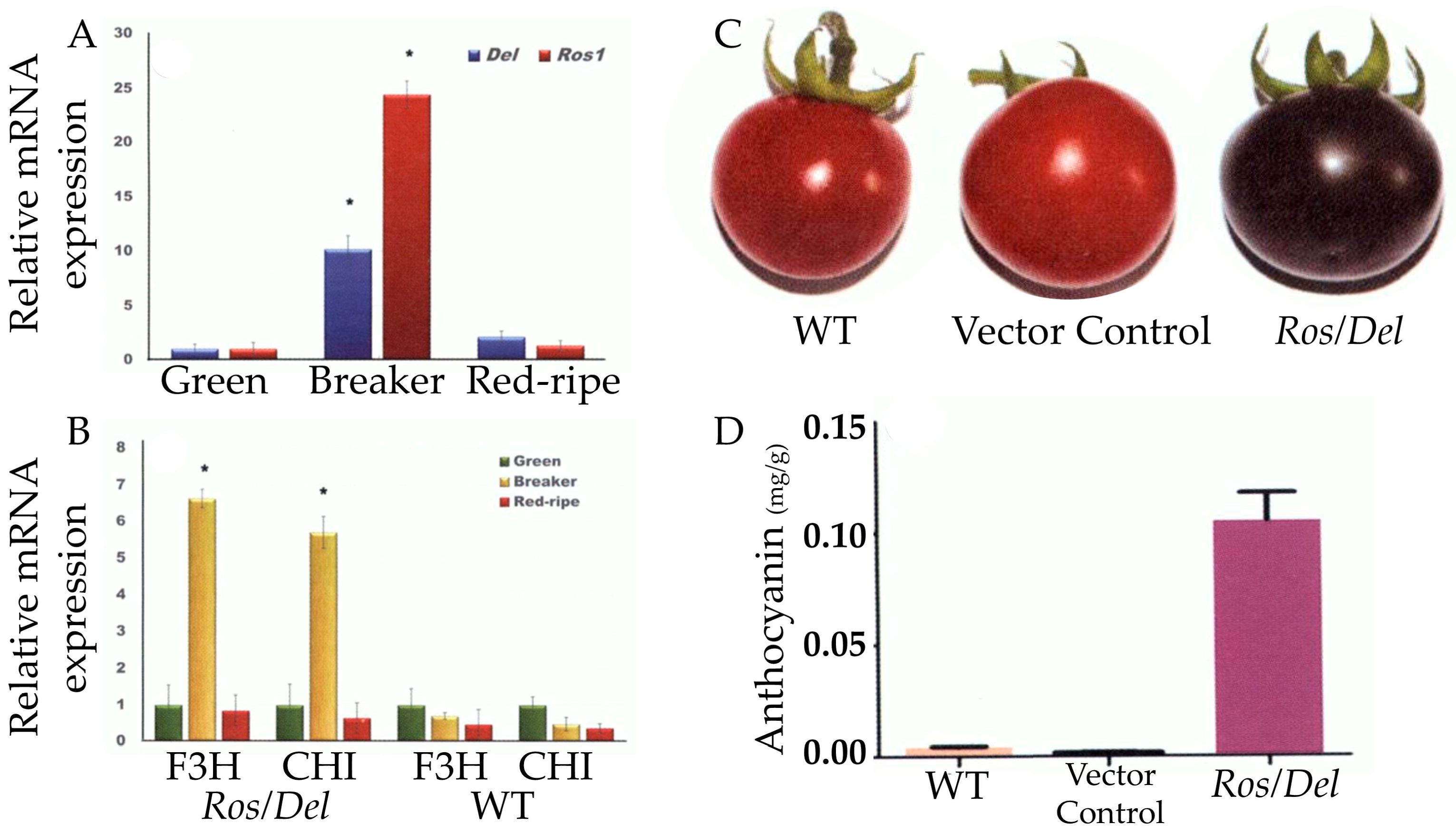
4.1. Introduce Delila (Del) and Rosea1 (Ros1) Genes in Tomato

Del and Ros1 play crucial roles as regulatory genes in the process of anthocyanin biosynthesis, which is responsible for the vibrant red, purple, and blue colors observed in plants. These particular genes are also responsible for encoding transcription factors that serve to trigger the activation of a range of structural genes within the anthocyanin biosynthetic pathway. Del functions as a transcription factor of the basic helix–loop–helix (bHLH) type, whereas Ros1 operates as a transcription factor of the R2R3-MYB type. Research has demonstrated that the simultaneous expression of both Del and Ros1 leads to a notable increase in anthocyanin accumulation across various plant species, such as snapdragon, tomato, petunia, and tobacco [178,179,180]. Two adopted schematic diagrams of cloning the anthocyanin genes for transformation into tomatoes are shown in Figure 5.
A study was conducted to add anthocyanin to tomato fruit through the activation of specific transcription factors. The Del and Ros1 genes from snapdragon were introduced into the fruit of var. MicroTom [18]. The transgenic fruit exhibited normal development and manifested purple pigmentation towards the end of the mature green stage. Upon reaching maturity, various transgenic tomato lines displayed different phenotypes, ranging from medium (Del/Ros1Z) to high (Del/Ros1C and Del/Ros1Y) and very high anthocyanin accumulation (Del/Ros1N), with the highest concentrations averaging 2.83 ± 0.46 mg/g FW. In contrast, wild-type fruit showed minimal levels of anthocyanins. Purple fruit exhibited high anthocyanin levels in both the peel and flesh. The upregulation of the Del and Ros1 genes’ expression increased the transcript levels of the majority of genes implicated in the biosynthesis of anthocyanin, along with the genes associated with side-chain alteration, such as a hypothetical anthocyanin acyltransferase, and two genes that could potentially play a role in the transport of anthocyanin into the vacuole, including a putative anthocyanin transporter [18].
Another group of researchers introduced Ros1 and Del through Agrobacterium-mediated transformation into cv. Arka Vikas. This initiative helped to accumulate anthocyanin in transgenic lines of up to 0.1 mg/g (FW), which was 70–100 times higher than non-transgenic tomatoes [173]. In addition to anthocyanin, antioxidant capacity and carotenoid content were increased considerably in Del and Ros1 transgenic tomatoes. They also revealed that the expression of the CHI and F2H genes became several-fold higher during the mature stage in transgenic fruits (Figure 6) [173]. CHI and F3H are key enzymes in flavonoid biosynthesis in plants [181,182].
To evaluate the organoleptic acceptance of transgenic tomatoes by end users, a study created CHI, Ros1, and Del transgenic Solanum lycopersicum L. tomato [174]. On average, the total flavanol content of the peel was elevated by 2.3-fold in the Del/Ros lines and 9.8-fold in the CHI lines compared to the wild-type tomatoes. The anthocyanin content of the peel ranged from 0.5 to 0.9 mg/g, whereas that of the flesh ranged from 0.03 to 0.08 mg/g in the transgenic line, which is significantly higher than wild-type and CHI tomatoes 165. In terms of sensory properties, the study did not find any difference between Del/Ros1 and WT tomato in color, flavor, texture, and overall section [174]. It was indicated that Ros/Del transgenic tomato enhanced anthocyanin and flavanol without altering organoleptic properties.
The PAP1 gene can enhance anthocyanin content in tomato shoots and also possibly produce anthocyanin production in fruits [183]. An individual study conducted on the PAP1 gene alone with crossing the CHI transgenic line found that the skin of the transgenic tomato line that only introduced the PAP1 gene showed a relatively higher abundance of anthocyanin, but was not significantly different than WT—12.3 µg/g and 8.17 µg/g, accordingly. In contrast, the generations of transgenic lines between the CHI × PAP1 lines showed significantly higher total anthocyanin: 48.11 µg/g of tomato skin [183]. In addition to PAP1, PAP2 expression also helped to produce anthocyanin. A study introduced MYB90/PAP2 into tomato to enhance anthocyanin accumulation [171]. The expression of anthocyanin biosynthetic genes (AtPAP2) was increased. This resulted in the accumulation of anthocyanin in leaves (0.21 units/g FW) and flowers (0.3 units/g FW), but not in the fruits in the transgenic line. They concluded that AtPAP2’s only interaction with SlJAF13 resulted in the prevention of anthocyanin accumulation in fruits [171]. This study suggested that the organ-specific expression of the anthocyanin accumulation gene and interaction with other genes also influence anthocyanin accumulation.

4.2. Introduce the SlMYB75 Gene in Tomato

MYB transcription factors play a role in different aspects of fruit quality by altering primary, secondary, and organic acid metabolism [184]. A research endeavor was designed to explore the potential function of SlMYB75 (also known as SlAN2) in enhancing tomato fruit quality, particularly in terms of anthocyanin accumulation. The overexpression of SlAN2 in fruits resulted in a distinct orange color, rapid softening, and elevated levels of ethylene [185]. The researchers introduced SlMYB75 into Solanum lycopersicum cv. Micro-Tom and conducted qRT-PCR analyses, which confirmed the presence of SlMYB75 transcripts in all tissues, although their expression in fruits was relatively low [175]. To address this issue, a sense construct of SlMYB75 was introduced into a tomato cultivar “Micro-Tom”, resulting in the development of three distinct transgenic homozygous lines. Certain generations of transgenic lines exhibited significantly elevated levels of SlMYB75 expression in fruits, along with robust phenotypic characteristics. The overexpression of SMYB75 found in transgenic homozygous lines resulted in a higher accumulation of anthocyanin (approximately 2.0 mg/g FW), whereas anthocyanin was absent in the wild-type tomato line [175].
Del and Ros1 genes are key regulators in anthocyanin biosynthesis, enhancing the red, purple, and blue pigmentation in plants. Their simultaneous expression boosts anthocyanin accumulation significantly in various plants, including tomatoes, without affecting the organoleptic properties. Introducing Del and Ros1 into tomatoes has resulted in increased anthocyanin, antioxidant capacity, and carotenoid content, with different phenotypic expressions across transgenic lines. Additionally, other genes like PAP1 and MYB transcription factors also contribute to anthocyanin production, although their effects can vary depending on gene interactions and expression patterns. The overexpression of SlMYB75 in tomatoes has shown a marked increase in anthocyanin content, demonstrating the potential for genetic enhancements to improve fruit quality.

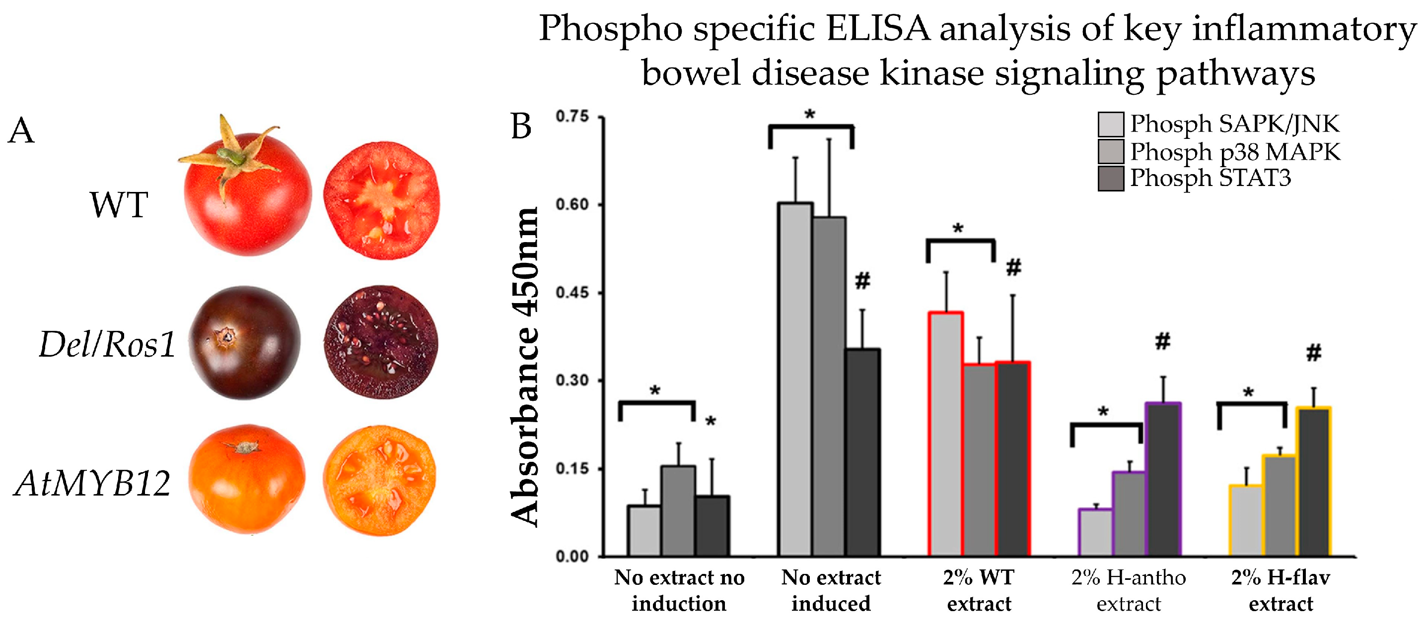
5. Evidence of Anthocyanin-Enriched Tomato Extract as a Therapeutic Agent in CRC

Numerous studies proved the role of anthocyanin extracted from different sources against CRC. To understand the importance of bioengineered anthocyanin-enriched tomatoes to fight against CRC, a few studies have been conducted [94]. For instance, a study has been conducted to investigate the consequence of extract from bioengineered anthocyanin-enriched tomato generated by breeding tomato varieties of anthocyanin fruit known as atroviolaceum (atv) against CRC. They treated colorectal adenocarcinoma cell HT-29 with five different anthocyanin concentrations (8.8, 17.5, 28.0, 35.0, and 52.5 mg/mL) of extracts from bioengineered tomato peel [186]. The extracts inhibited HT-29 cell proliferation in a dose-dependent manner. The maximum inhibition was observed after 24 h exposure to anthocyanin-enriched extract, which contains 28 μg/mL of anthocyanin. On the contrary, the survival rate of HT-29 cells exposed to an equivalent amount of wild-type tomato extract consistently hovered at 80–90% [186] (Figure 7). Through this finding, it has been demonstrated that bioengineered anthocyanin-enriched tomatoes can be a potential candidate fruit to supply anthocyanin and mitigate anthocyanin deficiency-related disease.
A further investigation was carried out on the colonic epithelial cell (CEC) of male C57BL/6 mice. The CEC cells were subjected to treatment either with and/or without the addition of tomato extracts (2% v/v) derived from Del/Ros1 (H-antho) and AtMYB12 (H-flav) transgenic anthocyanin-enriched tomato lines [184]. Utilizing an ELISA assay with phospho-epitope specific capture antibodies, in conjunction with primary murine CEC lysates, the activation status of NF-κB, SAPK/JNK, p38 MAPK, and STAT3 kinases was assessed. The introduction of high anthocyanin tomato extract resulted in an 87% decrease in SAPK/JNK activity in comparison to controls with no tomato extract. Similarly, the high anthocyanin extracts led to a 75% reduction in p38 MAPK activation, when compared to the control group without tomato extract (Figure 8). Notably, the high anthocyanin extract exhibited a significant impact on p38 MAPK activation as well [187]. In summary, the outcomes of the study unequivocally showcased the influence of anthocyanin on the signaling pathway within colon cells.

6. Implications, Limitations, and Future Research

Bioengineering tomatoes that are enriched in high anthocyanins could provide a widely accessible and cost-effective dietary intervention to mitigate the global burden of CRC. This approach also aligns with the increasing demand for functional foods that offer therapeutic benefits. By integrating anthocyanin-enriched tomatoes into the diet, we can harness their preventive and therapeutic abilities, offering a promising strategy to reduce CRC incidence and improve patient outcomes worldwide.
However, there are several challenges associated with the bioengineering of tomatoes to produce higher levels of anthocyanins. The total anthocyanin concentration in existing bioengineered anthocyanin-enriched tomatoes may not be high enough. Therefore, it is necessary to identify and introduce new anthocyanin-synthesizing genes or establish a genome-editing technology of existing anthocyanin-synthesizing genes in tomatoes, which is challenging. Additionally, open-field environmental factors (soil pH, temperature, and nutrition availability) for large production could be a threat to maximizing anthocyanin accumulation in bioengineered tomatoes. This requires comprehensive studies to understand how different environmental factors influence anthocyanin biosynthesis and to develop best practices for cultivation. On top of that, bioengineered crops have already been accepted by several governments, but some are still in the process of approval, and a few countries did not approve them at all. Thus, acceptance by the general public remains a concern.
Future research on the effect of anthocyanin-enriched bioengineered tomatoes on CRC should focus on the key areas to maximize the therapeutic potential and health benefits of these bioengineered plants. Firstly, identifying and introducing new genes that significantly enhance anthocyanin accumulation in tomatoes is crucial. Advanced genetic engineering techniques, such as genome editing with CRISPR-Cas9, can be utilized to modify regulatory genes involved in anthocyanin biosynthesis, ensuring higher concentrations of these beneficial compounds. Secondly, comprehensive studies should be conducted to evaluate all potential health outcomes associated with the consumption of bioengineered anthocyanin-rich tomatoes. This includes examining antioxidant activity, anti-inflammatory effects, and potential impacts on gut microbiota. Thirdly, a deep investigation into the molecular pathways influenced by high anthocyanin levels in bioengineered tomato extracts is necessary, particularly focusing on their role in reducing inflammation and other mechanisms in CRC. This could involve the integration of omics (transcriptomic, proteomic, epigenomic, and metabolomic) techniques to uncover the interactions between anthocyanins and cellular pathways. Lastly, conducting studies on large animal models and eventually human clinical trials will be essential to establish the efficacy and safety of these bioengineered tomatoes, ensuring wider acceptance and potential integration into dietary recommendations for cancer prevention and therapy.

7. Conclusions

Given the critical need for affordable and effective solutions to combat CRC, it is essential to explore protective measures against inflammation. Among natural compounds, anthocyanins have demonstrated significant potential in fighting CRC. Tomatoes enriched with anthocyanins through bioengineering present a promising source of this beneficial compound due to their affordability, worldwide availability, and ease of cultivation.
Bioengineered anthocyanin-enriched tomatoes offer a viable option for delivering the health benefits of anthocyanins to a broad population, including those who may not have access to other anthocyanin-rich fruits. The widespread acceptance and use of these high anthocyanin bioengineered tomatoes could play a crucial role in the global fight against CRC.

Author Contributions

Conceptualization, M.S. and W.W.; writing—original draft preparation, M.S.; writing—review and editing, M.S., K.K., S.P., X.S.S. and W.W.; supervision, W.W.; funding acquisition, W.W. All authors have read and agreed to the published version of the manuscript.

Funding

This research was supported in part by USDA-NIFA 2024-67017-42463 (W.W.) and USDA-NIFA Cooperative Project KS23-2170HA7005337 (W.W.). Contribution No. 25-021-J from the Kansas Agricultural Experiment Station.

Data Availability Statement

The original contributions presented in the study are included in the article, further inquiries can be directed to the corresponding author.

Conflicts of Interest

The authors declare no conflicts of interest.

References

  1. Chen, H.; Wang, Z.; Yu, Y.; Qiu, M.; Yang, L.; Meng, W.; Wang, C.; Li, Y.; Li, L.; Xu, H.; et al. Global trend of colorectal cancer, prevention, and control in China: Challenges and strategies. Sci. Sin. Vitae 2022, 52, 1612–1625. [Google Scholar] [CrossRef]
  2. Liu, Y.; Zhang, C.; Wang, Q.; Wu, K.; Sun, Z.; Tang, Z.; Zhang, B. Temporal Trends in the Disease Burden of Colorectal Cancer with Its Risk Factors at the Global and National Level from 1990 to 2019, and Projections Until 2044. Clin. Epidemiol. 2023, 15, 55–71. [Google Scholar] [CrossRef] [PubMed]
  3. Sharma, R.; Abbasi-Kangevari, M.; Abd-Rabu, R.; Abidi, H.; Abu-Gharbieh, E.; Acuna, J.; Adhikari, S.; Advani, S.; Afzal, M.; Aghaie Meybodi, M.; et al. Global, regional, and national burden of colorectal cancer and its risk factors, 1990–2019: A systematic analysis for the Global Burden of Disease Study 2019. Lancet Gastroenterol. Hepatol. 2022, 7, 627–647. [Google Scholar] [CrossRef] [PubMed]
  4. Tudosie, M.S.; Pauna, A.; Stefani, C.; Staicu, I.M. Diet and Food chemicals increasing the risk of colorectal cancer–literature review. J. Mind Med. Sci. 2022, 9, 118–124. [Google Scholar] [CrossRef]
  5. Dariya, B.; Chalikonda, G.; Nagaraju, G.P. Epidemiology of Colorectal Cancer. In Colon Cancer Diagnosis and Therapy; Nagaraju, G.P., Shukla, D., Vishvakarma, N.K., Eds.; Springer International Publishing: Cham, Switzerland, 2021; Volume 1, pp. 1–13. [Google Scholar] [CrossRef]
  6. Debela, D.T.; Muzazu, S.G.; Heraro, K.D.; Ndalama, M.T.; Mesele, B.W.; Haile, D.C.; Kitui, S.K.; Manyazewal, T. New approaches and procedures for cancer treatment: Current perspectives. SAGE Open Med. 2021, 9, 20503121211034366. [Google Scholar] [CrossRef]
  7. Heber, D.; Li, Z. Nutrition Intervention in Cancer. Med. Clin. N. Am. 2016, 100, 1329–1340. [Google Scholar] [CrossRef]
  8. Mori, T.; Kido, A.; Kawahara, I.; Nuaga, S.; Miyagawa, Y.; Goto, K.; Mori, S.; Kishi, S.; Fujii, K.; Fujiwara-Tani, R. Nutritional intervention for cancer sarcopenia. Ann. Musculoskelet. Med. 2021, 5, 001–004. [Google Scholar]
  9. Pak, H.; Maghsoudi, L.H.; Soltanian, A.; Gholami, F. Surgical complications in colorectal cancer patients. Ann. Med. Surg. 2020, 55, 13–18. [Google Scholar] [CrossRef]
  10. Birgisson, H.; Påhlman, L.; Gunnarsson, U.; Glimelius, B. Late adverse effects of radiation therapy for rectal cancer—A systematic overview. Acta Oncol. 2007, 46, 504–516. [Google Scholar] [CrossRef]
  11. Esmeeta, A.; Adhikary, S.; Dharshnaa, V.; Swarnamughi, P.; Ummul Maqsummiya, Z.; Banerjee, A.; Pathak, S.; Duttaroy, A.K. Plant-derived bioactive compounds in colon cancer treatment: An updated review. Biomed. Pharmacother. 2022, 153, 113384. [Google Scholar] [CrossRef]
  12. Colobatiu, L.; Gavrilas, L.; Mocan, A. Chapter 10—Natural compounds as chemosensitizers: A lesson from plants. In pH-Interfering Agents as Chemosensitizers in Cancer Therapy; Supuran, C.T., Carradori, S., Eds.; Academic Press: Cambridge, MA, USA, 2021; pp. 147–165. [Google Scholar]
  13. Khoo, H.E.; Azlan, A.; Tang, S.T.; Lim, S.M. Anthocyanidins and anthocyanins: Colored pigments as food, pharmaceutical ingredients, and the potential health benefits. Food Nutr. Res. 2017, 61, 1361779. [Google Scholar] [CrossRef] [PubMed]
  14. Hudlikar, R.; Wu, R.; Cheng, D.; Kuo, D.H.-C.; Wang, L.; Peter, R.; Yin, R.; Li, S.; Kong, A.-N. Anthocyanins and Cancer Prevention. In Natural Products for Cancer Chemoprevention: Single Compounds and Combinations; Pezzuto, J.M., Vang, O., Eds.; Springer International Publishing: Cham, Switzerland, 2020; pp. 351–373. [Google Scholar] [CrossRef]
  15. Thomasset, S.; Berry, D.P.; Cai, H.; West, K.; Marczylo, T.H.; Marsden, D.; Brown, K.; Dennison, A.; Garcea, G.; Miller, A.; et al. Pilot study of oral anthocyanins for colorectal cancer chemoprevention. Cancer Prev. Res. 2009, 2, 625–633. [Google Scholar] [CrossRef]
  16. Lim, S.; Xu, J.; Kim, J.; Chen, T.Y.; Su, X.; Standard, J.; Carey, E.; Griffin, J.; Herndon, B.; Katz, B.; et al. Role of anthocyanin-enriched purple-fleshed Sweetpotato P40 in colorectal cancer prevention. Mol. Nutr. Food Res. 2013, 57, 1908–1917. [Google Scholar] [CrossRef] [PubMed]
  17. Medic, N.; Tramer, F.; Passamonti, S. Anthocyanins in Colorectal Cancer Prevention. A Systematic Review of the Literature in Search of Molecular Oncotargets. Front. Pharmacol. 2019, 10, 675. [Google Scholar] [CrossRef]
  18. Butelli, E.; Titta, L.; Giorgio, M.; Mock, H.-P.; Matros, A.; Peterek, S.; Schijlen, E.G.W.M.; Hall, R.D.; Bovy, A.G.; Luo, J.; et al. Enrichment of tomato fruit with health-promoting anthocyanins by expression of select transcription factors. Nat. Biotechnol. 2008, 26, 1301–1308. [Google Scholar] [CrossRef]
  19. Naing, A.H.; Kim, C.K. Roles of R2R3-MYB transcription factors in transcriptional regulation of anthocyanin biosynthesis in horticultural plants. Plant Mol. Biol. 2018, 98, 1–18. [Google Scholar] [CrossRef]
  20. Thilmony, R.; Dasgupta, K.; Shao, M.; Harris, D.; Hartman, J.; Harden, L.A.; Chan, R.; Thomson, J.G. Tissue-specific expression of Ruby in Mexican lime (C. aurantifolia) confers anthocyanin accumulation in fruit. Front. Plant Sci. 2022, 13, 945738. [Google Scholar] [CrossRef]
  21. Menconi, J.; Perata, P.; Gonzali, S. In pursuit of purple: Anthocyanin biosynthesis in fruits of the tomato clade. Trends Plant Sci. 2024, 29, 589–604. [Google Scholar] [CrossRef]
  22. Cammareri, M.; Frary, A.; Frary, A.; Grandillo, S. Genetic and Biotechnological Approaches to Improve Fruit Bioactive Content: A Focus on Eggplant and Tomato Anthocyanins. Int. J. Mol. Sci. 2024, 25, 6811. [Google Scholar] [CrossRef]
  23. Choo, W.S.; Saik, A.Y.H. Chapter 4—Valorization of fruit and vegetable waste for bioactive pigments: Extraction and utilization. In Valorization of Agri-Food Wastes and By-Products; Bhat, R., Ed.; Academic Press: Cambridge, MA, USA, 2021; pp. 61–81. [Google Scholar] [CrossRef]
  24. Alappat, B.; Alappat, J. Anthocyanin Pigments: Beyond Aesthetics. Molecules 2020, 25, 5500. [Google Scholar] [CrossRef]
  25. Dong, W.; Yang, X.; Zhang, N.; Chen, P.; Sun, J.; Harnly, J.M.; Zhang, M. Study of UV–Vis molar absorptivity variation and quantitation of anthocyanins using molar relative response factor. Food Chem. 2024, 444, 138653. [Google Scholar] [CrossRef] [PubMed]
  26. Tian, S.; Ali, M.M.; Ke, M.; Lu, Y.; Zheng, Y.; Cai, X.; Fang, S.; Wu, J.; Lin, Z.; Chen, F. Novel R2R3-MYB Transcription Factor LhMYB1 Promotes Anthocyanin Accumulation in Lilium concolor var. pulchellum. Horticulturae 2024, 10, 509. [Google Scholar] [CrossRef]
  27. Wallace, T.C.; Giusti, M.M. Anthocyanins—Nature’s Bold, Beautiful, and Health-Promoting Colors. Foods 2019, 8, 550. [Google Scholar] [CrossRef] [PubMed]
  28. Goulas, V.; Vicente, A.; Manganaris, G. Structural diversity of anthocyanins in fruits. In Anthocyanins: Structure, Biosynthesis and Health Benefits; Nova Sciences: Hauppauge, NY, USA, 2012; pp. 225–250. [Google Scholar]
  29. Kumar, S.; Mitharwal, S.; Kumar, S.; Bashir, K.; Jan, K.; Kaushik, A. Commercial Production of Anthocyanins from Subtropical Fruits. In Anthocyanins in Subtropical Fruits; CRC Press: Boca Raton, FL, USA, 2023; pp. 71–90. [Google Scholar]
  30. Romualdo, G.R.; Fragoso, M.F.; Borguini, R.G.; de Araújo Santiago, M.C.P.; Fernandes, A.A.H.; Barbisan, L.F. Protective effects of spray-dried açaí (Euterpe oleracea Mart) fruit pulp against initiation step of colon carcinogenesis. Food Res. Int. 2015, 77, 432–440. [Google Scholar] [CrossRef]
  31. da Costa, D.S.; Bragotto, A.P.A.; de Carvalho, L.M.; Amado, L.L.; Lima, R.R.; Rogez, H. Analysis of polyphenols, anthocyanins and toxic elements in Açaí Juice (Euterpe oleracea Mart.): Quantification and in vivo assessment of the antioxidant capacity of clarified Açaí juice. Meas. Food 2024, 14, 100149. [Google Scholar] [CrossRef]
  32. Šimerdová, B.; Bobríková, M.; Lhotská, I.; Kaplan, J.; Křenová, A.; Šatínský, D. Evaluation of Anthocyanin Profiles in Various Blackcurrant Cultivars over a Three-Year Period Using a Fast HPLC-DAD Method. Foods 2021, 10, 1745. [Google Scholar] [CrossRef]
  33. Tian, Y.; Karhu, S.; Virtanen, M.; Linderborg, K.M.; Yang, B.; Laaksonen, O. Variation of chemical and sensory profiles of blackcurrant (Ribes nigrum) juices produced from different cultivars of European origins. LWT 2023, 173, 114353. [Google Scholar] [CrossRef]
  34. Johnson, J.B.; Collins, T.; Mani, J.S.; Naiker, M. Nutritional Quality and Bioactive Constituents of Six Australian Plum Varieties. Int. J. Fruit Sci. 2021, 21, 115–132. [Google Scholar] [CrossRef]
  35. Kodagoda, G.; Hong, H.T.; O’Hare, T.J.; Sultanbawa, Y.; Topp, B.; Netzel, M.E. Effect of Storage on the Nutritional Quality of Queen Garnet Plum. Foods 2021, 10, 352. [Google Scholar] [CrossRef]
  36. Wang, S.; Wang, B.; Dong, K.; Li, J.; Li, Y.; Sun, H. Identification and quantification of anthocyanins of 62 blueberry cultivars via UPLC-MS. Biotechnol. Biotechnol. Equip. 2022, 36, 587–597. [Google Scholar] [CrossRef]
  37. Yang, L.-C.; Hsu, S.-H.; Meng, Y.-Y.; Chen, S.-F. Quantification of anthocyanins in blueberries (Vaccinium spp.) by modified QuEChERS and liquid chromatography-mass spectrometry. J. Chin. Chem. Soc. 2022, 69, 1070–1078. [Google Scholar] [CrossRef]
  38. Wang, F.; Zhang, S.; Deng, G.; Xu, K.; Xu, H.; Liu, J. Extracting Total Anthocyanin from Purple Sweet Potato Using an Effective Ultrasound-Assisted Compound Enzymatic Extraction Technology. Molecules 2022, 27, 4344. [Google Scholar] [CrossRef] [PubMed]
  39. Wang, L.; Zhao, Y.; Zhou, Q.; Luo, C.-L.; Deng, A.-P.; Zhang, Z.-C.; Zhang, J.-L. Characterization and hepatoprotective activity of anthocyanins from purple sweet potato (Ipomoea batatas L. cultivar Eshu No. 8). J. Food Drug Anal. 2017, 25, 607–618. [Google Scholar] [CrossRef] [PubMed]
  40. Szot, I.; Łysiak, G.P.; Sosnowska, B. The Beneficial Effects of Anthocyanins from Cornelian Cherry (Cornus mas L.) Fruits and Their Possible Uses: A Review. Agriculture 2024, 14, 52. [Google Scholar] [CrossRef]
  41. Toshima, S.; Hirano, T.; Kunitake, H. Comparison of anthocyanins, polyphenols, and antioxidant capacities among raspberry, blackberry, and Japanese wild Rubus species. Sci. Hortic. 2021, 285, 110204. [Google Scholar] [CrossRef]
  42. Teng, H.; Fang, T.; Lin, Q.; Song, H.; Liu, B.; Chen, L. Red raspberry and its anthocyanins: Bioactivity beyond antioxidant capacity. Trends Food Sci. Tech. 2017, 66, 153–165. [Google Scholar] [CrossRef]
  43. Anirban, A.; Hong, H.T.; O’Hare, T.J. Profiling and Quantification of Anthocyanins in Purple-Pericarp Sweetcorn and Purple-Pericarp Maize. Molecules 2023, 28, 2665. [Google Scholar] [CrossRef]
  44. Zhao, X.; Corrales, M.; Zhang, C.; Hu, X.; Ma, Y.; Tauscher, B. Composition and Thermal Stability of Anthocyanins from Chinese Purple Corn (Zea mays L.). J. Agric. Food Chem. 2008, 56, 10761–10766. [Google Scholar] [CrossRef]
  45. Araújo, A.C.d.; Gomes, J.P.; Silva, F.B.d.; Nunes, J.S.; Santos, F.S.d.; Silva, W.P.d.; Ferreira, J.P.d.L.; Queiroz, A.J.d.M.; Figueirêdo, R.M.F.d.; Lima, G.S.d.; et al. Optimization of Extraction Method of Anthocyanins from Red Cabbage. Molecules 2023, 28, 3549. [Google Scholar] [CrossRef]
  46. Wiczkowski, W.; Szawara-Nowak, D.; Topolska, J. Red cabbage anthocyanins: Profile, isolation, identification, and antioxidant activity. Food Res. Int. 2013, 51, 303–309. [Google Scholar] [CrossRef]
  47. Blando, F.; Berland, H.; Maiorano, G.; Durante, M.; Mazzucato, A.; Picarella, M.E.; Nicoletti, I.; Gerardi, C.; Mita, G.; Andersen, Ø.M. Nutraceutical Characterization of Anthocyanin-Rich Fruits Produced by “Sun Black” Tomato Line. Front. Nutr. 2019, 6, 133. [Google Scholar] [CrossRef] [PubMed]
  48. Jian, W.; Ou, X.; Sun, L.; Chen, Y.; Liu, S.; Lu, W.; Yang, X.; Zhao, Z.; Li, Z. Characterization of anthocyanin accumulation, nutritional properties, and postharvest attributes of transgenic purple tomato. Food Chem. 2023, 408, 135181. [Google Scholar] [CrossRef] [PubMed]
  49. Li, J.; Shi, C.; Shen, D.; Han, T.; Wu, W.; Lyu, L.; Li, W. Composition and Antioxidant Activity of Anthocyanins and Non-Anthocyanin Flavonoids in Blackberry from Different Growth Stages. Foods 2022, 11, 2902. [Google Scholar] [CrossRef]
  50. Taghavi, T.; Patel, H.; Rafie, R. Extraction Solvents Affect Anthocyanin Yield, Color, and Profile of Strawberries. Plants 2023, 12, 1833. [Google Scholar] [CrossRef] [PubMed]
  51. Ahmed, T.; Suzauddula, M.; Akter, K.; Hossen, M.; Islam, M.N. Green Technology for Fungal Protein Extraction—A Review. Separations 2024, 11, 186. [Google Scholar] [CrossRef]
  52. Taghavi, T.; Patel, H.; Rafie, R. Anthocyanin Extraction Method and Sample Preparation Affect Anthocyanin Yield of Strawberries. Nat. Prod. Commun. 2022, 17, 1934578X221099970. [Google Scholar] [CrossRef]
  53. Ngoc Nhon, H.T.; Diem My, N.T.; Tuong Vi, V.N.; Kim Lien, P.T.; Thao Minh, N.T.; Doan Duy, L.N.; Hong Anh, L.T.; Anh Dao, D.T. Enhancement of extraction effectiveness and stability of anthocyanin from Hibiscus sabdariffa L. J. Agric. Food Res. 2022, 10, 100408. [Google Scholar] [CrossRef]
  54. Nunes Mattos, G.; Pessanha de Araújo Santiago, M.C.; Sampaio Doria Chaves, A.C.; Rosenthal, A.; Valeriano Tonon, R.; Correa Cabral, L.M. Anthocyanin Extraction from Jaboticaba Skin (Myrciaria cauliflora Berg.) Using Conventional and Non-Conventional Methods. Foods 2022, 11, 885. [Google Scholar] [CrossRef]
  55. Ahmed, T.; Rana, M.R.; Hossain, M.A.; Ullah, S.; Suzauddula, M. Optimization of ultrasound-assisted extraction using response surface methodology for total anthocyanin content, total phenolic content, and antioxidant activities of Roselle (Hibiscus sabdariffa L.) calyces and comparison with conventional Soxhlet extraction. Biomass Convers. Biorefinery 2023, 14, 17127–17148. [Google Scholar] [CrossRef]
  56. Sri Raghavi, R.; Visalakshi, M.; Karthikeyan, S.; Amutha Selvi, G.; Thamaraiselvi, S.; Gurusamy, K. Standardisation of anthocyanin extraction techniques from hibiscus (Hibiscus rosa-sinensis) petals for biocolour utilisation. J. Pharm. Innov. 2022, 11, 303–309. [Google Scholar]
  57. Thornton, D.; Barton, L.; Hsu, L. The development of an automated countercurrent chromatography process for isolation of anthocyanins. J. Chromatogr. A 2018, 1575, 66–71. [Google Scholar] [CrossRef] [PubMed]
  58. Zou, H.; Ma, Y.; Xu, Z.; Liao, X.; Chen, A.; Yang, S. Isolation of strawberry anthocyanins using high-speed counter-current chromatography and the copigmentation with catechin or epicatechin by high pressure processing. Food Chem. 2018, 247, 81–88. [Google Scholar] [CrossRef] [PubMed]
  59. Harahap, A.A.; Letare, S.K.; Hendrianie, N. The effect of solvents and extraction time on anthocyanin extraction from butterfly pea (Clitoria ternatea L.). AIP. Conf. Proc. 2023, 2667, 020003. [Google Scholar] [CrossRef]
  60. Han, X.; Huang, J.; Cai, J.; Yu, C.; Jiang, S.; Ye, Q.; Yang, S.; Pan, W.; Zhang, Q.; Wang, Y. Microwave-ultrasonic technique development coupled with natural deep eutectic solvents in anthocyanin extraction from perilla leaves (Perilla frutescens var. Acuta). J. Sci. Food Agric. 2023, 103, 3006–3016. [Google Scholar] [CrossRef]
  61. Jovanović, M.; Krgović, N.; Radan, M.; Ćujić-Nikolić, N.; Mudrić, J.; Drinić, Z.; Šavikin, K. Extraction of chokeberry anthocyanins using natural deep eutectic solvents. Planta Med. 2022, 88, P-314. [Google Scholar] [CrossRef]
  62. Vannuchi, N.; Braga, A.R.C.; De Rosso, V.V. High-Performance Extraction Process of Anthocyanins from Jussara (Euterpe edulis) Using Deep Eutectic Solvents. Processes 2022, 10, 615. [Google Scholar] [CrossRef]
  63. Jusoh, Y.M.M.; Idris, A.A.; Khairuddin, N.; Zaidel, D.N.A.; Hashim, Z.; Mahmooda, N.A.N.; Zakaria, Z.Y.; Muhamad, I.I. Effect of solvent pH, microwave power and extraction time on microwave-assisted extraction of Hibiscus rosa-sinensis. Chem. Eng. Trans. 2018, 63, 541–546. [Google Scholar]
  64. Adhikari, B.; Shrestha, O.K. Effect of Processing Variables on Anthocyanin and Total Polyphenol Extraction from Water Caltrop (Trapa bispinosa) Hull. Himal. J. Sci. Technol. 2018, 2, 76–83. [Google Scholar] [CrossRef]
  65. Chen, W.; Karangwa, E.; Yu, J.; Duhoranimana, E.; Xia, S.; Feng, B.; Zhang, X.; Jia, C. Coupling effects of preheating time and extraction medium pH on red radish anthocyanin yield, glucosinolate degradation and off-odour removal. Int. J. Food Sci. Technol. 2018, 53, 709–718. [Google Scholar] [CrossRef]
  66. Zhao, Y.; Gao, L.; Wang, J.; Xue, Z.; Zhang, M.; Ma, X.; Wang, G.; Lv, S. Preparation and Application of pH-Sensitive Film Containing Anthocyanins Extracted from Lycium ruthenicum Murr. Materials 2023, 16, 3828. [Google Scholar] [CrossRef]
  67. Nguyen, H.M.; Putterill, J.; Dare, A.P.; Plunkett, B.J.; Cooney, J.; Peng, Y.; Souleyre, E.J.F.; Albert, N.W.; Espley, R.V.; Günther, C.S. Two genes, ANS and UFGT2, from Vaccinium spp. are key steps for modulating anthocyanin production. Front. Plant Sci. 2023, 14, 1082246. [Google Scholar] [CrossRef] [PubMed]
  68. Ferrari, M.; Muto, A.; Bruno, L.; Muzzalupo, I.; Chiappetta, A. Modulation of Anthocyanin Biosynthesis-Related Genes during the Ripening of Olea europaea L. cvs Carolea and Tondina Drupes in Relation to Environmental Factors. Int. J. Mol. Sci. 2023, 24, 8770. [Google Scholar] [CrossRef] [PubMed]
  69. Massa, S.; Pagliarello, R.; Cemmi, A.; Di Sarcina, I.; Bombarely, A.; Demurtas, O.C.; Diretto, G.; Paolini, F.; Petzold, H.E.; Bliek, M.; et al. Modifying Anthocyanins Biosynthesis in Tomato Hairy Roots: A Test Bed for Plant Resistance to Ionizing Radiation and Antioxidant Properties in Space. Front. Plant Sci. 2022, 13, 830931. [Google Scholar] [CrossRef] [PubMed]
  70. Kang, S.-I.; Rahim, M.A.; Afrin, K.S.; Jung, H.-J.; Kim, H.-T.; Park, J.-I.; Nou, I.-S. Expression of anthocyanin biosynthesis-related genes reflects the peel color in purple tomato. Horticult. Environ. Biotechnol. 2018, 59, 435–445. [Google Scholar] [CrossRef]
  71. Lin-Wang, K.; McGhie, T.K.; Wang, M.; Liu, Y.; Warren, B.; Storey, R.; Espley, R.V.; Allan, A.C. Engineering the anthocyanin regulatory complex of strawberry (Fragaria vesca). Front. Plant Sci. 2014, 5, 651. [Google Scholar] [CrossRef]
  72. Zhang, Y.; Chu, G.; Hu, Z.; Gao, Q.; Cui, B.; Tian, S.; Wang, B.; Chen, G. Genetically engineered anthocyanin pathway for high health-promoting pigment production in eggplant. Mol. Breed 2016, 36, 54. [Google Scholar] [CrossRef]
  73. Xie, R. Anthocyanin biosynthesis in fruit tree crops: Genes and their regulation. Afr. J. Biotechnol. 2011, 10, 19890–19897. [Google Scholar] [CrossRef]
  74. Gao, X.; Chen, Q.; Yang, Y.; Zhu, B.; Xiao, J. Transcriptomic analysis reveals anthocyanin biosynthesis regulation in blueberry (Vaccinium ashei) fruit. Can. J. Plant Sci. 2022, 102, 195–206. [Google Scholar] [CrossRef]
  75. Feng, S.; Wang, Y.; Yang, S.; Xu, Y.; Chen, X. Anthocyanin biosynthesis in pears is regulated by a R2R3-MYB transcription factor PyMYB10. Planta 2010, 232, 245–255. [Google Scholar] [CrossRef]
  76. Tiwari, V.; Sharma, S.; Tiwari, A.; Sheoran, B.; Kaur, S.; Sharma, A.; Yadav, M.; Bhatnagar, A.; Garg, M. Effect of dietary anthocyanins on biomarkers of type 2 diabetes and related obesity: A systematic review and meta-analysis. Crit. Rev. Food Sci. Nutr. 2023, 64, 7517–7534. [Google Scholar] [CrossRef]
  77. Ugwu, P.; Ubom, R.; Madueke, P.; Okorie, P.; Nwachukwu, D. Anti-Hypertensive Effects of Anthocyanins from Hibiscus sabdarifa Calyx on the Renin-Angiotensin-Aldoslestrone System in Wistar Rats. Niger. J. Physiolog. Sci. 2022, 37, 113–117. [Google Scholar] [CrossRef] [PubMed]
  78. Xu, C.; Zhu, J.; Gong, G.; Guo, L.; Zhang, Y.; Zhang, Z.; Ma, C. Anthocyanin attenuates high salt-induced hypertension via inhibiting the hyperactivity of the sympathetic nervous system. Clin. Exp. Hypertens. 2023, 45, 2233717. [Google Scholar] [CrossRef] [PubMed]
  79. Park, S.; Choi, M.; Lee, M. Effects of anthocyanin supplementation on reduction of obesity criteria: A systematic review and meta-analysis of randomized controlled trials. Nutrients 2021, 13, 2121. [Google Scholar] [CrossRef] [PubMed]
  80. Xiao, T.; Luo, Z.; Guo, Z.; Wang, X.; Ding, M.; Wang, W.; Shen, X.; Zhao, Y. Multiple roles of black raspberry anthocyanins protecting against alcoholic liver disease. Molecules 2021, 26, 2313. [Google Scholar] [CrossRef] [PubMed]
  81. Trushina, E.; Mustafina, O.; Aksenov, I.; Tutelyan, V. Anthocyanins as a factor in the alimentary restoration of cellular immunity in diet induced obesity in rats. Med. Immunol. 2023, 25, 703–708. [Google Scholar] [CrossRef]
  82. Fan, M.J.; Yeh, P.H.; Lin, J.P.; Huang, A.C.; Lien, J.C.; Lin, H.Y.; Chung, J.G. Anthocyanins from black rice (Oryza sativa) promote immune responses in leukemia through enhancing phagocytosis of macrophages in vivo. Exp. Ther. Med. 2017, 14, 59–64. [Google Scholar] [CrossRef]
  83. Tan, S.; Pang, S.; Zhu, S.; Wei, W.; Sun, D. Protective Effect of Anthocyanins from Blueberry on Fluoride-Induced Immune System Injury in Wistar Rats. Fluoride 2023, 56, 217–243. [Google Scholar]
  84. Lin, B.W.; Gong, C.C.; Song, H.F.; Cui, Y.Y. Effects of anthocyanins on the prevention and treatment of cancer. Br. J. Pharmacol. 2017, 174, 1226–1243. [Google Scholar] [CrossRef]
  85. Usha, S.; Murugan, K.; Krishnan, R. Antimetastatic potential of anthocyanins from Cordyline australis (G. Forst.) Endl. Red star variety on MCF onco cell lines. J. Appl. Nat. Sci. 2022, 14, 777–783. [Google Scholar]
  86. Shi, N.; Chen, X.; Chen, T. Anthocyanins in Colorectal Cancer Prevention Review. Antioxidants 2021, 10, 1600. [Google Scholar] [CrossRef]
  87. Pan, D.; Huang, B.; Gan, Y.; Gao, C.; Liu, Y.; Tang, Z. Phycocyanin ameliorates colitis-associated colorectal cancer by regulating the gut microbiota and the IL-17 signaling pathway. Mar. Drugs 2022, 20, 260. [Google Scholar] [CrossRef] [PubMed]
  88. Kay, C.D.; Pereira-Caro, G.; Ludwig, I.A.; Clifford, M.N.; Crozier, A. Anthocyanins and Flavanones Are More Bioavailable than Previously Perceived: A Review of Recent Evidence. Annu. Rev. Food Sci. Technol. 2017, 8, 155–180. [Google Scholar] [CrossRef] [PubMed]
  89. Ogawa, Y.; Donlao, N.; Thuengtung, S.; Tian, J.; Cai, Y.; Reginio, F.C.; Ketnawa, S.; Yamamoto, N.; Tamura, M. Impact of food structure and cell matrix on digestibility of plant-based food. Curr. Opin. Food Sci. 2018, 19, 36–41. [Google Scholar] [CrossRef]
  90. Zhang, H.; Hassan, Y.I.; Renaud, J.; Liu, R.; Yang, C.; Sun, Y.; Tsao, R. Bioaccessibility, bioavailability, and anti-inflammatory effects of anthocyanins from purple root vegetables using mono- and co-culture cell models. Mol. Nutr. Food Res. 2017, 61, 1600928. [Google Scholar] [CrossRef]
  91. Li, H.; Deng, Z.; Liu, R.; Loewen, S.; Tsao, R. Bioaccessibility, in vitro antioxidant activities and in vivo anti-inflammatory activities of a purple tomato (Solanum lycopersicum L.). Food Chem. 2014, 159, 353–360. [Google Scholar] [CrossRef]
  92. Milbury, P.E.; Cao, G.; Prior, R.L.; Blumberg, J. Bioavailablility of elderberry anthocyanins. Mech. Ageing Dev. 2002, 123, 997–1006. [Google Scholar] [CrossRef]
  93. Mallery, S.R.; Budendorf, D.E.; Larsen, M.P.; Pei, P.; Tong, M.; Holpuch, A.S.; Larsen, P.E.; Stoner, G.D.; Fields, H.W.; Chan, K.K.; et al. Effects of human oral mucosal tissue, saliva, and oral microflora on intraoral metabolism and bioactivation of black raspberry anthocyanins. Cancer Prev. Res. 2011, 4, 1209–1221. [Google Scholar] [CrossRef]
  94. Gonzali, S.; Perata, P. Anthocyanins from Purple Tomatoes as Novel Antioxidants to Promote Human Health. Antioxidants 2020, 9, 1017. [Google Scholar] [CrossRef]
  95. Wanyo, P.; Chamsai, T.; Toontom, N.; Nghiep, L.K.; Tudpor, K. Differential Effects of In Vitro Simulated Digestion on Antioxidant Activity and Bioaccessibility of Phenolic Compounds in Purple Rice Bran Extracts. Molecules 2024, 29, 2994. [Google Scholar] [CrossRef]
  96. McDougall, G.J.; Dobson, P.; Smith, P.; Blake, A.; Stewart, D. Assessing Potential Bioavailability of Raspberry Anthocyanins Using an in Vitro Digestion System. J. Agric. Food Chem. 2005, 53, 5896–5904. [Google Scholar] [CrossRef]
  97. Li, X.; Wang, Y.; Jiang, Y.; Liu, C.; Zhang, W.; Chen, W.; Tian, L.; Sun, J.; Lai, C.; Bai, W. Microencapsulation with fructooligosaccharides and whey protein enhances the antioxidant activity of anthocyanins and their ability to modulate gut microbiota in vitro. Food Res. Int. 2024, 181, 114082. [Google Scholar] [CrossRef] [PubMed]
  98. Zhang, W.; Li, X.; Ma, X.; Li, H.; Liu, J.; Zeng, Y.; Cai, D.; Xu, Q.; Chen, G.; Tian, L.; et al. Microencapsulation of anthocyanins extracted from black soybean peels by whey protein/fructo-oligosaccharide contributes to improved stability, bioavailability, and ability to regulate glycolipid metabolism. Food Front. 2024, 5, 570–583. [Google Scholar] [CrossRef]
  99. Dobre, M.; Trandafir, B.; Milanesi, E.; Salvi, A.; Bucuroiu, I.A.; Vasilescu, C.; Niculae, A.M.; Herlea, V.; Hinescu, M.E.; Constantinescu, G. Molecular profile of the NF-κB signalling pathway in human colorectal cancer. J. Cell. Mol. Med. 2022, 26, 5966–5975. [Google Scholar] [CrossRef] [PubMed]
  100. Zhu, Y.; Li, X. Advances of Wnt signalling pathway in colorectal cancer. Cells 2023, 12, 447. [Google Scholar] [CrossRef] [PubMed]
  101. He, K.; Gan, W.J. Wnt/β-Catenin Signaling Pathway in the Development and Progression of Colorectal Cancer. Cancer Manag. Res. 2023, 15, 435–448. [Google Scholar] [CrossRef] [PubMed]
  102. Fleming-de-Moraes, C.D.; Rocha, M.R.; Tessmann, J.W.; de Araujo, W.M.; Morgado-Diaz, J.A. Crosstalk between PI3K/Akt and Wnt/β-catenin pathways promote colorectal cancer progression regardless of mutational status. Cancer Biol. Ther. 2022, 23, 1–13. [Google Scholar] [CrossRef]
  103. Arqués, O.; Chicote, I.; Puig, I.; Tenbaum, S.P.; Argilés, G.; Dienstmann, R.; Fernandez, N.; Caratu, G.; Matito, J.; Silberschmidt, D. Tankyrase inhibition blocks Wnt/β-catenin pathway and reverts resistance to PI3K and AKT inhibitors in the treatment of colorectal cancer. Clin. Cancer Res. 2016, 22, 644–656. [Google Scholar] [CrossRef]
  104. Silva, V.R.; Santos, L.d.S.; Dias, R.B.; Quadros, C.A.; Bezerra, D.P. Emerging agents that target signaling pathways to eradicate colorectal cancer stem cells. Cancer Commun. 2021, 41, 1275–1313. [Google Scholar] [CrossRef]
  105. Zhong, J.; Ding, S.; Zhang, X.; Di, W.; Wang, X.; Zhang, H.; Chen, Y.; Zhang, Y.; Hu, Y. To investigate the occurrence and development of colorectal cancer based on the PI3K/AKT/mTOR signaling pathway. Front. Biosci.-Landmark 2023, 28, 37. [Google Scholar] [CrossRef]
  106. Ghobashi, A.H.; Vuong, T.T.; Kimani, J.W.; Ladaika, C.A.; Hollenhorst, P.C.; O’Hagan, H.M. Activation of AKT induces EZH2-mediated β-catenin trimethylation in colorectal cancer. Iscience 2023, 26, 107630. [Google Scholar] [CrossRef]
  107. Jeong, J.-W.; Lee, W.S.; Shin, S.C.; Kim, G.-Y.; Choi, B.T.; Choi, Y.H. Anthocyanins Downregulate Lipopolysaccharide-Induced Inflammatory Responses in BV2 Microglial Cells by Suppressing the NF-κB and Akt/MAPKs Signaling Pathways. Int. J. Mol. Sci. 2013, 14, 1502–1515. [Google Scholar] [CrossRef] [PubMed]
  108. Yu, L.L.; Yu, H.G.; Yu, J.P.; Luo, H.S.; Xu, X.M.; Li, J.H. Nuclear factor-kappaB p65 (RelA) transcription factor is constitutively activated in human colorectal carcinoma tissue. World J. Gastroenterol. 2004, 10, 3255–3260. [Google Scholar] [CrossRef] [PubMed]
  109. Gambhir, S.; Vyas, D.; Hollis, M.; Aekka, A.; Vyas, A. Nuclear factor kappa B role in inflammation associated gastrointestinal malignancies. World J. Gastroenterol. 2015, 21, 3174–3183. [Google Scholar] [CrossRef] [PubMed]
  110. Sunami, Y.; Wirth, T. Intestinal carcinogenesis: IKK can go all the way. J. Clin. Investig. 2011, 121, 2551–2553. [Google Scholar] [CrossRef]
  111. Zhang, Z.; Pan, Y.; Zhao, Y.; Ren, M.; Li, Y.; Lu, G.; Wu, K.; He, S. Delphinidin modulates JAK/STAT3 and MAPKinase signaling to induce apoptosis in HCT116 cells. Environ. Toxicol. 2021, 36, 1557–1566. [Google Scholar] [CrossRef]
  112. Corvinus, F.M.; Orth, C.; Moriggl, R.; Tsareva, S.A.; Wagner, S.; Pfitzner, E.B.; Baus, D.; Kaufman, R.; Huber, L.A.; Zatloukal, K. Persistent STAT3 activation in colon cancer is associated with enhanced cell proliferation and tumor growth. Neoplasia 2005, 7, 545–555. [Google Scholar] [CrossRef]
  113. Takayama, T.; Miyanishi, K.; Hayashi, T.; Sato, Y.; Niitsu, Y. Colorectal cancer: Genetics of development and metastasis. J. Gastroenterol. 2006, 41, 185–192. [Google Scholar] [CrossRef]
  114. Iacopetta, B. TP53 mutation in colorectal cancer. Hum. Mutat. 2003, 21, 271–276. [Google Scholar] [CrossRef]
  115. Ha, U.S.; Bae, W.J.; Kim, S.J.; Yoon, B.I.; Hong, S.H.; Lee, J.Y.; Hwang, T.K.; Hwang, S.Y.; Wang, Z.; Kim, S.W. Anthocyanin induces apoptosis of DU-145 cells in vitro and inhibits xenograft growth of prostate cancer. Yonsei Med. J. 2015, 56, 16–23. [Google Scholar] [CrossRef]
  116. Cooks, T.; Pateras, I.S.; Tarcic, O.; Solomon, H.; Schetter, A.J.; Wilder, S.; Lozano, G.; Pikarsky, E.; Forshew, T.; Rosenfeld, N.; et al. Mutant p53 prolongs NF-κB activation and promotes chronic inflammation and inflammation-associated colorectal cancer. Cancer Cell 2013, 23, 634–646. [Google Scholar] [CrossRef]
  117. Haas, M.J.; Onstead-Haas, L.; Naem, E.; Arnold, A.; Rohrbaugh, N.; Flowers, M.; Mooradian, A.D. The effect of black seed (Nigella sativa) extract on FOXO3 expression in HepG2 cells. Phytother. Res. 2014, 28, 873–879. [Google Scholar] [CrossRef] [PubMed]
  118. Choi, Y.M.; Yoon, H.; Lee, S.; Ko, H.C.; Shin, M.J.; Lee, M.C.; Hur, O.S.; Ro, N.Y.; Desta, K.T. Isoflavones, anthocyanins, phenolic content, and antioxidant activities of black soybeans (Glycine max (L.) Merrill) as affected by seed weight. Sci. Rep. 2020, 10, 19960. [Google Scholar] [CrossRef] [PubMed]
  119. Khaleghpour, K.; Li, Y.; Banville, D.; Yu, Z.; Shen, S.H. Involvement of the PI 3-kinase signaling pathway in progression of colon adenocarcinoma. Carcinogenesis 2004, 25, 241–248. [Google Scholar] [CrossRef] [PubMed]
  120. Itoh, N.; Semba, S.; Ito, M.; Takeda, H.; Kawata, S.; Yamakawa, M. Phosphorylation of Akt/PKB is required for suppression of cancer cell apoptosis and tumor progression in human colorectal carcinoma. Cancer 2002, 94, 3127–3134. [Google Scholar] [CrossRef]
  121. Chen, D.; Yuan, M.; Ye, Q.; Wang, X.; Xu, J.; Shi, G.; Hu, Z. Cyanidin-3-O-glucoside inhibits epithelial-to-mesenchymal transition, and migration and invasion of breast cancer cells by upregulating KLF4. Food Nutr. Res. 2020, 64. [Google Scholar] [CrossRef]
  122. Zhao, W.; Hisamuddin, I.M.; Nandan, M.O.; Babbin, B.A.; Lamb, N.E.; Yang, V.W. Identification of Krüppel-like factor 4 as a potential tumor suppressor gene in colorectal cancer. Oncogene 2004, 23, 395–402. [Google Scholar] [CrossRef]
  123. Xu, J.; Lü, B.; Xu, F.; Gu, H.; Fang, Y.; Huang, Q.; Lai, M. Dynamic down-regulation of Krüppel-like factor 4 in colorectal adenoma-carcinoma sequence. J. Cancer Res. Clin. Oncol. 2008, 134, 891–898. [Google Scholar] [CrossRef]
  124. Imamura, Y.; Hibi, K.; Koike, M.; Fujiwara, M.; Kodera, Y.; Ito, K.; Nakao, A. RUNX3 promoter region is specifically methylated in poorly-differentiated colorectal cancer. Anticancer Res. 2005, 25, 2627–2630. [Google Scholar]
  125. Paramanantham, A.; Kim, M.J.; Jung, E.J.; Nagappan, A.; Yun, J.W.; Kim, H.J.; Shin, S.C.; Kim, G.S.; Lee, W.S. Pretreatment of Anthocyanin from the Fruit of Vitis coignetiae Pulliat Acts as a Potent Inhibitor of TNF-α Effect by Inhibiting NF-κB-Regulated Genes in Human Breast Cancer Cells. Molecules 2020, 25, 2396. [Google Scholar] [CrossRef]
  126. Lee, D.-Y.; Yun, S.-M.; Song, M.-Y.; Jung, K.; Kim, E.-H. Cyanidin Chloride Induces Apoptosis by Inhibiting NF-κB Signaling through Activation of Nrf2 in Colorectal Cancer Cells. Antioxidants 2020, 9, 285. [Google Scholar] [CrossRef]
  127. Yamamoto, Y.; Gaynor, R.B. Therapeutic potential of inhibition of the NF-kappaB pathway in the treatment of inflammation and cancer. J. Clin. Investig. 2001, 107, 135–142. [Google Scholar] [CrossRef] [PubMed]
  128. Patel, M.; Horgan, P.G.; McMillan, D.C.; Edwards, J. NF-κB pathways in the development and progression of colorectal cancer. Transl. Res. 2018, 197, 43–56. [Google Scholar] [CrossRef] [PubMed]
  129. Yun, J.-M.; Afaq, F.; Khan, N.; Mukhtar, H. Delphinidin, an anthocyanidin in pigmented fruits and vegetables, induces apoptosis and cell cycle arrest in human colon cancer HCT116 cells. Mol. Carcinog. 2009, 48, 260–270. [Google Scholar] [CrossRef] [PubMed]
  130. He, W.-L.; Weng, X.-T.; Wang, J.-L.; Lin, Y.-K.; Liu, T.-W.; Zhou, Q.-Y.; Hu, Y.; Pan, Y.; Chen, X.-L. Association Between c-Myc and Colorectal Cancer Prognosis: A Meta-Analysis. Front. Physiol. 2018, 9, 1549. [Google Scholar] [CrossRef] [PubMed]
  131. de Sousa Moraes, L.F.; Sun, X.; Peluzio, M.; Zhu, M.J. Anthocyanins/anthocyanidins and colorectal cancer: What is behind the scenes? Crit. Rev. Food Sci. Nutr. 2019, 59, 59–71. [Google Scholar] [CrossRef]
  132. Tian, Y.; Qi, P.; Hu, X. Downregulated FOXO3a associates with poor prognosis and promotes cell invasion and migration via WNT/β-catenin signaling in cervical carcinoma. Front. Oncol. 2020, 10, 903. [Google Scholar] [CrossRef]
  133. Sun, L.; Liu, J.; Bao, D.; Hu, C.; Zhao, Y.; Chen, S. Progress in the study of FOXO3a interacting with microRNA to regulate tumourigenesis development. Front. Oncol. 2023, 13, 1293968. [Google Scholar] [CrossRef]
  134. Lee, C.W.L.; Ito, K.; Ito, Y. Role of RUNX3 in bone morphogenetic protein signaling in colorectal cancer. Cancer Res. 2010, 70, 4243–4252. [Google Scholar] [CrossRef]
  135. Jin, Q.; Qu, H.; Quan, C. New insights into the regulation of METTL3 and its role in tumors. Cell Commun Signal. 2023, 21, 334. [Google Scholar] [CrossRef]
  136. Wei, J.; Yu, W.; Hao, R.; Fan, J.; Gao, J. Anthocyanins from Aronia melanocarpa Induce Apoptosis in Caco-2 Cells through Wnt/β-Catenin Signaling Pathway. Chem. Biodivers. 2020, 17, e2000654. [Google Scholar] [CrossRef]
  137. Dharmawansa, K.V.S.; Hoskin, D.W.; Rupasinghe, H.P.V. Chemopreventive Effect of Dietary Anthocyanins against Gastrointestinal Cancers: A Review of Recent Advances and Perspectives. Int. J. Mol. Sci. 2020, 21, 6555. [Google Scholar] [CrossRef] [PubMed]
  138. Charepalli, V.; Reddivari, L.; Radhakrishnan, S.; Vadde, R.; Agarwal, R.; Vanamala, J.K. Anthocyanin-containing purple-fleshed potatoes suppress colon tumorigenesis via elimination of colon cancer stem cells. J. Nutr. Biochem. 2015, 26, 1641–1649. [Google Scholar] [CrossRef] [PubMed]
  139. Zhang, Y.; Wang, X. Targeting the Wnt/β-catenin signaling pathway in cancer. J. Hematol. Oncol. 2020, 13, 165. [Google Scholar] [CrossRef] [PubMed]
  140. Wang, Z.; Sun, Y.; Wu, M.; Zhou, L.; Zheng, Y.; Ren, T.; Li, M.; Zhao, W. Hawthorn Proanthocyanidin Extract Inhibits Colorectal Carcinoma Metastasis by Targeting the Epithelial-Mesenchymal Transition Process and Wnt/β-Catenin Signaling Pathway. Foods 2024, 13, 1171. [Google Scholar] [CrossRef]
  141. Ahmed, H.H.; El-Abhar, H.S.; Hassanin, E.A.K.; Abdelkader, N.F.; Shalaby, M.B. Punica granatum suppresses colon cancer through downregulation of Wnt/β-Catenin in rat model. Rev. Bras. Farmacogn. 2017, 27, 627–635. [Google Scholar] [CrossRef]
  142. Xue, C.; Yao, Q.; Gu, X.; Shi, Q.; Yuan, X.; Chu, Q.; Bao, Z.; Lu, J.; Li, L. Evolving cognition of the JAK-STAT signaling pathway: Autoimmune disorders and cancer. Signal Transduct. Target. Ther. 2023, 8, 204. [Google Scholar] [CrossRef]
  143. Xiong, H.; Zhang, Z.G.; Tian, X.Q.; Sun, D.F.; Liang, Q.C.; Zhang, Y.J.; Lu, R.; Chen, Y.X.; Fang, J.Y. Inhibition of JAK1, 2/STAT3 signaling induces apoptosis, cell cycle arrest, and reduces tumor cell invasion in colorectal cancer cells. Neoplasia 2008, 10, 287–297. [Google Scholar] [CrossRef]
  144. Pourshahidi, S.; Davari, M. Anthocyanins: Promising Natural Compounds for Prevention and Treatment of Oral Squamous Cell Carcinoma. Middle East J. Rehabil. Health Stud. 2020, 7, e105844. [Google Scholar] [CrossRef]
  145. Luo, L.N.; Yang, P.; Huang, W. The effect and mechanism of anthocyanin on hepatic ischemia reperfusion injury in rats. J. Xi’an Jiaotong Univ. 2016, 37, 594–598. [Google Scholar] [CrossRef]
  146. Liebl, M.C.; Hofmann, T.G. The role of p53 signaling in colorectal cancer. Cancers 2021, 13, 2125. [Google Scholar] [CrossRef]
  147. Zhang, S.; Zhou, L.; Hong, B.; van den Heuvel, A.P.J.; Prabhu, V.V.; Warfel, N.A.; Kline, C.L.B.; Dicker, D.T.; Kopelovich, L.; El-Deiry, W.S. Small-molecule NSC59984 restores p53 pathway signaling and antitumor effects against colorectal cancer via p73 activation and degradation of mutant p53. Cancer Res. 2015, 75, 3842–3852. [Google Scholar] [CrossRef] [PubMed]
  148. Tomicic, M.T.; Dawood, M.; Efferth, T. Epigenetic alterations upstream and downstream of p53 signaling in colorectal carcinoma. Cancers 2021, 13, 4072. [Google Scholar] [CrossRef]
  149. Ghaleb, A.M.; Yang, V.W. Krüppel-like factor 4 (KLF4): What we currently know. Gene 2017, 611, 27–37. [Google Scholar] [CrossRef] [PubMed]
  150. Choi, B.J.; Cho, Y.G.; Song, J.W.; Kim, C.J.; Kim, S.Y.; Nam, S.W.; Yoo, N.J.; Lee, J.Y.; Park, W.S. Altered expression of the KLF4 in colorectal cancers. Pathol. Res. Pract. 2006, 202, 585–589. [Google Scholar] [CrossRef] [PubMed]
  151. He, Z.; He, J.; Xie, K. KLF4 transcription factor in tumorigenesis. Cell Death Discov. 2023, 9, 118. [Google Scholar] [CrossRef] [PubMed]
  152. Selvamuthukumaran, M. Natural Anthocyanins from Subtropical Fruits for Cancer Prevention. In Anthocyanins in Subtropical Fruits; CRC Press: Boca Raton, FL, USA, 2023; pp. 97–103. [Google Scholar]
  153. Lee, E.; Cheung, J.; Bialkowska, A.B. Krüppel-like Factors 4 and 5 in Colorectal Tumorigenesis. Cancers 2023, 15, 2430. [Google Scholar] [CrossRef]
  154. Wangensteen, H.; Bräunlich, M.; Nikolic, V.; Malterud, K.E.; Slimestad, R.; Barsett, H. Anthocyanins, proanthocyanidins and total phenolics in four cultivars of aronia: Antioxidant and enzyme inhibitory effects. J. Funct. Foods 2014, 7, 746–752. [Google Scholar] [CrossRef]
  155. Dobros, N.; Zielińska, A.; Siudem, P.; Zawada, K.D.; Paradowska, K. Profile of Bioactive Components and Antioxidant Activity of Aronia melanocarpa Fruits at Various Stages of Their Growth, Using Chemometric Methods. Antioxidants 2024, 13, 462. [Google Scholar] [CrossRef]
  156. Malik, M.; Zhao, C.; Schoene, N.; Guisti, M.M.; Moyer, M.P.; Magnuson, B.A. Anthocyanin-rich extract from Aronia meloncarpa E induces a cell cycle block in colon cancer but not normal colonic cells. Nutr. Cancer 2003, 46, 186–196. [Google Scholar] [CrossRef]
  157. Asahi, Y.; Okuno, K.; Xu, C.; Taketomi, A.; Goel, A. Abstract 3822: Novel evidence for the role of the p53 signaling pathway in mediating the anticancer effects of aronia berry extract in colorectal cancer cells. Cancer Res. 2023, 83, 3822. [Google Scholar] [CrossRef]
  158. Ghaleb, A.M.; Elkarim, E.A.; Bialkowska, A.B.; Yang, V.W. KLF4 Suppresses Tumor Formation in Genetic and Pharmacological Mouse Models of Colonic Tumorigenesis. Mol. Cancer Res. 2016, 14, 385–396. [Google Scholar] [CrossRef] [PubMed]
  159. Yun-Kyoung, L.; Park, S.Y.; Kim, Y.-M.; Lee, W.S.; Park, O.J. Anthocyanins target AMPK/mTOR and AMPK/Wnt pathways in exerting anti-tumor effects in colon cancer or hepatocarcinoma cells. FASEB J. 2010, 24, lb259. [Google Scholar] [CrossRef]
  160. Fan, H.; Ji, Y.; Wang, Y.; Liu, D.; Wei, T.; Cao, X.; Yang, M.; Bai, C.; Wang, Z. Anthocyanins from Lycium ruthenicum Murray Inhibit HepG2 Cells Growth, Metastasis and Promote Apoptosis and G2/M Phase Cycle Arrest by Activating the AMPK/mTOR Autophagy Pathway. Evid. Based Complement. Altern. Med. 2022, 2022, 9609596. [Google Scholar] [CrossRef] [PubMed]
  161. Rascio, F.; Spadaccino, F.; Rocchetti, M.T.; Castellano, G.; Stallone, G.; Netti, G.S.; Ranieri, E. The Pathogenic Role of PI3K/AKT Pathway in Cancer Onset and Drug Resistance: An Updated Review. Cancers 2021, 13, 3949. [Google Scholar] [CrossRef]
  162. Hornsveld, M.; Dansen, T.B.; Derksen, P.W.; Burgering, B.M.T. Re-evaluating the role of FOXOs in cancer. Semin. Cancer Biol. 2018, 50, 90–100. [Google Scholar] [CrossRef]
  163. Zhao, X.; Feng, P.; He, W.; Du, X.; Chen, C.; Suo, L.; Liang, M.; Zhang, N.; Na, A.; Zhang, Y. The prevention and inhibition effect of anthocyanins on colorectal cancer. Curr. Pharm. Des. 2019, 25, 4919–4927. [Google Scholar] [CrossRef]
  164. Anwar, S.; Fratantonio, D.; Ferrari, D.; Saija, A.; Cimino, F.; Speciale, A. Berry anthocyanins reduce proliferation of human colorectal carcinoma cells by inducing caspase-3 activation and p21 upregulation. Mol. Med. Rep. 2016, 14, 1397–1403. [Google Scholar] [CrossRef]
  165. Tsai, M.C.; Chen, C.C.; Tseng, T.H.; Chang, Y.C.; Lin, Y.J.; Tsai, I.N.; Wang, C.C.; Wang, C.J. Hibiscus Anthocyanins Extracts Induce Apoptosis by Activating AMP-Activated Protein Kinase in Human Colorectal Cancer Cells. Nutrients 2023, 15, 3972. [Google Scholar] [CrossRef]
  166. Li, X.; Wu, Y.; Tian, T. TGF-β Signaling in Metastatic Colorectal Cancer (mCRC): From Underlying Mechanism to Potential Applications in Clinical Development. Int. J. Mol. Sci. 2022, 23, 14436. [Google Scholar] [CrossRef]
  167. Rashmita, S.; Govinda, B.; Arvind, S. A study on marketing behaviour of tomato growers in Shivpuri District MP, India. Int. J. Curr. Microbiol. Appl. Sci. 2020, 9, 331–334. [Google Scholar] [CrossRef]
  168. Kaboré, K.; Konaté, K.; Sanou, A.; Dakuyo, R.; Sama, H.; Santara, B.; Compaoré, E.W.R.; Dicko, M.H. Tomato By-Products, a Source of Nutrients for the Prevention and Reduction of Malnutrition. Nutrients 2022, 14, 2871. [Google Scholar] [CrossRef] [PubMed]
  169. Mattoo, A.K.; Dwivedi, S.L.; Dutt, S.; Singh, B.; Garg, M.; Ortiz, R. Anthocyanin-rich vegetables for human consumption—Focus on potato, sweetpotato and tomato. Int. J. Mol. Sci. 2022, 23, 2634. [Google Scholar] [CrossRef] [PubMed]
  170. Menconi, J.; Perata, P.; Gonzali, S. Novel R2R3 MYB transcription factors regulate anthocyanin synthesis in Aubergine tomato plants. BMC Plant Biol. 2023, 23, 148. [Google Scholar] [CrossRef] [PubMed]
  171. Li, N.; Wu, H.; Ding, Q.; Li, H.; Li, Z.; Ding, J.; Li, Y. The heterologous expression of Arabidopsis PAP2 induces anthocyanin accumulation and inhibits plant growth in tomato. Funct. Integr. Genom. 2018, 18, 341–353. [Google Scholar] [CrossRef]
  172. Lim, W.; Li, J. Synergetic effect of the Onion CHI gene on the PAP1 regulatory gene for enhancing the flavonoid profile of tomato skin. Sci. Rep. 2017, 7, 12377. [Google Scholar] [CrossRef]
  173. Maligeppagol, M.; Chandra, G.S.; Navale, P.M.; Deepa, H.; Rajeev, P.R.; Asokan, R.; Babu, K.P.; Babu, C.S.B.; Rao, V.K.; Kumar, N.K.K. Anthocyanin enrichment of tomato (Solanum lycopersicum L.) fruit by metabolic engineering. Curr. Sci. 2013, 105, 72–80. [Google Scholar]
  174. Lim, W.; Miller, R.; Park, J.; Park, S. Consumer Sensory Analysis of High Flavonoid Transgenic Tomatoes. J. Food Sci. 2014, 79, S1212–S1217. [Google Scholar] [CrossRef]
  175. Jian, W.; Cao, H.; Yuan, S.; Liu, Y.; Lu, J.; Lu, W.; Li, N.; Wang, J.; Zou, J.; Tang, N.; et al. SlMYB75, an MYB-type transcription factor, promotes anthocyanin accumulation and enhances volatile aroma production in tomato fruits. Horticult. Res. 2019, 6, 22. [Google Scholar] [CrossRef]
  176. Su, X.; Xu, J.; Rhodes, D.; Shen, Y.; Song, W.; Katz, B.; Tomich, J.; Wang, W. Identification and quantification of anthocyanins in transgenic purple tomato. Food Chem. 2016, 202, 184–188. [Google Scholar] [CrossRef]
  177. Hassanin, A.A.; Saad, A.M.; Bardisi, E.A.; Salama, A.; Sitohy, M.Z. Transfer of Anthocyanin Accumulating Delila and Rosea1 Genes from the Transgenic Tomato Micro-Tom Cultivar to Moneymaker Cultivar by Conventional Breeding. J. Agric. Food Chem. 2020, 68, 10741–10749. [Google Scholar] [CrossRef]
  178. Naing, A.H.; Ai, T.N.; Lim, K.B.; Lee, I.J.; Kim, C.K. Overexpression of Rosea1 from Snapdragon Enhances Anthocyanin Accumulation and Abiotic Stress Tolerance in Transgenic Tobacco. Front. Plant Sci. 2018, 9, 1070. [Google Scholar] [CrossRef] [PubMed]
  179. Naing, A.H.; Park, K.I.; Ai, T.N.; Chung, M.Y.; Han, J.S.; Kang, Y.-W.; Lim, K.B.; Kim, C.K. Overexpression of snapdragon Delila (Del) gene in tobacco enhances anthocyanin accumulation and abiotic stress tolerance. BMC Plant Biol. 2017, 17, 65. [Google Scholar] [CrossRef]
  180. Lim, W.; Li, J. Co-expression of onion chalcone isomerase in Del/Ros1-expressing tomato enhances anthocyanin and flavonol production. Plant Cell Tissue Organ Cult. 2017, 128, 113–124. [Google Scholar] [CrossRef]
  181. Zhu, J.; Zhao, W.; Li, R.; Guo, D.; Li, H.; Wang, Y.; Mei, W.; Peng, S. Identification and Characterization of Chalcone Isomerase Genes Involved in Flavonoid Production in Dracaena cambodiana. Front. Plant Sci. 2021, 12, 616396. [Google Scholar] [CrossRef] [PubMed]
  182. Han, Y.; Huang, K.; Liu, Y.; Jiao, T.; Ma, G.; Qian, Y.; Wang, P.; Dai, X.; Gao, L.; Xia, T. Functional Analysis of Two Flavanone-3-Hydroxylase Genes from Camellia sinensis: A Critical Role in Flavonoid Accumulation. Genes 2017, 8, 300. [Google Scholar] [CrossRef] [PubMed]
  183. Zuluaga, D.L.; Gonzali, S.; Loreti, E.; Pucciariello, C.; Degl’Innocenti, E.; Guidi, L.; Alpi, A.; Perata, P. Arabidopsis thaliana MYB75/PAP1 transcription factor induces anthocyanin production in transgenic tomato plants. Funct. Plant Biol. 2008, 35, 606–618. [Google Scholar] [CrossRef]
  184. Zhang, Y.; Butelli, E.; Alseekh, S.; Tohge, T.; Rallapalli, G.; Luo, J.; Kawar, P.G.; Hill, L.; Santino, A.; Fernie, A.R.; et al. Multi-level engineering facilitates the production of phenylpropanoid compounds in tomato. Nat. Commun. 2015, 6, 8635. [Google Scholar] [CrossRef]
  185. Meng, X.; Yang, D.; Li, X.; Zhao, S.; Sui, N.; Meng, Q. Physiological changes in fruit ripening caused by overexpression of tomato SlAN2, an R2R3-MYB factor. Plant Physiol. Biochem. 2015, 89, 24–30. [Google Scholar] [CrossRef]
  186. Mazzucato, A.; Willems, D.; Bernini, R.; Picarella, M.E.; Santangelo, E.; Ruiu, F.; Tilesi, F.; Soressi, G.P. Novel phenotypes related to the breeding of purple-fruited tomatoes and effect of peel extracts on human cancer cell proliferation. Plant Physiol. Biochem. 2013, 72, 125–133. [Google Scholar] [CrossRef]
  187. Tomlinson, M.L.; Butelli, E.; Martin, C.; Carding, S.R. Flavonoids from Engineered Tomatoes Inhibit Gut Barrier Pro-inflammatory Cytokines and Chemokines, via SAPK/JNK and p38 MAPK Pathways. Front. Nutr. 2017, 4, 61. [Google Scholar] [CrossRef]
Figure 1. Molecular structure of common anthocyanins and anthocyanidins.
Foods 13 02991 g001
Figure 2. Effect of anthocyanin on NFκB signaling pathway in CRC. Adopted and modified from [125,126,128].
Foods 13 02991 g002
Figure 4. Effect of anthocyanin on initiating apoptosis in human colon cancer line. Black arrows indicate activation or promotion of processes. Green arrows represent the promotion of apoptosis, indicating pathways leading to cell death. Red blunt-ended lines indicate inhibition or blocking. Adopted form [165].
Foods 13 02991 g004
Figure 5. Map of T-DNA region of the binary vectors used for transformation. Schematic diagram expressing both Delila (Del) and Rosea1 (Ros1) under the control of the tomato fruit-specific E8 promoter in pGree II (A) [173,177] and pDEL.ROS (B) [18]. LB, left T-DNA border region; RB, right T-DNA border region; Kanr, nptII gene conferring kanamycin resistance under the control of the nopaline synthase (nos) promoter; Nos-T, nos terminator region; and Cmv3′, cauliflower mosaic virus terminator region.
Foods 13 02991 g005
Figure 6. Del/Ros1l transgenic tomato. mRNA expression of Del and Ros1 at different stages of transgenic tomato (A). mRNA expression of F3H and CHI in transgenic and WT tomatoes at different stages (B). WT, vector control, and transgenic fruits at mature stage (C). Anthocyanin content of WT, vector control, and transgenic tomato (D). The statistical significance was based on Student’s t-test, * p < 0.05. Adopted from [173].
Foods 13 02991 g006
Figure 7. Wild-type and SB (Sun Black) tomato (A). Effect of tomato extract on CRC cell viability (B). Bars represent mean values SEM; * and ** indicate significant differences between genotypes within dose for p ≤ 0.05 and p ≤ 0.01, respectively. Adopted and modified from [186].
Foods 13 02991 g007
Figure 8. Bioengineered anthocyanin- and anthocyanin-enriched tomato (A). Effect of anthocyanin- and flavonoid-enriched tomato extract on C57BL/6 mice’s colonic epithelial cell (B). * Statistically significant (p < 0.05). # Not statistically significant (p > 0.05). Adopted and modified from [184,187].
Foods 13 02991 g008
Table 1. Major sources of anthocyanin.
SchemeSubsets of AnthocyaninReferences
FruitsTACName of the CompoundsConcentration
Açaí732 mg/100 gCyanidin-3-O-glucoside133.25 mg/100 g DW[30,31]
Cyanidin-3-O-rutinoside225.61 mg/100 g DW
Blackcurrant294.38 mg/100 gDelphinidin-3-O-glucoisde8.58 mg/100 g FW[32,33]
Delphinidin-3-O-rutinoside42.73 mg/100 g FW
Cyanidin-3-O-glucoside2.99 mg/100 g FW
Cyanidin-3-O-rutinoside30.11 mg/100 g FW
Queen Garnet plum277 mg/100 gCyanidin-3-O-glucoside17.00 mg/100 g FW[34,35]
Blueberry275.86 mg/100 gCyanidin-3-O-glucoside0.01 mg/100 g[36,37]
Sweet potato223 mg/100 gCyanidin 3-p-hydroxybenzoyl sophoroside-5-glucoside85.80 mg/100 g[38,39]
Cyanidin 3-(6‴-caffeoyl sophoroside)-5-glucoside33.90 mg/100 g
Peonidin 3-p-hydroxybenzoyl sophoroside-5-glucoside710.00 mg/100 g
Peonidin 3-(6‴-caffeoyl sophoroside)-5-glucoside229.00 mg/100 g
Cyanidin 3-feruloyl sophoroside-5-glucoside204.00 mg/100 g
Peonidin 3-feruloyl sophoroside-5-glucoside712.00 mg/100 g
Cyanidin 3-caffeoyl sophoroside-5-glucoside1310.00 mg/100 g
Cyanidin 3-sophoroside-5-glucoside444.00 mg/100 g
Cyanidin 3-dicaffeoyl sophoroside-5-glucoside1220.00 mg/100 g
Cyanidin 3-caffeoyl-p-gydroxybenzoyl sophoroside-5-glucoside1480.00 mg/100 g
Peonidin-3-caffeoyl sophoroside-5-glucoside3250.00 mg/100 g
Cyanidin 3-caffeoyl-feruloyl sophoroside-5-glucoside1620.00 mg/100 g
Peonidin 3-dicaffeoyl sophoroside-5-glucoside5790.00 mg/100 g
Peonidin 3-caffeoyl-p-hydroxybenzoyl sophoroside-5-glucoside7570.00 mg/100 g
Peonidin 3-caffeoyl-feruloyl sophoroside-5-glucoside6920.00 mg/100 g
Peonidin 3-caffeoyl-p-coumaryl sophoroside-5-glucoside559.00 mg/100 g
Peonidin 3-feruloyl-p-hydroxybenzoyl sophoroside-5-glucoside581.00 mg/100 g
Peonidin 3-coumaryl-p-hydroxybenzoyl sophoroside-5-glucoside181.00 mg/100 g
Peonidin 3-(6″, 6‴-diferuloyl sophoroside)-5-glucoside243.00 mg/100 g
Cherry223 mg/100 gCyanidin 3-O-galactoside22.62 mg/100 g DW[40]
Raspberries211.3 mg/100 gCyanidin-3-O-sophoroside25.40 mg/100 g[41,42]
Purple corn 194.47 mg/100 gCyanidin-3-O-glucoside41.45 mg/100 g DW[43,44]
Red cabbage191.37 mg/100 gCyanidin-3-diglucoside-5-glucoside58.00 mg/100 g DW[45,46]
Cyanidin-3-(sinapoyl)(sinapoyl)-diglucoside-5-glucoside26.00 mg/100 g DW
Cyanidin-3-(feruloyl)(sinapoyl)-diglucoside-5-glucoside18.00 mg/100 g DW
Cyanidin-3-(feruloyl)(feruloyl)-diglucoside-5-glucoside17.00 mg/100 g DW
Cyanidin-3-(sinapoyl)-diglucoside-5-glucoside18.00 mg/100 g DW
Cyanidin-3-(p-coumaroyl)-diglucoside-5-glucoside19.00 mg/100 g DW
Tomato120 mg/100 gCyanidin-3-O-galactoside0.03 mg/100 g FW[47,48]
Cyanidin-3-O-rutinoside0.11 mg/100 g FW
Cyanidin-3-(6-caffeoyl)-glucoside0.09 mg/100 g FW
Delphinidin-3-O-glucoside2.00 mg/100 g FW
Delphinidin-3-rutinoside-5-glucoside0.10 mg/100 g FW
Delphinidin-3,5-O-diglucoside0.20 mg/100 g FW
Delphinidin-3-O-rutinoside7.50 mg/100 g FW
Peonidin-3-O-rutinoside0.07 mg/100 g FW
Peonidin-3-O-(6-O-p-counmaryl)-glucoside0.07 mg/100 g FW
Petunidin-3-O-glucoside0.19 mg/100 g FW
Petunidin-3-O-rutinoside0.51 mg/100 g FW
Malvidin-3-)-glucoside0.00 mg/100 g FW
Malvidin-3-O-rutinoside0.09 mg/100 g FW
Cyanidin-3-(sinapoyl)-diglucoside-5-glucoside12.00 mg/100 g DW
Cyanidin-3-(feruloyl)-diglucoside-5-glucoside14.00 mg/100 g DW
Blackberry102.7 mg/100 gCyanidin-3-O-glucoside40.43 mg/100 g[49]
Cyanidin-3-O-sophoroside42.30 mg/100 g
Cyanidin-3-O-xyloside0.11 mg/100 g
Pelargonidin-3-O-glucoside0.79 mg/100 g
Petunidin-3-O-glucoside0.01 mg/100 g
Cyanidin-3-O-rutinoside18.63 mg/100 g
Peonidin-3-O-galactoside0.07 mg/100 g
Peonidin-3-O-glucoside0.36 mg/100 g
Table 2. Effect of anthocyanin on modulation of transcription factors involved in CRC.
Pathways in CRCTranscription FactorEffect of Anthocyanin on the Transcription FactorsReferences
NFκB signaling pathwayNFκB
Anthocyanin increased IκBα and interrupted NFκB alpha activity.
Study model: BV2 cell.
Dose: 100 µg/mL.
[107,108,109,110]
Wnt/β-catenin signaling pathwayNFκB
JAK/STAT signaling pathwaySTAT3
Anthocyanin decreased STAT3 activity.
Study model: HCT116.
Dose: 80–120 µM.
[111,112]
p53 signaling pathwayp53
Anthocyanin increased p53 expressions and apoptosis.
Study model: DU-145 cells.
Dose: 8 mg/kg body weight.
[113,114,115,116]
NF-κB signaling pathwayp53
PI3K-AKT signaling pathwayFOXO3a
Black seed rich in anthocyanin treatment decreased FOXO3 levels by 74% and induced FOXO3 phosphorylation.
Study model: HepG2 Cell.
[117,118,119,120]
Wnt/β-catenin signaling pathwayFOXO3a
mTOR signaling pathwayKLF4
Anthocyanin (Cy3G) increased KLF4 protein level.
Study model: MDA-MB-231 and MDA-MB-468 cells.
Dose: 20 μM.
[121,122,123]
Wnt/β-catenin signaling pathwayKLF4
p53 signaling pathwayKLF4
TGF-β/BMPs signaling pathwayRUNX3-[124]
Wnt/β-catenin signaling pathwayRUNX3-
Table 3. Bioengineered tomato and anthocyanin accumulation.
Transgenic LinesMaximum Anthocyanin ContentsOther Function EnhancedReferences
Delila and Rosea1 transgenic Solanum lycopersicum cv. MicroTomFruits 2.83 ± 0.46 mg/g FW
Increased the activity of the water-soluble fraction.
[18]
AftAft/atvatv purple line (SB)Whole Mature fruit 1.2 mg/g DW and 7.1 mg/100 g FW.
Increased chlorogenic, rutin, carotenoid, phenolic, vitamin C, and antioxidant activities.
[47]
MYB90/PAP2 transgenic tomato cv. Micro-TomLeaves 0.21. units/g FW.
Flower > 0.3 unit/g FW.
Reduced fruit size.
Decreased length and number of roots.
[171]
CHI × PAP1 transgenic Solanum lycopersicum L. cv. RubionFruits skin: 48.11 µg/g
Increased flavanol.
[172]
Del and Ros1 tomato cv. Arka VikasFruits 0.01 mg/g FW
Other biochemicals remained unchanged.
[173]
CHI, Delila and Rosea1 transgenic Solanum lycopersicum L.Peel 0.5–0.9 mg/g
Flesh 0.03–0.08 mg/g
Increased flavanol.
[174]
SlMYB75-OE Solanum lycopersicum cv. Micro-TomFruits 2.0 mg/g FW
Delayed ripening.
Better Stress responses, secondary metabolism, and phytohormone signaling pathways.
Increased volatile compounds.
[175]
Del/Ros1 transgenic purple tomatoesFruit 5.2 g/kg DW, Peel 5.1 g/kg DW, and Flesh 5.8 g/kg DW.
Not observed.
[176]
Del/Ros1 bred Moneymaker tomatoFruits 0.7–0.8 g/kg DW (green) and Fruits 1.3–3.0 g/kg DW (mature)
Increased antibacterial activity.
[177]
Disclaimer/Publisher’s Note: The statements, opinions and data contained in all publications are solely those of the individual author(s) and contributor(s) and not of MDPI and/or the editor(s). MDPI and/or the editor(s) disclaim responsibility for any injury to people or property resulting from any ideas, methods, instructions or products referred to in the content.

