
A Canine Case of Nocardia africana Infection Detected by Matrix-Assisted Laser Desorption Ionization—Time-of-Flight Mass Spectrometry

 ,
,
Abstract
:1. Introduction
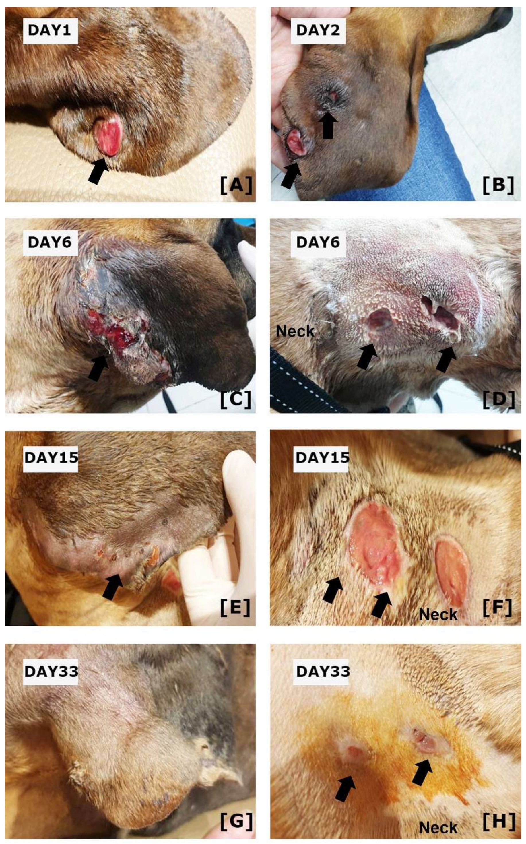
2. Case Description
3. Discussion
4. Conclusions
Author Contributions
Funding
Institutional Review Board Statement
Informed Consent Statement
Data Availability Statement
Conflicts of Interest
References
- Kirpensteijn, J.; Fingland, R.B. Cutaneous actinomycosis and nocardiosis in dogs: 48 cases (1980–1990). J. Am. Vet. Med. Assoc. 1992, 201, 917–920. [Google Scholar] [PubMed]
- Malik, R.; Krockenberger, M.B.; O’Brien, C.R.; White, J.D.; Foster, D.; Tisdall, P.L.; Gunew, M.; Carr, P.D.; Bodell, L.; McCowan, C.; et al. Nocardia infections in cats: A retrospective multi-institutional study of 17 cases. Aust. Vet. J. 2006, 84, 235–245. [Google Scholar] [CrossRef] [PubMed]
- Wilson, J.W. Nocardiosis: Updates and clinical overview. Mayo Clin. Proc. 2012, 87, 403–407. [Google Scholar] [CrossRef] [PubMed] [Green Version]
- Lipovan, I.; Burlacu, M.R.; Mareš, M.; Vulpe, V. Multiple dermal ulcers in a case of feline nocardial mycetoma. Sci. Work. Ser. C Vet. Med. 2015, 61, 139–142. [Google Scholar]
- Ribeiro, M.G.; Salerno, T.; Mattos-Guaraldi, A.L.; Camello, T.C.; Langoni, H.; Siqueira, A.K.; Paes, A.C.; Fernandes, M.C.; Lara, G.H. Nocardiosis: An overview and additional report of 28 cases in cattle and dogs. Rev. Inst. Med. Trop. Sao Paulo 2008, 50, 177–185. [Google Scholar] [CrossRef] [PubMed] [Green Version]
- Brown-Elliott, B.A.; Brown, J.M.; Conville, P.S.; Wallace, R.J., Jr. Clinical and laboratory features of the Nocardia spp. based on current molecular taxonomy. Clin. Microbiol. Rev. 2006, 19, 259–282. [Google Scholar] [CrossRef] [Green Version]
- Hattori, Y.; Kano, R.; Kunitani, Y.; Yanai, T.; Hasegawa, Y. Nocardia africana isolated from a feline mycetoma. J. Clin. Microbiol. 2003, 41, 908–910. [Google Scholar] [CrossRef] [PubMed] [Green Version]
- de Farias, M.R.; Werner, J.; Ribeiro, M.G.; Rodigheri, S.M.; Cavalcante, C.Z.; Chi, K.D.; Condas, L.A.; Gonoi, T.; Matsuzama, T.; Yazama, K. Uncommon mandibular osteomyelitis in a cat caused by Nocardia africana. BMC Vet. Res. 2012, 8, 239. [Google Scholar] [CrossRef] [PubMed] [Green Version]
- Singhal, N.; Kumar, M.; Kanaujia, P.K.; Virdi, J.S. MALDI-TOF mass spectrometry: An emerging technology for microbial identification and diagnosis. Front. Microbiol. 2015, 6, 791. [Google Scholar] [CrossRef] [Green Version]
- Girard, V.; Mailler, S.; Polsinelli, S.; Jacob, D.; Saccomani, M.C.; Celliere, B.; Monnin, V.; van Belkum, A.; Hagen, F.; Meis, J.F.; et al. Routine identification of Nocardia species by MALDI-TOF mass spectrometry. Diagn. Microbiol. Infect. Dis. 2017, 87, 7–10. [Google Scholar] [CrossRef] [PubMed]
- Durand, T.; Vautrin, F.; Bergeron, E.; Girard, V.; Polsinelli, S.; Monnin, V.; Durand, G.; Dauwalder, O.; Dumitrescu, O.; Laurent, F.; et al. Assessment of VITEK® MS IVD database V3.0 for identification of Nocardia spp. using two culture media and comparing direct smear and protein extraction procedures. Eur. J. Clin. Microbiol. Infect. Dis. 2020, 39, 559–567. [Google Scholar] [CrossRef] [PubMed]
- Hamid, M.E.; Maldonado, L.; Sharaf Eldin, G.S.; Mohamed, M.F.; Saeed, N.S.; Goodfellow, M. Nocardia africana sp. nov., a new pathogen isolated from patients with pulmonary infections. J. Clin. Microbiol. 2001, 39, 625–630. [Google Scholar] [CrossRef] [PubMed] [Green Version]
- Zhao, P.; Zhang, X.; Du, P.; Li, G.; Li, L.; Li, Z. Susceptibility profiles of Nocardia spp. to antimicrobial and antituberculotic agents detected by a microplate Alamar Blue assay. Sci. Rep. 2017, 7, 43660. [Google Scholar] [CrossRef] [PubMed]
- Yaemsiri, S.; Sykes, J.E. Sykes Successful Treatment of Disseminated Nocardiosis Caused by Nocardia veterana in a Dog. J. Vet. Intern. Med. 2018, 32, 418–422. [Google Scholar] [CrossRef] [Green Version]
- Uhde, A.K.; Kilwinski, J.; Peters, M.; Verspohl, J.; Feßler, A.T.; Schwarz, S.; Wohlsein, P. Fatal nocardiosis in a dog caused by multiresistant Nocardia veterana. Vet. Microbiol. 2016, 183, 78–84. [Google Scholar] [CrossRef] [PubMed]
- Saksena, R.; Rynga, D.; Rajan, S.; Gaind, R.; Dawar, R.; Sardana, R.; Sen, M.K.; Suri, J.C. Fatal pulmonary infection by trimethoprim-sulfamethoxazole resistant Nocardia otitidiscaviarum: Report of two cases and review. J. Infect. Dev. Ctries. 2020, 14, 214–222. [Google Scholar] [CrossRef] [PubMed] [Green Version]


| Antibiotics | Sensitivity | Antibiotics | Sensitivity |
|---|---|---|---|
| Amikacin | S | Cefalothin | R |
| Cefazolin | S | Cefotaxime | R |
| Cefotetan | S | Cefoxitin | R |
| Gentamicin | S | Ciprofloxacin | R |
| Imipenem | S | Doxycycline | R |
| Kanamycin | S | Penicillin | R |
| Minocycline | S | Sulphamethox/Trimethoprim | R |
| Amoxycillin/Clavulanic acid | R | Vancomycin | R |
| Ampicillin | R |
Publisher’s Note: MDPI stays neutral with regard to jurisdictional claims in published maps and institutional affiliations. |
© 2022 by the authors. Licensee MDPI, Basel, Switzerland. This article is an open access article distributed under the terms and conditions of the Creative Commons Attribution (CC BY) license (https://creativecommons.org/licenses/by/4.0/).
Share and Cite
Yoon, J.-S.; So, H.; Joo, B.; Park, J.; Jeong, I.-S.; Lee, G.-J.; Park, J. A Canine Case of Nocardia africana Infection Detected by Matrix-Assisted Laser Desorption Ionization—Time-of-Flight Mass Spectrometry. Vet. Sci. 2022, 9, 265. https://doi.org/10.3390/vetsci9060265
Yoon J-S, So H, Joo B, Park J, Jeong I-S, Lee G-J, Park J. A Canine Case of Nocardia africana Infection Detected by Matrix-Assisted Laser Desorption Ionization—Time-of-Flight Mass Spectrometry. Veterinary Sciences. 2022; 9(6):265. https://doi.org/10.3390/vetsci9060265
Chicago/Turabian StyleYoon, Ji-Seon, Hyungjae So, Beomsung Joo, Jihong Park, In-Seong Jeong, Gi-Jong Lee, and Jinho Park. 2022. "A Canine Case of Nocardia africana Infection Detected by Matrix-Assisted Laser Desorption Ionization—Time-of-Flight Mass Spectrometry" Veterinary Sciences 9, no. 6: 265. https://doi.org/10.3390/vetsci9060265