Article

Characterization and Pathogenicity of Colletotrichum truncatum Causing Hylocereus undatus Anthracnose through the Changes of Cell Wall-Degrading Enzymes and Components in Fruits

1
Biocontrol Engineering Laboratory of Crop Diseases and Pests of Gansu Province, College of Plant Protection, Gansu Agricultural University, Lanzhou 730070, China
2
Gansu Provincial Key Laboratory of Arid Land Crop Science, Gansu Agricultural University, Lanzhou 730070, China
*
Author to whom correspondence should be addressed.
J. Fungi 2024, 10(9), 652; https://doi.org/10.3390/jof10090652
Submission received: 15 August 2024 / Revised: 5 September 2024 / Accepted: 11 September 2024 / Published: 13 September 2024
(This article belongs to the Special Issue Control of Postharvest Fungal Diseases)

Abstract

:
Anthracnose is one of the destructive diseases of pitaya that seriously affects the plant growth and fruit quality and causes significant yield and economic losses worldwide. However, information regarding the species of pathogens that cause anthracnose in pitaya (Hylocereus undatus) fruits in Gansu Province, China, and its pathogenic mechanism is unknown. Thus, the purposes of our present study were to identify the species of pathogens causing H. undatus fruits anthracnose based on the morphological and molecular characteristics and determine its pathogenic mechanism by physiological and biochemical methods. In our present study, forty-six isolates were isolated from the collected samples of diseased H. undatus fruits and classified as three types (named as H-1, H-2, and H-3), according to the colony and conidium morphological characteristics. The isolation frequencies of H-1, H-2, and H-3 types were 63.04%, 21.74%, and 15.22%, respectively. The representative single-spore isolate of HLGTJ-1 in H-1 type has significant pathogenicity, and finally we identified Colletotrichum truncatum as the pathogen based on the morphological characteristics as well as multi-locus sequence analysis. Moreover, the H. undatus fruits inoculated with C. truncatum had a significantly increased activity of cell wall-degrading enzymes (CWDEs) cellulase (Cx), β-glucosidase (β-Glu), polygalacturonase (PG), and pectin methylgalacturonase (PMG), while having a decreased level of cell wall components of original pectin and cellulose in comparison to control. The average increased activities of Cx, β-Glu, PG, and PMG were 30.73%, 40.40%, 51.55%, and 32.23% from day 0 to 6 after inoculation, respectively. In contrast, the average decreased contents of original pectin and cellulose were 1.82% and 16.47%, respectively, whereas the average increased soluble pectin content was 38.31% in comparison to control. Our results indicate that C. truncatum infection increased the activities of CWDEs in H. undatus fruits to disassemble their cell wall components, finally leading to the fruits’ decay and deterioration. Thus, our findings will provide significant evidence in the controlling of pitaya anthracnose in the future.

1. Introduction

Pitaya (Hylocereus spp.) is known as a representative and important tropical and sub-tropical fruit due to its high nutrients, medicinal properties, and high economic values [1,2,3]. Now, it is widely cultivated in America, Vietnam, China, Mexico, Nicaragua, Malaysia, Colombia, Thailand, Ecuador, and other countries [4]. However, with increasing of cultivation area, pitaya fruit diseases are becoming a key problem, resulting in the fruits’ decay, shortened shelf life, and economic losses [5]. Anthracnose caused by Colletotrichum spp. are one of the most devastating preharvest and postharvest diseases of pitaya fruits [6,7,8]. Some studies reported that the disease incidence of pitaya anthracnose was 50% in an orchard in Fortaleza, Ceará, Brazil, in 2018 [9], and also up to 35% in the municipality of Pelotas, Rio Grande do Sul state, Brazil, in 2017 [10]. The yield losses due to it in pitaya were up to 20–80% in Malaysia [11] and approximately 50% losses in pitaya were caused by Colletotrichum gloeosporioides [12]. In 2020, we found that anthracnose occurred in the greenhouse of pitaya (H. undatus) fruits in Wuwei city, Gansu Province, China, with a field incidence of 15.6%. However, the species of pathogens that cause pitaya anthracnose are different in different regions, and related work on species identification is still lacking in the world. In previous studies, the species of pathogens causing pitaya fruit anthracnose have been reported as C. gloeosporioides (Malaysia, USA, Japan, and Brazil) [11,13,14,15], C. truncatum (Malaysia and India) [16,17] and C. siamense (India) [18], C. tropicale (Mexico) [19], C. karstii (Brazil) [10], C. aenigma and C. siamense (Thailand) [20], and C. fructicola (Philippines) [21]. In China, the species of C. gloeosporioides [22], C. siamense [23], and C. truncatum [24] that cause pitaya fruit anthracnose have been reported in Guangzhou Province, Hainan Province, Yunnan Province, and other regions. Meanwhile, the species of C. gloeosporioides, C. truncatum, and C. boninense that can cause H. polyrhizus, H. undatus, and H. costaricensis anthracnose have been reported in Taiwan [25]. However, information on the pathogens of pitaya fruit anthracnose in China is still lacking, especially information regarding the species of pathogens that cause anthracnose in H. undatus fruits in Gansu Province, China, and their pathogenic mechanisms remains unknown.
As is well known, the cell wall is the first mechanical barrier for the plants against fungal pathogen invasion and infection [26], but the cell wall-degrading enzymes (CWDEs) that are secreted by different species of Colletotrichum play a key role in penetrating the barrier of the plant cell wall [27], infecting the plant tissue, and leading to the infected tissue’s death, maceration, decay, and deterioration [28,29]. Similarly, we have found that the pathogenic isolate can cause the H. undatus fruit’s tissue maceration and decay, even resulting in the whole fruits softening finally in greenhouse or pathogenicity testing in the present study. Moreover, a previous study reported that the gradually increased activities of CWDEs (polygalacturonase (PG), pectin polysaccharide (PME), β-galactosidase, and cellulase) play the important role in modifying the pectic and hemi-cellulosic components in the cell wall of pitaya fruits during fruit softening [30]. To date, there is still a lack of research regarding the effect of Colletotrichum sp. infection on the activities of the CWDEs and the contents of pectin and cellulase in pitaya fruits.
Thus, the aims of our present study were to identify the species of the pathogens causing H. undatus fruit anthracnose in Gansu Province, China, based on the morphological and molecular characteristics, and investigate the impact of the pathogens on the changes in CWDE activities and cell wall components in H. undatus fruits.

2. Materials and Methods

2.1. Fruit Sample Collection

In 2020, twenty-eight Hylocereus undatus fruit samples with typical anthracnose symptoms were collected from the greenhouse in Wuwei city, Gansu Province, China. The diseased fruit samples were labeled and stored at 4 °C for pathogen isolation.

2.2. Pathogen Isolation and Purification

The pathogens were isolated from H. undatus fruits with typical anthracnose symptoms by the method of tissue isolation. The diseased fruit samples were rinsed with sterile water three times and then dried using sterile paper for isolating the pathogens. The fruit sections were prepared by cutting the junction between the diseased and healthy fruit areas (0.5 cm long × 0.5 cm wide), and then disinfecting with 75% ethanol for 30 s, followed by rinsing with sterile water three times. Thereafter, the sterilized fruits sections were dried on the sterile paper and then inoculated with potato dextrose agar (PDA) medium with five sections for each. The inoculation PDA plates were incubated with a constant temperature of 25 °C and light/dark (12 h/12 h) for 3 days. Isolates with different morphological characteristics of colony and conidium were purified by cutting the mycelial discs (d = 0.5 cm) from the edge of colony at 3 days after incubation, and inoculated on the center of new PDA medium for 7 days. Finally, the single-spore isolates were purified by inoculating the single spore from the 7-day-old isolate colony on the PDA media. The morphological characteristics of the single-spore isolates were recorded, and their isolation frequencies were calculated at 7 days after incubation. The representative single-spore isolates were also used for further pathogenicity determination and morphological and molecular characteristics identification.

2.3. Isolate Pathogenicity Determination

For the pathogenicity testing, three representative isolates were selected from types H-1, H-2, and H-3 (one isolate for each type), named as HLGTJ-1, HLGTJ-2, and HLGTJ-3. The pathogenicity of the representative single-spore isolates was determined by inoculating the mycelial discs of the isolates on the detached and healthy H. undatus fruit samples according to Koch’s postulates. The uniform maturity and healthy H. undatus fruits were disinfected with 75% ethanol for 30 s, and then rinsed with sterile water three times and air-dried on a bench for pathogenicity testing. The specific methods for the representative single-spore isolate’s pathogenicity testing are described by Zhang et al. (2022) [31]. The fruits inoculated with the mycelial discs of the isolates and the PDA medium discs without mycelia were regarded as the treatment and control, respectively. Thereafter, the inoculation fruit samples were placed in a plastic container at a constant temperature of 25 °C with 60% relative humidity, and incubated under a light/dark (12 h/12 h) photoperiod. The pathogenicity and symptoms of the inoculation fruits were determined and observed from 1 to 7 days after the inoculation for 2 days internally. At day 7, the pathogenic isolates were re-isolated and incubated on PDA media to observe the morphological characteristics in comparison to the inoculation isolates. The experiments in the treatment and control group were repeated three times, and three fruit samples were used for each replicate and representative isolate, respectively.

2.4. Morphological Characteristics of the Isolate Identification

The diameter of mycelial discs (d = 0.5 cm) were cut from the edge of the 7-day-old single-spore isolate colony, inoculated individually on the center of PDA media, and incubated at 25 °C. The morphological characteristics of the colony, acervulus, conidium, seta, appressorium, and number of acervuli and setae formed were observed and recorded. Additionally, the sizes of acervulus, seta, and conidium samples (100 for each replicate), and appressorium (50 for each replicate) were measured for each of the three replicates, and then the mean value was determined and calculated for the isolate. Finally, the morphological characteristics of the isolate was identified and based primarily on the species description by Damm et al. (2009) [32] and Guo et al. (2014) [24] in previous studies.

2.5. Molecular Characteristics of the Isolate Identification

For the identification of the isolate species, the fresh mycelia of the representative single-spore isolate were collected at 7 days after incubation on PDA media, and quickly frozen with liquid nitrogen for the genomic DNA extraction. The genomic DNA was extracted by the modified CTAB method and stored in the refrigerator at −20 °C for further experiment. The genomic DNA of the representative single-spore isolate was used as a template to amplify the target internal transcribed spacers (ITSs), glyceraldehyde-3-phosphate dehydrogenase (GAPDH), and histone 3 (HIS3) gene fragments by using the Polymerase Chain Reaction (PCR) amplification. The sequences of the ITS region and the GAPDH and HIS3 genes of the representative single-spore isolate were amplified using the primers of HJ-ITS5/HJ-ITS4, HJ-GAPDHF/HJ-GAPDHR, and HJ-HIS3F/HJ-HIS3R [33,34] (Table 1). The primers and PCR products used in the present study were synthesized and sequenced by Sangon Biotech Co., Ltd. (Shanghai, China), respectively. The sequences of the HJ-ITS region and HJ-GAPDH and HJ-HIS3 genes were aligned on the NCBI website using BLAST search, and then the related sequences were downloaded and submitted to multi-locus phylogenetic analyses based on maximum likelihood (ML) by combining the dataset of HJ-ITS region and HJ-GAPDH and HJ-HIS3 sequences. Multi-locus phylogenetic tree construction was performed using the IQ-TREE (v1.6.12) software, with a boot-strap value of 1000.

2.6. Effect of Isolate Infection on the Physiological-Biochemical Characteristics of H. undatus Fruits

The uniformly mature and healthy H. undatus fruits with the same size were disinfected with 75% ethanol for 2 min and finally rinsed with sterile water and air-dried by placing on a bench for inoculating. The mycelial discs (d = 0.5 cm) were prepared at the edge of the isolate colony at 7 days after inoculation. The specific method for the inoculation is the same as the pathogenicity of isolate determination. Each fruit was inoculated with three mycelial discs (treatment) or the PDA medium discs without mycelia (control). After inoculation, the fruits in the treatment and control group were placed in a plastic container and incubated at 25 °C in an incubator with a constant relative humidity of 60% and a light/dark (12 h/12 h) photoperiod. For the determination of the effect of the isolate infection on the physiological-biochemical characteristics of H. undatus fruits, the junction tissues between the diseased and healthy fruit area in the treatment fruits and the junction tissues between the inoculation site and healthy fruit area in the control fruits were collected at 0, 1, 2, 3, 4, 5, and 6 days after inoculation. Each experiment in the treatment and control group was repeated three times and with three H. undatus fruits in each replicate.

2.6.1. Effect of the Isolate Infection on the Activities of Cell Wall-Degrading Enzymes of H. undatus Fruits

The methods for the extraction and activity determination of the CWDEs cellulase (Cx), β-glucosidase (β-Glu), polygalacturonase (PG), and pectin methylgalacturonase (PMG) were according to the instructions for the plant CWDEs assay kit (Shanghai Optimal Biotechnology Co., Ltd., Shanghai, China), and three replicates were set for each treatment and control.

2.6.2. Effect of the Isolate Infection on the Contents of Cell Wall Components of H. undatus Fruits

The cell wall components of original pectin, soluble pectin, and cellulose content in H. undatus fruits were determined according to the instructions for the plant cell wall components assay kit (Shanghai Optimal Biotechnology Co., Ltd., Shanghai, China), and three replicates were set for each treatment and control.

2.7. Statistical Analysis

The experiments in the present study were repeated three times, and data were the mean of the three replicates. The values of CWDEs activities and cell wall components in H. undatus fruits at different days after inoculation with the isolate or not were expressed as mean and standard errors. One-way ANOVA analysis was carried out and tested for the significant differences among the treatments using statistical analysis system SPSS version 16.0 (SPSS Inc., Chicago, IL, USA), and Duncan’s multiple range test at p < 0.05.

3. Results

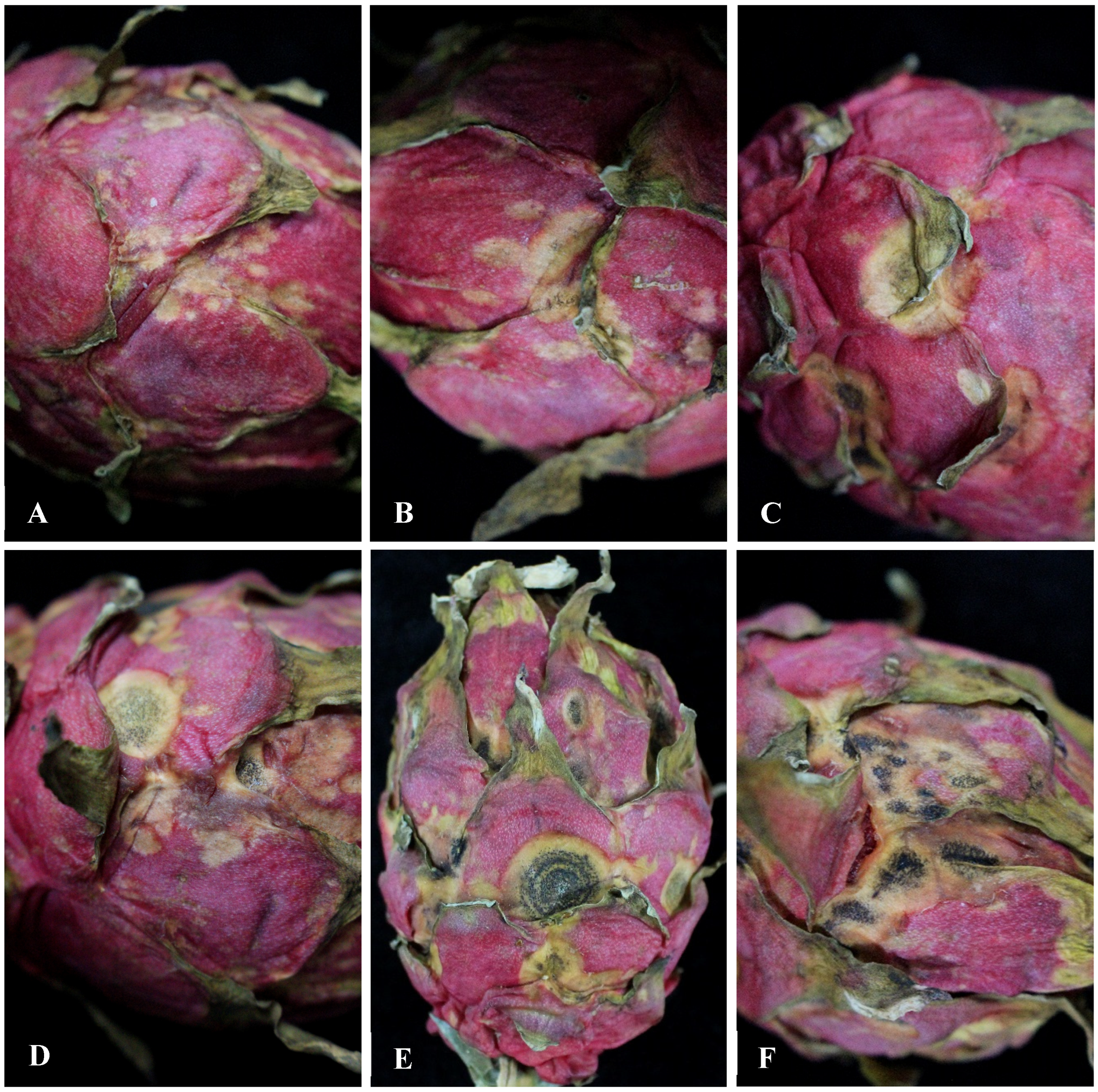
3.1. Observation of the Symptoms of H. undatus Fruits Anthracnose in Greenhouse

The symptoms initially observed in the greenhouse appeared as reddish-orange spots, which later became larger and changed to pale brown lesions in H. undatus fruits (Figure 1A,B). As the lesions progressed, the centers of the lesions were gray-white with brown border (Figure 1C), and a large number of black dots (black acervuli) were formed (some with concentric circles) on the lesions center of H. undatus fruits (Figure 1D–F).

3.2. Isolation and Determination of the Pathogenicity of the Isolates

Forty-six isolates were isolated from the collected samples of the diseased H. undatus fruits, and primary classified as three types according to the colony and conidium morphological characteristics (named as H-1, H-2, and H-3). Among the 46 isolates, 29, 10, and 7 isolates were identified as types H-1, H-2, and H-3, respectively. The isolation frequencies of the H-1, H-2, and H-3 types were 63.04%, 21.74%, and 15.22%, respectively.
The in vitro pathogenicity testing of the representative isolate HLGTJ-1 resulted in significant symptoms on the inoculated fruits that were similar to the anthracnose symptoms found in the greenhouse among the representative isolates HLGTJ-1, HLGTJ-2, and HLGTJ-3. The brown lesion formed on the inoculated area of the fruit with white or light grey mycelia on the center at 3 days after inoculation (Figure 2B). Subsequently, with the continuous expansion of the lesion, typical anthracnose symptoms of the disease lesion occurred at a later stage after inoculation. The lesion center became gray-white, and a large number of black dots formed on the surface of the lesion at 7 days (Figure 2C). In contrast, the fruit in the control group did not show any disease symptoms (Figure 2A). The morphology of the re-isolates from the infected fruit was consistent with the inoculated isolate, while no isolates were isolated from the control fruit. Thus, the representative isolate of HLGTJ-1 in the H1 type was identified as the pathogenic isolate that causes H. undatus fruit anthracnose according to Koch’s postulates.

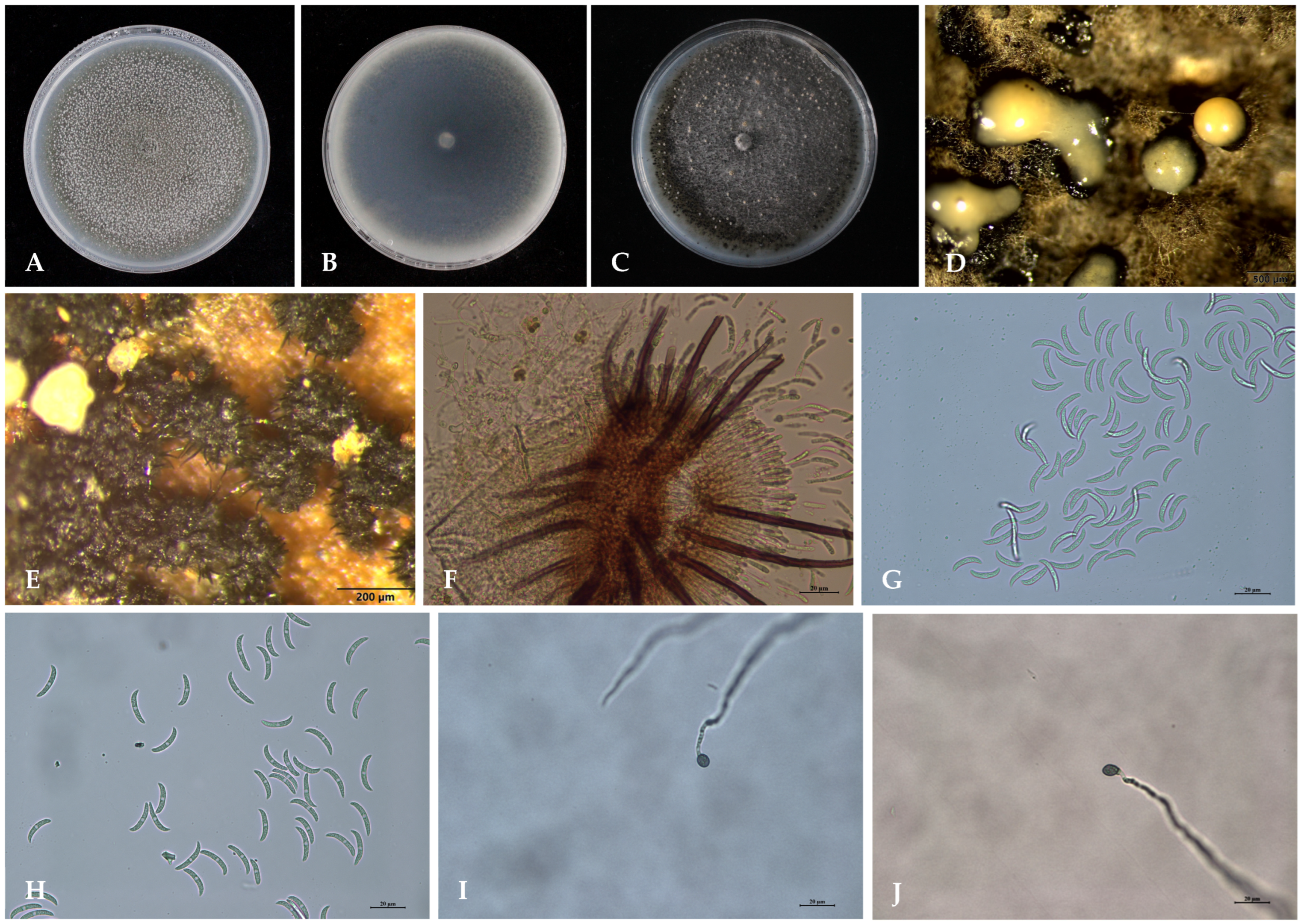
3.3. Morphological Characteristics of the Isolate

The colony of HLGTJ-1 on PDA medium exhibited white to light grey mycelia initially, and then changed to dark grey in the front, whereas thick dark grey mycelia with distinct zonation was observed in the reverse of the plate. Thereafter, the colony on PDA medium exhibited abundant black acervuli (Figure 3A) and pinkish-orange conidial mass (Figure 3C,D) in the front, while dark gray color was observed in the reverse of the plate (Figure 3B) after incubation. The acervulus was round or oval, and dark brown (238.05 × 457.49 μm) (Figure 3E,F). The seta was dark brown to black, rigid and straight, with a swollen base and a tapered apex (Figure 3E,F). The conidium (21.27 μm × 4.04 μm) was colorless, crescent or sickle-shaped, with an acute apex and a narrow truncate base (Figure 3G,H). A single germ tube was germinated from the middle or top of each conidium and formed a spherical to ovate appressorium (8.13 μm × 11.75 μm) (Figure 3I,J). Finally, the morphological characteristics indicates that the HLGTJ-1 isolate had the same characteristics as previously described for Colletotrichum truncatum.

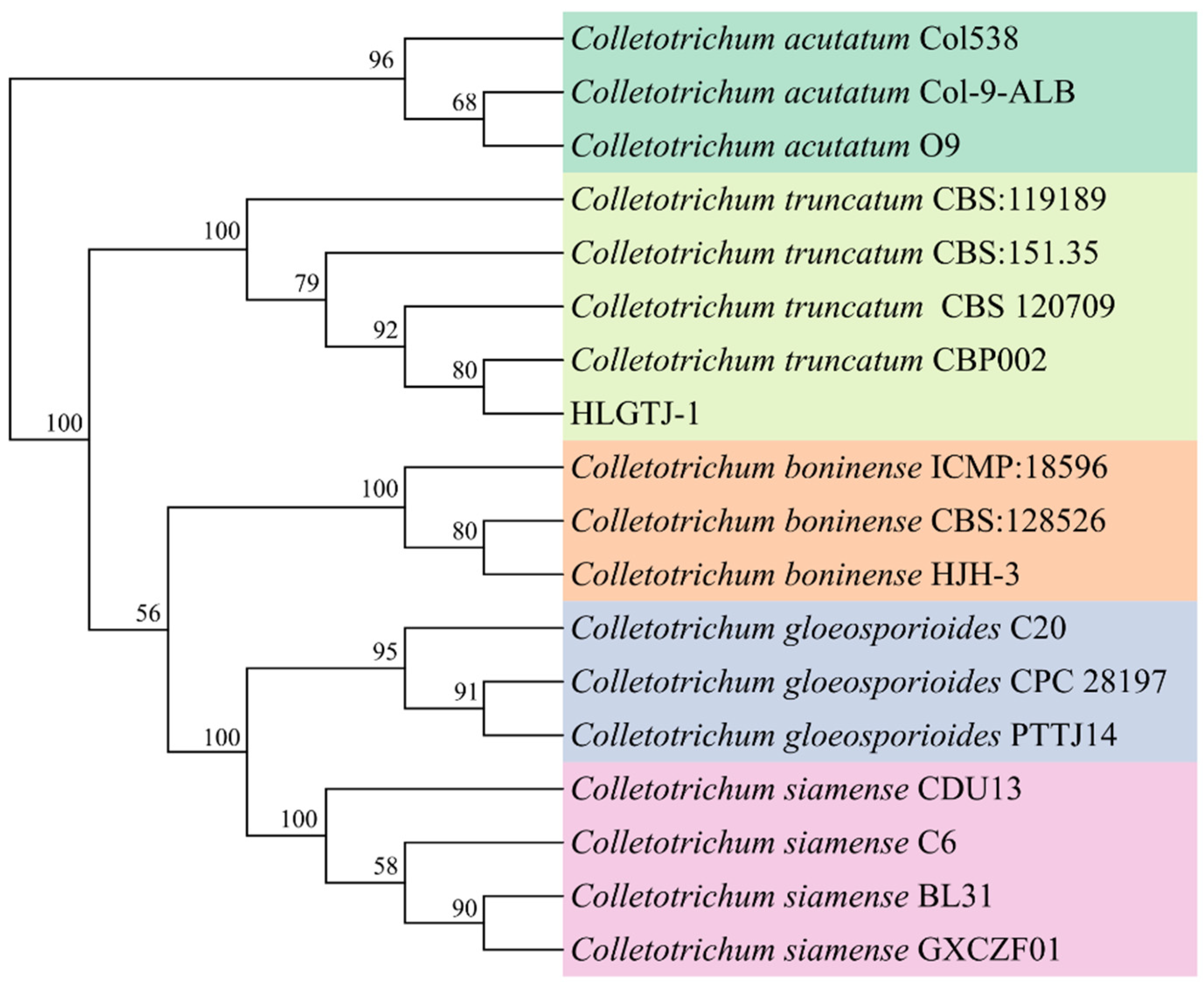
3.4. Molecular Identification of the Isolate

The fragments of the HJ-ITS region and HJ-GAPDH and HJ-HIS3 genes were obtained by PCR amplification, and the sequences of the HJ-ITS region and HJ-GAPDH and HJ-HIS3 genes were more than 99% identical to Colletotrichum truncatum previously deposited in the NCBI database. The combined dataset of the HJ-ITS region and HJ-GAPDH and HJ-HIS3 genes sequences of the HLGTJ-1 isolate was grouped within the C. truncatum CBP002 (KF300886.1) clade and with support rates of 80% (Figure 4). Thus, the isolate of HLGTJ-1 was identified as C. truncatum, based on the multi-locus sequences of the HJ-ITS region and HJ-GAPDH and HJ-HIS3 genes.

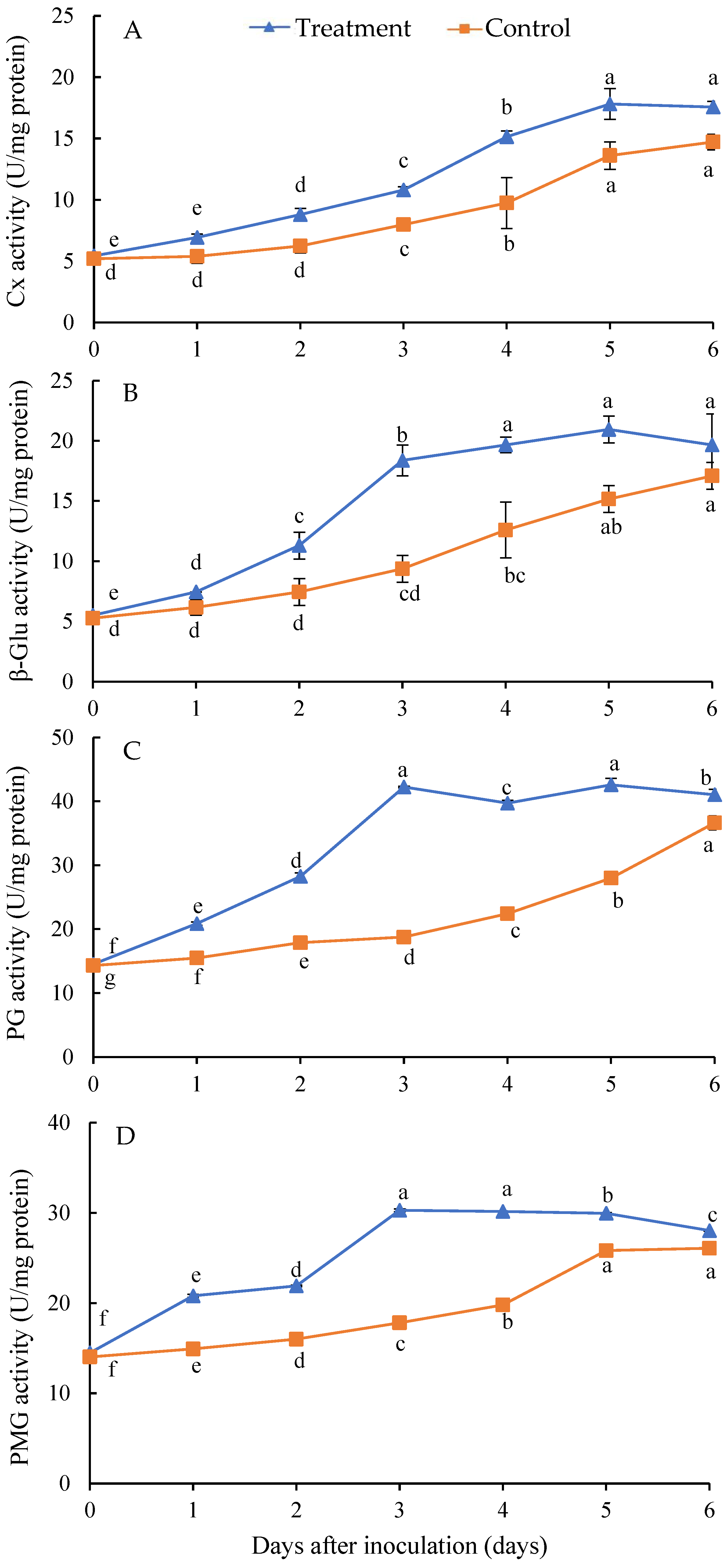
3.5. Effect of Isolate Infection on the Activities of Cell Wall-Degrading Enzymes of H. undatus Fruits

The activities of Cx (Figure 5A) and β-Glu (Figure 5B) in the healthy and diseased fruits’ tissue showed a rapid increase from day 0 to 5 after inoculation, whereas the activity of PG (Figure 5C) and PMG (Figure 5D) showed a rapid increase from day 0 to 3 after inoculation. The average activities of Cx (Figure 5A), β-Glu (Figure 5B), PG (Figure 5C), and PMG (Figure 5D) were significantly increased from day 0 to 6 after inoculation with the isolate in comparison to the control healthy fruits. Compared with the control, the average increased activities of Cx, β-Glu, PG, and PMG were 30.73%, 40.40%, 51.55%, and 32.23% from day 0 to 6 after inoculation, respectively. In addition, the activities of Cx and β-Glu reached a peak on the fifth day, whereas those of PG and PMG did so on the third day in the treatment group after inoculation.

3.6. Effect of Isolate Infection on the Contents of Cell Wall Components of H. undatus Fruits

Compared with the control, the original pectin content in the inoculated fruits’ tissue was decreased from day 0 to 3 after inoculation. Initially, the original pectin content gradually declined, followed by an increase and subsequent decrease. The average decrease in original pectin content was 1.82% from day 0 to 6 after inoculation in comparison to the control (Figure 6A). Conversely, the soluble pectin content was increased from day 0 to 6 in the control and treatment groups. Compared with the control, the average content of soluble pectin in the inoculated fruits’ tissue was significantly higher than the healthy fruits from day 0 to 6 after inoculation. The average increased content of soluble pectin was 38.31% from day 0 to 6 after inoculation in comparison to the control (Figure 6B).
The isolate infection had a significant effect on the cellulose content in H. undatus fruits. Compared with the control, the average cellulose content in the inoculated fruits’ tissue was significantly decreased from day 0 to 6 after inoculation. The peak of cellulose content was 35.28 and 28.58 mg/g in the control and treatment at 1 and 2 days after inoculation. The average decreased cellulose content was 16.47% from day 0 to 6 after inoculation in comparison to the control (Figure 6C).

4. Discussion

Colletotrichum spp. are some of the most prevalent fungal pathogens causing anthracnose in diverse tropical and subtropical fruits [35]. Seven species of Colletotrichum spp., including C. gloeosporioides, C. aenigma, C. tropicale, C. siamense, C. truncatum, C. karstii, and C. fructicola, that can cause pitaya anthracnose have been reported in Brazil, China, the US, Thailand, the Philippines, Malaysia, and other regions. Among the seven Colletotrichum species, C. gloeosporioides has been reported as the major anthracnose pathogen in Hylocereus spp. around the world [15], and several members within the C. gloeosporioides complex have been identified and reported by the application of multiple genes or regions. Recently, the species C. truncatum has been reported as causing anthracnose on different types of beans and lentils [36], guar [37], tomato [38], strawberry [39], papaya [40], and chilli [41], except the species within the C. gloeosporioides complex. In the present study, we discovered the species of C. truncatum causing H. undatus fruit anthracnose in Wuwei city, Gansu Province, China, according to the morphological characteristics and multi-locus sequence analysis of the ITS region and GAPDH and HIS3 genes. Similarly, C. truncatum was reported as causing anthracnose in H. polyrhizus stem in Malaysia [16], H. undatus and H. polyrhizus plants in India [17], and H. undatus fruits in a market in Yunnan Province, China [24]. However, C. truncatum causing H. undatus fruit anthracnose at harvest time has never been reported in Gansu Province, China. To our knowledge, this is the first report of C. truncatum causing anthracnose on H. undatus fruits at harvest time in Gansu Province, China.
Furthermore, the activities of Cx, β-Glu, PG, and PMG in H. undatus fruits infected by C. truncatum were significantly higher than those in healthy fruits. Similarly, some studies revealed that the pathogen of Phomopsis longanae infection significantly increased the activities of the CWDEs pectinesterase, polygalacturonase, β-galactosidase in the pericarp of harvested “Fuyan” longan (Dimocarpus longan Lour. cv. Fuyan) fruits [42]; Macrophomina phaseolina infection can induce the cell wall degradation of maize and sunflower [43]. In addition, the substances pectin, hemicelluloses, and cellulose are the main components of the cell wall which contribute to the mechanical properties of the cell wall and the strength of the pericarp [44,45]. However, the degradation of pectin can disassemble the cellulose and hemicellulose in the cell wall, resulting in the tissue of pericarp loosening or the fruit softening [46,47,48]. Our results found that the soluble pectin content in the inoculated fruits significantly increased after inoculation, and was higher than that in the control fruits, whereas the original pectin and cellulose contents in the inoculated fruits were lower than those in the control fruits. Likewise, other studies revealed that the pathogenic Botryodiplodia theobromae caused mango stem-end rot through producing PG, PMG, and CX to disrupt the fruits’ tissue in the deterioration process [49]; Phomopsis longanae inoculation treatment accelerated the degradation of the cell wall components including chelate-soluble pectin, Na2CO3-soluble pectin, hemicellulose, and cellulose in the longan pericarp cell wall and middle lamella, whereas it elevated the water-soluble pectin content [42].

5. Conclusions

In the present study, C. truncatum was first reported as the agent that caused anthracnose in H. undatus fruits at harvest time in Gansu Province, China, based on the morphological characteristics as well as multi-locus sequence analysis. Moreover, the H. undatus fruits inoculated with C. truncatum had significantly increased activity of Cx, β-Glu, PG, and PMG, and content of soluble pectin, while the average contents of the cell wall components of cellulose and original pectin were decreased in comparison to the control. Our results indicate that C. truncatum infection can increase the activities of H. undatus fruit CWDEs to disassemble the cell wall components of original pectin and cellulose, finally leading to the infected fruits’ tissue death, maceration, decay, and deterioration.

Author Contributions

Conceptualization, S.Z. and B.X.; data curation, J.L. and Y.L.; formal analysis, Y.L.; funding acquisition, S.Z.; methodology, Y.L. and S.Z.; project administration, S.Z. and B.X.; software, Y.L. and E.L.; supervision, B.X.; writing—original draft, S.Z.; writing—review and editing, B.X. All authors have read and agreed to the published version of the manuscript.

Funding

This work was supported by the Gansu Provincial Major Science and Technology Project (23ZDNA008); the Fuxi Outstanding Talent Cultivation Program, Gansu Agricultural University (Gaufx-03J03); and the Lanzhou Science and Technology Project (2023-QN-171).

Institutional Review Board Statement

Not applicable.

Informed Consent Statement

Not applicable.

Data Availability Statement

The original contributions presented in the study are included in the article, and further inquiries can be directed to the corresponding author.

Conflicts of Interest

The authors declare no conflicts of interest. The authors declare that the research was conducted in the absence of any commercial or financial relationships that could be construed as a potential conflict of interest.

References

  1. Elobeidy, A.A. Mass propagation of pitaya (dragon fruit). Fruits 2006, 61, 313–319. [Google Scholar] [CrossRef]
  2. Jitareerat, P.; Sripong, K.; Masaya, K.; Aiamla, S.; Uthairatanakij, A. Combined effects of food additives and heat treatment on fruit rot disease and quality of harvested dragon fruit. Agric. Nat. Resour. 2018, 52, 543–549. [Google Scholar] [CrossRef]
  3. Fan, P.; Huber, D.J.; Su, Z.; Hu, M.J.; Gao, Z.; Li, M.; Shi, X.; Zhang, Z. Effect of postharvest spray of apple polyphenols on the quality of fresh-cut red pitaya fruit during shelf life. Food Chem. 2018, 243, 19–25. [Google Scholar] [CrossRef] [PubMed]
  4. Chen, N.J.; Paull, R.E. Overall Dragon Fruit Production and Global Marketing. FFTC Agricultural Policy Platform (FFTC-AP). 2018. Available online: https://ap.fftc.org.tw/article/1596 (accessed on 14 August 2024).
  5. Luu, T.; Phi, Q.; Nguyen, T.; Dinh, V.; Pham, B.; Do, T. Antagonistic activity of endophytic bacteria isolated from weed plant against stem end rot pathogen of pitaya in Vietnam. Egypt. J. Biol. Pest Control 2021, 31, 14. [Google Scholar] [CrossRef]
  6. Ali, A.; Zahid, N.; Manickam, N.; Siddiqui, Y.; Alderson, P.G.; Maqbool, M. Effectiveness of submicron chitosan dispersions in controlling anthracnose and maintaining quality of dragon fruit. Postharvest Biol. Technol. 2013, 86, 147–153. [Google Scholar] [CrossRef]
  7. Ali, A.; Zahid, N.; Manickam, S.; Siddiqui, Y.; Alderson, P.G.; Maqbool, M. Induction of lignin and pathogenesis related proteins in dragon fruit plants in response to submicron chitosan dispersions. Crop Prot. 2014, 63, 83–88. [Google Scholar] [CrossRef]
  8. Zahid, N.; Ali, A.; Manickam, S.; Siddiqui, Y.; Alderson, P.G.; Maqbool, M. Efficacy of curative applications of SCD on anthracnose intensity and vegetative growth of dragon fruit plants. Crop Prot. 2014, 62, 129–134. [Google Scholar] [CrossRef]
  9. Inokuti, E.M.; Silva, D.E.M.; Almeida, M.M.M.; Oliveira, J.R.; Andrade, I.L.; Silva, C.F.B.; Corrêa, M.C.M.; Lima, C.S. First report of Colletotrichum tropicale causing anthracnose on pitaya (Hylocereus costaricensis) in Brazil. Plant Dis. 2024, 108, 797. [Google Scholar] [CrossRef]
  10. Nascimento, M.B.; Bellé, C.; Azambuja, R.M.; Maich, S.L.P.; Neves, C.G.; Souza-Junior, I.T.; Jacobsen, C.R.F.; Barros, D.R. First report of Colletotrichum karstii causing anthracnose spot on pitaya (Hylocereus undatus) in Brazil. Plant Dis. 2019, 103, 2137. [Google Scholar] [CrossRef]
  11. Masyahit, M.; Sijam, K.; Awang, Y.; Mohd Satar, M.G. The first report of the occurrence of anthracnose disease caused by Colletotrichum gloeosporioides (penz.) Penz. & Sacc. on dragon fruit (Hylocereus spp.) in Peninsular Malaysia. Am. J. Appl. Sci. 2009, 6, 902–912. [Google Scholar]
  12. Zahid, N.; Ali, A.; Manickam, S.; Siddiqui, Y.; Maqbool, M. Potential of chitosan loaded nanoemulsions to control different Colletotrichum spp. and maintain quality of tropical fruits during cold storage. J. Appl. Microbiol. 2012, 113, 925–939. [Google Scholar] [CrossRef] [PubMed]
  13. Palmateer, A.J.; Ploetz, R.C.; van Santen, E.; Correll, J.C. First occurrence of anthracnose caused by Colletotrichum gloeosporioides on Pitahaya. Plant Dis. 2007, 91, 631. [Google Scholar] [CrossRef]
  14. Taba, S.; Mikami, D.; Takaesu, K.; Ooshiro, A.; Moromizato, Z.; Nakasone, S.; Kawano, S. Anthracnose of pitaya (Hylocereus undatus) by Colletotrichum gloeosporioides. Jpn. J. Phytopathol. 2006, 72, 25–27. [Google Scholar] [CrossRef]
  15. Takahashi, L.M.; Rosa, D.D.; Basseto, M.A.; de Souza, H.G.; Furtado, E.L. First report of Colletotrichum gloeosporioides on Hylocereus megalanthus in Brazil. Australas. Plant Dis. 2008, 3, 96–97. [Google Scholar]
  16. Suzianti, I.V.; Intan Sakinah, M.A.; Latiffah, Z. Characterization and pathogenicity of Colletotrichum truncatum causing stem anthracnose of red-fleshed dragon fruit (Hylocereus polyrhizus) in Malaysia. J. Phytopathol. 2015, 163, 67–71. [Google Scholar]
  17. Salunkhe, V.N.; Bhagat, Y.S.; Lonkar, S.G.; Kakade, V.D.; Chavan, S.B.; Kochewad, S.A.; Nangare, D.D. First report of Colletotrichum truncatum causing anthracnose of dragon fruit (Hylocereus spp.) in India. Plant Dis. 2022, 106, 2754. [Google Scholar] [CrossRef]
  18. Abirami, K.; Sakthivel, K.; Sheoran, N.; Baskaran, V.; Gautam, R.K.; Jerard, B.A.; Kumar, A. Occurrence of anthracnose disease caused by Colletotrichum siamense on dragon fruit (Hylocereus undatus) in Andaman Islands, India. Plant Dis. 2019, 103, 768. [Google Scholar] [CrossRef]
  19. Nuñez-García, P.R.; Carrillo-Fasio, J.A.; Márquez-Licona, G.; Leyva-Madrigal, K.Y.; Lagunes-Fortiz, E.; Tovar-Pedraza, J.M. First report of Colletotrichum tropicale causing anthracnose on Pitahaya fruit in Mexico. Plant Dis. 2023, 106, 1750. [Google Scholar] [CrossRef] [PubMed]
  20. Meetum, P.; Leksomboon, C.; Kanjanamaneesathian, M. First report of Colletotrichum aenigma and C. siamense, the causal agents of anthracnose disease of dragon fruit in Thailand. J. Plant Pathol. 2015, 97, 391–403. [Google Scholar]
  21. Edzel, E.; John Darby, T.; Jennelyn, B.; Rodel, M.; Mark Angelo, B. First report of Colletotrichum fructicola, causing anthracnose of Hylocereus plants, in the Philippines. Czech Mycol. 2021, 73, 79–90. [Google Scholar]
  22. Ma, W.J.; Yang, X.; Wang, X.R.; Zeng, Y.S.; Liao, M.D.; Chen, C.J.; Sun, S.; Jia, D.M. First report of anthracnose disease on young stems of bawanghua (Hylocereus undatus) caused by Colletotrichum gloeosporioides in China. Plant Dis. 2014, 98, 991. [Google Scholar] [CrossRef] [PubMed]
  23. Zhao, H.J.; Chen, S.C.; Chen, Y.F.; Zou, C.C.; Wang, X.L.; Wang, Z.H.; Liu, A.R.; Ahammed, G.J. First report of red dragon fruit (Hylocereus polyrhizus) anthracnose caused by Colletotrichum siamense in China. Plant Dis. 2018, 6, 862. [Google Scholar] [CrossRef]
  24. Guo, L.W.; Wu, Y.X.; Ho, H.H.; Su, Y.Y.; Mao, Z.C.; He, P.F.; He, Y.Q. First report of dragon fruit (Hylocereus undatus) anthracnose caused by Colletotrichum truncatum in China. J. Phytopathol. 2014, 162, 272–275. [Google Scholar] [CrossRef]
  25. Lin, C.P.; Ann, P.J.; Huang, H.C.; Chang, T.T.; Tsai, J.N. Anthracnose of pitaya (Hylocereus spp.) caused by Colletotrichum spp., a new postharvest disease in Taiwan. J. Taiwan Agric. Res. 2017, 66, 171–183. [Google Scholar]
  26. Kubicek, C.P.; Starr, T.L.; Glass, N.L. Plant cell wall degrading enzymes and their secretion in plant-pathogenic fungi. Annu. Rev. Phytopathol. 2014, 52, 427–451. [Google Scholar] [CrossRef]
  27. Anand, T.; Bhaskaran, R.; Karthikeyan, T.R.G.; Rajesh, M.; Senthilraja, G. Production of cell wall degrading enzymes and toxins by Colletotrichum capsici and Alternaria alternata causing fruit rot of chillies. J. Plant Prot. Res. 2008, 48, 437–451. [Google Scholar] [CrossRef]
  28. Fernando, T.H.P.S.; Jayasinghe, C.K.; Wijesundera, R.L.C. Cell wall degrading enzyme secretion by Colletotrichum acutatum, the causative fungus of secondary leaf fall of Hevea brasiliensis. Mycol. Res. 2001, 105, 195–201. [Google Scholar] [CrossRef]
  29. Kimaru, S.K.; Monda, E.; Cheruiyot, R.C.; Mbaka, J.; Alakonya, A. Morphological and molecular identification of the causal agent of anthracnose disease of avocado in Kenya. Int. J. Microbiol. 2018, 10, 1925. [Google Scholar] [CrossRef] [PubMed]
  30. Almeida, D.P.F.; Huber, D.J. In vivo pectin solubility in ripening and chill-injured tomato fruit. Physiol. Plant 2007, 174, 174–182. [Google Scholar] [CrossRef]
  31. Zhang, S.; Xiang, D.; Sun, C.; Han, K.; Li, T.; Zhou, J.; Xu, B. Morphological and molecular identification of peach brown rot disease in Tibet and exploration of the biocontrol efficiency of Trichoderma. J. Fungi 2022, 8, 1174. [Google Scholar] [CrossRef]
  32. Damm, U.; Woudenberg, J.H.C.; Cannon, P.F.; Crous, P.W. Colletotrichum species with curved conidia from herbaceous hosts. Fung. Diver. 2009, 39, 45–87. [Google Scholar]
  33. White, T.J.; Bruns, T.; Lee, S.; Taylor, J. Amplification and direct sequencing of fungal ribosomal RNA genes for phylogenetics. In PCR Protocols; Academic Press: New York, NY, USA, 1990; Volume 35, pp. 315–322. [Google Scholar]
  34. Shi, M.; Xue, S.M.; Zhang, M.Y.; Li, S.P.; Huang, B.Z.; Huang, Q.; Liu, Q.B.; Liao, X.L.; Li, Y.Z. Colletotrichum truncatum-a new etiological anthracnose agent of sword bean (Canavalia gladiata) in southwestern China. Pathogens 2022, 11, 1463. [Google Scholar] [CrossRef] [PubMed]
  35. Siddiqui, Y.; Ali, A. Colletotrichum gloeosporioides (Anthracnose). In Postharvest Decay Control Strategies; Elsevier Inc.: Amsterdam, The Netherlands, 2014; pp. 337–371. [Google Scholar]
  36. Ford, R.; Banniza, S.; Photita, W.; Taylor, P.W.J. Morphological and molecular discrimination of Colletotrichum truncatum causing anthracnose on lentil in Canada. Australas. Plant Pathol. 2004, 33, 559–569. [Google Scholar] [CrossRef]
  37. García-León, E.; Cota-Barreras, G.A.; Beltrán-Peña, H.; Leyva-Madrigal, K.Y.; Valenzuela-Escoboza, F.A.; Cota-Barreras, C.I.; Tovar-Pedraza, J.M. First report of Colletotrichum truncatum causing anthracnose of guar (Cyamopsis tetragonoloba) in Mexico. Plant Dis. 2022, 106, 6. [Google Scholar] [CrossRef]
  38. Diao, Y.Z.; Zhang, C.; Lin, D.; Liu, X.L. First report of Colletotrichum truncatum causing anthracnose of tomato in China. Plant Dis. 2023, 98, 5. [Google Scholar] [CrossRef] [PubMed]
  39. Bi, Y.; Guo, W.; Zhang, G.J.; Liu, S.C.; Chen, Y. First report of Colletotrichum truncatum causing anthracnose of strawberry in China. Plant Dis. 2017, 101, 5. [Google Scholar] [CrossRef]
  40. Maharaj, A.; Rampersand, S.N. Genetic differentiation of Colletotrichum gloeosporioides and C. truncatum associated with anthracnose disease of papaya (Carica papaya L.) and bell pepper (Capsium annuum L.) based on ITS PCR-RFLP fingerprinting. Mol. Biotechnol. 2012, 50, 237–249. [Google Scholar] [CrossRef]
  41. Than, P.P.; Jeewon, R.; Hyde, K.D.; Pongsupasamit, S.; Mongkolporn, O.; Taylor, P.W.J. Characterization and pathogenicity of Colletotrichum species associated with anthracnose on chili (Capsicum spp.) in Thailand. Plant Pathol. 2008, 57, 562–572. [Google Scholar] [CrossRef]
  42. Chen, Y.; Zhang, S.; Lin, H.; Sun, J.; Lin, Y.; Wang, H.; Lin, M.; Shi, J. Phomopsis longanae chi-induced changes in activities of cell wall-degrading enzymes and contents of cell wall components in pericarp of harvested longan fruit and its relation to disease development. Front. Microbiol. 2018, 9, 1051. [Google Scholar] [CrossRef]
  43. Ramos, A.M.; Gally, M.; Szapiro, G.; Itzcovich, T.; Carabajal, M.; Levin, L. In vitro growth and cell wall degrading enzyme production by Argentinean isolates of Macrophomina phaseolina, the causative agent of charcoal rot in corn. Rev. Argent. Microbiol. 2016, 48, 267–273. [Google Scholar] [CrossRef]
  44. Huang, X.M.; Wang, H.C.; Gao, F.F.; Huang, H.B. A comparative study of the pericarp of litchi cultivars susceptible and resistant to fruit cracking. J. Hortic. Sci. Biotechnol. 1999, 74, 351–354. [Google Scholar] [CrossRef]
  45. Vorwerk, S.; Somerville, S.; Somerville, C. The role of plant cell wall polysaccharide composition in disease resistance. Trends Plant Sci. 2004, 4, 203–209. [Google Scholar] [CrossRef] [PubMed]
  46. Duan, X.W.; Cheng, G.P.; Yang, E.; Yi, C.; Ruenroengklin, N.; Lu, W.J.; Luo, Y.B.; Jiang, Y.M. Modification of pectin polysaccharides during ripening of postharvest banana fruit. Food Chem. 2008, 111, 144–149. [Google Scholar] [CrossRef]
  47. Zhou, R.; Li, Y.F.; Yan, L.P.; Xie, J. Effect of edible coatings on enzymes, cell-membrane integrity, and cell-wall constituents in relation to brittleness and firmness of Huanghua pears (Pyrus pyrifolia Nakai cv. Huanghua) during storage. Food Chem. 2011, 124, 569–575. [Google Scholar] [CrossRef]
  48. Chen, Y.H.; Hung, Y.C.; Chen, M.Y.; Lin, H.T. Effects of acidic electrolyzed oxidizing water on retarding cell wall degradation and delaying softening of blueberries during postharvest storage. LWT Food Sci. Technol. 2017, 84, 650–657. [Google Scholar] [CrossRef]
  49. Li, M.; Gao, Z.Y.; Hu, M.J.; Zhou, S.; Yang, D.P.; Yang, B.; Yi, J.X.; Yang, F.Z. Pathogenicity of cell wall degrading enzymes produced by Botryodiplodia theobromae Pat. against mangoes. Agric. Biotechnol. 2012, 1, 18–21. [Google Scholar]
Figure 1. Symptoms of Hylocereus undatus fruit anthracnose at different time periods in Wuwei city, China. (A) and (B): the symptoms at the initial stage; (CF): the symptoms at a later stage.
Jof 10 00652 g001
Figure 2. The pathogenicity test of the representative isolate of HLGTJ-1 on Hylocereus undatus fruit after inoculation. (A) Fruit inoculation with the PDA discs without the HLGTJ-1 isolate (control); (B,C) fruits inoculation with the mycelial discs of the HLGTJ-1 isolate at 3 and 7 days after inoculation, respectively.
Jof 10 00652 g002
Figure 3. Morphological characteristics of the representative isolate of HLGTJ-1. (A,B) The front and reverse views of the colony, respectively; (C) the conidial mass produced on the front of the colony; (D) the conidial mass observed under a stereoscope; (E) the acervuli and setae observed under a stereoscope; (F) the acervulus and setae observed under a microscope using the hand-sliced method; (G,H) conidia; (I,J) appressorium.
Jof 10 00652 g003
Figure 4. Multi-locus phylogenetic tree of the single-spore isolate of HLGTJ-1 based on the combined sequences (HJ-ITS region and HJ-GAPDH and HJ-HIS3 genes) by the maximum likelihood (ML) method. Bootstraps supporting values higher than 50% from the 1000 replicates are presented at the nodes.
Jof 10 00652 g004
Figure 5. Changes in activities of Cx (A), β-Glu (B), PG (C), and PMG (D) in Hylocereus undatus fruits at different days after inoculation with the isolate. Different letters in Figure are significantly different at p < 0.05.
Jof 10 00652 g005
Figure 6. Changes in original pectin (A), soluble pectin (B), and cellulose (C) contents in Hylocereus undatus fruits at different days after inoculation with the isolate. Different letters in Figure are significantly different at p < 0.05.
Jof 10 00652 g006
Table 1. Primers for the PCR amplification in the present study.
Region or GenesPrimer NamePrimer Sequence (5′–3′)Annealing Temperature (°C)
ITSHJ-ITS4
HJ-ITS5
TCCTCCGCTTATTGATATGC
GGAAGTAAAAGTCGTAACAAGG
56
GAPDHHJ-GAPDHF
HJ-GAPDHR
GCCGTCAACGACCCCTTCATTG
GGGTGGAGTCGTACTTGAGCAT
56
HIS3HJ-HIS3F
HJ-HIS3R
AGGTCCACTGGTGGCAAG
AGCTGGATGTCCTTGGACTG
54
Disclaimer/Publisher’s Note: The statements, opinions and data contained in all publications are solely those of the individual author(s) and contributor(s) and not of MDPI and/or the editor(s). MDPI and/or the editor(s) disclaim responsibility for any injury to people or property resulting from any ideas, methods, instructions or products referred to in the content.

