

Central Role of the Actomyosin Ring in Coordinating Cytokinesis Steps in Budding Yeast


Abstract
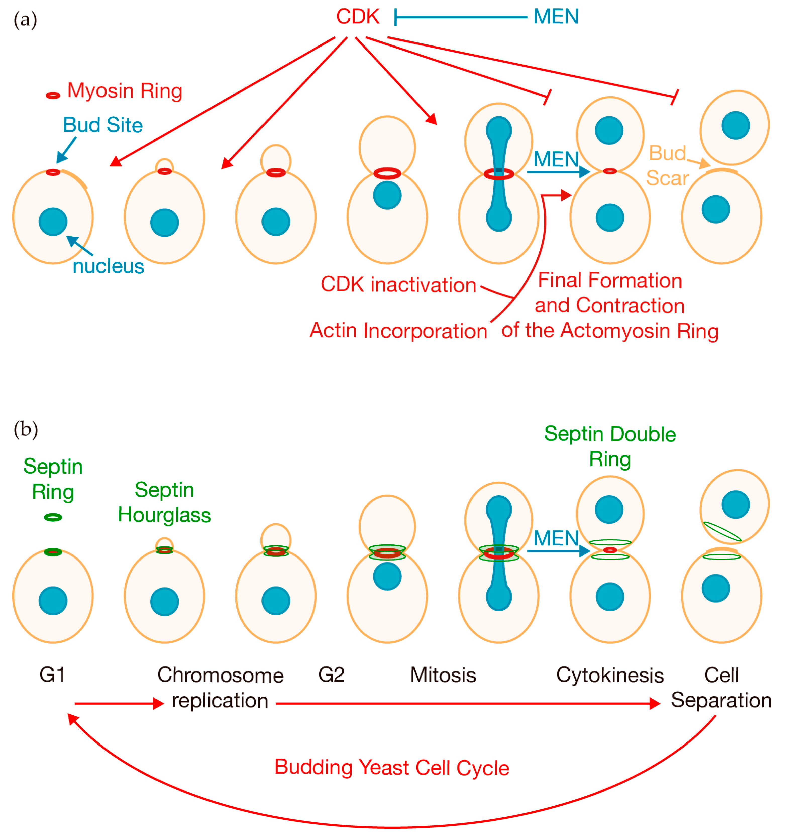
:1. Introduction
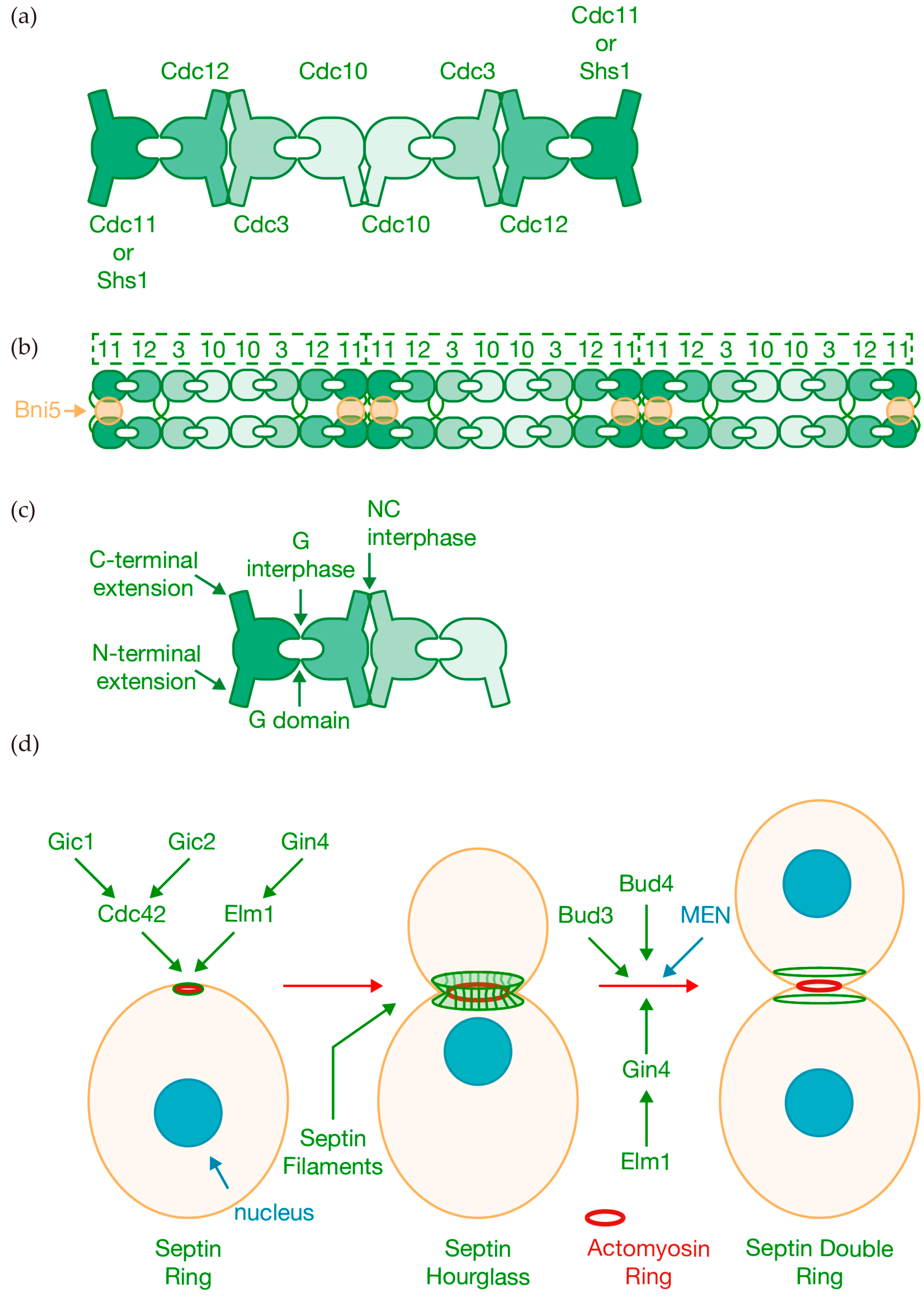
2. Positioning the Contractile Ring in the Cell: The Septin Scaffold
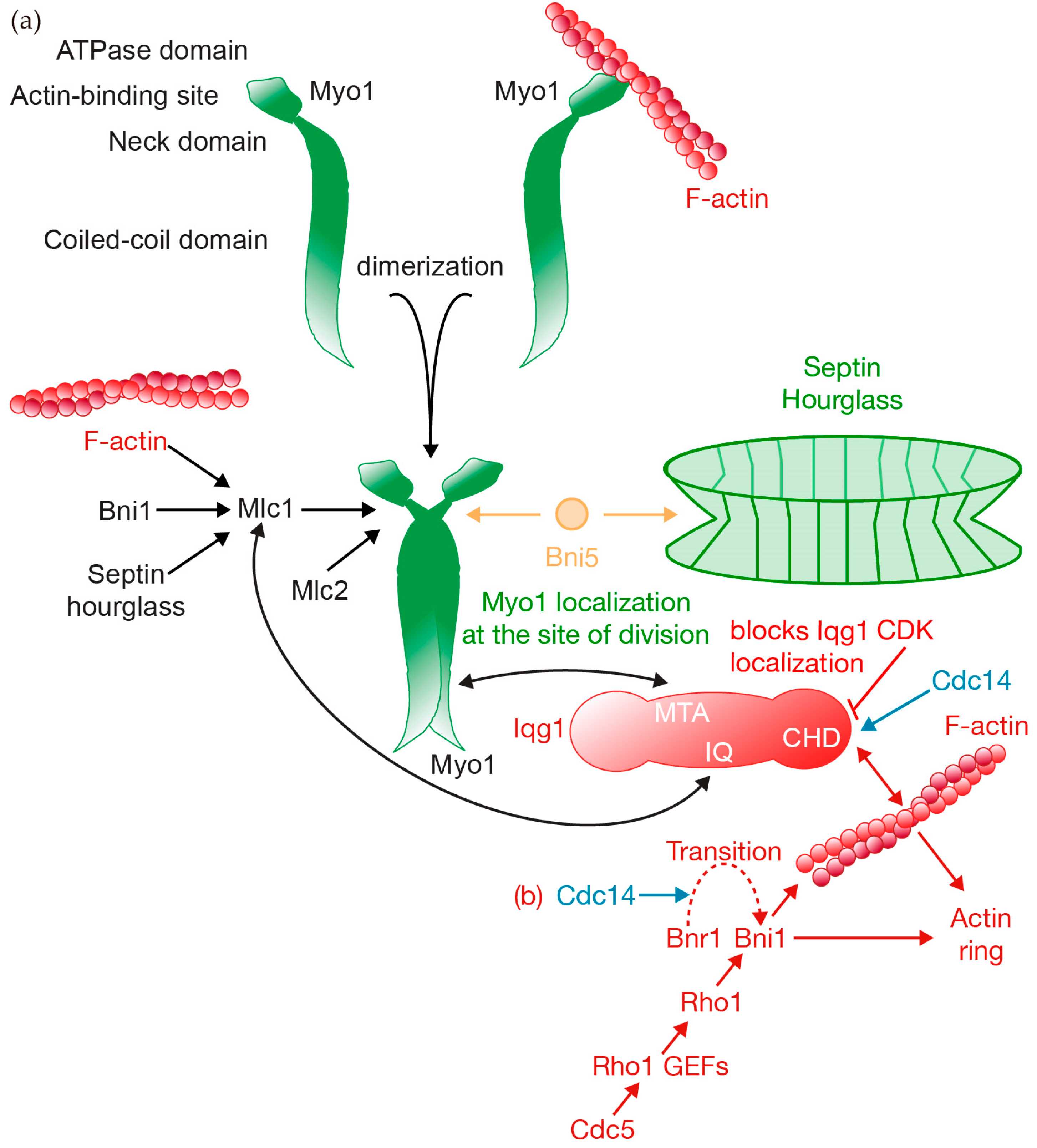
3. Building a Contractile Ring
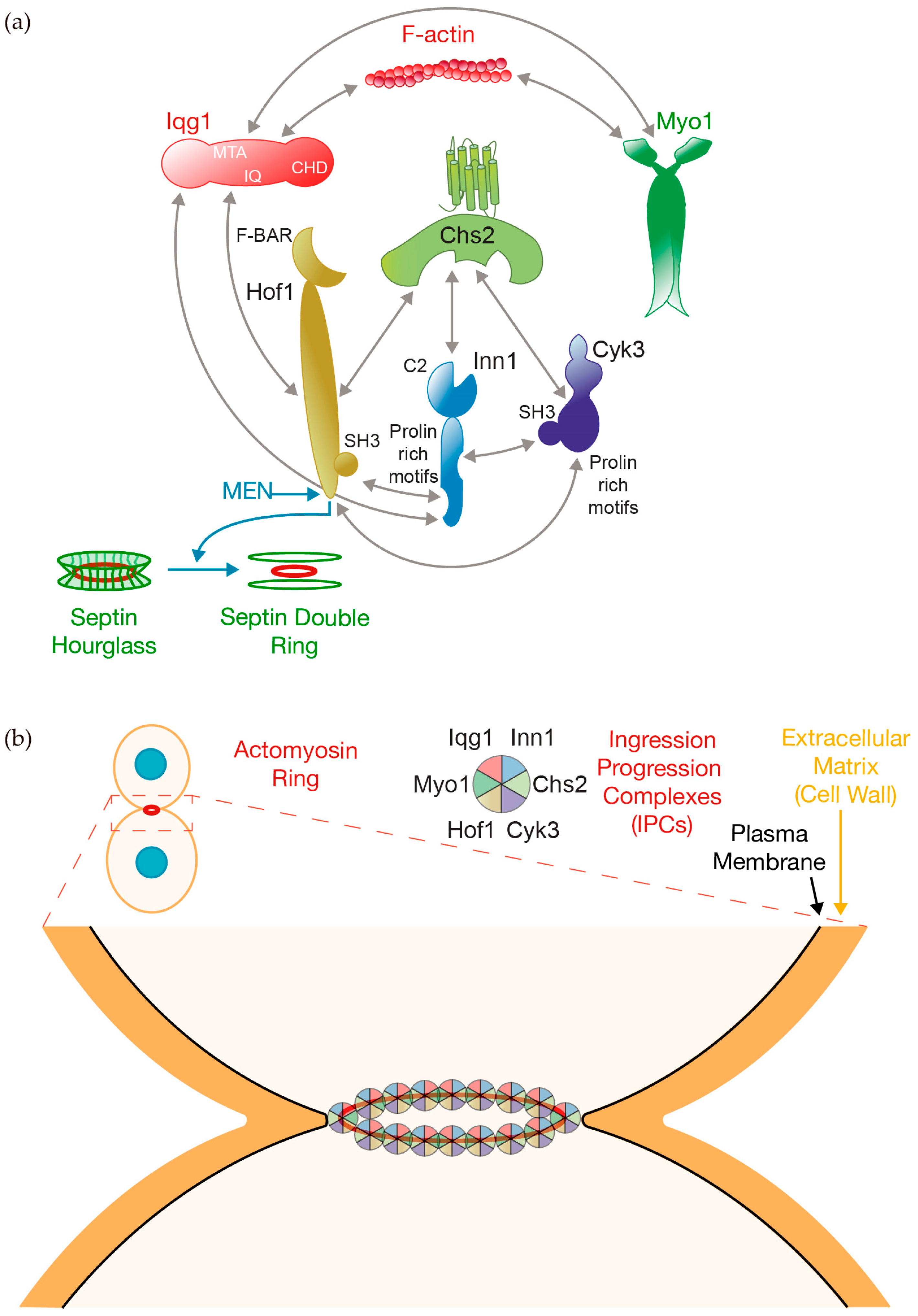
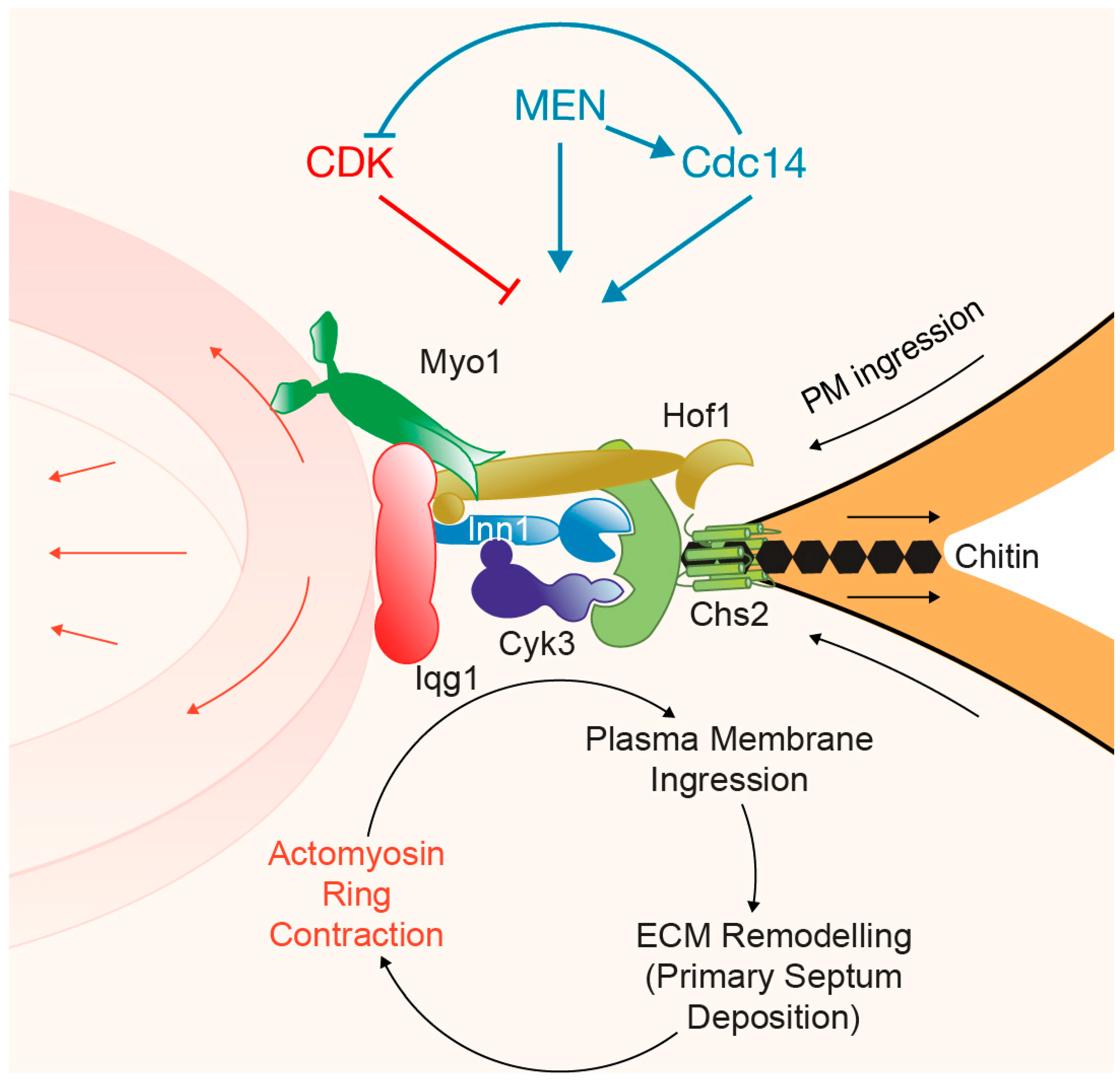
4. Ingression Progression Complexes (IPCs)
5. Actomyosin Ring Constriction
6. Mechanism and Regulation of Primary Septum Deposition
7. Concluding Remarks

| S. cerevisiae | Type of Protein/Function | S. pombe | C. albicans | Candida albicans Virulence Studies |
|---|---|---|---|---|
| BNI1 | Formin | cdc12 | BNI1 | mouse intravenous infection [140] |
| BNI5 | Bud protein | nda | BNI5 | nda |
| BUD3 | Bud protein | nda | BUD3 | nda |
| BUD4 | Bud protein | nda | INT1 | nda |
| CDC28 | CDK1 | cdc2 | CDC28 | nda |
| CDC3 | Septin | spn1 | CDC3 | nda |
| CDC5 | Polo kinase | plo1 | CDC5 | |
| CDC10 | Septin | spn2 | CDC10 | mouse intravenous infection [141] |
| CDC11 | Septin | spn3 | CDC11 | mouse intravenous infection [142] |
| CDC12 | Septin | spn4 spn6 | CDC12 | nda |
| CDC14 | MEN pathway | clp1 | CDC14 | nda |
| CDC15 | MEN pathway | cdc7 | CDC15 | nda |
| CDC42 | GTPase | cdc42 | CDC42 | nda |
| CDC55 | Phosphatase PP2A | pab1 | CDC55 | nda |
| CHS2 | Chitin synthase | chs1 chs2 | CHS1 | mouse intravenous infection [143,144] |
| CYK3 | Contractile ring Assembly | cyk3 | CYK3 | nda |
| DBF2 | MEN pathway | sid2 | DBF2 | nda |
| DBF20 | MEN pathway | sid2 | nda | nda |
| ELM1 | Morphogenesis | ssp1 | ELM1 | nda |
| GIC1 | GTPase binding | nda | nda | nda |
| GIC2 | GTPase binding | nda | nda | nda |
| GIN4 | Septin ring assembly | cdr2 cdr1 | GIN4 | nda |
| HOF1 | Contractile ring assembly | cdc15 imp2 | HOF1 | nda |
| INN1 | Contractile ring assembly | fic1 | INN1 | nda |
| IQG1 | IQGAP | rng2 | IQG1 | nda |
| MLC1 | Myosin essential light chain | cdc4 | MLC1 | nda |
| MLC2 | Myosin regulatory light chain | rlc1 | C5_03370C_A | nda |
| MYO1 | Myosin II | myo2 myp2 | MYO1 | nda |
| RVS167 | Actin binding | hob1 | RVS167 | mouse oropharyngeal infection [145], mouse intravenous infection [146] |
| SHS1 | Septin | spn3 | SEP7 | nda |
| SPA2 | Polarisome component | spa2 | SPA2 | mouse intravenous infection [147] |
| TEM1 | MEN pathway | spg1 | TEM1 | mouse intravenous infection [144] |
Author Contributions
Funding
Institutional Review Board Statement
Data Availability Statement
Acknowledgments
Conflicts of Interest
References
- Fujiwara, T.; Bandi, M.; Nitta, M.; Ivanova, E.V.; Bronson, R.T.; Pellman, D. Cytokinesis failure generating tetraploids promotes tumorigenesis in p53-null cells. Nature 2005, 437, 1043–1047. [Google Scholar] [CrossRef] [PubMed]
- Sagona, A.P.; Stenmark, H. Cytokinesis and cancer. FEBS Lett. 2010, 584, 2652–2661. [Google Scholar] [CrossRef] [PubMed]
- Lens, S.M.A.; Medema, R.H. Cytokinesis defects and cancer. Nat. Rev. Cancer 2019, 19, 32–45. [Google Scholar] [CrossRef]
- Meitinger, F.; Palani, S. Actomyosin ring driven cytokinesis in budding yeast. Semin. Cell Dev. Biol. 2016, 53, 19–27. [Google Scholar] [CrossRef]
- Bhavsar-Jog, Y.P.; Bi, E. Mechanics and regulation of cytokinesis in budding yeast. Semin. Cell Dev. Biol. 2017, 66, 107–118. [Google Scholar] [CrossRef] [PubMed]
- Pollard, T.D.; O’Shaughnessy, B. Molecular Mechanism of Cytokinesis. Annu. Rev. Biochem. 2019, 88, 661–689. [Google Scholar] [CrossRef] [PubMed]
- Marquardt, J.; Chen, X.; Bi, E. Septin Assembly and Remodeling at the Cell Division Site during the Cell Cycle. Front. Cell Dev. Biol. 2021, 9, 793920. [Google Scholar] [CrossRef]
- Foltman, M.; Molist, I.; Arcones, I.; Sacristan, C.; Filali-Mouncef, Y.; Roncero, C.; Sanchez-Diaz, A. Ingression Progression Complexes Control Extracellular Matrix Remodelling during Cytokinesis in Budding Yeast. PLoS Genet. 2016, 12, e1005864. [Google Scholar] [CrossRef]
- Foltman, M.; Filali-Mouncef, Y.; Crespo, D.; Sanchez-Diaz, A. Cell polarity protein Spa2 coordinates Chs2 incorporation at the division site in budding yeast. PLoS Genet. 2018, 14, e1007299. [Google Scholar] [CrossRef]
- Roncero, C.; Sanchez, Y. Cell separation and the maintenance of cell integrity during cytokinesis in yeast: The assembly of a septum. Yeast 2010, 27, 521–530. [Google Scholar] [CrossRef]
- Weiss, E.L. Mitotic Exit and Separation of Mother and Daughter Cells. Genetics 2012, 192, 1165–1202. [Google Scholar] [CrossRef] [PubMed]
- Foltman, M.; Mendez, I.; Bech-Serra, J.J.; de la Torre, C.; Brace, J.L.; Weiss, E.L.; Lucas, M.; Queralt, E.; Sanchez-Diaz, A. TOR complex 1 negatively regulates NDR kinase Cbk1 to control cell separation in budding yeast. PLoS Biol. 2023, 21, e3002263. [Google Scholar] [CrossRef] [PubMed]
- Foltman, M.; Sanchez-Diaz, A. TOR Complex 1: Orchestrating Nutrient Signaling and Cell Cycle Progression. Int. J. Mol. Sci. 2023, 24, 15745. [Google Scholar] [CrossRef] [PubMed]
- Morgan, D.O. Principles of CDK regulation. Nature 1995, 374, 131–134. [Google Scholar] [CrossRef]
- Morgan, D.O. Cyclin-dependent kinases: Engines, clocks, and microprocessors. Annu. Rev. Cell Dev. Biol. 1997, 13, 261–291. [Google Scholar] [CrossRef]
- Nurse, P. A long twentieth century of the cell cycle and beyond. Cell 2000, 100, 71–78. [Google Scholar] [CrossRef]
- Swaffer, M.P.; Jones, A.W.; Flynn, H.R.; Snijders, A.P.; Nurse, P. CDK Substrate Phosphorylation and Ordering the Cell Cycle. Cell 2016, 167, 1750–1761.e16. [Google Scholar] [CrossRef]
- Ghiara, J.B.; Richardson, H.E.; Sugimoto, K.; Henze, M.; Lew, D.J.; Wittenberg, C.; Reed, S.I. A cyclin B homolog in S. cerevisiae: Chronic activation of the Cdc28 protein kinase by cyclin prevents exit from mitosis. Cell 1991, 65, 163–174. [Google Scholar] [CrossRef]
- Lew, D.J.; Reed, S.I. Morphogenesis in the yeast cell cycle: Regulation by Cdc28 and cyclins. J. Cell Biol. 1993, 120, 1305–1320. [Google Scholar] [CrossRef]
- Stegmeier, F.; Amon, A. Closing mitosis: The functions of the Cdc14 phosphatase and its regulation. Annu. Rev. Genet. 2004, 38, 203–232. [Google Scholar] [CrossRef]
- Sanchez-Diaz, A.; Nkosi, P.J.; Murray, S.; Labib, K. The Mitotic Exit Network and Cdc14 phosphatase initiate cytokinesis by counteracting CDK phosphorylations and blocking polarised growth. EMBO J. 2012, 31, 3620–3634. [Google Scholar] [CrossRef] [PubMed]
- Meitinger, F.; Palani, S.; Pereira, G. The power of MEN in cytokinesis. Cell Cycle 2012, 11, 219–228. [Google Scholar] [CrossRef] [PubMed]
- Bouchoux, C.; Uhlmann, F. A quantitative model for ordered Cdk substrate dephosphorylation during mitotic exit. Cell 2011, 147, 803–814. [Google Scholar] [CrossRef] [PubMed]
- Palani, S.; Meitinger, F.; Boehm, M.E.; Lehmann, W.D.; Pereira, G. Cdc14-dependent dephosphorylation of Inn1 contributes to Inn1-Cyk3 complex formation. J. Cell Sci. 2012, 125, 3091–3096. [Google Scholar] [CrossRef] [PubMed]
- Manzano-Lopez, J.; Monje-Casas, F. The Multiple Roles of the Cdc14 Phosphatase in Cell Cycle Control. Int. J. Mol. Sci. 2020, 21, 709. [Google Scholar] [CrossRef]
- Meitinger, F.; Petrova, B.; Lombardi, I.M.; Bertazzi, D.T.; Hub, B.; Zentgraf, H.; Pereira, G. Targeted localization of Inn1, Cyk3 and Chs2 by the mitotic-exit network regulates cytokinesis in budding yeast. J. Cell Sci. 2010, 123, 1851–1861. [Google Scholar] [CrossRef]
- Oh, Y.; Chang, K.J.; Orlean, P.; Wloka, C.; Deshaies, R.; Bi, E. Mitotic exit kinase Dbf2 directly phosphorylates chitin synthase Chs2 to regulate cytokinesis in budding yeast. Mol. Biol. Cell 2012, 23, 2445–2456. [Google Scholar] [CrossRef]
- Hartwell, L.H.; Culotti, J.; Reid, B. Genetic control of the cell-division cycle in yeast. I. Detection of mutants. Proc. Natl. Acad. Sci. USA 1970, 66, 352–359. [Google Scholar] [CrossRef]
- Hartwell, L.H. Genetic control of the cell division cycle in yeast. IV. Genes controlling bud emergence and cytokinesis. Exp. Cell Res. 1971, 69, 265–276. [Google Scholar] [CrossRef]
- Spiliotis, E.T.; McMurray, M.A. Masters of asymmetry-lessons and perspectives from 50 years of septins. Mol. Biol. Cell 2020, 31, 2289–2297. [Google Scholar] [CrossRef]
- Carroll, C.W.; Altman, R.; Schieltz, D.; Yates, J.R.; Kellogg, D. The septins are required for the mitosis-specific activation of the Gin4 kinase. J. Cell Biol. 1998, 143, 709–717. [Google Scholar] [CrossRef] [PubMed]
- Mino, A.; Tanaka, K.; Kamei, T.; Umikawa, M.; Fujiwara, T.; Takai, Y. Shs1p: A novel member of septin that interacts with spa2p, involved in polarized growth in saccharomyces cerevisiae. Biochem. Biophys. Res. Commun. 1998, 251, 732–736. [Google Scholar] [CrossRef]
- Pan, F.; Malmberg, R.L.; Momany, M. Analysis of septins across kingdoms reveals orthology and new motifs. BMC Evol. Biol. 2007, 7, 103. [Google Scholar] [CrossRef]
- Shuman, B.; Momany, M. Septins From Protists to People. Front. Cell Dev. Biol. 2021, 9, 824850. [Google Scholar] [CrossRef] [PubMed]
- Dolat, L.; Hu, Q.; Spiliotis, E.T. Septin functions in organ system physiology and pathology. Biol. Chem. 2014, 395, 123–141. [Google Scholar] [CrossRef]
- Bi, E.; Maddox, P.; Lew, D.J.; Salmon, E.D.; McMillan, J.N.; Yeh, E.; Pringle, J.R. Involvement of an actomyosin contractile ring in Saccharomyces cerevisiae cytokinesis. J. Cell Biol. 1998, 142, 1301–1312. [Google Scholar] [CrossRef]
- Byers, B.; Goetsch, L. A highly ordered ring of membrane-associated filaments in budding yeast. J. Cell Biol. 1976, 69, 717–721. [Google Scholar] [CrossRef] [PubMed]
- Bertin, A.; McMurray, M.A.; Grob, P.; Park, S.S.; Garcia, G., 3rd; Patanwala, I.; Ng, H.L.; Alber, T.; Thorner, J.; Nogales, E. Saccharomyces cerevisiae septins: Supramolecular organization of heterooligomers and the mechanism of filament assembly. Proc. Natl. Acad. Sci. USA 2008, 105, 8274–8279. [Google Scholar] [CrossRef]
- Garcia, G., 3rd; Bertin, A.; Li, Z.; Song, Y.; McMurray, M.A.; Thorner, J.; Nogales, E. Subunit-dependent modulation of septin assembly: Budding yeast septin Shs1 promotes ring and gauze formation. J. Cell Biol. 2011, 195, 993–1004. [Google Scholar] [CrossRef]
- Booth, E.A.; Vane, E.W.; Dovala, D.; Thorner, J. A Forster Resonance Energy Transfer (FRET)-based System Provides Insight into the Ordered Assembly of Yeast Septin Hetero-octamers. J. Biol. Chem. 2015, 290, 28388–28401. [Google Scholar] [CrossRef]
- Sirajuddin, M.; Farkasovsky, M.; Hauer, F.; Kuhlmann, D.; Macara, I.G.; Weyand, M.; Stark, H.; Wittinghofer, A. Structural insight into filament formation by mammalian septins. Nature 2007, 449, 311–315. [Google Scholar] [CrossRef] [PubMed]
- Brausemann, A.; Gerhardt, S.; Schott, A.K.; Einsle, O.; Grosse-Berkenbusch, A.; Johnsson, N.; Gronemeyer, T. Crystal structure of Cdc11, a septin subunit from Saccharomyces cerevisiae. J. Struct. Biol. 2016, 193, 157–161. [Google Scholar] [CrossRef] [PubMed]
- Marques da Silva, R.; Christe Dos Reis Saladino, G.; Antonio Leonardo, D.; D’Muniz Pereira, H.; Andrea Sculaccio, S.; Paula Ulian Araujo, A.; Charles Garratt, R. A key piece of the puzzle: The central tetramer of the Saccharomyces cerevisiae septin protofilament and its implications for self-assembly. J. Struct. Biol. 2023, 215, 107983. [Google Scholar] [CrossRef]
- Weems, A.; McMurray, M. The step-wise pathway of septin hetero-octamer assembly in budding yeast. Elife 2017, 6, e23689. [Google Scholar] [CrossRef]
- Glomb, O.; Gronemeyer, T. Septin Organization and Functions in Budding Yeast. Front. Cell Dev. Biol. 2016, 4, 123. [Google Scholar] [CrossRef]
- Marquardt, J.; Chen, X.; Bi, E. Architecture, remodeling, and functions of the septin cytoskeleton. Cytoskeleton 2019, 76, 7–14. [Google Scholar] [CrossRef]
- Bertin, A.; McMurray, M.A.; Thai, L.; Garcia, G., 3rd; Votin, V.; Grob, P.; Allyn, T.; Thorner, J.; Nogales, E. Phosphatidylinositol-4,5-bisphosphate promotes budding yeast septin filament assembly and organization. J. Mol. Biol. 2010, 404, 711–731. [Google Scholar] [CrossRef] [PubMed]
- Bridges, A.A.; Jentzsch, M.S.; Oakes, P.W.; Occhipinti, P.; Gladfelter, A.S. Micron-scale plasma membrane curvature is recognized by the septin cytoskeleton. J. Cell Biol. 2016, 213, 23–32. [Google Scholar] [CrossRef] [PubMed]
- Beber, A.; Taveneau, C.; Nania, M.; Tsai, F.C.; Di Cicco, A.; Bassereau, P.; Levy, D.; Cabral, J.T.; Isambert, H.; Mangenot, S.; et al. Membrane reshaping by micrometric curvature sensitive septin filaments. Nat. Commun. 2019, 10, 420. [Google Scholar] [CrossRef] [PubMed]
- Cannon, K.S.; Woods, B.L.; Crutchley, J.M.; Gladfelter, A.S. An amphipathic helix enables septins to sense micrometer-scale membrane curvature. J. Cell Biol. 2019, 218, 1128–1137. [Google Scholar] [CrossRef]
- Caviston, J.P.; Longtine, M.; Pringle, J.R.; Bi, E. The role of Cdc42p GTPase-activating proteins in assembly of the septin ring in yeast. Mol. Biol. Cell 2003, 14, 4051–4066. [Google Scholar] [CrossRef] [PubMed]
- Dobbelaere, J.; Gentry, M.S.; Hallberg, R.L.; Barral, Y. Phosphorylation-dependent regulation of septin dynamics during the cell cycle. Dev. Cell 2003, 4, 345–357. [Google Scholar] [CrossRef]
- Cid, V.J.; Adamikova, L.; Sanchez, M.; Molina, M.; Nombela, C. Cell cycle control of septin ring dynamics in the budding yeast. Microbiology 2001, 147, 1437–1450. [Google Scholar] [CrossRef] [PubMed]
- Gladfelter, A.S.; Bose, I.; Zyla, T.R.; Bardes, E.S.; Lew, D.J. Septin ring assembly involves cycles of GTP loading and hydrolysis by Cdc42p. J. Cell Biol. 2002, 156, 315–326. [Google Scholar] [CrossRef] [PubMed]
- Iwase, M.; Luo, J.; Nagaraj, S.; Longtine, M.; Kim, H.B.; Haarer, B.K.; Caruso, C.; Tong, Z.; Pringle, J.R.; Bi, E. Role of a Cdc42p effector pathway in recruitment of the yeast septins to the presumptive bud site. Mol. Biol. Cell 2006, 17, 1110–1125. [Google Scholar] [CrossRef] [PubMed]
- Sadian, Y.; Gatsogiannis, C.; Patasi, C.; Hofnagel, O.; Goody, R.S.; Farkasovsky, M.; Raunser, S. The role of Cdc42 and Gic1 in the regulation of septin filament formation and dissociation. Elife 2013, 2, e01085. [Google Scholar] [CrossRef]
- Barral, Y.; Mermall, V.; Mooseker, M.S.; Snyder, M. Compartmentalization of the cell cortex by septins is required for maintenance of cell polarity in yeast. Mol. Cell 2000, 5, 841–851. [Google Scholar] [CrossRef]
- Takizawa, P.A.; DeRisi, J.L.; Wilhelm, J.E.; Vale, R.D. Plasma membrane compartmentalization in yeast by messenger RNA transport and a septin diffusion barrier. Science 2000, 290, 341–344. [Google Scholar] [CrossRef]
- Vrabioiu, A.M.; Mitchison, T.J. Structural insights into yeast septin organization from polarized fluorescence microscopy. Nature 2006, 443, 466–469. [Google Scholar] [CrossRef]
- DeMay, B.S.; Bai, X.; Howard, L.; Occhipinti, P.; Meseroll, R.A.; Spiliotis, E.T.; Oldenbourg, R.; Gladfelter, A.S. Septin filaments exhibit a dynamic, paired organization that is conserved from yeast to mammals. J. Cell Biol. 2011, 193, 1065–1081. [Google Scholar] [CrossRef]
- Bertin, A.; McMurray, M.A.; Pierson, J.; Thai, L.; McDonald, K.L.; Zehr, E.A.; Garcia, G., 3rd; Peters, P.; Thorner, J.; Nogales, E. Three-dimensional ultrastructure of the septin filament network in Saccharomyces cerevisiae. Mol. Biol. Cell 2012, 23, 423–432. [Google Scholar] [CrossRef] [PubMed]
- Ong, K.; Wloka, C.; Okada, S.; Svitkina, T.; Bi, E. Architecture and dynamic remodelling of the septin cytoskeleton during the cell cycle. Nat. Commun. 2014, 5, 5698. [Google Scholar] [CrossRef] [PubMed]
- Marquardt, J.; Yao, L.L.; Okada, H.; Svitkina, T.; Bi, E. The LKB1-like Kinase Elm1 Controls Septin Hourglass Assembly and Stability by Regulating Filament Pairing. Curr. Biol. 2020, 30, 2386–2394.e4. [Google Scholar] [CrossRef]
- Finnigan, G.C.; Booth, E.A.; Duvalyan, A.; Liao, E.N.; Thorner, J. The Carboxy-Terminal Tails of Septins Cdc11 and Shs1 Recruit Myosin-II Binding Factor Bni5 to the Bud Neck in Saccharomyces cerevisiae. Genetics 2015, 200, 843–862. [Google Scholar] [CrossRef]
- Marquardt, J.; Chen, X.; Bi, E. Reciprocal regulation by Elm1 and Gin4 controls septin hourglass assembly and remodeling. J. Cell Biol. 2024, 223, e202308143. [Google Scholar] [CrossRef] [PubMed]
- Chen, X.; Wang, K.; Svitkina, T.; Bi, E. Critical Roles of a RhoGEF-Anillin Module in Septin Architectural Remodeling during Cytokinesis. Curr. Biol. 2020, 30, 1477–1490.e3. [Google Scholar] [CrossRef]
- Tamborrini, D.; Juanes, M.A.; Ibanes, S.; Rancati, G.; Piatti, S. Recruitment of the mitotic exit network to yeast centrosomes couples septin displacement to actomyosin constriction. Nat. Commun. 2018, 9, 4308. [Google Scholar] [CrossRef]
- Vallen, E.A.; Caviston, J.; Bi, E. Roles of Hof1p, Bni1p, Bnr1p, and myo1p in cytokinesis in Saccharomyces cerevisiae. Mol. Biol. Cell 2000, 11, 593–611. [Google Scholar] [CrossRef]
- Meitinger, F.; Boehm, M.E.; Hofmann, A.; Hub, B.; Zentgraf, H.; Lehmann, W.D.; Pereira, G. Phosphorylation-dependent regulation of the F-BAR protein Hof1 during cytokinesis. Gene Dev. 2011, 25, 875–888. [Google Scholar] [CrossRef]
- Meitinger, F.; Palani, S.; Hub, B.; Pereira, G. Dual function of the NDR-kinase Dbf2 in the regulation of the F-BAR protein Hof1 during cytokinesis. Mol. Biol. Cell 2013, 24, 1290–1304. [Google Scholar] [CrossRef]
- Varela Salgado, M.; Adriaans, I.E.; Touati, S.A.; Ibanes, S.; Lai-Kee-Him, J.; Ancelin, A.; Cipelletti, L.; Picas, L.; Piatti, S. Phosphorylation of the F-BAR protein Hof1 drives septin ring splitting in budding yeast. Nat. Commun. 2024, 15, 3383. [Google Scholar] [CrossRef] [PubMed]
- Wloka, C.; Vallen, E.A.; The, L.; Fang, X.; Oh, Y.; Bi, E. Immobile myosin-II plays a scaffolding role during cytokinesis in budding yeast. J. Cell Biol. 2013, 200, 271–286. [Google Scholar] [CrossRef] [PubMed]
- Fang, X.; Luo, J.; Nishihama, R.; Wloka, C.; Dravis, C.; Travaglia, M.; Iwase, M.; Vallen, E.A.; Bi, E. Biphasic targeting and cleavage furrow ingression directed by the tail of a myosin II. J. Cell Biol. 2010, 191, 1333–1350. [Google Scholar] [CrossRef]
- Wang, K.; Okada, H.; Bi, E. Comparative Analysis of the Roles of Non-muscle Myosin-IIs in Cytokinesis in Budding Yeast, Fission Yeast, and Mammalian Cells. Front. Cell Dev. Biol. 2020, 8, 593400. [Google Scholar] [CrossRef]
- Lord, M.; Laves, E.; Pollard, T.D. Cytokinesis depends on the motor domains of myosin-II in fission yeast but not in budding yeast. Mol. Biol. Cell 2005, 16, 5346–5355. [Google Scholar] [CrossRef]
- Luo, J.; Vallen, E.A.; Dravis, C.; Tcheperegine, S.E.; Drees, B.; Bi, E. Identification and functional analysis of the essential and regulatory light chains of the only type II myosin Myo1p in Saccharomyces cerevisiae. J. Cell Biol. 2004, 165, 843–855. [Google Scholar] [CrossRef]
- Okada, H.; MacTaggart, B.; Ohya, Y.; Bi, E. The kinetic landscape and interplay of protein networks in cytokinesis. iScience 2021, 24, 101917. [Google Scholar] [CrossRef] [PubMed]
- Feng, Z.; Okada, S.; Cai, G.; Zhou, B.; Bi, E. Myosin-II heavy chain and formin mediate the targeting of myosin essential light chain to the division site before and during cytokinesis. Mol. Biol. Cell 2015, 26, 1211–1224. [Google Scholar] [CrossRef] [PubMed]
- Epp, J.A.; Chant, J. An IQGAP-related protein controls actin-ring formation and cytokinesis in yeast. Curr. Biol. 1997, 7, 921–929. [Google Scholar] [CrossRef]
- Lippincott, J.; Li, R. Sequential assembly of myosin II, an IQGAP-like protein, and filamentous actin to a ring structure involved in budding yeast cytokinesis. J. Cell Biol. 1998, 140, 355–366. [Google Scholar] [CrossRef]
- Lee, P.R.; Song, S.; Ro, H.S.; Park, C.J.; Lippincott, J.; Li, R.; Pringle, J.R.; De Virgilio, C.; Longtine, M.S.; Lee, K.S. Bni5p, a septin-interacting protein, is required for normal septin function and cytokinesis in Saccharomyces cerevisiae. Mol. Cell. Biol. 2002, 22, 6906–6920. [Google Scholar] [CrossRef] [PubMed]
- Shannon, K.B.; Li, R. The multiple roles of Cyk1p in the assembly and function of the actomyosin ring in budding yeast. Mol. Biol. Cell 1999, 10, 283–296. [Google Scholar] [CrossRef] [PubMed]
- Shannon, K.B.; Li, R. A myosin light chain mediates the localization of the budding yeast IQGAP-like protein during contractile ring formation. Curr. Biol. 2000, 10, 727–730. [Google Scholar] [CrossRef] [PubMed]
- Tian, C.; Wu, Y.; Johnsson, N. Stepwise and cooperative assembly of a cytokinetic core complex in Saccharomyces cerevisiae. J. Cell Sci. 2014, 127, 3614–3624. [Google Scholar] [CrossRef] [PubMed]
- Wang, K.; Okada, H.; Wloka, C.; Bi, E. Unraveling the mechanisms and evolution of a two-domain module in IQGAP proteins for controlling eukaryotic cytokinesis. Cell Rep. 2023, 42, 113510. [Google Scholar] [CrossRef]
- Naylor, S.G.; Morgan, D.O. Cdk1-dependent phosphorylation of Iqg1 governs actomyosin ring assembly prior to cytokinesis. J. Cell Sci. 2014, 127, 1128–1137. [Google Scholar] [CrossRef]
- Shannon, K.B. IQGAP Family Members in Yeast, Dictyostelium, and Mammalian Cells. Int. J. Cell Biol. 2012, 2012, 894817. [Google Scholar] [CrossRef]
- Pruyne, D.; Evangelista, M.; Yang, C.; Bi, E.; Zigmond, S.; Bretscher, A.; Boone, C. Role of formins in actin assembly: Nucleation and barbed-end association. Science 2002, 297, 612–615. [Google Scholar] [CrossRef]
- Sagot, I.; Rodal, A.A.; Moseley, J.; Goode, B.L.; Pellman, D. An actin nucleation mechanism mediated by Bni1 and profilin. Nat. Cell Biol. 2002, 4, 626–631. [Google Scholar] [CrossRef]
- Tolliday, N.; VerPlank, L.; Li, R. Rho1 directs formin-mediated actin ring assembly during budding yeast cytokinesis. Curr. Biol. 2002, 12, 1864–1870. [Google Scholar] [CrossRef]
- Juanes, M.A.; Piatti, S. The final cut: Cell polarity meets cytokinesis at the bud neck in S. cerevisiae. Cell Mol. Life Sci. 2016, 73, 3115–3136. [Google Scholar] [CrossRef] [PubMed]
- Pruyne, D.; Gao, L.; Bi, E.; Bretscher, A. Stable and dynamic axes of polarity use distinct formin isoforms in budding yeast. Mol. Biol. Cell 2004, 15, 4971–4989. [Google Scholar] [CrossRef] [PubMed]
- Buttery, S.M.; Yoshida, S.; Pellman, D. Yeast formins Bni1 and Bnr1 utilize different modes of cortical interaction during the assembly of actin cables. Mol. Biol. Cell 2007, 18, 1826–1838. [Google Scholar] [CrossRef] [PubMed]
- Bloom, J.; Cristea, I.M.; Procko, A.L.; Lubkov, V.; Chait, B.T.; Snyder, M.; Cross, F.R. Global analysis of Cdc14 phosphatase reveals diverse roles in mitotic processes. J. Biol. Chem. 2011, 286, 5434–5445. [Google Scholar] [CrossRef] [PubMed]
- Yoshida, S.; Kono, K.; Lowery, D.M.; Bartolini, S.; Yaffe, M.B.; Ohya, Y.; Pellman, D. Polo-like kinase Cdc5 controls the local activation of Rho1 to promote cytokinesis. Science 2006, 313, 108–111. [Google Scholar] [CrossRef]
- Kohno, H.; Tanaka, K.; Mino, A.; Umikawa, M.; Imamura, H.; Fujiwara, T.; Fujita, Y.; Hotta, K.; Qadota, H.; Watanabe, T.; et al. Bni1p implicated in cytoskeletal control is a putative target of Rho1p small GTP binding protein in Saccharomyces cerevisiae. EMBO J. 1996, 15, 6060–6068. [Google Scholar] [CrossRef]
- Imamura, H.; Tanaka, K.; Hihara, T.; Umikawa, M.; Kamei, T.; Takahashi, K.; Sasaki, T.; Takai, Y. Bni1p and Bnr1p: Downstream targets of the Rho family small G-proteins which interact with profilin and regulate actin cytoskeleton in Saccharomyces cerevisiae. EMBO J. 1997, 16, 2745–2755. [Google Scholar] [CrossRef]
- Liu, H.P.; Bretscher, A. Disruption of the single tropomyosin gene in yeast results in the disappearance of actin cables from the cytoskeleton. Cell 1989, 57, 233–242. [Google Scholar] [CrossRef]
- Drees, B.; Brown, C.; Barrell, B.G.; Bretscher, A. Tropomyosin is essential in yeast, yet the TPM1 and TPM2 products perform distinct functions. J. Cell Biol. 1995, 128, 383–392. [Google Scholar] [CrossRef]
- Oh, Y.; Schreiter, J.; Nishihama, R.; Wloka, C.; Bi, E. Targeting and functional mechanisms of the cytokinesis-related F-BAR protein Hof1 during the cell cycle. Mol. Biol. Cell 2013, 24, 1305–1320. [Google Scholar] [CrossRef]
- Oh, Y.; Schreiter, J.H.; Okada, H.; Wloka, C.; Okada, S.; Yan, D.; Duan, X.; Bi, E. Hof1 and Chs4 Interact via F-BAR Domain and Sel1-like Repeats to Control Extracellular Matrix Deposition during Cytokinesis. Curr. Biol. CB 2017, 27, 2878–2886.e5. [Google Scholar] [CrossRef] [PubMed]
- Nkosi, P.J.; Targosz, B.S.; Labib, K.; Sanchez-Diaz, A. Hof1 and Rvs167 have redundant roles in actomyosin ring function during cytokinesis in budding yeast. PLoS ONE 2013, 8, e57846. [Google Scholar] [CrossRef] [PubMed]
- Sanchez-Diaz, A.; Marchesi, V.; Murray, S.; Jones, R.; Pereira, G.; Edmondson, R.; Allen, T.; Labib, K. Inn1 couples contraction of the actomyosin ring to membrane ingression during cytokinesis in budding yeast. Nat. Cell Biol. 2008, 10, 395–406. [Google Scholar] [CrossRef]
- Nishihama, R.; Schreiter, J.H.; Onishi, M.; Vallen, E.A.; Hanna, J.; Moravcevic, K.; Lippincott, M.F.; Han, H.; Lemmon, M.A.; Pringle, J.R.; et al. Role of Inn1 and its interactions with Hof1 and Cyk3 in promoting cleavage furrow and septum formation in S. cerevisiae. J. Cell Biol. 2009, 185, 995–1012. [Google Scholar] [CrossRef]
- Jendretzki, A.; Ciklic, I.; Rodicio, R.; Schmitz, H.P.; Heinisch, J.J. Cyk3 acts in actomyosin ring independent cytokinesis by recruiting Inn1 to the yeast bud neck. Mol. Genet. Genom. 2009, 282, 437–451. [Google Scholar] [CrossRef] [PubMed]
- Labedzka, K.; Tian, C.; Nussbaumer, U.; Timmermann, S.; Walther, P.; Muller, J.; Johnsson, N. Sho1p connects the plasma membrane with proteins of the cytokinesis network through multiple isomeric interaction states. J. Cell Sci. 2012, 125, 4103–4113. [Google Scholar] [CrossRef]
- Tolliday, N.; Pitcher, M.; Li, R. Direct evidence for a critical role of myosin II in budding yeast cytokinesis and the evolvability of new cytokinetic mechanisms in the absence of myosin II. Mol. Biol. Cell 2003, 14, 798–809. [Google Scholar] [CrossRef]
- Cabib, E.; Schmidt, M. Chitin synthase III activity, but not the chitin ring, is required for remedial septa formation in budding yeast. FEMS Microbiol. Lett. 2003, 224, 299–305. [Google Scholar] [CrossRef] [PubMed]
- Rancati, G.; Pavelka, N.; Fleharty, B.; Noll, A.; Trimble, R.; Walton, K.; Perera, A.; Staehling-Hampton, K.; Seidel, C.W.; Li, R. Aneuploidy underlies rapid adaptive evolution of yeast cells deprived of a conserved cytokinesis motor. Cell 2008, 135, 879–893. [Google Scholar] [CrossRef]
- Mendes Pinto, I.; Rubinstein, B.; Kucharavy, A.; Unruh, J.R.; Li, R. Actin depolymerization drives actomyosin ring contraction during budding yeast cytokinesis. Dev. Cell 2012, 22, 1247–1260. [Google Scholar] [CrossRef]
- Tully, G.H.; Nishihama, R.; Pringle, J.R.; Morgan, D.O. The anaphase-promoting complex promotes actomyosin-ring disassembly during cytokinesis in yeast. Mol. Biol. Cell 2009, 20, 1201–1212. [Google Scholar] [CrossRef] [PubMed]
- Sburlati, A.; Cabib, E. Chitin synthetase 2, a presumptive participant in septum formation in Saccharomyces cerevisiae. J. Biol. Chem. 1986, 261, 15147–15152. [Google Scholar] [CrossRef] [PubMed]
- Cabib, E. The septation apparatus, a chitin-requiring machine in budding yeast. Arch. Biochem. Biophys. 2004, 426, 201–207. [Google Scholar] [CrossRef] [PubMed]
- Wloka, C.; Bi, E. Mechanisms of cytokinesis in budding yeast. Cytoskeleton 2012, 69, 710–726. [Google Scholar] [CrossRef] [PubMed]
- Schmidt, M.; Bowers, B.; Varma, A.; Roh, D.H.; Cabib, E. In budding yeast, contraction of the actomyosin ring and formation of the primary septum at cytokinesis depend on each other. J. Cell Sci. 2002, 115, 293–302. [Google Scholar] [CrossRef]
- VerPlank, L.; Li, R. Cell cycle-regulated trafficking of Chs2 controls actomyosin ring stability during cytokinesis. Mol. Biol. Cell 2005, 16, 2529–2543. [Google Scholar] [CrossRef]
- Chuang, J.S.; Schekman, R.W. Differential trafficking and timed localization of two chitin synthase proteins, Chs2p and Chs3p. J. Cell Biol. 1996, 135, 597–610. [Google Scholar] [CrossRef]
- Spellman, P.T.; Sherlock, G.; Zhang, M.Q.; Iyer, V.R.; Anders, K.; Eisen, M.B.; Brown, P.O.; Botstein, D.; Futcher, B. Comprehensive identification of cell cycle-regulated genes of the yeast Saccharomyces cerevisiae by microarray hybridization. Mol. Biol. Cell 1998, 9, 3273–3297. [Google Scholar] [CrossRef]
- Zhang, G.; Kashimshetty, R.; Ng, K.E.; Tan, H.B.; Yeong, F.M. Exit from mitosis triggers Chs2p transport from the endoplasmic reticulum to mother-daughter neck via the secretory pathway in budding yeast. J. Cell Biol. 2006, 174, 207–220. [Google Scholar] [CrossRef]
- Teh, E.M.; Chai, C.C.; Yeong, F.M. Retention of Chs2p in the ER requires N-terminal CDK1-phosphorylation sites. Cell Cycle 2009, 8, 2964–2974. [Google Scholar] [CrossRef]
- Chin, C.F.; Bennett, A.M.; Ma, W.K.; Hall, M.C.; Yeong, F.M. Dependence of Chs2 ER export on dephosphorylation by cytoplasmic Cdc14 ensures that septum formation follows mitosis. Mol. Biol. Cell 2012, 23, 45–58. [Google Scholar] [CrossRef] [PubMed]
- Moyano-Rodriguez, Y.; Vaquero, D.; Vilalta-Castany, O.; Foltman, M.; Sanchez-Diaz, A.; Queralt, E. PP2A-Cdc55 phosphatase regulates actomyosin ring contraction and septum formation during cytokinesis. Cell Mol. Life Sci. 2022, 79, 165. [Google Scholar] [CrossRef] [PubMed]
- Martinez-Rucobo, F.W.; Eckhardt-Strelau, L.; Terwisscha van Scheltinga, A.C. Yeast chitin synthase 2 activity is modulated by proteolysis and phosphorylation. Biochem. J. 2009, 417, 547–554. [Google Scholar] [CrossRef]
- Korinek, W.S.; Bi, E.; Epp, J.A.; Wang, L.; Ho, J.; Chant, J. Cyk3, a novel SH3-domain protein, affects cytokinesis in yeast. Curr. Biol. 2000, 10, 947–950. [Google Scholar] [CrossRef] [PubMed]
- Devrekanli, A.; Foltman, M.; Roncero, C.; Sanchez-Diaz, A.; Labib, K. Inn1 and Cyk3 regulate chitin synthase during cytokinesis in budding yeasts. J. Cell Sci. 2012, 125, 5453–5466. [Google Scholar] [CrossRef]
- Wang, M.; Nishihama, R.; Onishi, M.; Pringle, J.R. Role of the Hof1-Cyk3 interaction in cleavage-furrow ingression and primary-septum formation during yeast cytokinesis. Mol. Biol. Cell 2018, 29, 597–609. [Google Scholar] [CrossRef]
- Miller, D.P.; Hall, H.; Chaparian, R.; Mara, M.; Mueller, A.; Hall, M.C.; Shannon, K.B. Dephosphorylation of Iqg1 by Cdc14 regulates cytokinesis in budding yeast. Mol. Biol. Cell 2015, 26, 2913–2926. [Google Scholar] [CrossRef]
- Onishi, M.; Ko, N.; Nishihama, R.; Pringle, J.R. Distinct roles of Rho1, Cdc42, and Cyk3 in septum formation and abscission during yeast cytokinesis. J. Cell Biol. 2013, 202, 311–329. [Google Scholar] [CrossRef]
- Hedman, A.C.; Smith, J.M.; Sacks, D.B. The biology of IQGAP proteins: Beyond the cytoskeleton. EMBO Rep. 2015, 16, 427–446. [Google Scholar] [CrossRef]
- Adachi, M.; Kawasaki, A.; Nojima, H.; Nishida, E.; Tsukita, S. Involvement of IQGAP family proteins in the regulation of mammalian cell cytokinesis. Genes. Cells 2014, 19, 803–820. [Google Scholar] [CrossRef]
- Spencer, S.; Dowbenko, D.; Cheng, J.; Li, W.; Brush, J.; Utzig, S.; Simanis, V.; Lasky, L.A. PSTPIP: A tyrosine phosphorylated cleavage furrow-associated protein that is a substrate for a PEST tyrosine phosphatase. J. Cell Biol. 1997, 138, 845–860. [Google Scholar] [CrossRef] [PubMed]
- Akram, Z.; Ahmed, I.; Mack, H.; Kaur, R.; Silva, R.C.; Castilho, B.A.; Friant, S.; Sattlegger, E.; Munn, A.L. Yeast as a Model to Understand Actin-Mediated Cellular Functions in Mammals-Illustrated with Four Actin Cytoskeleton Proteins. Cells 2020, 9, 672. [Google Scholar] [CrossRef] [PubMed]
- Angers-Loustau, A.; Cote, J.F.; Charest, A.; Dowbenko, D.; Spencer, S.; Lasky, L.A.; Tremblay, M.L. Protein tyrosine phosphatase-PEST regulates focal adhesion disassembly, migration, and cytokinesis in fibroblasts. J. Cell Biol. 1999, 144, 1019–1031. [Google Scholar] [CrossRef]
- Rossi, A.M.; Bohnert, K.A.; Gould, K.L. The fission yeast cytokinetic ring component Fic1 promotes septum formation. Biol. Open 2023, 12, bio059957. [Google Scholar] [CrossRef]
- Pollard, L.W.; Onishi, M.; Pringle, J.R.; Lord, M. Fission yeast Cyk3p is a transglutaminase-like protein that participates in cytokinesis and cell morphogenesis. Mol. Biol. Cell 2012, 23, 2433–2444. [Google Scholar] [CrossRef]
- Cortes, J.C.; Konomi, M.; Martins, I.M.; Munoz, J.; Moreno, M.B.; Osumi, M.; Duran, A.; Ribas, J.C. The (1,3)beta-D-glucan synthase subunit Bgs1p is responsible for the fission yeast primary septum formation. Mol. Microbiol. 2007, 65, 201–217. [Google Scholar] [CrossRef]
- Arasada, R.; Pollard, T.D. Contractile ring stability in S. pombe depends on F-BAR protein Cdc15p and Bgs1p transport from the Golgi complex. Cell Rep. 2014, 8, 1533–1544. [Google Scholar] [CrossRef]
- Mizuguchi, S.; Uyama, T.; Kitagawa, H.; Nomura, K.H.; Dejima, K.; Gengyo-Ando, K.; Mitani, S.; Sugahara, K.; Nomura, K. Chondroitin proteoglycans are involved in cell division of Caenorhabditis elegans. Nature 2003, 423, 443–448. [Google Scholar] [CrossRef] [PubMed]
- Izumikawa, T.; Kanagawa, N.; Watamoto, Y.; Okada, M.; Saeki, M.; Sakano, M.; Sugahara, K.; Sugihara, K.; Asano, M.; Kitagawa, H. Impairment of embryonic cell division and glycosaminoglycan biosynthesis in glucuronyltransferase-I-deficient mice. J. Biol. Chem. 2010, 285, 12190–12196. [Google Scholar] [CrossRef]
- Li, C.R.; Wang, Y.M.; De Zheng, X.; Liang, H.Y.; Tang, J.C.; Wang, Y. The formin family protein CaBni1p has a role in cell polarity control during both yeast and hyphal growth in Candida albicans. J. Cell Sci. 2005, 118, 2637–2648. [Google Scholar] [CrossRef]
- Noble, S.M.; French, S.; Kohn, L.A.; Chen, V.; Johnson, A.D. Systematic screens of a Candida albicans homozygous deletion library decouple morphogenetic switching and pathogenicity. Nat. Genet. 2010, 42, 590–598. [Google Scholar] [CrossRef] [PubMed]
- Warenda, A.J.; Kauffman, S.; Sherrill, T.P.; Becker, J.M.; Konopka, J.B. Candida albicans septin mutants are defective for invasive growth and virulence. Infect. Immun. 2003, 71, 4045–4051. [Google Scholar] [CrossRef] [PubMed]
- Munro, C.A.; Winter, K.; Buchan, A.; Henry, K.; Becker, J.M.; Brown, A.J.; Bulawa, C.E.; Gow, N.A. Chs1 of Candida albicans is an essential chitin synthase required for synthesis of the septum and for cell integrity. Mol. Microbiol. 2001, 39, 1414–1426. [Google Scholar] [CrossRef] [PubMed]
- Becker, J.M.; Kauffman, S.J.; Hauser, M.; Huang, L.; Lin, M.; Sillaots, S.; Jiang, B.; Xu, D.; Roemer, T. Pathway analysis of Candida albicans survival and virulence determinants in a murine infection model. Proc. Natl. Acad. Sci. USA 2010, 107, 22044–22049. [Google Scholar] [CrossRef]
- Naseem, S.; Douglas, L.M.; Konopka, J.B. Candida albicans rvs161Delta and rvs167Delta Endocytosis Mutants Are Defective in Invasion into the Oral Cavity. MBio 2019, 10, e02503-19. [Google Scholar] [CrossRef] [PubMed]
- Douglas, L.M.; Martin, S.W.; Konopka, J.B. BAR domain proteins Rvs161 and Rvs167 contribute to Candida albicans endocytosis, morphogenesis, and virulence. Infect. Immun. 2009, 77, 4150–4160. [Google Scholar] [CrossRef]
- Zheng, X.D.; Wang, Y.M.; Wang, Y. CaSPA2 is important for polarity establishment and maintenance in Candida albicans. Mol. Microbiol. 2003, 49, 1391–1405. [Google Scholar] [CrossRef]

Disclaimer/Publisher’s Note: The statements, opinions and data contained in all publications are solely those of the individual author(s) and contributor(s) and not of MDPI and/or the editor(s). MDPI and/or the editor(s) disclaim responsibility for any injury to people or property resulting from any ideas, methods, instructions or products referred to in the content. |
© 2024 by the authors. Licensee MDPI, Basel, Switzerland. This article is an open access article distributed under the terms and conditions of the Creative Commons Attribution (CC BY) license (https://creativecommons.org/licenses/by/4.0/).
Share and Cite
Foltman, M.; Sanchez-Diaz, A. Central Role of the Actomyosin Ring in Coordinating Cytokinesis Steps in Budding Yeast. J. Fungi 2024, 10, 662. https://doi.org/10.3390/jof10090662
Foltman M, Sanchez-Diaz A. Central Role of the Actomyosin Ring in Coordinating Cytokinesis Steps in Budding Yeast. Journal of Fungi. 2024; 10(9):662. https://doi.org/10.3390/jof10090662
Chicago/Turabian StyleFoltman, Magdalena, and Alberto Sanchez-Diaz. 2024. "Central Role of the Actomyosin Ring in Coordinating Cytokinesis Steps in Budding Yeast" Journal of Fungi 10, no. 9: 662. https://doi.org/10.3390/jof10090662