1. Introduction
Peanut, also known as groundnut (
Arachis hypogaea L.; family Fabaceae), is the fourth most important leguminous oil crop in the world after soybean, oilseed rape, and cotton [
1,
2]. It is cultivated in tropical and sub-tropical areas throughout North and South America, Africa, Europe, and Asia [
3]. Globally, 41% of peanuts are used as food and 49% are for the extraction of edible oil. Besides oil content, the peanut seed contains protein (22–30% dry weight), many minerals (P, Ca, Mg, K, Se, Mn, and Cu), vitamins (niacin, folate, thiamin, riboflavin, pantothenic acid, pyridoxine, and vitamin E), fiber, and essential amino acids [
4,
5,
6,
7]. The peanut is also a rich source of bioactive compounds such as resveratrol, caffeic acid, polyphenols, flavonoids, phytosterols, phenolic acids, stilbene, chlorogenic acid, ferulic acid, and coumaric acid that could be used for disease prevention, including metabolic syndrome [
6,
7,
8,
9].
Peanut production is highly affected by various biotic and abiotic stress factors, including insect pest infestation, drought, salinity, and temperature fluctuations, causing a reduction in yield and quality [
1,
10]. As an alternative, the in vitro propagation of the peanut may provide efficient and rapid systems for commercial applications. However, the peanut is highly recalcitrant to in vitro manipulations, and thus the regeneration success obtained so far in tissue culture conditions is very low [
11,
12]. Moreover, storage through drying or freezing of peanut seeds is a major problem that affects seed viability and reduces in vitro seed germination [
13,
14]. The recalcitrant nature of seeds depends on the peanut genotypes, specific protocols being required for each type [
12,
15]. Accordingly, it possesses a recalcitrant nature to in vitro regeneration procedures, just like the other legumes. Indeed, although several protocols of somatic embryogenesis, organogenesis, and micropropagation of peanuts have been developed up to date, plant regeneration remained relatively low. Moreover, the current in vitro protocols for peanuts are labor-intensive and costly, and an automation for mass production is extremely difficult. However, recently developed innovative bioreactors allow for liquid culture in a temporary immersion system (TIS), leading to the explants interacting with the medium intermittently, thus renewing the atmosphere and supplying nutrients to fulfill the growth needs of the plants [
16]. In fact, a TIS not only combines the advantages of liquid and semi-solid cultures, but also decreases to a minimum the difficulties (such as low regeneration rate, limited uptake of the nutrients from semi-solid media, hyperhydricity, asphyxia) of such classic propagation techniques. Presently, the TIS exhibits better performance to increase plant biomass and bioactive compounds for several plant species [
17,
18]. Among several options of TIS available in the market, the present study tested the performance of the Plantform
TM bioreactor for the de novo regeneration of peanuts. Plantform
TM was designed for large-scale in vitro plant propagation. Its advantages include having light, transparent, autoclavable, and easy to manipulate polycarbonate containers (180 mm × 150 mm × 150 mm), as well as allowing air exchange controlled by a separate pump and timer [
19]. Recently, the TIS Plantform™ has been used to improve the micropropagation of the date palm (
Phoenix dactylifera [
20]) and marihuana (
Cannabis sativa [
21]) and provided new perspectives and opportunities for the mass propagation of these challenging species. Plantform™ has also been adapted for the large-scale multiplication of bananas (
Musa spp. [
22]).
Peanut cv. Virginia is a major cash crop with desirable agronomic characteristics and yield stability, especially in the Virginia–Carolina region of the United States. The desirability of the cv. Virginia peanut is associated with its large pod and kernel size, as well as its inshell for gourmet industries. However, the greater productivity of the large-kernelled type such as Virginia requires additional costs [
23,
24]. For instance, it requires more calcium and soil moisture for germination, which increases the costs [
23,
25,
26]. The main aim of this study was to test the efficiency of liquid culture in TIS, using the Plantform™ bioreactor, for the large-scale propagation of the peanut germplasm. In order to achieve this goal, a range of genotypes were used in two separate experiments, one testing the cv. Virginia, and a second one the cv. NC7 (cv of American origin) as well as two valuable Turkish autochthonous cultivars, namely 7 × 77 and Com74. To the best of our knowledge, the present study is the first that addresses the use of a TIS in peanuts.
2. Materials and Methods
2.1. Plant Material
Certified mature seeds of
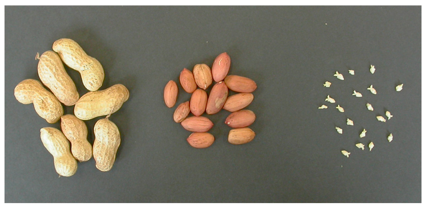
Arachis hypogaea L. cv. Virginia were provided by Florsilva Ansaloni (Italy). The oblong pods of about 3–7 cm long contained 2 seeds about 1–1.5 cm long, covered with yellowish-brown shells. In the seed, a 0.5–0.7 cm long embryo was located at the edge of the cotyledons, with the plumule (embryonic shoot) located toward the inner part of the seed and the radicle (embryonic root) toward the bottom of the cotyledon (
Figure 1). In a second experiment, optimized protocols were tested with the cvs. NC7, 7 × 77 and Com74, of which the mature seeds were provided by Akdeniz University (Antalya, Turkey). Among these, cv. NC7 was structurally the most similar to the cv. Virginia, while the pods, as well as rotund seeds of cvs. 7 × 77 and Com74, were slightly smaller in size and were covered with a brownish shell.
For conservation, pods were removed, bare seeds were checked for apparent symptoms of mold. Clean seeds were transferred to tightly closed glass jars and maintained at 4 °C in darkness until they were used. Maximum conservation time of the seeds never exceeded 2 years.
2.2. Decontamination of the Seeds and Explant Preparation
The seeds were soaked in 70% (v/v) ethanol solution for 5 min for surface sterilization. Then, the seeds were subjected decontamination with 10% (w/v) H2O2 (hydrogen peroxide) for 5 min followed by 20% (v/v) NaOCl for 20 min with 2–3 drops of Tween 20, both were followed by minimum 5-min rinse in sterile dH2O, repeated at least twice. Both cotyledons of the de-embryonated seeds served as explant source.
2.3. Determination of Seed Viability and Germination Rate
Decontaminated seeds were evaluated for viability, immediately after their supply as well as following their conservation, by TTC (2,3,5 tryphenyl tetrazolium chloride) test [
27]. Randomly selected seed samples of all varieties were soaked in water and maintained in darkness for 24 h. They were then soaked in 1% TTC solution (pH 7.4) and maintained at 30 °C for 24 h. Afterward, the seeds were washed with dH
2O, and the cotyledons were separated longitudinally to observe both the cotyledons and the embryo for the coloring. Accordingly, the viability of the tissues can be determined rapidly, within 48 h, by observing their color. In the present study, seeds (both the cotyledons and the embryos) that reflected red color (totally or partially) upon TTC test were recorded as ‘alive’. The percentage (%) of seed viability was calculated accordingly.
As different typologies of seed/embryo coloring were observed with TTC test of cv. Virginia, germination trials were conducted to confirm and support the results of the test. Therefore, decontaminated seeds, as well as excised embryos, were germinated in vitro on hormone-free MS medium [
28] at 22 ± 1 °C. They were maintained in darkness during the first 3 days and then under a 16/8 h photoperiod, with a light intensity of 60 µmol m
−2 s
−1, for the rest of the germination period. Both the seeds and the excised embryos were recorded as ‘germinated’ upon the emergence of the root and shoot primordia, respectively. De-embryonated cotyledons were then transferred to TIS. As in accordance with the TTC test, the germination trials were conducted upon the arrival of the seeds and at the end of their conservation.
2.4. In Vitro Propagation via Temporary Immersion System (TIS)
De-embryonated cotyledons of cv. Virginia were transferred into liquid media contained in PlantformTM bioreactor for the culture in TIS. To the MS media tested for the induction of direct organogenesis were added of 30 g/L sucrose, and one of two cytokinins, i.e., benzyladenine (BA) at 27.5, 55 or 100 µM, or thidiazuron (TDZ) at 5, 7.5 or 10 µM. Semi-solid MS media (solidified by the inclusion of 7 g/L agar) of the same compositions served as controls.
Several combinations of dry (in every 6, 8, 16, or 24 h) and medium immersion periods (for 8, 16, or 24 min) of TIS, corresponding to a total of 8, 12, 16, 24, 32, 36, 48, 64, 72, and 96 min daily immersions, were tested for the regeneration of de novo shoots on de-embryonated cotyledons. In addition to periodic immersions in liquid medium, the cultures were ventilated for 10 min every 4 h. For rooting, 4 weeks after the culture initiation, the shoots were detached from the cotyledons, and multiple shoots were separated and transferred to hormone-free semi-solid MS medium. After 3 weeks, rooted shoots were washed gently under running tap water and were transferred to commercial soil for outdoor plants (which was autoclaved previously at 121 °C for 20 min). Plastic containers were maintained in the climatic chamber at 22 ± 1 °C, under 16/8 h photoperiod for a week, during which the relative humidity was maintained by covering the plastic containers with a plastic bag and irrigating the plants once in every two days. Plastic bags were then removed gradually for the acclimatization of the plants, which were then transferred to greenhouse and irrigated regularly.
Optimized protocols of TIS culture with cv. Virginia were subsequently tested with the de-embryonated cotyledons of the other three varieties. All in vitro cultures were maintained in climatic chamber at 22 ± 1 °C, under 16/8 h photoperiod (60 µmol m−2 s−1) provided by cool, daylight fluorescent lights.
2.5. Data Collection and Statistical Analysis
A total of 20 randomly selected seeds of each variety were used for TTC test upon arrival and after conservation. Similarly, 40 randomly selected decontaminated seeds of cv. Virginia and 20 decontaminated seeds of NC7 and Turkish autochthonous varieties were used for germination trials upon arrival and after conservation.
Two replicates of 25 de-embryonated cotyledons of cv. Virginia were used for each cycle of TIS, and the experiment was repeated twice. Three replicates of 12 de-embryonated cotyledons of cv. Virginia were used as the control on semi-solid media, and the experiment was repeated twice. As for the other three varieties, the optimized protocols were tested using a sample of 30 explants both in TIS and semi-solid media.
During in vitro multiplication, the collected data consisted of (i) percentage of regenerating explants, (ii) average shoot number per regenerating explant, and (iii) average shoot length (cm). In addition, the Shoot Forming Capacity Index (SFC), formulated as SFC = (average shoot number per regenerating explant) × (% of regenerating explants)/100 [
29], was calculated to highlight the overall multiplication potential of each experiment. For rooting, the shoots were assembled in two separate groups according to their origin of multiplication; the ones that were propagated on semi-solid media and the ones that were propagated in TIS. Rooting data consisted of (i) percentage of rooting shoots and (ii) average root number. Rooted shoots were then transferred to soil and their acclimatization rate (%) was recorded after 1 month.
Statistical analysis of percentages was carried out by means of the test for homogeneity of proportions, and significant treatment differences selected by a non-parametric statistical test, the post hoc multiple comparisons test [
30], to compare multiple percentages. Discrete data were subjected to ANOVA, followed by the least significant difference (LSD) test at
p ≤ 0.05 to compare means.
4. Discussion
The type of the explant used in this study was de-embryonated cotyledons. The TTC test was applied to confirm the viability of the explants, which resulted in four different typologies of coloring: cotyledons and the embryo were colored entirely; cotyledons were colored entirely while the embryo was colored only at the plumule; cotyledons were colored partially while the embryo was colored entirely; and cotyledons were colored partially, and the embryo was colored only at the plumule. The increase in the percentage of partially colored cotyledons following seed storage at 4 °C in darkness can be attributed to the degradation of lipid and oil components of the seeds. This phenomenon was also investigated and discussed earlier by others, e.g., [
32,
33]. Nevertheless, the results of the present study demonstrate that even if the storage tissues were damaged, this did not interfere or impair the germination ability of peanut seeds and they germinated independently of the presence or integrity of the cotyledons.
In order to investigate the effect of the TIS bioreactor system on the de novo regeneration of
Arachis hypogeae L., de-embryonated cotyledons of cv. Virginia were transferred to the containers of Plantform
TM and subjected to several medium immersion regimes. Although one may expect a de-embryonated cotyledon to have minor morphogenic response due to its nutrient-storage nature for the zygotic embryo, as suggested also by [
34], it has been indeed reported, for a number of oilseed crops including peanut, that the cotyledons have a significant capacity for an organogenic growth response, see, e.g., [
34,
35,
36,
37,
38,
39,
40,
41,
42,
43,
44,
45]. However, previous studies with some other species (such as almond and cherry) also demonstrated that this capacity differs according to the location of the cotyledonary tissue and proximal region of the cotyledon (i.e., where the detached embryo was in contact with the cotyledon) displaying the highest regeneration capacity in comparison to the distal region [
46,
47,
48]. This phenomenon is in correlation with the outcomings of the present study, in which adventitious shoot primordia induction was always obtained in the proximal region. This can be attributed to the persistence of the regeneration capacity of the remaining cells and tissues of the detached embryo that are likely to be able to readily dedifferentiate into meristematic cells. Hence, previous reports and the outcomings of the present study support the idea that, just like the well-known explants such as apical and axillary nodes, internodes, leaf portions and roots, the cotyledon tissues can also be easily manipulated for in vitro regeneration.
The liquid media in the containers of TIS were MS media, added of one of the three concentrations of either BA (27.5, 55 or 110 µM) or TDZ (5, 7.5 or 10 µM). These concentrations of growth regulators, which are slightly higher than the standard value, were chosen inspired by other studies, e.g., [
30,
39,
43]. For instance, Pestana et al. [
39] cultivated cotyledons of cv. Tatu on MS medium supplemented with 110 µM BA. Burns et al. [
43] tested the effect of 10–80 µM BA for cotyledons of cvs. Frorida-07, Georgia Green, and VC-2, 160–640 µM for cv. New Mexico Valencia A, 160–320 µM for cv. Georgia Brown. Similarly, McKently et al. [
38] also reported that the optimum concentration of BA for shoot induction from the cotyledons of cv. Forigiant were higher than what is usually used in other plant species. In the present study, although there are some reverse examples, in general TDZ seems to provide a relatively higher percentage of regenerating explants and average shoot numbers; however, this distinction is not so clear for the average shoot length. As for different cytokinin concentrations, there is no linear correlation; increasing concentrations seems to be more efficient when applied in one immersion regime, and less efficient in another one. Yet, it can be concluded that an MS medium added to 110 µM BA or 10 µM TDZ are the most appropriate medium formulations in TIS.
During the preliminary experiments, liquid culture (in which the cotyledons were continuously and completely immersed in liquid media of the same composition and cultured on orbital shaker) was also tested. However, differently from the results of McKently et al. [
38], here no regeneration from those explants was achieved. The explants turned yellowish-brown and deteriorated shortly after the culture initiation. McKently and his co-workers, however, were able to obtain shoot formation both in liquid and solid media of the same composition, although the shoot production on solid media was approximately twice than in liquid and the shoots obtained in liquid media were vitreous.
The present study is the first example of the application of a TIS to peanuts. Thus, comparisons can be conducted on the results of the previous studies, where de-embryonated cotyledons of peanut were cultured on semi-solid MS media for de novo regeneration via direct shoot organogenesis. Among these, McKently et al. [
38] obtained 40% of regenerating explants of cv. Forigiant, with a maximum average shoot number of 12. When they applied the optimized protocol to other varieties, the shooting response ranged between 44 and 94%. Pestana et al. [
39] tested three different culture temperatures (25, 28, and 35 °C) for cv. Tatu and obtained the highest regeneration rate (56%) and average shoot number (24) when the highest culture temperature was applied. Maina et al. [
42] tested the regeneration efficiency of cotyledons of the African varieties and obtained the highest percentage of regeneration (35%) with cv. Chalimba. Radhakrishnan et al. [
40] obtained a maximum of 57% of regeneration from the de-embryonated cotyledons of cv. J11. Hoa et al. [
45] obtained 23.3% of regenerating explants with cv. L14, with 6.8 shoots in average for the regenerating explant. All these different results of the previous studies, as well as the results of the present study (which were conducted in a similar manner, using the same type of explant and similar growth regulator type and concentrations), not only reveal how the variety has an influence on shoot induction, but also the superiority of the TIS for de novo regeneration.
Rooting performance of the shoots was slightly higher for cv. Virginia and was significantly better for the other varieties when the shoots were previously multiplied in the TIS. This favorable influence was more evident in cvs. NC7 and 7 × 77. These results suggest that the culture conditions provided in the TIS increase the ability of the shoots to respond to external as well as internal hormonal stimuli, and thus a higher number of shoots readily gave rise to root meristemoids when they were cultured on an appropriate rooting medium. A similar trend was observed with other species as well (e.g., Anthurium spp., Carex oshimensis cv. Evergold, Ficus spp., and Nandina domestica; unpublished data).
5. Conclusions
Peanuts (Arachis hypogaea L.), a valuable source of herbal oil, proteins, minerals, vitamins, fibers, essential amino acids, and bioactive compounds, are an economically important species, consumed worldwide both for nutrition and pharmaceutical/medical uses. However, despite numerous attempts, this species still possesses a recalcitrant nature to in vitro manipulations. The present, study which aimed to develop an efficient protocol for the mass propagation of the species, tested the PlantformTM TIS for de novo regeneration using de-embryonated cotyledons of cv. Virginia, and obtained a significant enhancement in the percentage of regenerating explants and average number of shoots. The obtained shoots were healthy, without exhibiting any symptoms of hyperhydricity or any other abnormality associated with liquid culture, and were easily rooted and acclimatized. The selected medium immersion regimes of the TIS were then applied to cvs. NC7, 7 × 77, and Com74, and always provided a satisfactory result. Therefore, it can be concluded that, the present study, in which the use of a TIS was demonstrated for the first time in peanuts with very favorable results, will pave the way to further improve the in vitro propagation of not only peanuts but also other species with recalcitrant in vitro regeneration natures.


