Article

Analyzing Porcine Corneal Xenograft Compatibility: In Silico Insights on Graft Outcomes

by
Patricia Mae De Leon
1 and
Heherson Cabrera
2,*
1
School of Chemical, Biological, and Materials Engineering and Sciences, Mapúa University, Intramuros, Manila 1002, Metro Manila, Philippines
2
School of Health Sciences, Mapúa University, Brgy. Sta Cruz, Makati 1205, Metro Manila, Philippines
*
Author to whom correspondence should be addressed.
Transplantology 2024, 5(3), 193-207; https://doi.org/10.3390/transplantology5030019
Submission received: 16 June 2024 / Revised: 16 July 2024 / Accepted: 16 August 2024 / Published: 30 August 2024
(This article belongs to the Section Transplant Immunology and Immunosuppressive Drugs)

Abstract

:
Background: Corneal transplantation faces significant challenges due to the shortage in donor corneas. Porcine corneas have emerged as a potential solution due to their similarities in biomechanical properties with pigs, yet xenoimmune rejection poses an obstacle to their efficacy. Methods: In this study, in silico methods were employed to analyze the compatibility of porcine corneal xenografts, focusing on two key aspects: the comparison of corneal matrix proteins and investigation of the immunological mediators and pathways involved in corneal graft rejection. The amino acid sequences of the fourteen (14) most abundant proteins in the corneal matrix were compared to determine their structural and functional differences. The primary amino acid structures and compositions, theoretical pI, and grand average of hydropathicity were determined and compared between the two species. Results: In graft performance, similarities and differences between the donor and recipient tissues influence the success of transplantation. When the proteins closely resemble each other, in terms of structural characteristics and biochemical properties, the host’s immune system is less likely to recognize the tissue as foreign. The immunological mediators and pathways involved in corneal graft rejection were investigated, elucidating the mechanisms underlying xenograft incompatibility. Based on the results generated from STRING, the specific groups of molecules that are involved in the immune-mediated rejection process are costimulatory molecules, cytokines, immune checkpoint molecules, apoptosis regulators, cell adhesion molecules, growth factors, neuropeptides and hormones, certain receptors, the cytotoxic molecule GZMA, and the chemokine CCL5. Conclusions: The results of this study establish that the porcine cornea has a high suitability for corneal xenotransplantation into humans but requires immune-based therapeutic interventions to increase graft acceptance.

1. Introduction

Blindness and visual impairment are among the major health conditions worldwide, affecting over 2.2 billion individuals, half of whom harbor preventable or unaddressed eye-related concerns [1]. Corneal blindness, identified as the fourth leading cause of global blindness as per the World Health Organization, stands out as a prominent cause of preventable blindness [2]. Among the clinical treatments for corneal conditions, keratoplasty or corneal transplantation is the most common type of allogeneic transplantation, recognized for having high long-term success rates that are characterized by improvements in visual acuity, pain reduction, and eye structure maintenance [3,4]. The ultimate success of the surgical procedure, nonetheless, highly relies on the corneal state of the patient prior to transplantation. Recipients categorized as low- and high-risk receive different levels of treatment, with the latter sometimes denied of the medical operation or are required to follow successive immunosuppressive regimens [4,5].
While keratoplasty is a preferred treatment, an imbalance between the supply and demand of corneal tissues persists, particularly in developing countries. In the Philippines, for instance, only 500 to 800 transplantations a year are achieved, dependent upon the availability of corneal donors, as reported by the Eye Bank Foundation of the Philippines [6]. Corneal xenotransplantation, therefore, has been implemented using porcine corneas to address the donor shortage [5]. Numerous studies using acellular porcine corneal stroma (APCS) as xenografts suggest their potential as promising alternatives to traditional corneal grafts as they preserve the key architecture of the native corneal stroma [7,8]. They also facilitate the endogenous regeneration of corneal and neural cells, allowing for the maintenance of normal corneal transparency if matched with the existing extracellular matrix [7,9]. Confirming this efficacy, a similar study demonstrated APCS as an effective alternative to human corneal tissue as acellular implants enable the endogenous regeneration of corneal cells [7]. Nevertheless, the unavailability and inaccessibility of treatment and rehabilitation are significant barriers to managing corneal damage. Despite the diverse range of procedures available for corneal conditions, including keratectomy and keratoplasty, each approach poses several risks and limitations [8].
While considerable advancements have been made in the research on APCS, it is important to acknowledge that xenografts cannot perfectly replicate the biomechanical properties and microarchitecture of the native cornea. Despite anatomical, biomechanical, and chemical similarities between human and porcine cornea, xenogeneic tissue rejection persists due to the body’s innate and adaptive immune responses [10]. Corneal xenografts have the potential to prompt the recipient’s immune system to generate specific antibodies, subsequently mediating cytotoxic effects through complement-dependent mechanisms. Notably, xenograft rejection also exhibits more pronounced symptoms compared to allograft rejection [11]. To overcome immunological barriers, various methods of decellularization have been continually developed aimed at completely removing donor cells and antigens, thereby reducing the immune response from the host [12]. This essentially allows for the preservation of the biomechanical properties of the xenograft while diminishing the immune response associated with the antigenic properties of the xenogeneic components. However, the decellularization process does not entirely eliminate antigenic components, as evidenced by studies revealing that the remaining components are still capable of provoking an immune response [10]. This, in turn, can lead to xenograft failure.
Hence, this study proposes an in silico approach in determining the corneal compatibility between human and porcine corneas. The primary objective of this research was to comprehensively analyze the corneal matrix proteins and genetic markers of the donor and host corneas that are associated with corneal compatibility. Specifically, this study aimed to (a) analyze and compare the composition and distribution of corneal extracellular matrix (ECM) proteins in human and porcine corneas to elucidate the compatibility of porcine corneal grafts with the human corneal matrix, (b) characterize the immunological mediators and pathways associated with the graft rejection process to understand the underlying mechanisms, and (c) expound on the immunological mediators to guide the development of targeted interventions for the overall improvement of graft acceptance.
Pigs are remarkable in the sense that they more closely resemble the physiology and organs of humans compared to other animals, hence why they are often used in medical research for human health and diseases. With the global shortage of human donor corneas, pigs have emerged as a viable alternative source. This research holds significant implications on corneal xenotransplantation by understanding the intricacies of corneal matrix proteins and genetic markers both in human and porcine through the use of different bioinformatics tools. It is important to understand immunological similarities between the two species, by studying their matrix proteins and genetic markers, associated with corneal compatibility to mitigate the risk of immune rejection. Through elucidating the molecular and genetic facets, the findings of this research may provide a transformative path toward developing safe and effective porcine corneal alternatives, addressing the dire need for accessible and sustainable solutions in corneal transplantation.
This research centers on exploring corneal matrix proteins and genetic markers in human and porcine corneas utilizing bioinformatic tools, whether accessible online or as software applications. The bioinformatic approach allows for a detailed investigation into the molecular and genetic intricacies, potentially influencing the design of future xenotransplantation strategies. While this research offers valuable insights, certain limitations are inherent. Its primarily reliance on bioinformatic tools indicates that the accuracy of the results is contingent upon the quality of data and algorithms employed since in silico analyses might not fully replicate the dynamic in vivo conditions. Furthermore, since this study solely focused on the comparison of porcine against the human cornea, it may not fully encompass the complete spectrum of interspecies differences. The methods employed are limited to those mentioned in the methodology section.

2. Materials and Methods

2.1. Corneal Matrix Proteins

The corneal stroma constitutes a complex tissue predominantly composed of collagen fibrils, proteoglycans, and additional ECM constituents. The proteins within the stroma play a role in maintaining its transparency, mechanical strength, and structural integrity. Notable proteins abundant in the typical human corneal stroma include collagen types I, V, and VI, along with proteoglycans such as keratocan, lumican, decorin, and biglycan. Glycoproteins like fibronectin, laminin, and fibrillin-1 contribute significantly to maintaining the cornea’s structural stability and transparency. They also mediate cell adhesion, migration, and signaling processes for corneal homeostasis and wound healing [13,14]. Hence, the aforementioned proteins underwent the following analytical methodologies aimed at evaluating corneal compatibility.
For the collagen types, their main chain compositions were chosen. Collagen type I is composed of two pro-α1(I) and one pro-α2(I) chain; hence, COL1A1 and COL1A2 sequences were acquired. The COL5A1 and COL5A1 sequences were obtained since collagen type V is made up of three or two pro-α1(V) and one pro-α2(V) chains. As for collagen type VI, its basic structural unit involves a heterotrimer of the α1(VI), α2(VI), and α3(VI) chains, so COL6A1 to COL6A3 sequences were procured.

2.1.1. Acquisition of Protein Sequences

The amino acid sequences of the proteins mentioned, for both human (Homo sapiens) and porcine (Sus scrofa), were acquired from the UniProt database (https://www.uniprot.org/ (accessed on 12 January 2024)). The sequences of each protein were selected based on the first result listed on the website, as indicated by the default sorting of results. In cases where the canonical sequences are not available, the isoforms were obtained from the same database Table S1.

2.1.2. Sequence Alignment

Basic Local Alignment Search Tool (BLAST) is a widely used bioinformatic tool employed for comparative analyses of primary biological sequence data, which encompass nucleotide or protein sequences [15]. The amino acid sequences of each protein from the porcine were compared to those of the human using the BLAST algorithm (https://blast.ncbi.nlm.nih.gov/Blast.cgi (accessed on 15 January 2024)). The blastp algorithm modality was used since it specializes in the comparison of protein sequences. The parameters for the analyses using blastp were set to default.
The BLAST algorithm conducts a comparative analysis of the protein sequences and computes the statistical significance of the observed matches by finding local similarities between the sequences. For protein-to-protein sequence comparison, the specific program used was “blastp” [15]. Blastp was used to identify and compare the amino acid sequences of the most abundant proteins present in the corneal matrix. In interpreting the blastp results, it is important to understand that there are no universally accurate criteria as to determine which of parameters should be covered. Hence, considering the nature of this research paper, the following parameters were considered: query cover, expected (E) value, identity, positives, and gaps.

2.1.3. Physicochemical Analysis

For further analysis, the proteins were subjected to the ProtParam tool of the ExPASy Bioinformatics Resource Portal (https://web.expasy.org/protparam (accessed on 15 January 2024)) to calculate the physiochemical properties of the proteins. The calculated parameters that were considered relevant in the context of the study were the theoretical pI, amino acid composition, and the grand average hydropathicity (GRAVY). The GRAVY value is a numerical value that represents the overall hydrophobicity or hydrophilicity of a protein sequence. The theoretical isoelectric point (pI), on the other hand, refers to the estimated pH at which a protein carries no net electrical charge.

2.2. Immunological Descriptors Analysis

Corneal graft rejection primarily occurs due to alloimmune responses, where the recipient’s immune system recognizes the transplanted cornea as a foreign tissue. In the rejection process, immunological mediators orchestrate the activation and recruitment of immune cells involved in graft rejection [16]. Analyzing immunological mediators in the context of graft rejection provides insights into the underlying mechanisms of graft failure.
In this study, the proteins associated with corneal graft rejection were determined using the Search Tool for the Retrieval of Interacting Genes/Proteins (STRING) database (https://string-db.org/ (accessed on 18 January 2024)). STRING collects and integrates data from various sources to create a network of known and predicted protein interactions. In order to retrieve the proteins, the search database used was “Pathway/Process/Disease/Publication”, where the search term was “corneal graft rejection”, filtered for genesets found in Homo sapiens. The genesets were derived from the publication entitled Immunopathogenesis of corneal graft rejection [17]. The results derived from STRING in terms of the Kyoto Encyclopedia of Genes and Genomes (KEGG) pathways involved were narrowed down by selecting those that are involved in porcine corneal xenograft rejection (as listed in Figure S1).

3. Results

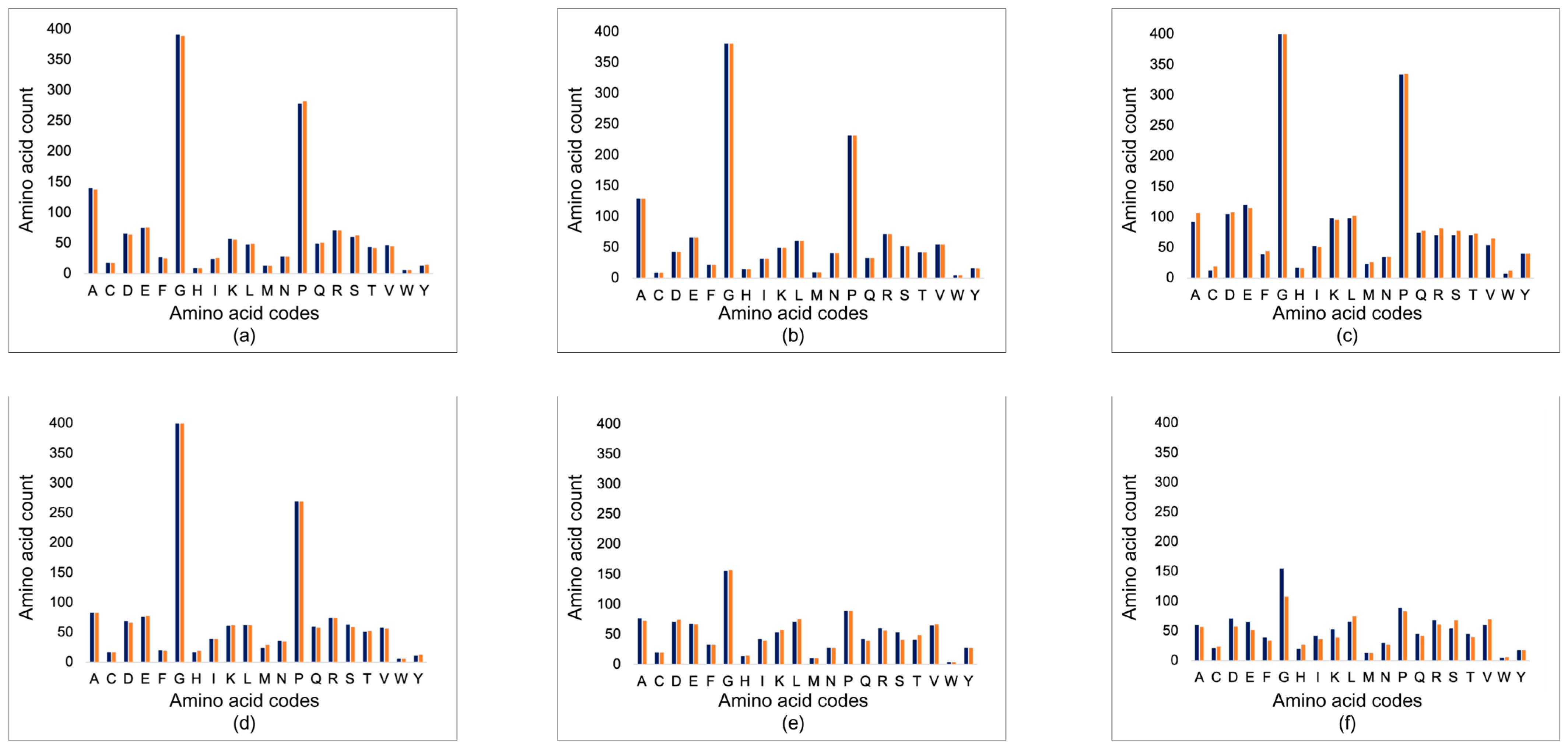
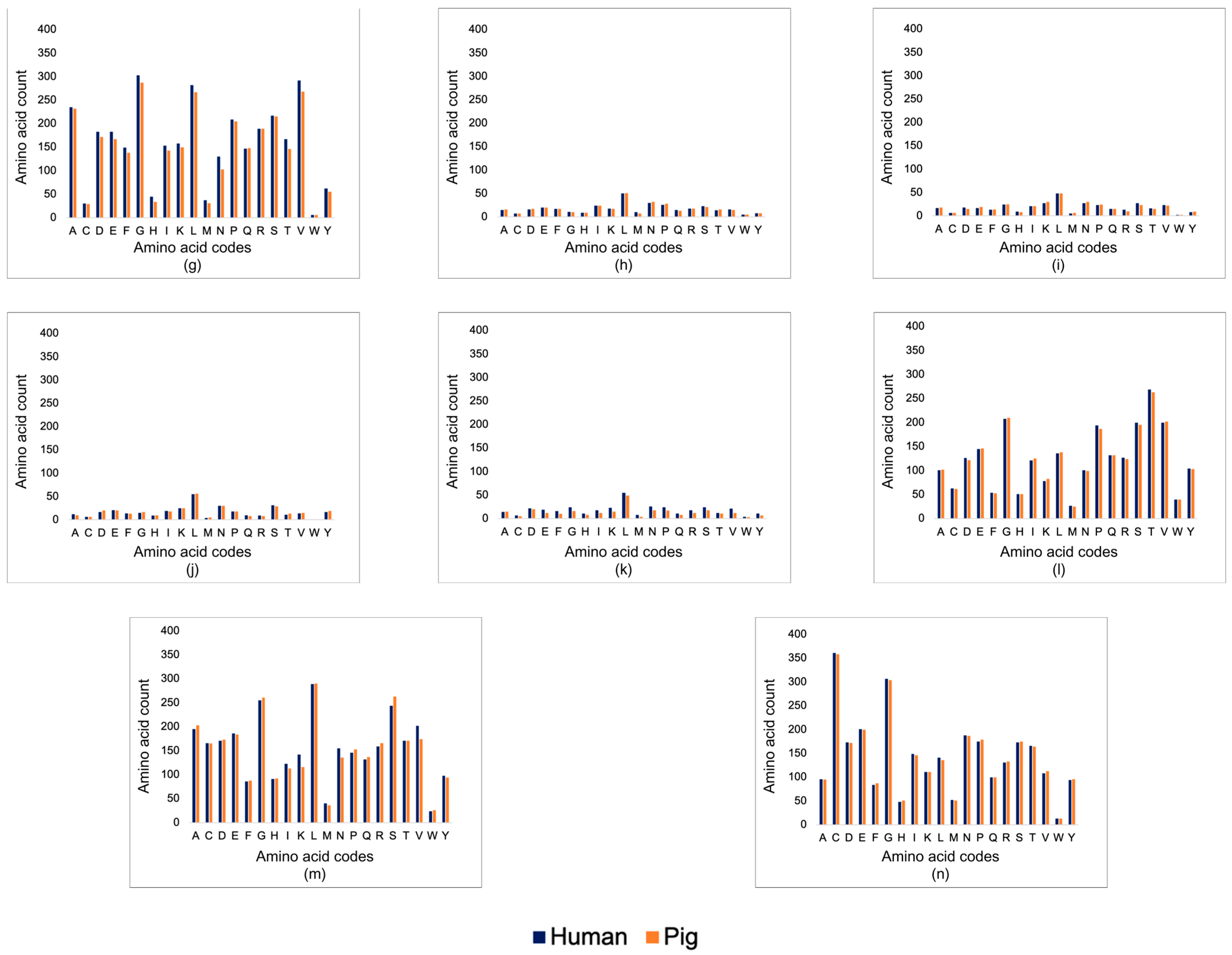
3.1. Corneal Matrix Protein Analysis

The specific proteins analyzed in the study were (a) COL1A1, (b) COL1A2, (c) COL5A1, (d) COL5A2, (e) COL6A1, (f) COL6A2, (g) COL6A3, (h) keratocan, (i) lumican, (j) decorin, (k) biglycan, (l) fibronectin, (m) laminin, and (n) fibrillin-1.

3.1.1. Basic Local Alignment Search Tool (BLAST)

A criterion for the identity parameter was established solely within the context of the objectives outlined in this research paper. As provided in Table 1, the range of identity values fell within specific categories to define the alignment between the query and subject sequences.
The query cover is the extent to which the query sequence aligns with the subject sequence. In this case, the query sequences are the protein sequences from the pig, whereas the subject sequences are those obtained from the human. A high query coverage indicates a substantial alignment between the compared sequences, while a low query coverage indicates either partial alignment or sequence divergence. The E-value, on the other hand, represents the number of alignments that are expected to occur by chance in a database. In simpler terms, the E-value essentially represents the “random background noise”. As the scores of the match increase, the E-value decreases. This is often used to assess the significance of sequence similarities. The similarities, however, are more specifically elucidated by the identity—the percentage of the protein that is the same between the two sequences. A more straightforward measure of sequence similarity is the proportion of identical amino acids in the aligned regions. Moreover, the positives and gaps are essential parameters in sequence alignment. The positives correspond to either the residues that are identical between the two sequences, or residues that are not necessarily identical but have similar chemical properties. In contrast, the gaps represent insertions or deletions (indels) in the alignment. A significant gap value may suggest sequence divergence or variation [18].
In terms of the query cover, a majority of the proteins had a sequence coverage of 99–100%, indicating that the query sequences share extensive similarities across their entire lengths with the subject sequences (as shown in Table 2).
As for the collagens, the percent identities of all chains of collagen type I (COL1A1, COL1A2) collagen type V (COL5A1, COL5A2), and the a-1 chain of collagen type VI (COL6A1) all had high sequence alignments (as shown in Table 2). The a-2 and a-3 chains of collagen type VI (COL6A2, COL6A3), conversely, had a relatively low percent identity that suggests sequence similarities falling within the moderate classification. As for the proteoglycans, all of them had lower percent identities compared to the collagens, with keratocan and decorin having percent identities that had high alignments. The biglycan, with a percent identity of 55%, suggests that only approximately half of the residues within the aligned regions between the query and subject sequences are identical. The glycoproteins fibronectin and fibrillin have high sequence similarities, whereas laminin has a moderate sequence alignment.

3.1.2. Structural Analysis

In terms of the collagens (as seen in Figure 1), the abundance of amino acids was relatively similar between human and pig, especially among the main amino acids that make up the different types of collagens (glycine, proline, and alanine, among others). However, for COL6A2 and COL6A3, the amino acid contents of pig have a significant difference compared to those of human. For the proteoglycans and glycoproteins, except biglycan, there are no significant differences between the amino acid composition between human and pig. The amino acids of the biglycan from pig are significantly lower compared to that from human, in terms of L, N, G, P, and S.
The ProtParam analysis revealed the GRAVY values of the pig and human sequences having negative values, indicating that the proteins are hydrophilic (as shown in Table 3). Upon comparison, the human and porcine corneal matrix proteins have relatively similar GRAVY values, suggesting the suitability of porcine corneal xenograft for transplantation. On the other hand, the theoretical pI of the proteins between human and pig has relatively similar values, except for COL6A2, COL6A3, keratocan, lumican, and biglycan.

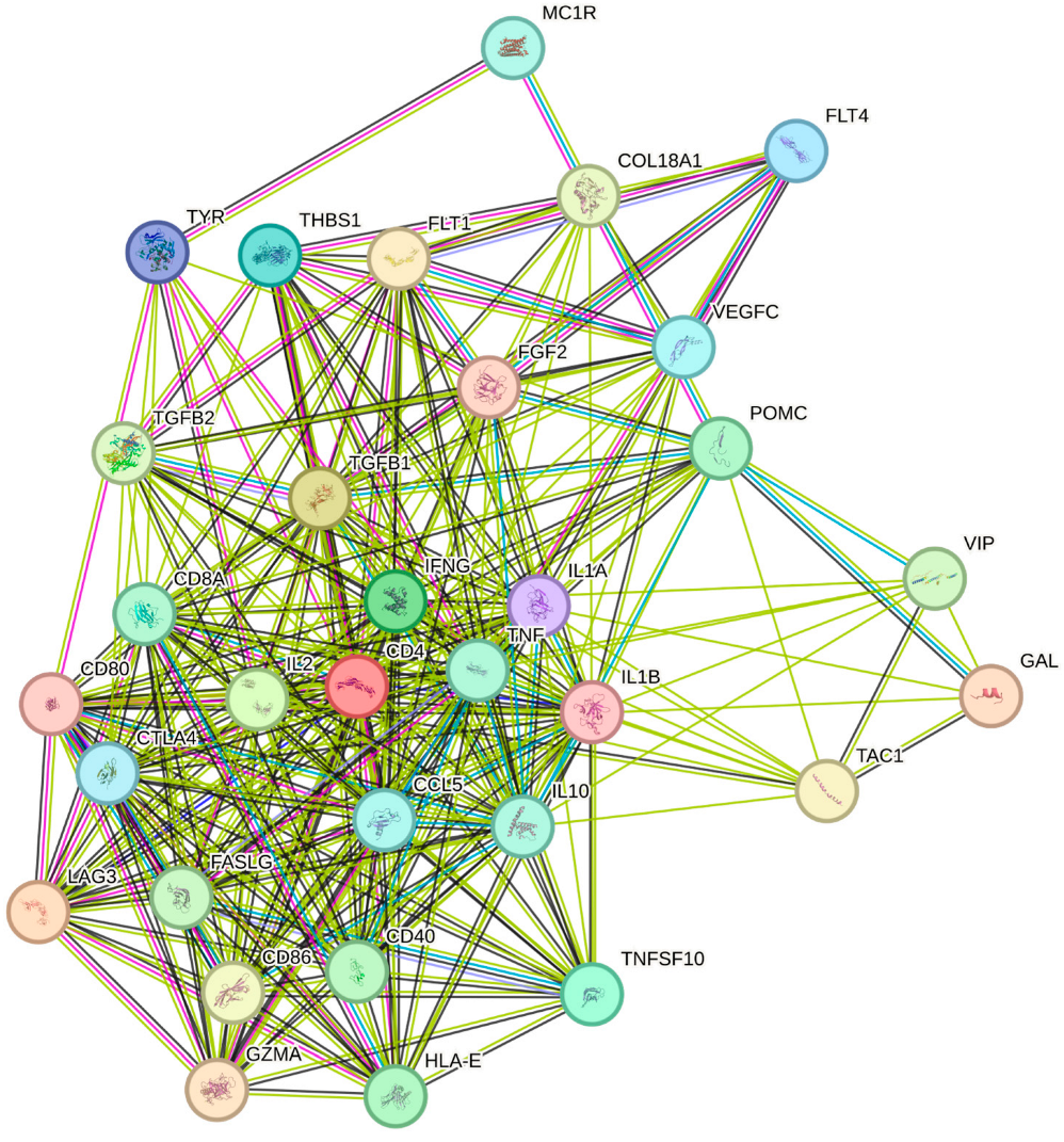
3.2. Immunological Mediators and Pathways

The STRING database shows the thirty-two (32) proteins involved in corneal graft rejection in human recipients. Among them are costimulatory molecules (CD40, CD80, CD86), cytokines (IL1A, IL1B, IL2, IL10, TNF, IFNG, VIP), immune checkpoint molecules (LAG3, CTLA4), apoptosis regulators (TNFSF10, FASLG), cell adhesion molecules (THBS1, COL18A1), growth factors (FGF2, VEGFC), neuropeptides and hormones (GAL, POMC, TAC1), receptors (FLT1, FLT4, TYR, MC1R, CD4, CD8A, HLA-E), cytotoxic molecules (GZMA), and chemokines (CCL5) (as depicted in Figure 2).
The specific groups of molecules that are involved in the immune-mediated rejection process are the costimulatory molecules, cytokines, immune checkpoint molecules, apoptosis regulators, receptors, chemokines, and growth factors. In graft rejection, they are related to multiple pathways. Among the results for the KEGG pathways from the STRING database, Table 4 shows the pathway descriptions related to xenograft rejection and the specific molecules involved. Immunological mediators as listed provide insights into the mechanisms underlying graft rejection.
The comparisons of other protein–protein association networks of other grafts shown in Figure 3 reveal distinct molecular landscapes, showing unique patterns or interactions for each type. This avoids the generalization of therapeutic approaches and underlines the specificity at which immune mediators or structural proteins must be targeted for successful integration.

4. Discussion

4.1. Corneal Matrix Protein Analysis—Sequence and Structure

While there are some sequences that range from 92 to 98%, this is still indicative of significant conservation between the sequences. This suggests that the important regions, both structurally and functionally, are more likely to have been represented in the alignment. Moreover, the E-values of close to or equal to 0.0 suggest that the query and subject sequences of the given proteins are an extremely significant match. In the context of determining graft compatibility, the aligned regions can potentially include functionally relevant domains or motifs that are associated with graft integration or rejection. Such domains within a protein allow particular functions, such as enzyme catalysis and protein–protein interactions, among others. Given the results of the query cover and E-value, this suggests that the conserved domains and motifs across the aligned region maintained the functional elements necessary for different biological processes. This is indicative of potential compatibility between the graft and recipient tissues, as the grafted tissue may retain essential functions and integrate effectively with the host environment.
A study explored the various characteristics of proteins that influence their ability to elicit antigenic and specific immune responses [19]. Their results suggested that a protein’s structure and composition, not the arrangement or sequence of amino acids within the protein, affect the protein’s interaction with the immune system. Thus, despite attempts at removing cells and debris from xenogeneic tissues through decellularization, the protein scaffold is still capable of expressing antigens that trigger the host immune response.
The comparative analysis of the amino acid sequences of the proteins abundant in the corneal stroma reveals areas within the sequences that exhibit conservation or divergence. In the context of graft performance, when a porcine corneal graft is transplanted into a human, similarities in the sequences between the donor and recipient tissues can influence the success of transplantation. When the sequences in the donor tissue closely resemble those of the recipient’s, the immune system is less likely to recognize the tissue as foreign. Despite differences in the sequence of amino acids, proteins can still retain similar functions or structural features as indicated by the aligned amino acids in the sequence. Moreover, it is important to consider the likelihood of cross-reactivity and immune tolerance. The concept of cross-reactivity dates to 1959, when a hypothesis was proposed to explain the function of immunological mechanisms in homograft rejection. Essentially, it states that the immune system of the recipient may recognize and respond to similar, not identical, antigens present in the donor proteins [20]. This assumption can be hypothesized for xenograft transplantations, wherein cross-reactivity may be responsible for reducing the risk of rejection. Higher percent identities indicate a stronger likelihood of cross-reactivity as opposed to those with lower identity values [21]. This can be further supported and verified using assays or sample-specific laboratory experiments.
However, the differences in the sequence alignments between the corneal proteins can potentially activate an immune response. The immune system is triggered when the differences in alignment are recognized as foreign, eliciting a response against the pig corneal tissue that leads to graft rejection. Hence, with the identity scores of the alignments ranging from low to high (as observed in Table 2), the transplantation of a corneal graft can result in the presentation of foreign peptides by antigen-presenting cells. This is more likely with the proteins having low to moderate sequence similarity scores—COL6A2, COL6A3, lumican, biglycan, and laminin.
While BLAST provides insights into homologous regions between the corneal matrix proteins of humans and pigs, the results are incapable of considering changes in the sequences that might induce alteration in protein structure and function. The structures of proteins are highly specialized to perform specific functions such that changes in a few atoms in one amino acid, or even changing a single amino acid, can cause such extensive disruption to the entire molecule that its functionality is completely compromised [22]. However, in some cases, changes in the amino acid sequences have no discernible effect on a given protein’s function.
In the context of xenograft transplantation, the recipient’s immune system recognizes foreign proteins as “non-self”, triggering an immune response aimed at eliminating the threat, which can result in the rejection of the transplant. Essentially, the immune system relies on specific shapes and configurations of the proteins to recognize them as foreign. If the proteins in the transplanted tissue are different enough from those of the recipient, the immune response can lead to the rejection of the xenograft, rendering the transplant ineffective or even harmful to the recipient. Hence, the amino acid constituents of the proteins were analyzed using ProtParam.
The lower G and P content and higher L content in COL6A2 of pig indicate changes in the structural motifs, where structural flexibility is reduced, and it becomes less hydrophilic (as indicated by the GRAVY value in Table 3). As for COL6A3, the G, V, and L of human is significantly higher than that of porcine. This means that the protein from pig is less hydrophilic (more hydrophobic); however, there is no significant difference between the GRAVY values of the two species. Although the higher V and L would usually indicate a difference in hydropathicity, it is possible that there might be compensatory changes with other amino acids that have similar properties. As for the proteoglycans and glycoproteins, the lower G and P content reduced conformational changes, and the L, N, and S content indicate more hydrophilicity. However, the GRAVY results show that the biglycan from human has a higher magnitude of hydrophilicity. This is because there is higher content of the hydrophilic amino acids (such as K, R, E, and D). Essentially, the hydropathicity of the proteins does not solely rely on the amino acid constituents, while they do have a significant effect, because of the compensatory changes with other amino acids or secondary structure differences. While the amino acid composition is a major determinant of the GRAVY value, the complexity of the protein structure and function elucidates that other factors influence the overall hydropathicity.
Having discussed the hydropathicity of the amino acids, the overall hydrophobicity or hydrophilicity of a protein sequence is represented by the Grand Average of Hydropathy (GRAVY) value. In a study, the authors used hydrophilicity analysis to determine potential antigenic determinants by analyzing the sequences of proteins. The study revealed that regions that have high local hydrophilicity are linked to antigenic determinants [23].
In this study, the ProtParam analysis reveals the GRAVY values of the pig and human sequences for the proteins. With all proteins having negative values, this indicates that the proteins are hydrophilic. Upon comparing the results, the human and porcine corneal matrix proteins have relatively similar GRAVY values, suggesting the suitability of porcine corneal xenograft for transplantation. The differences between the values can be explained by the amino acid constituents and their hydropathicity, as explained earlier. With the significant similarity between porcine and human in terms of their proteins, it is important to understand that the variations in amino acid contents influence intra- and inter-molecular interactions. Cellular response relies on spatial interactions between the cell-binding proteins and the functional groups present in the xenogeneic scaffold. Hence, if the scaffold’s surface properties closely match those of the native tissue, natural interactions may be promoted between the transplanted tissue and the host environment. The cells in the host environment will also less likely recognize the scaffold material as foreign if the recipient’s properties resemble those of the host tissue.
The theoretical pI, on the other hand, influences interactions through the surface charge properties of proteins that may be involved in the cell–scaffold interaction process. Like with GRAVY values, a study has revealed that the pI can be linked with the antigenicity of a protein [24]. According to the results in Table 3, the theoretical pI of the proteins between human and pig has relatively similar values, except for COL6A2, COL6A3, keratocan, lumican, and biglycan. The surface charge of proteins can influence their adsorption onto the scaffold material’s surface, which, in turn, can affect the behavior of cells interacting with the scaffold. To put it simply, the corneal matrix proteins can adsorb onto biomaterial surfaces, affecting cell adhesion, migration, proliferation, and differentiation. The adsorbed protein layer can then modulate host immune response and tissue integration of the biomaterial [25]. Therefore, significant differences in the pI values of the proteins may influence the overall success of corneal tissue transplantation.
Essentially, the results of the ProtParam analysis complement those of the sequence alignment results in BLAST. It is important to note, however, that the immune response is not solely based on the variations in amino acid composition, pI values, GRAVY values, and the like. The immune response to transplanted tissues involves a complex interplay of factors, including antigen presentation, inflammatory reactions, and tissue compatibility. While the results of the analyses expound on the antigenicity of the proteins based off similar and differences in the sequences, the approach in this study is solely dependent on the composition of corneal proteins between pig and human.

4.2. Immunological Mediator Analysis

The cornea is one of the structures of the eye that is considered to be an immune-privileged site. Immune privilege is a protective mechanism with which vital structures are safeguarded from the potentially damaging effects by inflammatory responses directed against pathogens. In the eyes, the normal immune response of the body is limited to maintain its vision despite being constantly exposed to potential threats from the external environment. The mechanisms involved are the absence of lymphatic vessels in certain parts, weak expression of major histocompatibility complex (MHC) antigens, expression of the Fas ligand (FASLG, CD95L0) and programmed death ligand 1 (PDL1), immature antigen-presenting cells (APCs), and the presence of immunomodulating molecules like IL10 and TGF-beta, among others. However, despite this, the immune privilege of the eye is relative. The mechanism falters due to several factors, such as surgical trauma, inflammation, infections, and neovascularization. As a result, this leads to the rejection of a graft, more so in the case of a corneal xenograft [26].
In analyzing the rejection process of corneal grafts, various molecular interactions and immune responses are involved. Transplants from pigs to humans are subject to vigorous immunologic rejection involving both innate and adaptive immune responses [27]. Figure 2 shows molecules that are considered immune components triggered by the introduction of a xenograft to the recipient. Essentially, what happens during the introduction of the donor graft is that the proteins present on the ECM, as discussed in earlier parts, are recognized as “non-self” or foreign due to their differences in terms of sequence alignments. The APCs encounter these foreign proteins, process them into smaller peptides, and present them on their surfaces using MHC molecules. These peptides are then recognized by the immune system as intruders and set off a chain reaction that leads to an immune response, leading to either hyperacute or acute graft rejection.
The immune-mediated destruction of corneal transplants, whether from the same species (allografts) or different species (xenografts), primarily involves the attack of CD4+ T cells on corneal endothelial cells. CD8+ T cells and natural killer T cells may also play a role, particularly if CD4+ T cell function is compromised. Unlike human endothelium, porcine endothelium consistently expresses certain molecules that can fully activate human T cells, possibly leading to more intense rejection reactions. In cases of allograft rejection, the dominance of genes encoding for IL-2 and IFN-γ highlights the involvement of the Th1 response, although there is also significant expression of Th2 cytokine genes.
Xenograft rejection, on the other hand, seems to be driven by proinflammatory cytokines, especially IFN-γ, released by CD4+ and/or CD8+ T cells at the graft site. The immune response occurs indirectly, where foreign antigens are presented to T cells by the host’s APCs. During corneal transplantation, the initial stage involves the presentation of donor antigens to naive T cells in lymph nodes by host APCs like dendritic cells expressing MHC-II and co-stimulatory molecules. The subsequent activation of T cells in draining lymph nodes, their migration to the cornea, the recognition of donor MHC antigens, and the release of proinflammatory cytokines lead to inflammation and tissue damage. This process involves CD8+ cytotoxic T lymphocytes and CD4+ T-helper lymphocytes secreting IL-2, IFN-gamma, and lymphotoxin, which recruit immune cells, ultimately causing tissue damage and the formation of memory T cells for a stronger immune response upon re-exposure to antigens [17,26].
These findings, supported by the results generated by STRING, can guide strategies to modulate or suppress the immune response to promote graft acceptance. For instance, a study by Sykes et al. discussed how the inhibition of C5, CD14, and TLR4 is capable of controlling the release of cytokines, thereby reducing inflammatory response during pig-to-human corneal xenotransplantation. The results concluded that C5 blockade inhibited cytokines in native porcine corneas, CD14 blockade inhibited cytokines in decellularized porcine corneas, complement C5-CD14 was able to reduce cytokines in all xenografts, and C5 blockade paired with TLR4 inhibitor showed the most promise in reducing inflammatory responses induced by corneal transplantation [28]. Another application is a study on how costimulatory blockades are used to induce immune tolerance in liver transplantation. Among well-studied costimulatory pathways are CD28/B7, ICOS/ICOSLG, and CD40/CD154, which are suppressed to inhibit T-cell activation and proliferation, thereby prolonging the survival of the liver transplant [29].
Hence, comparing the protein–protein association networks of other transplantations or grafts (as shown in Figure 3) reveals distinctive molecular landscapes inherent to each transplant type. The differences in molecular compositions and pathway configurations of each transplant imply that therapeutic strategies designed for one transplant may not be universally applicable for all types. This suggests tailored therapeutic intervention to induce immune tolerance during corneal xenotransplantation while minimizing off-target effects. Hence, given the immunological mediators and pathways in Table 4, effective interventions may include the costimulatory blockade of B7 (CD80/CD86) molecules and their T-cell ligands CD152 (CD28, CTLA4), the stimulation of FASLG for the activation of T-cell apoptosis, or the introduction of CTLA4-immunoglobulin to inhibit T cell activation, among others. However, the discussion of appropriate interventions for corneal xenograft rejections falls outside the scope of this study and requires separate and comprehensive investigations.

5. Conclusions

The in silico analyses conducted in this study elucidate the compatibility of porcine corneas as an alternative to human corneal grafts to address the shortage of donors. The comparison of corneal matrix proteins and investigation into the immunological mediators and pathways explain various factors that influence corneal xenograft compatibility. Structural and biochemical similarities enhance the prospects of a successful introduction of a graft, while the differences trigger alloimmune responses. The identified immune mediators and pathways offer insights into the complexities of the rejection process. The findings suggest that porcine corneas are suitable alternatives but require immune-based interventions to enhance acceptance.

Supplementary Materials

The following supporting information can be downloaded at: https://www.mdpi.com/article/10.3390/transplantology5030019/s1, Table S1: UniProt entry IDs for the protein sequences; Figure S1: KEGG pathways for corneal graft rejection from STRING database.

Author Contributions

Conceptualization, H.C.; methodology, P.M.D.L.; software, P.M.D.L.; validation, H.C.; formal analysis, P.M.D.L.; investigation, P.M.D.L.; data curation, P.M.D.L.; writing—original draft preparation, P.M.D.L.; writing—review and editing, H.C.; visualization, P.M.D.L.; supervision, H.C. All authors have read and agreed to the published version of the manuscript.

Funding

This research received no external funding.

Institutional Review Board Statement

Not applicable.

Informed Consent Statement

Not applicable.

Data Availability Statement

The data presented in this study are available in https://www.mdpi.com/article/10.3390/transplantology5030000/s1.

Conflicts of Interest

The authors declare no conflicts of interest.

References

  1. World Health Organization. Vision Impairment and Blindness; World Health Organization: Geneva, Switzerland, 2022; Available online: https://www.who.int/news-room/fact-sheets/detail/blindness-and-visual-impairment (accessed on 22 December 2023).
  2. Colby, K.; Dana, R. Foundations of Corneal Disease: Past, Present and Future, 1st ed.; Springer: Cham, Switzerland, 2020. [Google Scholar] [CrossRef]
  3. Amiri, F.; Ghiyasvandian, S.; Navab, E.; Zakerimoghadam, M. Corneal transplantation: A new view of life. Electron. Physician 2017, 9, 4055–4063. [Google Scholar] [CrossRef]
  4. Fuchsluger, T.A.; Kruse, F.E. High-Risk Corneal Transplantation. Cornea ESASO Course Ser. 2015, 6, 74–80. [Google Scholar] [CrossRef]
  5. Isidan, A.; Liu, S.; Li, P.; Lashmet, M.; Smith, L.J.; Hara, H.; Cooper, D.K.C.; Ekser, B. Decellularization methods for developing porcine corneal xenografts and future perspectives. Xenotransplantation 2019, 26, e12564. [Google Scholar] [CrossRef]
  6. Eye Bank Foundation of the Philippines. Who We Are. 2019. Available online: https://eyebankphil.org/about/who-we-are/ (accessed on 22 December 2023).
  7. Li, S.; Deng, Y.; Tian, B.; Huang, H.; Zhang, H.; Yang, R.; Zhong, J.; Wang, B.; Peng, L.; Yuan, J. Healing characteristics of acellular porcine corneal stroma following therapeutic keratoplasty. Xenotransplantation 2020, 27, e12566. [Google Scholar] [CrossRef]
  8. Zhou, Q.; Guaiquil, V.H.; Wong, M.; Escobar, A.; Ivakhnitskaia, E.; Yazdanpanah, G.; Jing, H.; Sun, M.; Sarkar, J.; Luo, Y.; et al. Hydrogels derived from acellular porcine corneal stroma enhance corneal wound healing. Acta Biomater. 2021, 134, 177–189. [Google Scholar] [CrossRef]
  9. Lin, Y.; Zheng, Q.; Hua, S.; Meng, Y.; Chen, W.; Wang, Y. Cross-linked decellularized porcine corneal graft for treating fungal keratitis. Sci. Rep. 2017, 7, 9955. [Google Scholar] [CrossRef] [PubMed]
  10. Sharifi, R.; Yang, Y.; Adibnia, Y.; Dohlman, C.H.; Chodosh, J.; Gonzalez-Andrades, M. Finding an Optimal Corneal Xenograft Using Comparative Analysis of Corneal Matrix Proteins Across Species. Sci. Rep. 2019, 9, 1876. [Google Scholar] [CrossRef] [PubMed]
  11. Song, Y.W.; Pan, Z.Q. Reducing porcine corneal graft rejection, with an emphasis on porcine endogenous retrovirus transmission safety: A review. Int. J. Ophthalmol. 2019, 12, 324–332. [Google Scholar] [CrossRef] [PubMed]
  12. Yoeruek, E.; Bayyoud, T.; Maurus, C.; Hofmann, J.; Spitzer, M.S.; Bartz-Schmidt, K.U.; Szurman, P. Decellularization of porcine corneas and repopulation with human corneal cells for tissue-engineered xenografts. Acta Ophthalmol. 2012, 90, e125–e131. [Google Scholar] [CrossRef]
  13. Dyrlund, T.F.; Poulsen, E.T.; Scavenius, C.; Nikolajsen, C.L.; Thøgersen, I.B.; Vorum, H.; Enghild, J.J. Human cornea proteome: Identification and quantitation of the proteins of the three main layers including epithelium, stroma, and endothelium. J. Proteome Res. 2012, 11, 4231–4239. [Google Scholar] [CrossRef]
  14. Espana, E.M.; Birk, D.E. Composition, structure and function of the corneal stroma. Exp. Eye Res. 2020, 198. [Google Scholar] [CrossRef] [PubMed]
  15. Wheeler, D. Blast Quickstart. In Comparative Genomics: Volumes 1 and 2; Humana Press: Totowa, NJ, USA, 2007. Available online: https://www.ncbi.nlm.nih.gov/books/NBK1734/#:~:text=The%20Basic%20Local%20Alignment%20Search,statistical%20significance%20of%20the%20matches (accessed on 15 January 2024).
  16. Hamrah, P.; Qazi, Y. Corneal Allograft Rejection: Immunopathogenesis to Therapeutics. J. Clin. Cell. Immunol. 2013, 2013 (Suppl. S9), 6. [Google Scholar] [CrossRef]
  17. Maharana, P.K.; Manda, S.; Kaweri, L.; Sahay, P.; Lata, S.; Asif, M.I.; Nagpal, R.; Sharma, N. Immunopathogenesis of corneal graft rejection. Indian J. Ophthalmol. 2023, 71, 1733–1738. [Google Scholar] [CrossRef] [PubMed]
  18. Chan Zuckerberg Initiative. A Guide to BLAST. Chan Zuckerberg ID. 2023. Available online: https://chanzuckerberg.zendesk.com/hc/en-us/articles/360050963352-A-Guide-to-BLAST#:~:text=Interpreting%20BLAST%20Results,accuracy%20of%20a%20given%20hit (accessed on 26 January 2024).
  19. Wang, Y.; Wu, W.; Negre, N.N.; White, K.P.; Li, C.; Shah, P.K. Determinants of antigenicity and specificity in immune response for protein sequences. BMC Bioinform. 2011, 12, 251. [Google Scholar] [CrossRef]
  20. Fujii, G.; Nelson, R.A. The cross-reactivity and transfer of antibody in transplantation immunity. J. Exp. Med. 1963, 118, 1037–1058. [Google Scholar] [CrossRef] [PubMed]
  21. Proteintech. How Do I Know if the Antibody Will Cross-React? Proteintech Group: Manchester, UK, 2023. Available online: https://www.ptglab.com/news/blog/how-do-i-know-if-the-antibody-will-cross-react/#:~:text=How%20do%20I%20check%20cross,sequence%20alignment%20using%20NCBI%2DBLAST (accessed on 27 January 2024).
  22. Alberts, B.; Johnson, A.; Lewis, J.; Raff, M.; Roberts, K.; Walter, P. The shape and structure of proteins. In Molecular Biology of the Cell, 4th ed.; Garland Science: New York, NY, USA, 2002. Available online: https://www.ncbi.nlm.nih.gov/books/NBK26830/ (accessed on 27 January 2024).
  23. Hopp, T.P.; Woods, K.R. Prediction of protein antigenic determinants from amino acid sequences. Proc. Natl. Acad. Sci. USA 1981, 78, 3824–3828. [Google Scholar] [CrossRef] [PubMed]
  24. Suzuki, Y. Predictability of antigenic evolution for H3N2 human influenza A virus. Genes Genet. Syst. 2013, 88, 225–232. [Google Scholar] [CrossRef] [PubMed]
  25. Batool, F.; Özçelik, H.; Stutz, C.; Gegout, P.Y.; Benkirane-Jessel, N.; Petit, C.; Huck, O. Modulation of immune-inflammatory responses through surface modifications of biomaterials to promote bone healing and regeneration. J. Tissue Eng. 2021, 12, 20417314211041428. [Google Scholar] [CrossRef]
  26. Hara, H.; Cooper, D.K.C. Xenotransplantation-the future of corneal transplantation? Cornea 2011, 30, 371–378. [Google Scholar] [CrossRef]
  27. Islam, R.; Islam, M.M.; Nilsson, P.H.; Mohlin, C.; Hagen, K.T.; Paschalis, E.I.; Woods, R.L.; Bhowmick, S.C.; Dohlman, C.H.; Espevik, T.; et al. Combined blockade of complement C5 and TLR co-receptor CD14 synergistically inhibits pig-to-human corneal xenograft induced innate inflammatory responses. Acta Biomater. 2021, 127, 169–179. [Google Scholar] [CrossRef]
  28. Sykes, M.; Sachs, D.H. Transplanting organs from pigs to humans. Sci. Immunol. 2019, 4, eaau6298. [Google Scholar] [CrossRef] [PubMed]
  29. Ding, M.; He, Y.; Zhang, S.; Guo, W. Recent Advances in Costimulatory Blockade to Induce Immune Tolerance in Liver Transplantation. Front. Immunol. 2021, 12, 537079. [Google Scholar] [CrossRef] [PubMed]
Figure 1. Overall amino acid compositions of proteins between human and pig for (a) COL1A1, (b) COL1A2, (c) COL5A1, (d) COL5A2, (e) COL6A1, (f) COL6A2, (g) COL6A3, (h) keratocan, (i) decorin, (j) lumican, (k) biglycan, (l), fibronectin, (m) laminin, and (n) fibrillin.
Transplantology 05 00019 g001aTransplantology 05 00019 g001b
Figure 2. Protein–protein association network associated with corneal graft rejection.
Transplantology 05 00019 g002
Figure 3. Protein–protein association networks associated with rejections of (a) liver transplant, (b) heart transplant, and (c) skin graft.
Transplantology 05 00019 g003
Table 1. Classification of percent identity criteria in BLAST analysis.
Identity Value (%)Sequence Alignment
100Identical
90–99High
60–89Moderate
<60Low
Table 2. BLAST results of protein comparison between human and porcine.
ProteinQuery Cover (%)Expected ValueIdentity (%)Positives (%)Gaps (%)
COL1A11000.096.9397.820.27
COL1A21000.093.4295.690.22
COL5A1990.093.3795.780.33
COL5A21000.097.2098.400.00
COL6A1980.091.1294.870.19
COL6A21000.067.9573.6511.10
COL6A31000.086.1356.204.46
Keratocan1000.091.7896.320.85
Decorin1000.091.1495.010.83
Lumican1000.088.5695.010.88
Biglycan921 × 10−855.2663.7421.05
Fibronectin990.093.7096.590.69
Laminin990.078.3286.801.43
Fibrillin1000.096.9398.260.00
Table 3. GRAVY and theoretical pI values of proteins.
GRAVYTheoretical pI
COL1A1Human−0.7865.60
Pig−0.7945.60
COL1A2Human−0.6489.08
Pig−0.6829.21
COL5A1Human−0.8734.94
Pig−0.8105.08
COL5A2Human−0.8136.07
Pig−0.8126.26
COL6A1Human−0.5255.26
Pig−0.5195.23
COL6A2Human−0.6245.85
Pig−0.4306.27
COL6A3Human−0.2276.26
Pig−0.2297.34
KeratocanHuman−0.2177.11
Pig−0.2366.51
DecorinHuman−0.2478.75
Pig−0.2438.84
LumicanHuman−0.2766.16
Pig−0.2825.82
BiglycanHuman−0.2497.16
Pig−0.1585.90
FibronectinHuman−0.5145.31
Pig−0.4935.39
LamininHuman−0.3355.93
Pig−0.3585.68
FibrillinHuman−0.4234.81
Pig−0.4344.85
Table 4. KEGG pathways and molecules involved.
KEGG PathwaysMoleculesStrength
Allograft rejectionIL22.21
IFNG
CD80
CD86
FASLG
CD40
HLA-E
TNF
IL10
Graft-versus-host diseaseIL22.19
IFNG
IL1A
IL1B
CD80
CD86
FASLG
HLA-E
TNF
Antigen processing and presentationCD41.68
IFNG
HLA-E
CD8A
TNF
T-cell receptor signaling pathwayCD41.63
IL2
IFNG
CD8A
TNF
IL10
CTLA4
Disclaimer/Publisher’s Note: The statements, opinions and data contained in all publications are solely those of the individual author(s) and contributor(s) and not of MDPI and/or the editor(s). MDPI and/or the editor(s) disclaim responsibility for any injury to people or property resulting from any ideas, methods, instructions or products referred to in the content.

