Comparison of Urea Determination in Biological Samples by EnFETs Based on pH and pNH4 Detection

Institute of Biocybernetics and Biomedical Engineering, Polish Academy of Sciences, ul. Trojdena 4, 02-109 Warsaw, Poland
*
Author to whom correspondence should be addressed.
Sensors 2003, 3(6), 160-165; https://doi.org/10.3390/s30600160
Submission received: 16 April 2003
/
Accepted: 16 June 2003
/
Published: 29 June 2003
Abstract
:In this paper, urea biosensors based on detection of pH and ammonium ions are presented. As transducers pH-sensitive ion-selective field effect transistors (ISFETs) and chemically modified FETs (ChemFETs) sensitive to ammonium ions were used. Results concerning urea determination by the biosensors in blood plasma and in dialysate show that the biosensors can be applied for urea monitoring in the effluent dialysate. However in the case of pNH4 based EnFETs a special pre-treatment (dilution with tris/HCl buffer) of the samples is necessary.
Introduction
The toxic substances removed by a dialysis are mostly small molecules. Since urea concentration is directly related to dietary proteins intake and distributes rapidly over body fluid once formed, the National Cooperative Dialysis Study Group suggested using of urea as a marker for monitoring the clearance of small molecules [1,2]. By measuring the urea in concentration in effluent dialysate during dialysis, information on the amount of urea removal is provided.
Many research groups have worked on different methods of urea determination in plasma. Classical approach is based on enzymatic and non-enzymatic spectrophotometric methods [3,4]. Although those methods are accurate and reliable, they are not applicable for an on-line urea monitoring analyzers. More suitable for the dialysis analyzers are biosensors.
This paper presents urea biosensors based on detection of pH and ammonium ions. As transducers pH-sensitive ion-selective field effect transistors (ISFETs) [5] and chemically modified FETs (ChemFETs) sensitive to ammonium ions [6] were used. The principle of operation of the enzymatic biosensors consists in the utilization of catalytic properties of enzyme immobilized on the surface of chemically sensitive layer. The substrate transported by diffusion to the enzymatic layer is hydrolyzed to products, which are detected by a basic transducer, according to the following reaction:
CO(NH2)2 +3H2O urease CO2 + 2NH4 + 2OH-
Results concerning urea determination by the biosensors in plasma and in dialysate are presented.
Experimental
Chemicals
For enzymatic layer preparation, urease from Jack beans (EC 3.5.1.5, type IX, activity 62 units/mg solid), bovine plasma albumin (BSA) and 25% glutaraldehyde obtained from SIGMA were used. Other reagents and chemicals were of analytical grade.
Preparation of Enzymatic Layer
Transducers pH-sensitive ISFETs and pNH4-ChemFETs used as basic structures for biosensors fabrication have been described previously [5,6]. The pH-sensitive Si3N4-gate-ISFETs, used in experiments, are characterized by the following parameters: sensitivity ca 45 mV/pH and response time 0.2 s (fabricated at the Institute of Electron Technology, Poland).
The ChemFETs with poly(hydroxyethyl methacrylate) (polyHEMA) hydrogel buffering layer and Siloprene ion selective membrane containing nonactine as ionophore, used as a basic structure for EnFETs, are characterized by the sensitivity about 55 mV/decade and a very good durability (ca 2 years).
Prior to hydration process of the silicon nitride surface, the ISFET gate area was cleaned with ethyl acetate. The glutaraldehyde layer was deposited twice from 2.5% water solution. Then, EnFETs were dried at room temperature for 1.5 hr and then washed with distilled water. Afterwards, the deposition of the GA layer was repeated. Next, the enzymatic layer was deposited by droplet coating method with urease solution (10 mg of the urease in 400 μl of the 5 mM phosphate buffer at the pH 6.0). After that, the EnFETs were dried at room temperature overnight and then non-attached enzyme molecules were washed out by vigorously stirred phosphate buffer solution. This chemical method of the urease immobilization is based on Schiff’s base formation between amino type groups on the silicon nitride surface, amino groups on the enzyme and aldehyde groups from glutaraldehyde.
The enzymatic membranes for ChemFETs were prepared in the following way: a portion of 5 µl aqueous solution of urease (concentration 10 mg/ml each) was deposited onto the ChemFET gate area covered with ion-selective membrane of Siloprene (dried at room temperature for 2-3 days) and then left under a cover for almost completely water evaporation for 2-3 hours at room temperature. Afterwards, 10 µl of 2.5% glutaraldehyde aqueous solution was placed onto the layer of enzyme and albumin mixture and left under a cover for crosslinking for 60 minutes at 4°C. The excess of glutaraldehyde was carefully washed off with deionized water and the sensor was washed for 1 hour in 0.005 M orthophosphate buffer at pH 6.
Measurements
The calibrations of the EnFETs were performed for different sodium ortophosphate (NaH2PO4) buffer solutions at the concentrations: 1, 5, 10, 25 and 100 mM and at pH 6.0, 7.0, and 8.0, containing 0.1 M NaCl. Before each calibration session, the EnFETs were conditioned in the buffer solution for 40 minutes. The subsequent additions of the standard urea solutions were added in the intervals of 10 minutes. To avoid dilution of the measuring buffer solution, the urea standards were prepared with use of the same measuring buffer.
The output signal, Ugs, (gate-source voltage) was recorded by computerized measurement setup with multi-channel amplifier system. The EnFETs were supplied with constant drain-source voltage, Uds=2.5 V and constant drain current, Id=100 μA. After each calibration session, the EnFETs were flushed with distilled water and dried at room temperature for 1 hr. Between the subsequent calibrations the EnFETs were stored dry at temperature +4°C. The calibration sessions were repeated twice or three times a week.
The urea determinations were performed in bovine blood plasma with anticoagulant and in hemodialysis fluid (concentrate of hemodialysis fluid, type D605, MTN Medizintechnik GmbH, Neubrandenburg, Germany, diluted in the ratio 1:35).
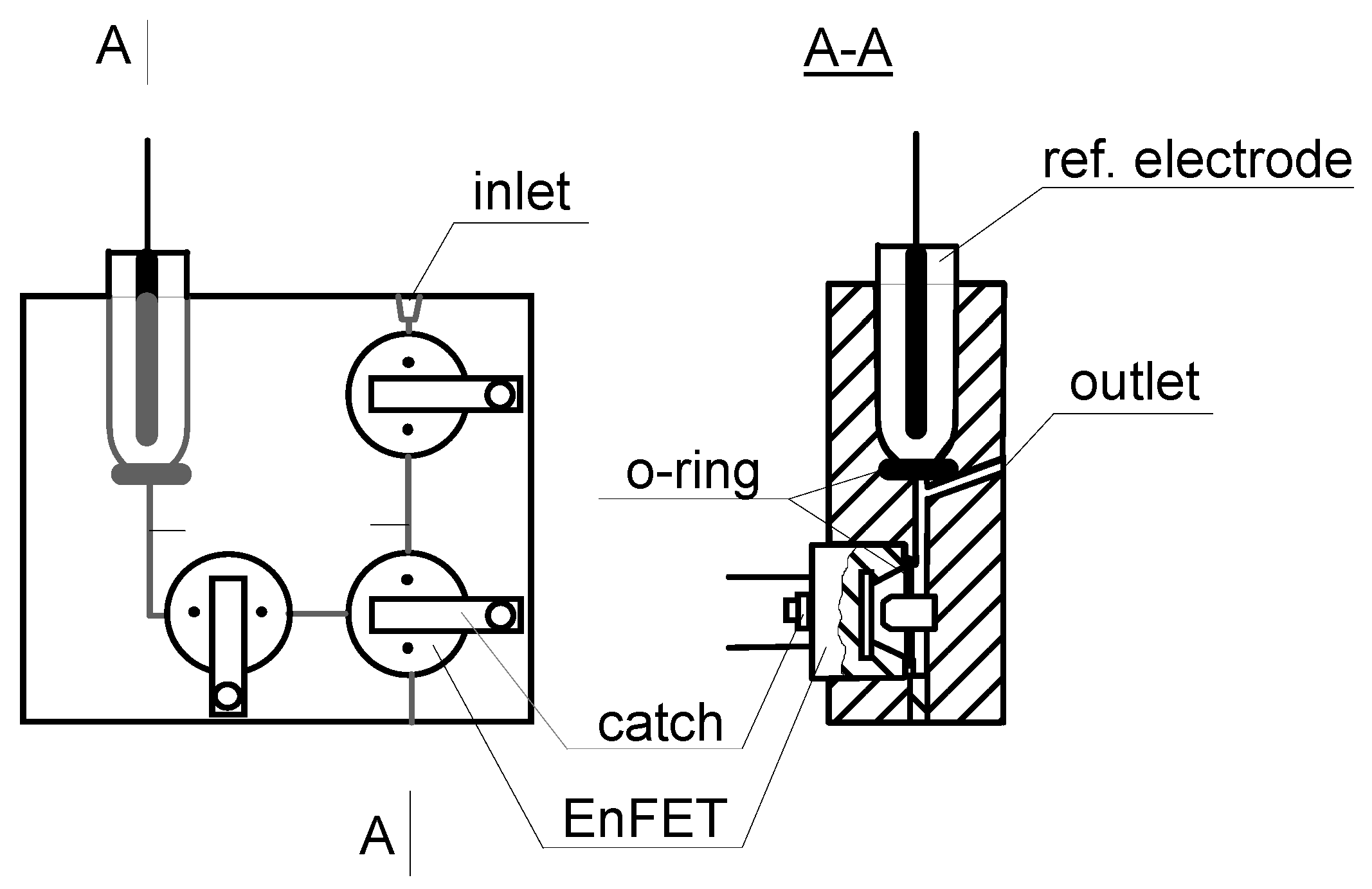
Figure 1.
Schematic diagram of the serial flow-through cells.

The pH-detection based EnFETs were placed in the serial flow-through cells (Fig. 1). Based on the calibration curves taken in plasma and dialysate, pairs of calibrations points were chosen for determinations of urea in the prepared samples. After each calibration, the subsequent determinations of urea in samples were repeated 5 times. Between changes of the sample in the measuring cells, the cells were washed with phosphate buffer at pH 6.0.
The ChemFET based biosensors were immersed in the solution to be measured (0.005 M orthophosphate buffer), connected to the measurement setup and conditioned for 2 hours (connected to the power supply). The calibration curves were obtained at room temperature by means of the standard addition method. The urea standard solutions were prepared by dissolving of a weighed amount of urea in following fluids: 50 mM tris/HCl buffer solution at pH 7.5, dialysate based on D-605 hemodialysis fluid concentrate (diluted concentrate), mixture of dialysate and the buffer, mixture of blood plasma and the buffer.
Results and Discussion
Urea biosensor based on pH-ISFET
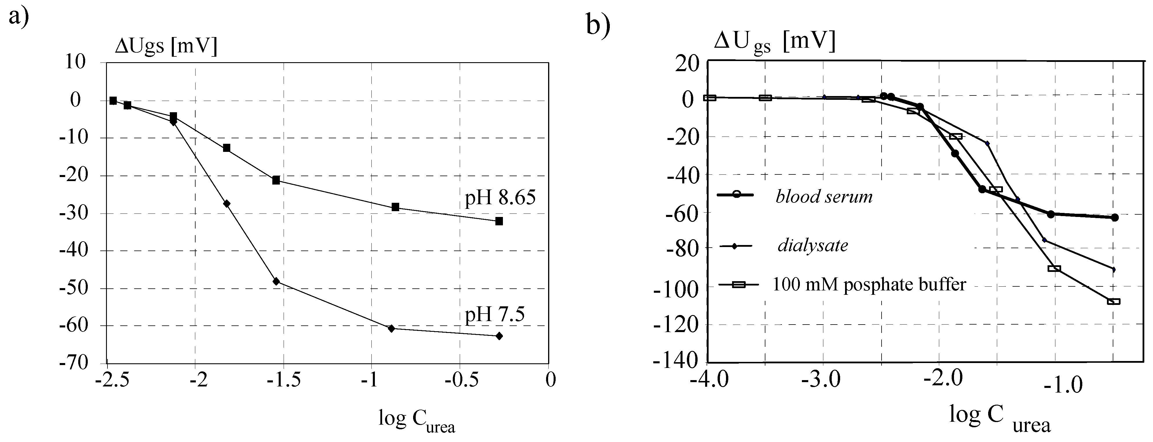
The output signal obtained from the EnFET strongly depends on the pH and buffer capacity (concentration) of the sample. The higher pH the lower output signal. Changing the buffer capacity results in shifting of output sensor's characteristic. Owning to first reason, to obtain grater analytical output signal, the pH of the blood plasma was lowered to 7.5 with 1 M HCl solution. Due to the second reason the buffer capacity should be constant during the measurements. Calibration curves carried out for undiluted blood plasma samples at pH 8.5 and 7.5 are shown in Fig. 2a.
Figure 2.
The EnFET response in blood plasma samples at different pH 8.65 and pH 7.5 (a), and in ortophosphate buffer solution of 100 mM concentration, blood plasma and dialysate (a concentrate of hemodialysis fluid D-605, diluted in the ratio 1:35) (b).
Figure 2.
The EnFET response in blood plasma samples at different pH 8.65 and pH 7.5 (a), and in ortophosphate buffer solution of 100 mM concentration, blood plasma and dialysate (a concentrate of hemodialysis fluid D-605, diluted in the ratio 1:35) (b).

It is known that for measurements taken in biological samples the problems of the protein fouling of the sensors as well as sample removing from the measuring cell become important. To find the best method of protein removing from the biosensor surface, the flow-through cell were washed with phosphate buffer solutions at different concentrations of sodium chloride. For estimation of the optimal cleaning procedure, the criterion of the shortest recovery time of the analytical output signal after the exposition to a plasma sample was taken. The shortest time of the analytical output signal recovery was for phosphate buffer of sodium chloride of 0.1 M concentration.
The physiological range of urea concentration expands between 2.5 to 6.7 mM, while pathophysiological range covers 30 to 150 mM [7]. To estimate linear range of an EnFET operation, the calibration was done for the wide range of urea concentration in dialysate pCurea (3; 0) (Fig. 2b). Based on the obtained graph, two linear subranges were chosen: (1) pCurea 2.19 to 1.72 (i.e. 6.45÷19.1 mM) and (2) pCurea 1.72 to 0.72 (i.e. 19.1÷190.5 mM). The first subrange is close to the physiological urea concentration in plasma whereas the second subrange covers the extent of the pathophysiology, which is characteristic for the renal insufficiency. The measurements were performed for EnFETs placed in the serial flow-through cells (Fig. 1). Between measurements the EnFETs were washed with 0.1 M phosphate buffer.
Summarizing, with the developed urea-biosensor it is possible to determine the urea concentration in blood plasma and in hemodialysis fluid for the physiological and the pathophysiological range. Comparison of the calibration curves obtained for pH-based EnFETs taken in the blood plasma, dialysate and 100 mM ortophosphate buffer solution are shown in Fig. 2b.
Urea Biosensor Based on pNH4-ChemFET
Due to complex composition of biological samples, especially high concentration of sodium and potassium, the application of the pNH4-based urea biosensors can be problematic mainly due to poor selectivity of the nonactine for ammonium ions over potassium and sodium ions. Therefore an attempt has been made to calibrate the biosensor in undiluted dialysate.
In analytical practice a preliminary sample modification by dilution with a proper solution is a commonly used method. So that, next measurements were taken in the mixture of tris/HCl buffer solution and the dialysate. The calibrations were performed in dialysate D-605 diluted with 0.05 M tris/HCl buffer (2:1 v/v) and with 0.1 M tris/HCl buffer (4:1 v/v). For comparison, similar measurements were performed in pure dialysate and pure 0.05 M tris/HCl buffer. The slope values obtained in these solutions are about 12, 38, 42 and 46 mV per decade, respectively. Examples of calibration curves of the urea EnFET obtained in mixtures of dialysate D-605 and tris/HCl buffer are presented in Fig. 3a.
Figure 3.
Calibration curves of the urea sensor in dialysate D-605, dialysate mixed with 0.05 M tris/HCl buffer (2:1 v/v), dialysate mixed with 0.1 M tris/HCl buffer (4:1 v/v) and in 0.05 M tris/HCl buffer (a) and in blood plasma mixed with 0.1 M tris/HCl buffer of pH 7.5 (1:2 v/v) (b).
Figure 3.
Calibration curves of the urea sensor in dialysate D-605, dialysate mixed with 0.05 M tris/HCl buffer (2:1 v/v), dialysate mixed with 0.1 M tris/HCl buffer (4:1 v/v) and in 0.05 M tris/HCl buffer (a) and in blood plasma mixed with 0.1 M tris/HCl buffer of pH 7.5 (1:2 v/v) (b).

In this case, the widest linear range of the calibration curve was obtained. It can be concluded that the best results are expected after dilution of the dialysate sample with the tris/HCl buffer. Since in the case of ammonium sensor based on ion-selective membrane containing nonactine, sodium ions may interfere with ammonium ions (indirect urea determination), another advantage of the using of the tris/HCl buffer is that the buffer does not contain sodium ions. Moreover, the linear range of the sensor can be extended towards higher urea.
In the case of urea determination in the blood plasma, the same method of sample dilution in the 50 mM tris/HCl buffer was applied. The calibration curves obtained in a mixture of blood plasma and tris/HCl buffer (1:2 v/v) recalculated to the real concentration of urea in the blood plasma are shown in Fig. 3b. As can be seen, the sensor sensitivity was about 53 mV per decade.
It was stated that for the blood plasma samples a preliminary dilution is always recommended in order to keep the urea concentration in the mixture of buffer and plasma in the linear range of the sensor operation. The urea sensor can be applied for urea determination in the dialysate and in the blood plasma using a method of sample pre-treatment, which can be realized both manually and in automatic systems.
Conclusions
Summarizing, with the developed urea-biosensors based on pH and pNH4 detection, it is possible to determine the urea concentration in blood plasma and in dialysate for the physiological and the pathophysiological range. The difference for both sensors consists in sample pre-treatment. For pNH4 based urea biosensors it is recommended to dilute both samples dialysate and blood plasma while for pH based urea biosensors dilution of the samples is not necessary.
References
- Lowrie, E.G.; Laird, N.M.; Henry, R.R. Protocol for the National Cooperative Dialysis Study. Kidney Int. 1983, 23 (Suppl. 13), 11–18. [Google Scholar]
- Gotch, F.A.; Sargent, A. A mechanistic analysis of the International Cooperative Dialysis Study (NCDS). Kidney Int. 1985, 28, 526–534. [Google Scholar] [CrossRef] [PubMed]
- Caraway, T.; Fanger, H. Am. J. Clin. Path. 1956, 26, 1475.
- Colasanti, G.; Arrigo, G.; Santoro, A.; et al. Biochemical aspects and clinical perspectives of continuous urea monitoring in plasma ultrafiltrate; Preliminary results of a multicenter study. The Int. J. Artificial Organs 1995, 18(9), 544–547. [Google Scholar]
- Pijanowska, D.G.; Torbicz, W. pH-ISFET based urea biosensor. Sensors and Actuators B Chem. 1997, B 44 No. 1-3, 370–376. [Google Scholar] [CrossRef]
- Brzózka, Z.; Dawgul, M.; Pijanowska, D.G.; Torbicz, W. Durable NH4+-sensitive CHEMFET. Sensors and Actuators B Chem. 1997, 44(1-3), 527–531. [Google Scholar] [CrossRef]
- Angielski, S. Biochemia kliniczna i anlityka, PZWL, Warszawa. 1990.
- Sample Availability: Available from the authors.
© 2003 by MDPI (http://www.mdpi.net). Reproduction is permitted for noncommercial purposes.
Share and Cite
MDPI and ACS Style
Bijanowska, D.G.; Dawgul, M.; Torbicz, W. Comparison of Urea Determination in Biological Samples by EnFETs Based on pH and pNH4 Detection. Sensors 2003, 3, 160-165. https://doi.org/10.3390/s30600160
AMA Style
Bijanowska DG, Dawgul M, Torbicz W. Comparison of Urea Determination in Biological Samples by EnFETs Based on pH and pNH4 Detection. Sensors. 2003; 3(6):160-165. https://doi.org/10.3390/s30600160
Chicago/Turabian StyleBijanowska, Dorota G., Marek Dawgul, and Wladyslaw Torbicz. 2003. "Comparison of Urea Determination in Biological Samples by EnFETs Based on pH and pNH4 Detection" Sensors 3, no. 6: 160-165. https://doi.org/10.3390/s30600160