2. Results and Discussion
The sponge
A. cavernosa was exhaustively extracted with acetone, and after being subjected to extensive column chromatography on silica gel, Sephadex LH-20, ODS, and semipreparative HPLC, compounds
1–12 were obtained. The known compounds, kalihipyran A (
8) [
16], 15-formamido-kalihinene (
9) [
6], 10-formamido-kalihinene (
10) [
6], and kalihinenes X (
11) and Y (
12) [
15], were identified by comparison of their spectroscopic data with literature values.
In the
1H and
13C NMR spectra of compounds
1–7, most signals were doubled in the ratio of ~1.8:1, 2.2:1, 1.1:1, 1.4:1, 2.1:1, 1.9:1, and 1.1:1, respectively. This suggested that 1-7 existed as equilibrium mixtures derived from the s-
trans and s-
cis forms of formamide groups, as in the cases of
8–12 [
6,
15,
16], which was supported by IR absorptions (
1, 1670;
2, 1681;
3, 1668;
4, 1681;
5, 1682;
6, 1686; and
7, 1668 cm
−1) [
16]. The assignments of the formamide groups (NH and CHO) in
1–7 for s-
trans isomers and s-
cis isomers were confirmed by HSQC and COSY data (
Table 1 and
Table 2).
Table 1.
1H NMR data of compounds 1–7 (CDCl3, J in Hz).
Table 1.
1H NMR data of compounds 1–7 (CDCl3, J in Hz).
| Position | 1
a, c | 2
b, c | 3
a, c | 4
a, c | 5
b, c | 6
a, c | 7
a, d |
|---|
| 1 | 1.24 m | 1.26 m
e | 1.65 m
e | 1.30 m
e | 1.30 m | 1.96 m | 2.40 m |
| 2a | 1.84 m | 1.34 m | 1.73 m | 1.85 m | 1.84 m
e | 1.87 m | 1.48 m
e |
| 2b | 1.30 m | 1.26 m
e | 1.65 m
e | 1.30 m
e | 1.28 m | 1.48 m | 1.48 m
e |
| 3a | 1.99 m
e | 1.97 m
e | 1.99 m
e | 2.00 m
e | 1.93 m
e | 2.01 m | 2.00 m
e |
| 3b | 1.99 m
e | 1.97 m
e | 1.99 m
e | 2.00 m
e | 1.93 m
e | 1.89 m | 2.00 m
e |
| 5 | 5.24 br s | 5.47 br s | 5.49 br d (4.0) | 5.69 br s | 6.37 br s | 5.70 br s | 5.39 br s |
| 6 | 2.05 m | 1.91 m | 2.15 m | 2.18 m | 2.11 m | 2.20 m | 2.20 m |
| 7 | 1.73 m | 1.14 m | 1.43 m
e | 1.17 m | 1.58 m | 1.62 m | 1.78 m
e |
| 8a | 1.66 m | 1.53 m | 1.43 m
e | 1.43 m | 1.10 m | 1.74 m | 1.55 m
e |
| 8b | 1.50 m | 1.26 m | 1.26 m | 1.57 m | 1.75 m | 1.94 m | 1.55m
e |
| 9a | 1.89 m | 1.88 m | 1.65 m
e | 1.90 dt (9.5, 3.5) | 1.89 m | 5.32 br s | 1.55 m
e |
| 9b | 1.62 m
e | 1.56 m | 1.55 m
e | 1.60 m | 1.62 m | 1.55 m
e |
| 11 | | 1.97 m
e | 1.75 m | | | | |
| 12a | 1.99 m
e | 1.26m
e | 1.24 m
e | 2.62 t (6.5) | 1.28 m
e | 1.78 m | 5.61 m
e |
| 12b | 1.99 m
e | 1.26 m
e | 1.24 m
e | 1.28 m
e | 1.63 m |
| 13a | 2.13 m | 1.97 m
e | 1.99 m
e | 2.23 m
e | 2.27 m | 1.83 m | 2.18 m |
| 13b | 1.99 m
e | 1.97 m
e | 1.90 m | 2.23 m
e | 1.97 m | 1.56 m | 2.13 m |
| 14 | 5.13 t (5.5) | 5.11 t (6.0) | 5.08 br s | 5.19 t (6.5) | 3.93 m | 3.75 t (7.0) | 3.89 br s |
| 16 | 1.69 s | 1.69 s | 1.67 s | 1.72 s | 1.39 s | 1.31 s | 4.88 s |
| 5.01 s |
| 17 | 1.62 s
e | 1.60 s | 1.59 s | 1.62 s | 1.31 s | 1.20 s | 1.78 s
e |
| 18 | 4.89 br s | 0.78 d (6.9) | 0.83 d (6.5) | 1.26 s | 1.22 s
e | 1.18 s | 4.16 br s
e |
| 4.80 br s | 4.16 br s
e |
| 19 | 1.62 s
e | 1.67 s | 1.65 s
e | 1.68 s | 1.65 s | 1.68 s | 1.62 s |
| 20 | 1.28 s | 1.22 s | 1.43 s
e | 1.27 s | 1.22 s
e | 1.68 s | 1.55 s
e |
| NH | 5.89 d (12.5) | 6.10 d (11.8) | 5.62 d (12.0) | 5.92 d (12.0) | 5.66 d (12.5) | 5.86 m | 5.61 m
e |
| CHO | 8.30 d (12.0) | 8.27 d (12.3) | 8.24 d (12.5) | 8.29 d (12.5) | 8.28 d (12.5) | 8.19 d (12.5) | 8.08 d (2.0) |
Cavernene A (1) was isolated as a colorless oil. A molecular formula of C
21H
33ON was established by the [M + Na]
+ ion peak at
m/z 338.2461 in the HRESIMS and supported by NMR data (
Table 1 and
Table 2), indicating six degrees of unsaturation. The
1H NMR spectrum showed the presence of four tertiary methyl [
δH 1.28 (3H, s), 1.62 (6H, s), and 1.69 (3H, s)] and four olefinic proton signals resonated at
δH 4.80 (1H, br s), 4.89 (1H, br s), 5.13 (1H, t,
J = 5.5 Hz), and 5.24 (1H, br s). The
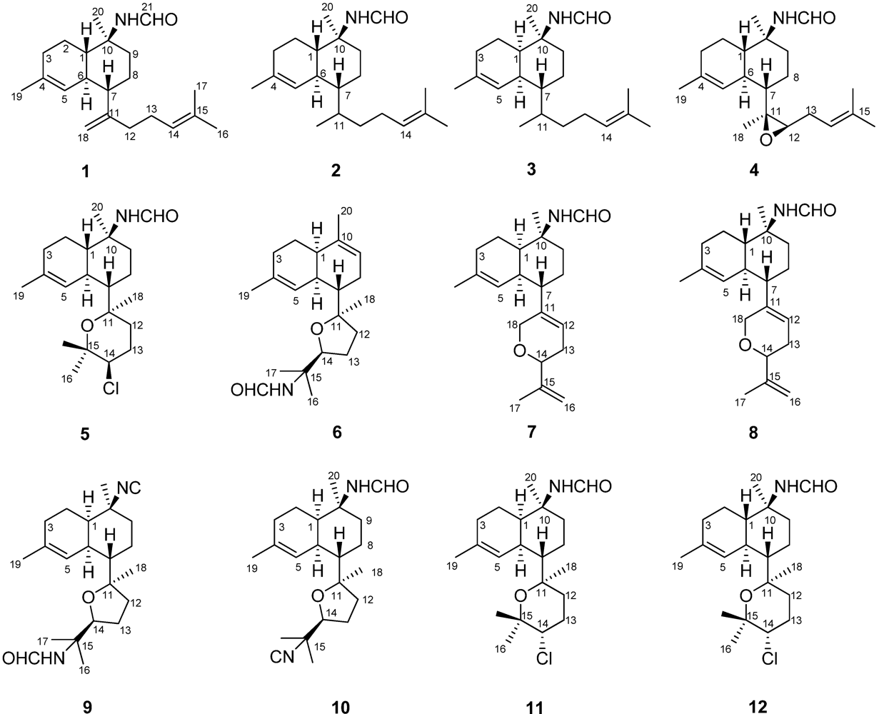
13C NMR and DEPT spectra of 1 displayed 21 carbon resonances for four methyls, seven methylenes (one olefinic), six methines (two olefinic and one formamido), and four quaternary carbons (three olefinic). The above moieties accounted for four of six degrees of unsaturation, indicating a bicyclic structure for 1. The COSY correlations of H-9/H-8/H-7/H-6/H-1/H-2/H-3/H-5/H-19, together with the HMBC correlations of H
3-19/C-3, C-4, and C-5, H
3-20/C-1, C-9, and C-10, H-5/C-1, C-3, C-6, and C-7, and CHO/C-10 indicated the presence of a decalin moiety (
Figure 2). The HMBC correlations from the geminal methyls (H
3-16 and H
3-17) to C-14 and C-15, from terminal olefinic protons (H
2-18) to C-7, C-11, and C-12, and from H-13 to C-11, C-12, C-14, and C-15 and COSY correlation between H-13 and H-14 revealed the presence of an isoprenoid unit homolog in 1 and connectivity of the two moieties between C-12 and C-7 through the quaternary carbon C-11.
Table 2.
13C NMR data (CDCl3, δ in ppm) of compounds 1–7.
Table 2.
13C NMR data (CDCl3, δ in ppm) of compounds 1–7.
| C | 1
a | 2
b | 3
a | 4
a | 5
b | 6
a | 7
a |
|---|
| s-
trans | s-
cis | s-
trans | s-
cis | s-
trans | s-
cis | s-
trans | s-
cis | s-
trans | s-
cis | s-
trans | s-
cis | s-
cis | s-
trans |
|---|
| 1 | 48.8 | 45.3 | 49.0 | 45.7 | 45.6 | 41.1 | 48.4 | 45.0 | 48.8 | 46.1 | 40.1 | 40.1 | 40.9 | 45.4 |
| 2 | 22.8 | 23.1 | 22.9 | 23.4 | 19.1 | 19.5 | 22.7 | 23.1 | 23.3 | 23.7 | 24.6 | 24.7 | 18.8 | 19.1 |
| 3 | 30.96 | 31.04 | 30.8 | 30.9 | 31.3 | 31.4 | 30.8 | 30.9 | 30.5 | 30.6 | 30.5 | 30.6 | 31.1 | 31.2 |
| 4 | 134.4 | 134.1 | 134.9 | 134.6 | 134.3 | 134.2 | 134.8 | 134.3 | 132.4 | 131.9 | 131.1 | 130.8 | 134.5 | 134.4 |
| 5 | 123.3 | 123.8 | 121.8 | 122.3 | 124.3 | 124.0 | 122.3 | 122.7 | 125.7 | 126.2 | 127.1 | 127.3 | 123.9 | 124.2 |
| 6 | 39.5 | 39.4 | 38.4 | 38.2 | 35.2 | 34.8 | 38.6 | 38.4 | 39.5 | 39.2 | 36.5 | 36.5 | 37.0 | 36.5 |
| 7 | 50.3 | 50.3 | 44.6 | 44.4 | 42.3 | 42.0 | 45.2 | 45.2 | 51.2 | 50.4 | 44.9 | 45.2 | 43.3 | 43.6 |
| 8 | 29.7 | 29.9 | 20.9 | 20.9 | 20.1 | 19.9 | 25.7 | 25.8 | 24.5 | 24.7 | 29.48 | 29.51 | 28.4 | 28.4 |
| 9 | 42.0 | 37.6 | 41.8 | 37.4 | 33.0 | 33.7 | 41.7 | 37.3 | 42.1 | 37.8 | 120.2 | 120.5 | 33.5 | 32.8 |
| 10 | 55.5 | 57.2 | 55.6 | 57.3 | 55.5 | 57.1 | 55.3 | 56.8 | 55.4 | 56.9 | 136.9 | 136.8 | 56.8 | 55.4 |
| 11 | 151.3 | 151.9 | 30.8 | 30.8 | 31.3 | 31.1 | 62.33 | 62.7 | 76.9 | 77.2 | 86.4 | 86.3 | 139.4 | 139.0 |
| 12 | 34.2 | 34.2 | 35.65 | 35.69 | 35.7 | 35.8 | 62.27 | 62.33 | 31.0 | 31.7 | 37.2 | 37.1 | 118.6 | 118.9 |
| 13 | 26.3 | 26.3 | 26.2 | 26.2 | 26.2 | 26.3 | 27.0 | 27.0 | 25.9 | 26.1 | 25.6 | 25.8 | 29.6 | 29.6 |
| 14 | 124.2 | 124.3 | 124.6 | 124.8 | 124.7 | 124.9 | 119.3 | 119.5 | 64.2 | 64.5 | 84.2 | 84.5 | 77.2 | 76.8 |
| 15 | 131.7 | 131.6 | 131.3 | 131.1 | 131.2 | 131.1 | 134.2 | 134.1 | 74.2 | 74.4 | 54.5 | 55.9 | 145.4 | 145.3 |
| 16 | 25.7 | 25.7 | 25.7 | 25.7 | 25.7 | 25.7 | 25.7 | 25.7 | 29.7 | 29.7 | 27.4 | 25.1 | 110.5 | 110.6 |
| 17 | 17.8 | 17.8 | 17.7 | 17.7 | 17.7 | 17.7 | 18.0 | 18.0 | 28.9 | 28.4 | 23.8 | 21.7 | 18.9 | 18.9 |
| 18 | 110.0 | 109.6 | 13.3 | 13.3 | 13.3 | 13.3 | 18.5 | 18.5 | 21.9 | 21.2 | 20.3 | 21.1 | 67.63 | 67.58 |
| 19 | 23.3 | 23.4 | 23.6 | 23.7 | 23.5 | 23.4 | 23.55 | 23.60 | 23.8 | 23.8 | 23.92 | 23.87 | 23.3 | 23.4 |
| 20 | 19.0 | 18.9 | 18.8 | 18.7 | 27.2 | 23.7 | 18.9 | 18.7 | 19.0 | 18.5 | 21.53 | 21.47 | 23.6 | 29.7 |
| 21 | 162.8 | 160.4 | 163.1 | 160.5 | 162.8 | 160.0 | 162.8 | 160.4 | 162.7 | 160.4 | 163.1 | 160.9 | 160.1 | 162.8 |
The relative configuration of 1 was established on the basis of NOESY data (
Figure 2). The NOESY correlations of H-1/H-7 and NH/H-1 indicated that these protons were on the same face of the decalin ring and arbitrarily assigned β-orientations. The α-orientation of H-6 was determined by the NOESY correlation between H
3-20 and H-6. In addition, the carbon resonances at
δC 48.8 (C-1), 39.5 (C-6), 42.0 (C-9), and 19.0 (C-20) in 1 further confirmed the
trans fusion of the decalin ring [
6,
8,
15]. Cavernene A (
1) can be envisaged as a decomposable intermediate product of isocyanobifloradiene epoxides in the plausible biogenetic pathway of kalihiprans [
3,
19].
Figure 2.
COSY (▬), key HMBC (→), and key NOESY correlations of 1.
Figure 2.
COSY (▬), key HMBC (→), and key NOESY correlations of 1.
Cavernene B (
2) was obtained as a colorless oil. Its molecular formula of C
21H
35ON was deduced from the HRESIMS (
m/z 340.2617 [M + Na]
+) combined with its NMR data (
Table 1 and
Table 2), indicating five degrees of unsaturation. The
1H and
13C NMR spectra of 1 and 2 were comparable except for a marked difference in the isoprenoid unit. Signals for a C-11–C-18 double bond were absent in
2 and replaced by saturated carbons resonated at
δC 30.8 and 13.3, respectively. The HMBC correlations of H
3-18/C-7, C-11, and C-12 and the COSY correlations of H
3-18/H-11/H-12 and H-11/H-7 supported the assignment of convernene B as
2. The relative configuration of the decalin ring in
2 was the same as in
1, with the observation of the NOESY correlations of NH/H-1, H
3-20/H-6, NH/H-9b, H-7/H-9b, and H
3-20/H-9a (Supporting Information). The relative configuration at C-11, however, could not be conclusively determined due to conformational flexibility between C-7 and C-11.
Cavernene C (
3), obtained as white needles, showed the same molecular formula of C
21H
35ON as
2 determined by pseudomolecular [M + Na]
+ ion peak at
m/z 340.2614 in HRESIMS. Its carbon skeleton was readily assignable as the same as
2 by HSQC, HMBC, and COSY spectra. In particular, the carbon resonances of the isoprenoid unit in
3 were almost superimposable on those of
2 (
Table 2), suggesting the same stereostructure of the isoprenoid unit for both
2 and
3. On the other hand, differences were observed for the signals of the decalin ring . Specifically, the carbon resonances at
δC 49.0 (C-1), 38.4 (C-6), 41.8 (C-9), and 18.8 (C-20) in
2 were replaced by resonances at
δC 45.6, 35.2, 33.0, and 27.2 in
3, respectively, indicating a
cis fusion of the decalin ring [
6,
8,
15]. The relative configuration at C-11, however, could not be conclusively determined due to the free rotation around the C-7–C-11 bond.
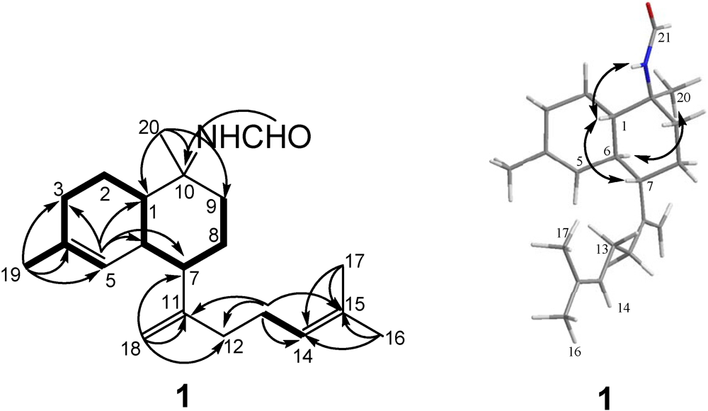
Cavernene D (
4) displayed a HRESIMS [M + Na]
+ peak at
m/z 354.2407 corresponding to a molecular formula of C
21H
33O
2N, implying six degrees of unsaturation. Many similarities of the
1H and
13C NMR data between
2 and
4 (
Table 1 and
Table 2) suggested they were structural analogs, with the main differences due to the presence of a trisubstituted epoxide (
δC 62.33 and 62.27) in
4 and the absence of two saturated carbons (
δC 30.8 and 35.65) in
2. The C-11/C-12 position of the epoxy group was determined by a COSY correlation of H-12/H-13 and HMBC correlations of H
3-18/C-7, C-11, and C-12 and H-13/C-11 and C-12. The relative configuration of the decalin ring of
4, found to agree with
2, was established by observation of NOESY correlations of NH/H-1, H-1/H-7, and H
3-20/H-6. The relative configurations of C-7 and C-11 between the conjoined bicyclic ring systems in
4 were assigned as 7
S* and 11
S*, respectively, based on NOESY correlations of H
3-18/H-6, H
3-18/H-8b, and H-8a/H-13, as shown in the Newman projection (
Figure 3). Thus, the epoxy group was determined as in β-orientation.
Figure 3.
Key NOESY correlations of 4.
Figure 3.
Key NOESY correlations of 4.
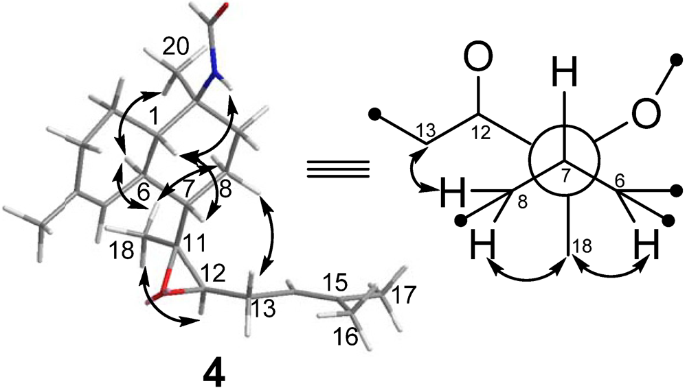
Kalihinene E (
5) was isolated as colorless needles (MeOH), and given a C
21H
34NO
2Cl molecular formula with five degrees of unsaturation, based on the HRESIMS (
m/z 368.2354 [M + H]
+) and NMR spectra. The ESIMS of
5 showed a cluster of isotopic [M + H]
+ ion peaks at
m/z 368/370 in a ratio of ~3:1, indicating the presence of a chlorine atom in the molecule. The NMR spectra of 5 revealed the presence of five methyls [
δH 1.22 (6H, s), 1.31 (3H, s), 1.39 (3H, s), and 1.65 (3H, s)], an olefinic methine [
δH 6.37 (1H, br s),
δC 125.7 (CH), and
δC 132.4 (qC)], a chlorine-bearing methine [
δH 3.93 (1H, m)/
δC 64.2], and two oxygenated quaternary carbons (
δC 74.2 and 76.9) for the major s-
trans isomer (
Table 1 and
Table 2). Analysis of the 2D NMR (HSQC, HMBC, and COSY) data (
Figure 4) revealed that
5 possessed the same carbon skeleton as kalihinene X (
11) and Y (
12) [
15].
Figure 4.
COSY (▬), key HMBC (→), and key NOESY correlations of 5.
Figure 4.
COSY (▬), key HMBC (→), and key NOESY correlations of 5.
The relative configuration of the decalin ring in
5 was the same as
12, inferred from the chemical shifts of C-1 to C-10 and NOESY correlations of H-1/H-7, H
3-20/H-6, and NH/H-1. A significant difference between
5 and
12 was found in the chemical shift of C-12 (
δC 31.0 in
5 instead of
δC 38.2 in
12), which was caused by the
γ-gauche effect [
20,
21,
22,
23,
24], indicating an
axial orientation of Cl–14. The relative configurations of C-7 and C-11 in
5 were determined as
S* and
R*, respectively, from NOESY correlations of H
3-18/H-6, H
3-18/H-8b, and H-8a/H-12, as shown in the Newman projection (
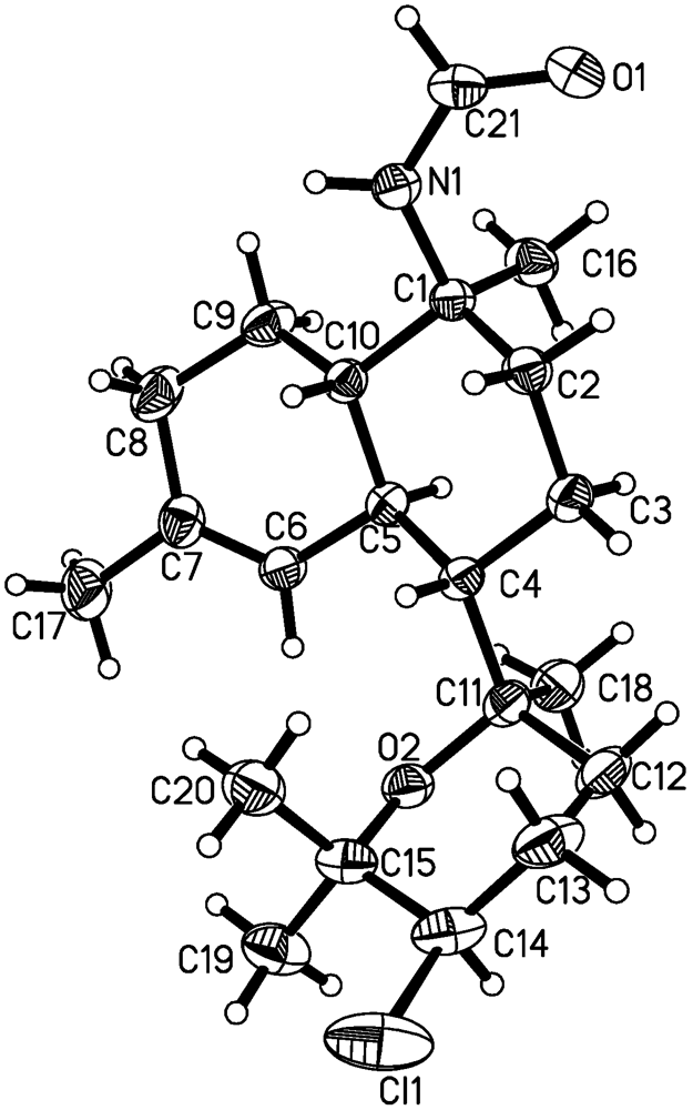
Figure 4). Finally, the absolute configuration of
5 was unambiguously determined as 1
S, 6
S, 7
S, 10
S, 11
R, and 14
R by single crystal X-ray diffraction using Cu Kα radiation (
Figure 5).
Figure 5.
ORTEP drawing of compound 5.
Figure 5.
ORTEP drawing of compound 5.
Kalihinene F (
6) was isolated as a colorless oil and given a C
21H
33NO
2 molecular formula based on HRESIMS measurements (
m/z 354.2406 [M + Na]
+) in combination with extensive NMR analysis. The NMR spectra of
6 (
Table 1 and
Table 2) revealed the presence of five methyls [
δH 1.18 (3H, s), 1.20 (3H, s), 1.31 (3H, s), and 1.68 (6H, s)], two trisubstituted double bonds [
δH 5.70 (br s),
δC 127.1 (CH), and
δC 131.1 (qC);
δH 5.32 (br s),
δC 120.2 (CH), and
δC 136.9 (qC)], an oxymethine [
δH 3.75 (t,
J = 7.0 Hz)/
δC 84.2], and an oxygenated quaternary carbons (
δC 86.4) for the major s-
trans isomer. The COSY correlations of H
3-20/H-9/H-8/H-7/H-6/H-1/H-2/H-3 and H-6/H-5/H
3-19, together with the HMBC correlations of H
3-19/C-3, C-4, and C-5, H
3-20/C-1, C-10, and C-9, H-5/C-1, C-3, and C-6, H-2 and H-3/C-4, and H-8/C-10 indicated the presence of a decalin moiety (
Figure 6). A tetrahydrofuran ring, attached to the decalin ring at C-7, was established by carbon resonances at
δC 84.2 (C-14) and 86.4 (C-11), COSY correlations of H-12/H-13/H-14, HMBC correlations of H
3-18/C-7, C-11, and C-12 and geminal methyls (H
3-16 and H
3-17)/C-14 and C-15, and NOESY correlation between H
3-18 and H-14. The location of the formamide functionality was assigned to be at C-15 by the observation of doubled singlets for H
3-16 [
δH 1.31 (s) and 1.38 (s) for s-
trans and s-
cis isomers, respectively] and H
3-17 [
δH 1.20 (s) and 1.34 (s) for s-
trans and s-
cis isomers, respectively] and doubled triplets for H-14 [
δH 3.75 (t, 7.0) and 3.80 (t, 7.0) for s-
trans and s-
cis isomers, respectively] [
6]. The relative configuration of
6 was determined by NOESY correlation of H-1/H-6 and carbon resonances at
δC 40.1 (C-1), 36.5 (C-6), and 44.9 (C-7) [
6]. NOESY correlations of H
3-18/H-6, H
3-18/H-8b, and H-8/H-12b defined the relative configurations of 7
S*, 11
R*, as shown in the Newman projection (
Figure 6).
Figure 6.
COSY (▬), key HMBC (→), and key NOESY (
![Marinedrugs 10 01445 i002]()
) correlations of
6.
Figure 6.
COSY (▬), key HMBC (→), and key NOESY (
![Marinedrugs 10 01445 i002]()
) correlations of
6.
Kalihypyran C (
7), a colorless oil, had a molecular formula of C
21H
31NO
2 established by HRESIMS at
m/z 352.2255 [M + Na]
+, indicating seven degrees of unsaturation. Its
1H and
13C NMR spectra (
Table 1 and
Table 2) showed the presence of three methyls [
δH 1.55 (3H, s), 1.62 (3H, s), 1.78 (3H, s)], two trisubstituted double bonds [
δH 5.39 (br s),
δC 123.9 (CH), and
δC 134.5 (qC);
δH 5.61 (br s),
δC 118.6 (CH), and
δC 139.0 (qC)], a disubstituted double bond [
δH 4.88 (br s), 5.01 (br s),
δC 110.5 (CH
2), and
δC 145.4 (qC)], an oxymethylene [
δH 4.16 (s, 2H)/
δC 67.63], and an oxymethine
δH [3.89 (br s)/δC 77.2] for s-
cis isomer. Many similarities of the
1H and
13C NMR data between
7 and
8 suggested they were structural analogs [
16], the COSY correlations of H
3-19/H-5/H-6, H
3-17/H-16, and H-18/H-12/H-13/H-14 and the HMBC correlations of H
3-19/C-3, C-4, and C-5, H
3-20/C-1, C-9, and C-10, H
3-17/C-14, C-15, and C-16, and H
2-16/C-14, C-15, and C-17 further confirmed
7 possessed the same carbon skeleton as
8. The chemical shifts of C-1, C-6, and C-20 for s-
trans isomers in
7 (
δC 45.4, 36.6, and 29.7, respectively) were different from those in
8 (
δC 48.9, 39.4, and 18.9, respectively), indicated
cis fusion of the decalin ring in
7 [
16]. The NOESY correlations of H-1/H-6, H-1/H
3-20, and H-6/H
3-20 confirmed the relative configurations as 7. However, the relative configuration of H-14 was not determined.
The isolated compounds were assessed for their cytotoxicity against a small panel of human cancer cell lines (human colon cancer cell line HCT-116, human lung epithelial cell line A549, human cervical carcinoma cell line HeLa, human hepatocellular carcinoma cell line QGY-7701, and human mammary cancer cell line MDA-MB-231) using a MTT method, and camptothecin (Shanghai Dibai Chemical Co., Shanghai, China; purity ≥98%) was used as positive control. Compounds 1 and 2 showed moderate cytotoxic activities against HCT-116 with IC
50 values of 6.31 and 8.99 μM, respectively. Compounds 5 showed cytotoxic activity against HCT-116, HeLa, QGY-7701 and MDA-MB-231 with IC
50 values of 14.36, 13.36, 17.78 and 12.84 μM, respectively (
Table 3). In addition, compounds
1–12 were tested for antifungal activity against fungi
Candida albicans,
Candida parapsilosis,
Candida glabrata,
Cryptococcus neoformans,
Trichophyton rubrum,
Microsporum gypseum, and
Aspergillus fumigatus. Compound
9 showed weak antifungal activity against
T. rubrum and
M. gypseum with MIC values of 8 and 32 μg/mL, respectively. Compound
10 displayed weak antifungal activity against fungi
C. albicans,
C. neoformans,
T. rubrum, and
M. gypseum with MIC values of 8, 8, 4, and 8 μg/mL, respectively. Ketoconazole (Shanghai Aiyan Chemical Co., Shanghai, China; purity ≥98%) was used as positive control with MIC value ≤0.25 μg/mL. It is worth noting that the isonitrile functionalities in the diterpenes play an important role in their antifungal activity.
Table 3.
Cytotoxicities of compounds 1–12 in five cancer cell lines.
Table 3.
Cytotoxicities of compounds 1–12 in five cancer cell lines.
| | Cytotoxicity IC50 (μM) |
|---|
| HCT-116 | A549 | HeLa | QGY-7701 | MDA-MB-231 |
|---|
| 1 | 6.31 | >50 | >50 | >50 | >50 |
| 2 | 8.99 | >50 | >50 | >50 | >50 |
| 3 | >50 | >50 | >50 | >50 | >50 |
| 4 | >50 | >50 | >50 | >50 | >50 |
| 5 | 14.36 | >50 | 13.36 | 17.78 | 12.84 |
| 6 | >50 | >50 | >50 | >50 | >50 |
| 7 | >50 | >50 | >50 | >50 | >50 |
| 8 | >50 | 13.09 | 11.19 | 13.53 | >50 |
| 9 | >50 | 17.53 | 14.74 | 16.39 | >50 |
| 10 | >50 | 6.98 | 13.30 | 14.53 | 6.84 |
| 11 | 12.25 | 8.55 | 10.59 | 13.02 | 7.46 |
| 12 | >50 | 17.12 | 10.05 | 14.41 | 15.23 |
| camptothecin | 9.25 | 2.32 | 6.98 | 4.05 | 0.50 |
3. Experimental Section
3.1. General Experimental Procedures
Optical rotation data were determined with a Perkin-Elmer 341 polarimeter (Perkin-Elmer, Inc., Waltham, MA, USA) with a 1 dm cell. UV spectra were collected using a Shimadzu UV 240 spectrophotometer (Shimadzu Corp., Kyoto, Japan). IR spectra were recorded on a Bruker vector 22 spectrometer (Bruker Optics, Inc., Billerica, MA, USA) with KBr pellets. NMR experiments were conducted on Bruker Avance-500 and AMX-400 spectrometers (Bruker Biospin Corp., Billerica, MA, USA). The HRESIMS spectra were acquired with a Waters Q-Tof micro YA019 mass spectrometer (Waters Corp., Milford, MA, USA). X-ray structure analysis was performed on a Bruker SMART APEX-II CCD diffractometer (Bruker Optics, Inc.). Melting points were obtained on an SGW X-4 melting point apparatus (Shanghai Precision & Scientific Instrument Co., Ltd, Shanghai, China). Reversed-phase HPLC was carried out on a YMC-Pack ODS-A column (250 × 10 mm, 5 µm; YMC Co., Ltd., Kyoto, Japan) using a Waters 1525 HPLC instrument with Waters 2998 UV detector and monitored at 210 nm. Silica gel (200–300 mesh, Qingdao Chengyang Ocean Chemical Co., Jinan, China), Sephadex LH-20 (Pharmacia Fine Chemicals, Piscataway, NJ, USA), and YMC ODS-A (50 μm, YMC Co., Ltd, Kyoto, Japan) were used as column packing materials. Fractions were monitored by TLC (HSGF 254, Yantai Huiyou Co., Yantai, China), and spots were visualized by heating silica gel plates sprayed with 12% H2SO4 in EtOH.
3.2. Animal Material
Samples of A. cavernosa were collected by hand using scuba around Xisha Islets in the South China Sea in March 2009 and identified by Professor Jin-He Li (Institute of Oceanology, Chinese Academy of Sciences, China). A voucher sample (JHQ-0901) was deposited in the Laboratory of Marine Drugs, Department of Pharmacy, Changzheng Hospital, Second Military Medical University, China.
3.3. Extraction and Isolation
The sponge (5.5 kg, wet weight) was extracted with acetone at room temperature five times (5 × 5 L) and the extract was concentrated to a brown oil, which was redissolved in H2O (2 L). The aqueous solution was extracted with CH2Cl2 (4 × 2 L) to afford a CH2Cl2-soluble extract (89 g). The resulting extract was partitioned between petroleum ether (4 × 1 L) and 90% aqueous MeOH (4 × 1 L) to yield a brownish red oil (49 g), which was subjected to column chromatography (80 × 7.0 cm) on silica gel (1000.5 g) eluting with petroleum ether-acetone gradient (stepwise, 0:1, 50:1, 30:1, 15:1, 8:1, 3:1 to 0:1, 40 L), and finally MeOH, to give eight fractions (Fractions 1–8). Fraction 5 (5.4 g) was fractionated over Sephadex LH-20 (40 × 2000 mm, eluted with CH2Cl2/MeOH 1:1, 1.5 L), and further fractionated by CC on ODS (RP-18, 30 × 500 mm), eluted with 70%–100% MeOH/H2O, to give four subfractions (Fractions 5a–d). Fraction 5a (203.4 mg) was purified by HPLC (YMC-Pack ODS-A, 5 µm, 10 × 250 mm, 2.0 mL/min, UV detection at 210 nm), using MeOH/H2O (88:12) as eluent, to yield 7 (2.1 mg, tR = 40.5 min) and 8 (4.2 mg, tR = 42.5 min). Fraction 5b (659 mg) was subjected to CC (15 × 300 mm) on silica gel (15 g), using CH2Cl2/MeOH with increasing polarity (1:0, 100:1, 50:1) as mobile phase, to yield compound 4 (13.3 mg). Fraction 5c (368.0 mg) was purified by HPLC (YMC-Pack ODS-A, 5 µm, 10 × 250 mm, 2.0 mL/min, UV detection at 210 nm), using MeOH/H2O (92:8) as eluent, to afford 1 (48.9 mg, tR = 36.6 min), 3 (4.2 mg, tR = 40.8 min), and 2 (50.7 mg, tR = 43.8 min). Fraction 5d (267.5 mg) was chromatographied (15 × 300 mm) on silica gel (15 g, eluted with CH2Cl2/MeOH 50:1), to obtain 10 (160 mg). Fraction 6 (7.4 g) was applied to Sephadex LH-20 (40 × 2000 mm) eluted with CH2Cl2/MeOH (1:1) to furnish three subfractions (Fractions 6a and c). Fraction 6b (1.3 g) was subjected to CC on ODS (RP-18, 30 × 500 mm), eluting with 80%–100% MeOH, to give five subfractions (Fractions 6b1–5). Fraction 6b2 (123 mg) was purified by HPLC (YMC-Pack ODS-A, 5 µm, 10 × 250 mm, 2.0 mL/min, UV detection at 210 nm) using MeOH/H2O (92:8) as eluent to 9 (1.0 mg, tR = 37.5 min). Fraction 6b3 (31.5 mg) was fractioned by a Sephadex LH-20 column (15 × 2000 mm) eluting with hexane/CH2Cl2/MeOH (5:4:1) to afford 6 (4.5 mg). Compound 5 (50.9 mg) was obtained from fraction 6b4 (478.5 mg) by a CC (15 × 300 mm) on silica gel (15 g, eluted with CH2Cl2/MeOH 30:1). Fraction 7 (8.3 g) was subjected to CC (40 × 2000 mm) on Sephadex LH-20 eluting with MeOH followed by HPLC purification (YMC-Pack ODS-A, 5 µm, 10 × 250 mm, 2.0 mL/min, UV detection at 210 nm) using MeOH/H2O (90:10) as eluent to yield 11 (201.5 mg, tR = 50.4 min) and 12 (197.6 mg, tR = 56.8 min).
Cavernene A (
1): colorless oil; [α]
![Marinedrugs 10 01445 i001]()
+25.0 (
c 0.06, MeOH); UV (CH
3CN) λ
max (log ε) < 200 (2.45) nm; IR (KBr) ν
max 3323, 3051, 2927, 2856, 1670, 1541, 1457, 1386 cm
−1;
1H and
13C NMR data, see
Table 1 and
Table 2; HRESIMS
m/z 338.2461 [M + Na]
+ (calcd for C
21H
33NONa, 338.2460).
Cavernene B (
2): colorless oil; [α]
![Marinedrugs 10 01445 i001]()
+46.2 (
c 0.07, MeOH); UV (CH
3CN) λ
max (log ε) < 200 (2.79), 234 (sh, 2.25) nm; IR (KBr) ν
max 3295, 3053, 2960, 2926, 2869, 1681, 1537, 1453, 1382 cm
−1;
1H and
13C NMR data, see
Table 1 and
Table 2; HRESIMS
m/z 340.2617 [M + Na]
+ (calcd for C
21H
35NONa, 340.2616).
Cavernene C (
3): white needles (MeOH); m.p. 98.0–102.0 °C; [α]
![Marinedrugs 10 01445 i001]()
+20.0 (
c 0.03, MeOH); UV (CH
3CN) λ
max (log ε) < 200 (2.95) nm; IR (KBr) ν
max 3304, 3062, 2958, 2926, 2855, 1668, 1538, 1455, 1381 cm
−1;
1H and
13C NMR data, see
Table 1 and
Table 2; HRESIMS
m/z 340.2614 [M + Na]
+ (calcd for C
21H
35NONa, 340.2616).
Cavernene D (
4): colorless needles (MeOH); m.p. 112.0–115.0 °C; [α]
![Marinedrugs 10 01445 i001]()
+51.4 (
c 0.04, MeOH); UV (CH
3CN) λ
max (log ε) < 200 (2.88) nm; IR (KBr) ν
max 3296, 3055, 2928, 2857, 1681, 1538, 1453, 1383 cm
−1;
1H and
13C NMR data, see
Table 1 and
Table 2; HRESIMS
m/z 354.2407 [M + Na]
+ (calcd for C
21H
33NO
2Na, 354.2409).
Kalihinene E (
5): colorless needles (MeOH); m.p. 185.0–190.0 °C; [α]
![Marinedrugs 10 01445 i001]()
+25.0 (
c 0.04, MeOH); UV (CH
3CN) λ
max (log ε) < 200 (2.94) nm; IR (KBr) ν
max 3295, 3074, 2929, 2868, 1682, 1541, 1451, 1381 cm
−1;
1H and
13C NMR data, see
Table 1 and
Table 2; HRESIMS
m/z 368.2354 [M + H]
+ (calcd for C
21H
35NO
2Cl, 368.2356).
Kalihinene F (
6): colorless oil; [α]
![Marinedrugs 10 01445 i001]()
+2.5 (
c 0.08, MeOH); UV (CH
3CN) λ
max (log ε) < 200 (2.63), 232 (sh, 2.12) nm; IR (KBr) ν
max 3216, 3057, 2966, 2929, 1686, 1451, 1379, 1311 cm
−1;
1H and
13C NMR data, see
Table 1 and
Table 2; HRESIMS
m/z 354.2406 [M + Na]
+ (calcd for C
21H
33NO
2Na, 354.2409).
Kalihipyran C (
7): colorless oil; [α]
![Marinedrugs 10 01445 i001]()
+24.6 (
c 0.07, MeOH); UV (CH
3CN) λ
max (log ε) < 200 (2.87) nm; IR (KBr) ν
max 3296, 3055, 2925, 2855, 1668, 1537, 1454, 1377 cm
−1;
1H and
13C NMR data, see
Table 1 and
Table 2; HRESIMS
m/z 352.2255 [M + Na]
+ (calcd for C
21H
31NO
2Na, 352.2252).
X-ray Crystallographic Analysis Data of Kalihinene E (5): Colorless needles, C21H34NO2Cl, MW = 367.94, monoclinic space group P21, a = 6.499 (2) Å, b = 7.952 (2) Å, c = 20.158 (4) Å, V = 1036.1 (4) Å3, Z = 2, Dcalcd = 1.179 g/cm3, and F (000) = 400. A single crystal of dimensions 0.05 × 0.13 × 0.18 mm was used for X-ray measurements and data collected on a Bruker SMART APEX-II CCD diffractometer using Cu Kα radiation and up to θ = 67.3 at 293 K. A total of 5789 reflections were collected, of which 3435 independent reflections were measured having an Rint of 0.0220, final R indices of I > 2σ (I), R1=0.0426, wR2 = 0.1163, R indices for all data R1 = 0.0445, and wR2 = 0.1184. The crystal structure solution was achieved using direct methods, as implemented with the SHELX-97 software program. The refinment method was full-matrix least-square on F2, goodness-of-fit on F2 was 1.042, and the largest difference peak and hole were 0.237 and −0.196 e. Å-3. The absolute structure was determined giving a Flack parameter of 0.07 (2). The X-ray diffraction material has been deposited in the Cambridge Crystallographic Data Center (CCCD No. 847695).
3.4. Cytotoxicity Assay
Cytotoxic activity was evaluated by a MTT method as described previously [
25]. Cells were cultured in RPMI-1640 supplemented with 10% fetal bovine serum in 5% CO
2 at 37 °C. An aliquot (200 μL) of these cell suspensions at a density of 5 × 10
−4 cell mL
−1 was plated in 96-well microtiter plates and incubated for 24 h under the above conditions. 2 μL of the test compound in DMSO at different concentrations was added to each well for 48 h, and then incubated with 1 mg/mL MTT for 4 h. The formazan dye product was measured by the absorbance at 570 nm on a microplate reader. IC
50 values were calculated by Reed and Muench’s method.
3.5. Antifungal Activity Assay
Antifungal activity was determined by the broth macrodilution method following the National Center for Clinical Laboratory Standards (NCCLS) recommendations against the following strains:
Candida albicans,
Candida parapsilosis,
Candida glabrata,
Cryptococcus neoformans,
Trichophyton rubrum,
Microsporum gypseum, and
Aspergillus fumigates [
26,
27]. Briefly, bacterial strains were grown aerobically at 30 °C in SDA for 16–20 h in an orbital shaker. A set of tube swith different concentrations of compounds
1–12 prepared in RPMI 1640 were next inoculated with the microorganisms and incubated 24 h for
C. albicans,
C. parapsilosis,
C. glabrata, and
A. fumigatus, 72 h for
C. neoformans, and 4–7 days for
T. rubrum and
M. gypseum. Broth tubes that appeared turbid were indicative of bacterial growth, while tubes that remained clear indicated no growth. The MIC, defined as the lowest concentration of inhibitory compound at which no growth was observed, was evaluated in triplicate for each compound (within the range 1.25–640 μg/mL). Cultures prepared under the same conditions but without compounds and cultures with the same proportions of DMSO (<1%) were used as controls. The growth of broth tubes without turbidity was further examined by counting the viable cells on the SDA plates.


 ) correlations of 6.
 +25.0 (c 0.06, MeOH); UV (CH3CN) λmax (log ε) < 200 (2.45) nm; IR (KBr) νmax 3323, 3051, 2927, 2856, 1670, 1541, 1457, 1386 cm−1; 1H and 13C NMR data, see Table 1 and Table 2; HRESIMS m/z 338.2461 [M + Na]+ (calcd for C21H33NONa, 338.2460).