
Antibacterial and Antifungal Compounds from Marine Fungi

Abstract
:1. Introduction
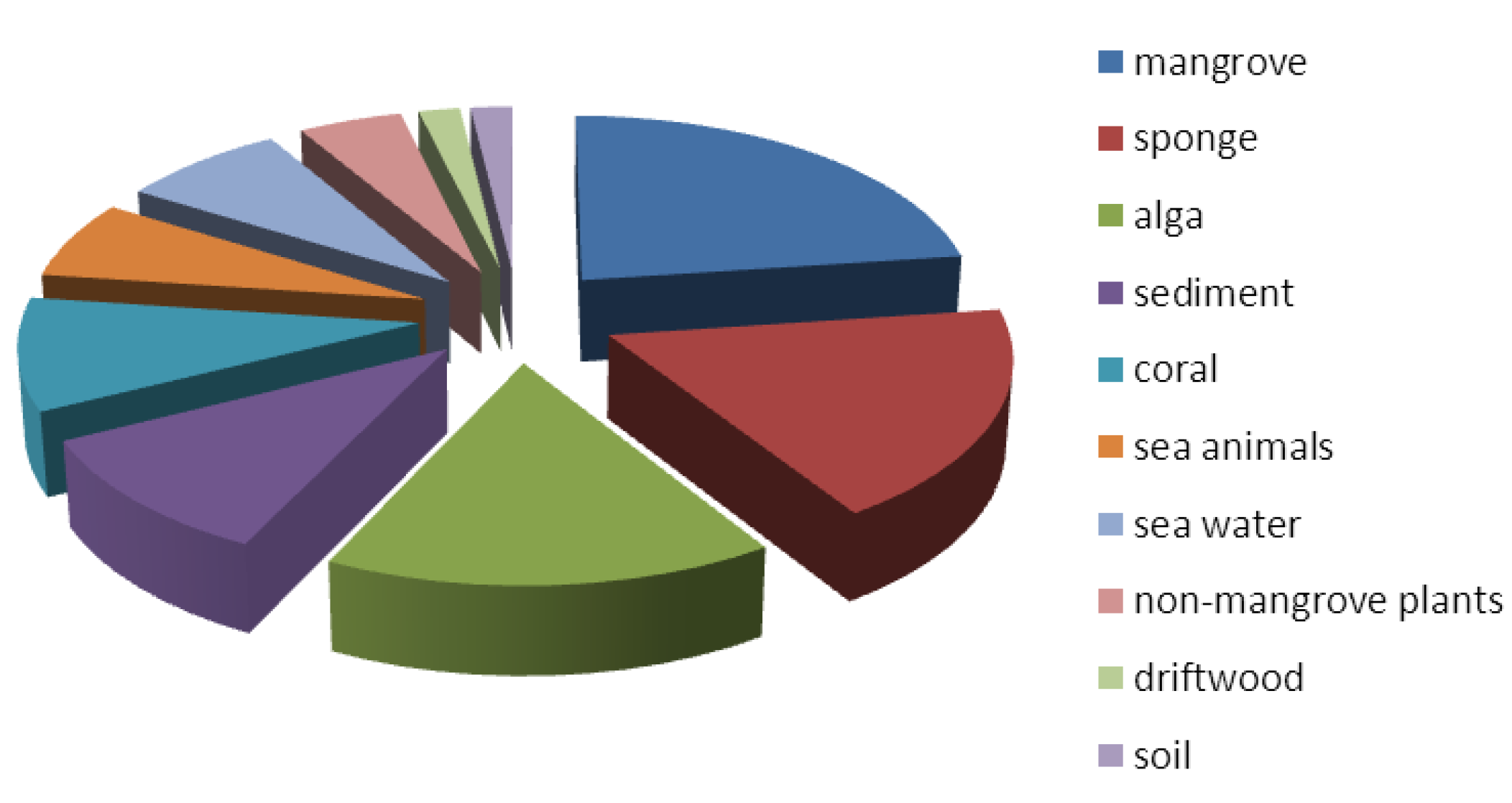
2. Sampling Location

3. Fungal Isolation and Identification

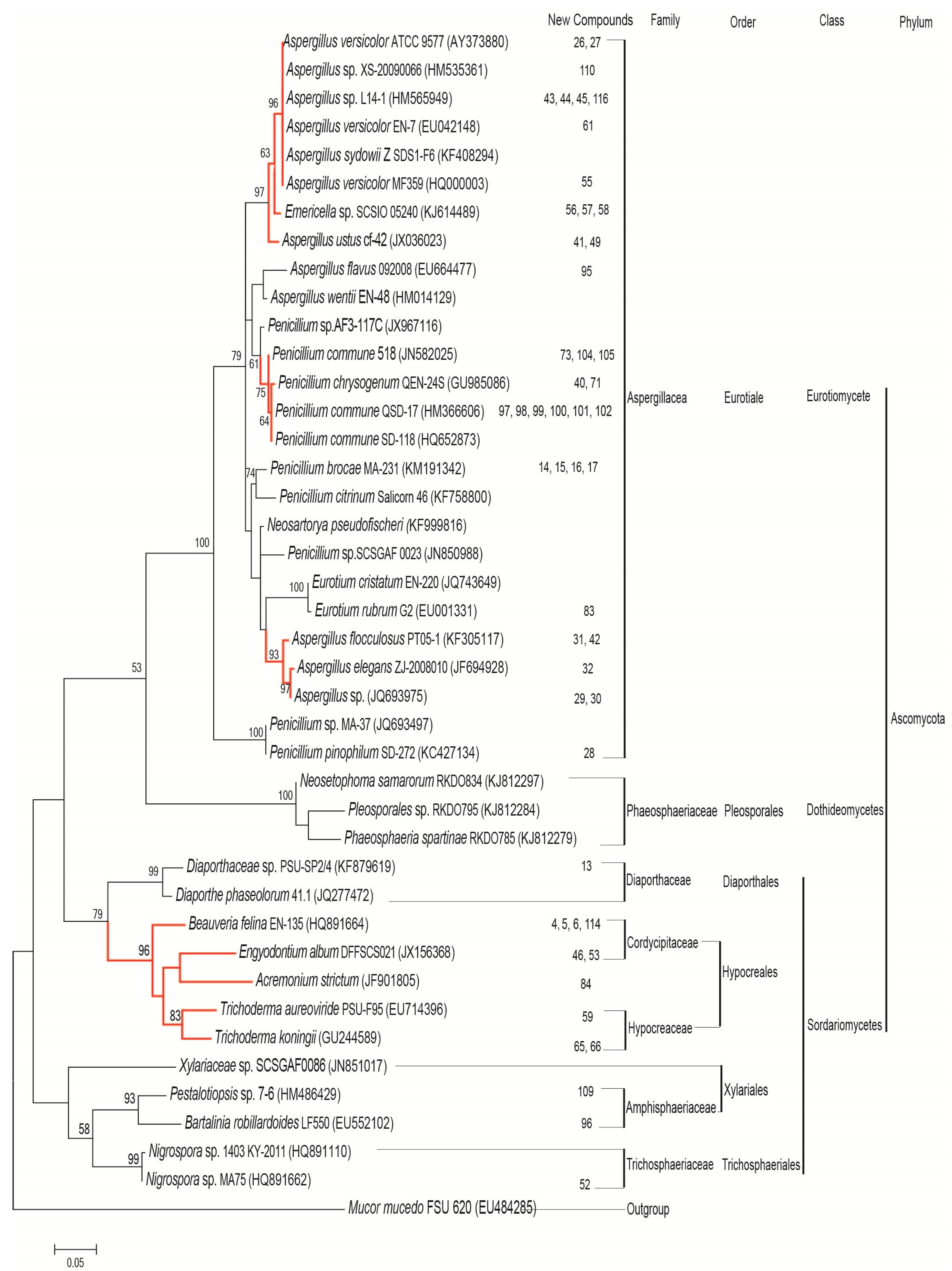
4. Phylogenetic Analysis

5. New Antibacterial and Antifungal Compounds from Marine Fungi
5.1. Nitrogen-Containing Compounds
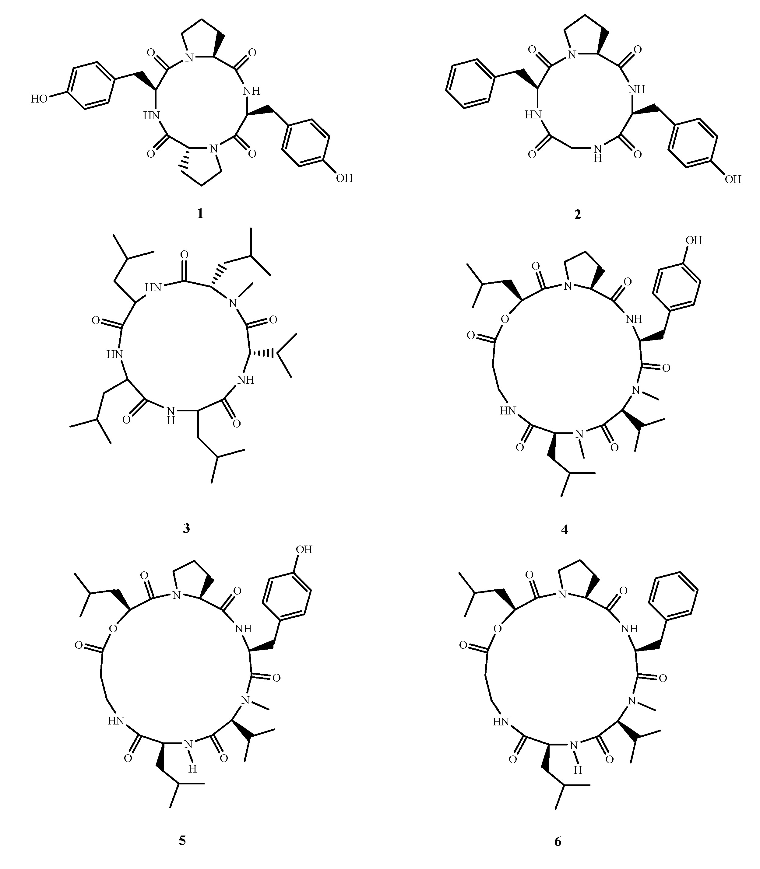
5.1.1. Peptides

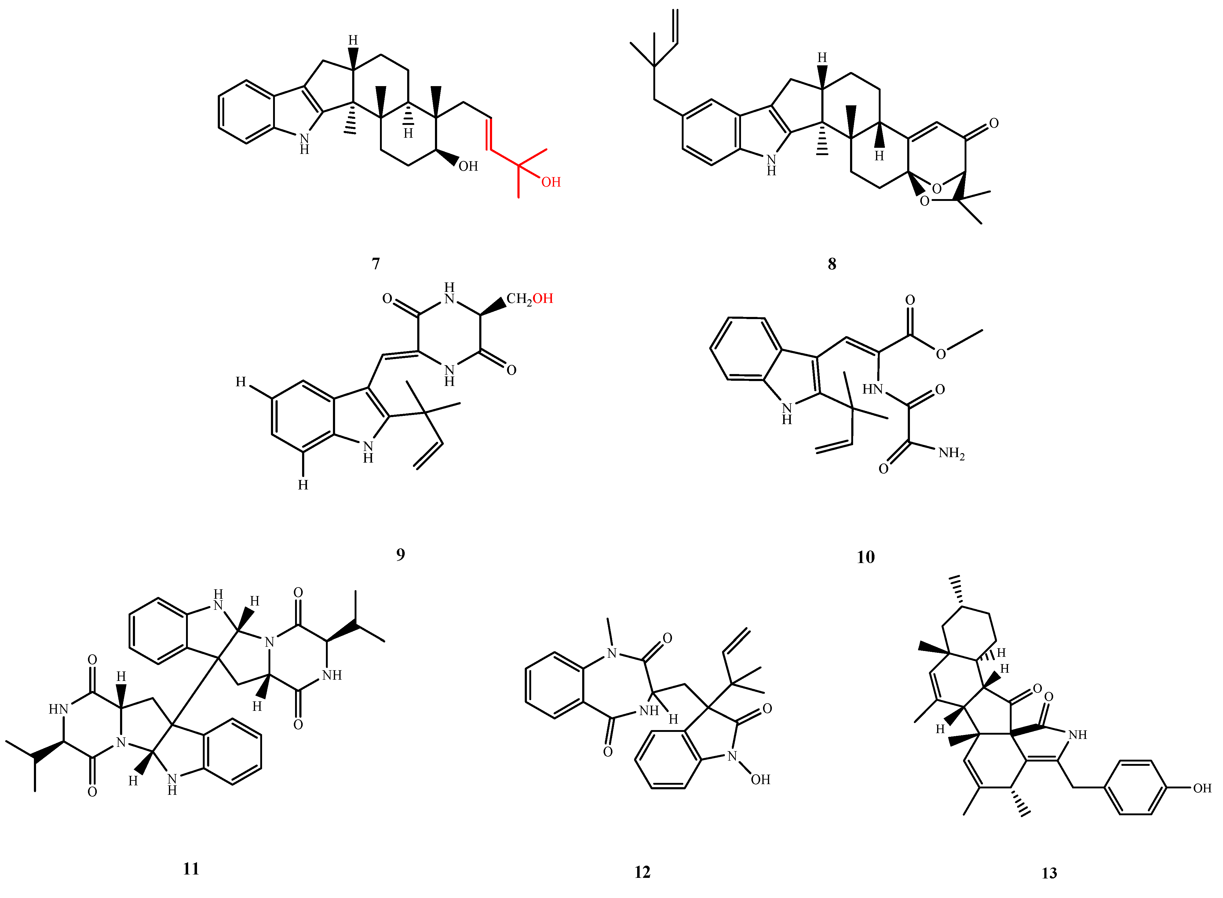
5.1.2. Indole-Alkaloids


5.1.3. Pyridines and Pyridinones

5.1.4. Piperazine/Diketopiperazine and Pyrimidine/Pyrimidinone

5.1.5. Other N-Containing Compounds
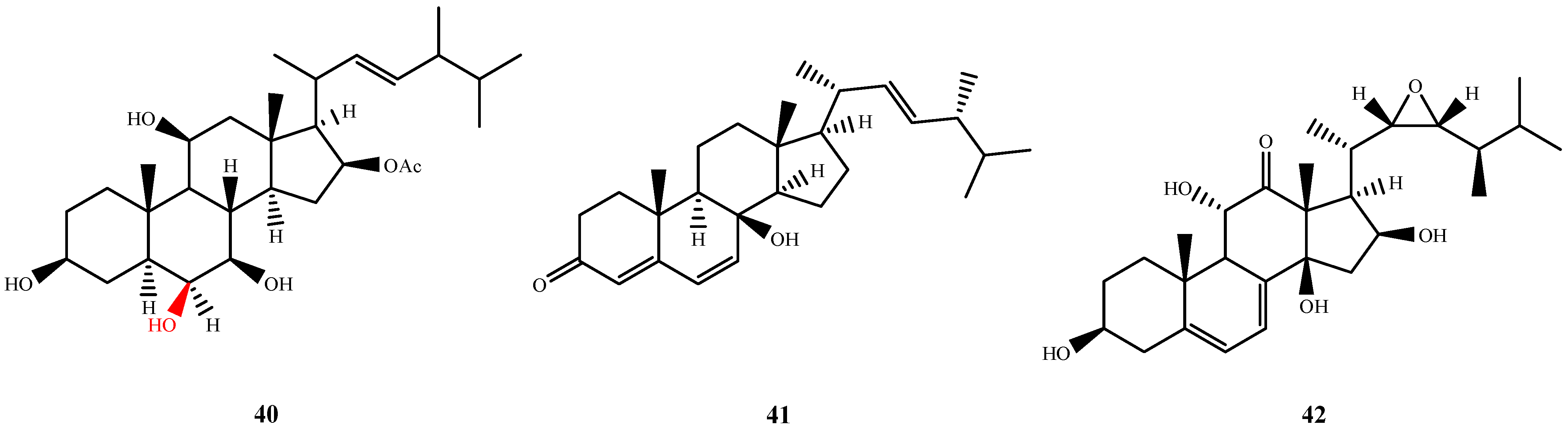
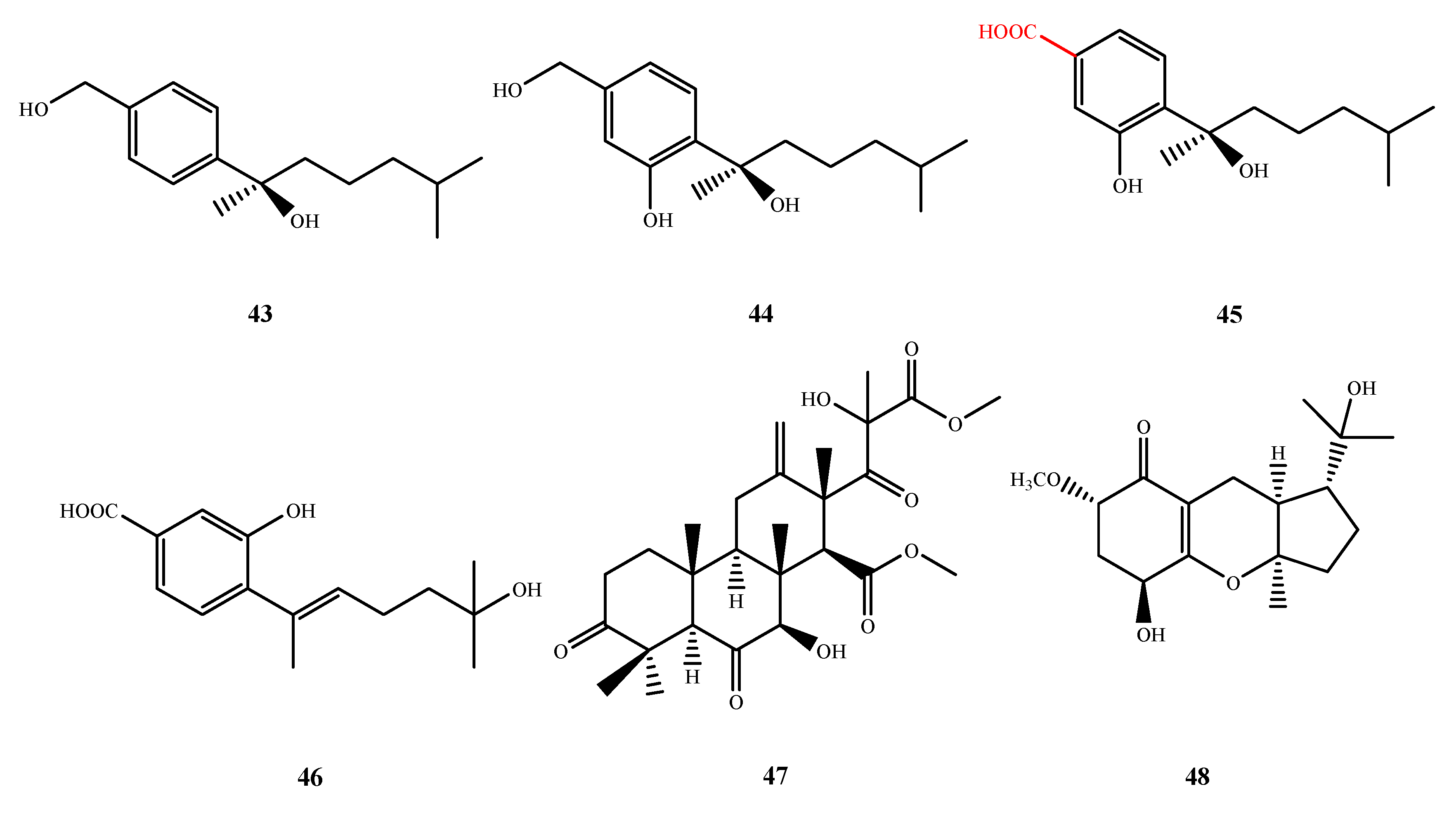
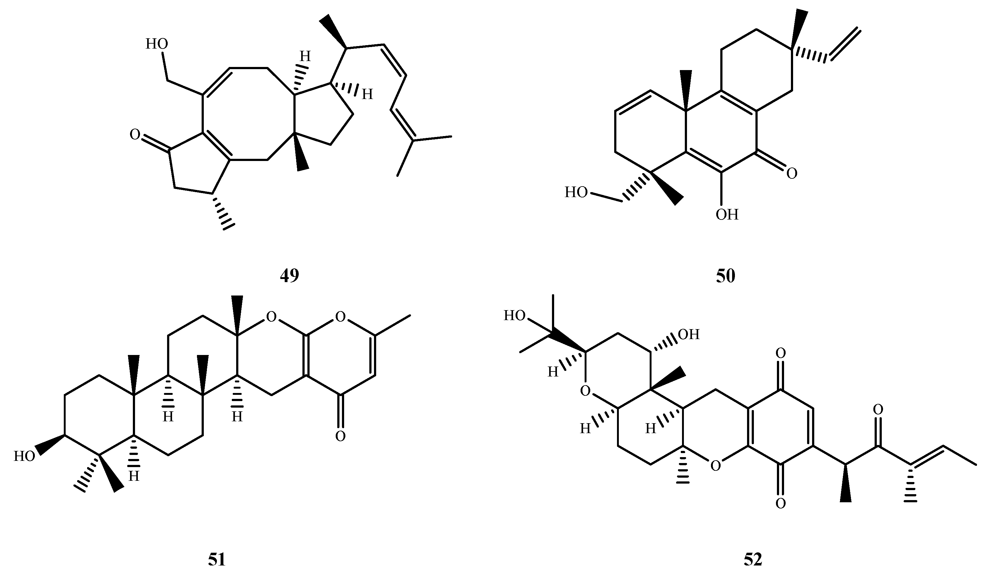
5.2. Steroids and Terpenoids


5.3. Polyketides
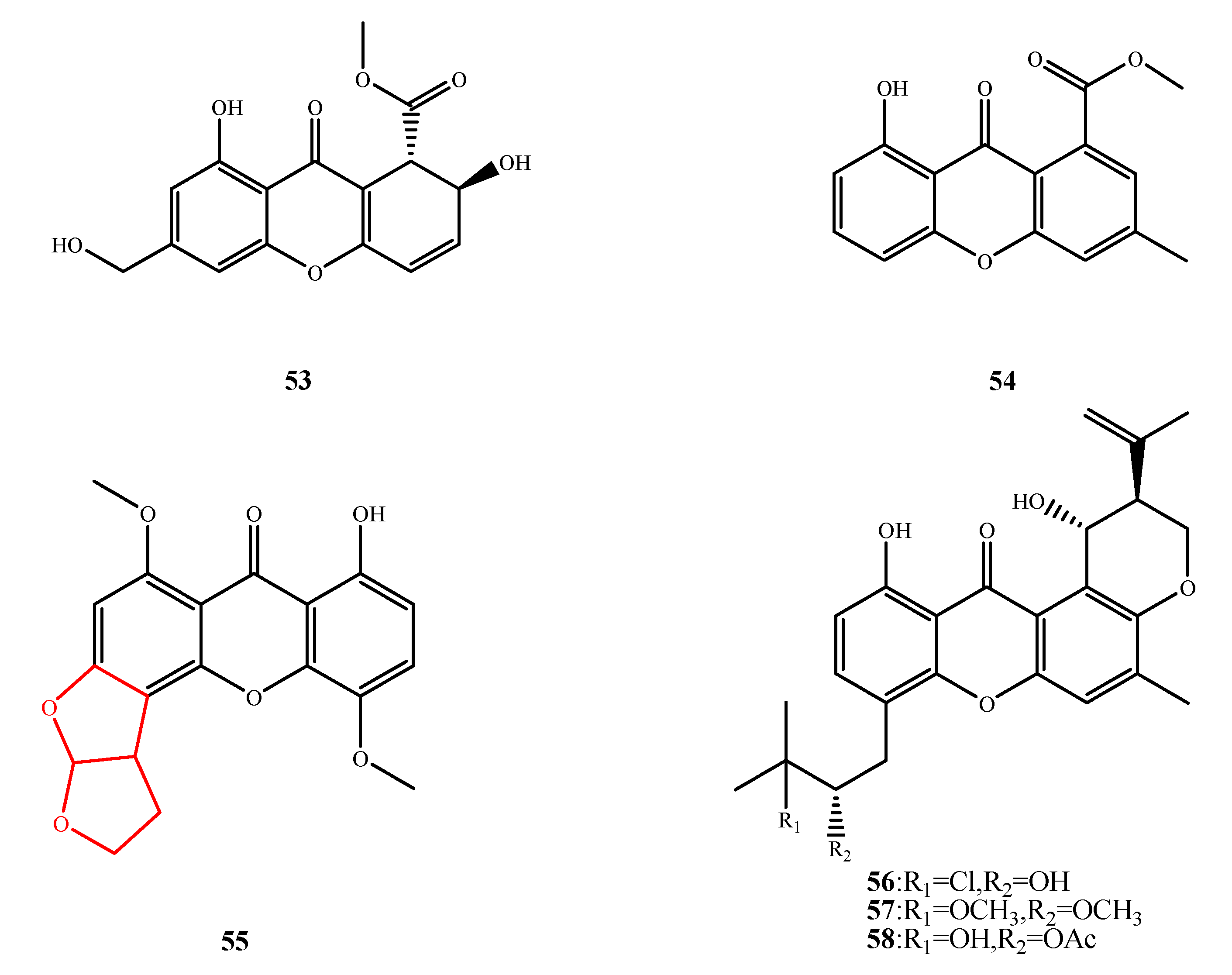
5.3.1. Xanthones

5.3.2. Anthraquinones

5.3.3. Quinones and Quinone Derivatives


5.4. Others


6. Known Antibacterial and Antifungal Compounds from Marine Fungi

| Compound name | Activity | Reference | Compound name | Activity | Reference |
|---|---|---|---|---|---|
| (−)-scleroderolide (117) | B+, Y | [67] | (−)-sclerodione (118) | B+, Y | [67] |
| (−)-sclerotiorin (119) | Y, F | [94] | (−)-stephacidin A (120) | B+ | [82] |
| (±)-pestalachloride C (121) | B− | [81] | (5α,6α)-ophiobolin H (122) | B− | [45] |
| 15G256β (123) | B | [73] | 15G256α (124) | B | [73] |
| 15G256π (125) | B+ | [73] | 1-methyl emodin (126) | B | [54] |
| 2,5-furandimethanol (127) | B+ | [36] | 3-HPA (128) | B+ | [90] |
| 4-deoxybostrycin (129) | B | [57] | 4-hydroxybenzaldehyde (130) | B− | [95] |
| 6,8-di-O-methylaverufin (131) | B | [96] | 6-epi-ophiobolin G (132) | B+ | [97] |
| 6-epi-ophiobolin K (133) | B+ | [97] | 6-O-methylaverufin (134) | B | [96] |
| 7-nor-ergosterolide (135) | Y, B | [33] | 8-acetyloxyaflatoxin B1 (136) | B− | [74] |
| acetylgliotoxin (137) | B+ | [87] | adenosine (138) | B− | [98] |
| aflatoxins B1 (149) | B− | [74] | aflatoxins B2 (140) | B− | [74] |
| AGI-B4 (141) | B | [42] | alternariol 2,4-dimethyl ether (142) | B− | [31] |
| anicequol (143) | F | [39] | AS-186c (144) | B+ | [72] |
| aspergillazine A (145) | B, Y | [83] | aspergillin PZ (146) | B+ | [34] |
| aspergillusene A (147) | B− | [99] | aspergillusidone C (148) | B+ | [100] |
| aspergillusone B (159) | B | [42] | asperphenamate (150) | B+ | [34] |
| aspochalasin E (151) | B, Y | [83] | aspochalasin D (152) | B | [34] |
| aspochalasin I (153) | B+ | [34] | aspulvinone E (154) | B, Y | [83] |
| averantin (155) | B+ | [101] | averufin (156) | B+ | [101] |
| bostrycin (157) | B | [56] | brefeldin A (158) | Y | [102] |
| brevianamide M (159) | B | [96] | butyrolactone I (160) | B+ | [88] |
| chlamydosporol (161) | B+ | [66] | cholesteryl linoleate (162) | B+ | [36] |
| chrysazin (163) | Y | [103] | cis-cyclo(Leu-Tyr) (164) | B+ | [104] |
| citrinin (165) | B | [105] | CJ-17665 (166) | Y | [32] |
| cladosporin (167) | B+ | [106] | conidiogenol (168) | B | [91] |
| conidiogenones B (169) | B, Y | [91] | coniothranthraquinone 1 (170) | B+ | [53] |
| cordyol C (171) | B | [82] | cpicpoformin (172) | B+ | [106] |
| cyclo(d)-Pro-(d)-Val (173) | B− | [31] | cyclosporine A (174) | Y, F | [107] |
| cytochalasin Z17 (175) | B− | [20] | cytosporone B (176) | B+ | [108] |
| cytosporone E (177) | B+ | [108] | deacetylsclerotiorin (178) | B+, Y | [75,76] |
| dechlorogriseofulvin (179) | Y | [48] | dicerandrol C (180) | B+ | [109] |
| dihydroisoflavipucine (181) | B | [20,71] | diorcinol (182) | B+ | [99,110] |
| djalonensone (183) | B− | [111] | echinulin (184) | B+ | [19] |
| epolones B (185) | Y | [102] | ergone (186) | B+,Y | [112] |
| eurorubrin (187) | B− | [92] | fonsecin (188) | F | [113] |
| fumitremorgin B (189) | B− | [114] | furandimethanol (190) | B+ | [98] |
| fusaric acid (191) | B+ | [115] | fusarielin A (192) | B+ | [66] |
| gliotoxin (193) | B | [87] | globosuxanthone A (194) | Y | [103] |
| glyantrypine (195) | B− | [114] | griseofulvin (196) | Y | [48] |
| griseophenone C (197) | B | [48] | guignardones B (198) | B+ | [44] |
| helicusin A (199) | Y | [76] | hydroxysydonic acid (200) | B | [42] |
| stachybocin A (201) | B+ | [23] | ilicicolin B (202) | B+ | [23] |
| isaridin E (203) | B− | [84] | isochaetochromin B2 (204) | B+ | [116] |
| isorhodoptilometrin (205) | B+ | [53] | lapatins B (206) | B− | [114] |
| malformins A1 (207) | B+ | [117] | malformins C (208) | B+ | [117] |
| meleagrin (209) | B+, Y | [95] | methylaverantin (210) | B+ | [101] |
| N-acetyldopamine (211) | F | [95] | neoaspergillic acid (212) | B | [118] |
| nidulin (213) | B+ | [100] | nidurufin (214) | B+ | [101] |
| nigrosporin B (215) | B | [119] | nornidulin (216) | B+ | [100] |
| notoamide B (217) | Y | [32] | notoamide R (218) | Y | [32] |
| ophiobolin K (219) | B+ | [97] | oxasetin (220) | B− | [120] |
| patulin (221) | B+ | [106] | penicillixanthone A (222) | B | [93] |
| pestalone (223) | B+, F | [121] | phomaligol A (224) | B+ | [35] |
| phomazine B (225) | F | [22] | phyllostine (226) | B+ | [106] |
| pycnidione (227) | Y | [102] | pyridoxatin (228) | B+, Y | [24] |
| pyrophen (229) | Y | [113] | reduced gliotoxin (230) | B | [87] |
| rubralide C (231) | B− | [31] | rubrofusarin B (232) | Y | [113] |
| sclerotiamide (233) | Y | [32] | secalonic acid B (234) | B | [93] |
| secalonic acid D (235) | B | [93] | siderin (236) | B | [54] |
| sporogen AO-1 (237) | Y | [122] | stachybocin B (238) | B+ | [23] |
| stephacidin A (239) | Y | [32] | stigmasterol (240) | B+ | [36] |
| tardioxopiperazine A (241) | B | [19] | tetrahydrobostrycin (242) | B | [48] |
| trichodermamide B (243) | B, Y | [83] | trichodermamides A (244) | B, Y | [83] |
| tyrosol (245) | B+ | [98] | ustilaginoidin D (246) | B+ | [116] |
| verruculogen (247) | B− | [114] | waikialides A (248) | Y | [32] |
| waikialides B (249) | Y | [32] | xanthocillin X (250) | B, F | [95] |
| Compound name | Activity | Reference | |||
| ω-hydroxyemodin (251) | B+ | [53] | |||
| (3β,5α,8α,22E)-5,8-epidioxyergosta-6,9,22-trien-3-ol (252) | B+ | [112] | |||
| (−)-7,8-dihydro-3,6-dihydroxy-1,7,7,8-tetramethyl-5H-furo-[2¢,3¢:5,6]naphtho[1,8-bc]furan-5-one (8) (253) | B+ | [67] | |||
| (Z)-5-(hydroxymenthyl)-2-(60)-methylhept-2′-en-2′-yl)-phenol (254) | B− | [41,99] | |||
| 1,2,3,4-tetrahydro-2-methyl-3-methylene-1,4-dioxopyrazino[1,2-α]indole (255) | B+ | [87] | |||
| 1,3,8-trihydroxy-6-methylanthracene-9,10-dione (256) | B | [53,54] | |||
| 2-(hydroxymethyl)benzene-1,4-diol (257) | B+ | [89] | |||
| 2-carboxymethyl-3-hexylmaleic acid anhydride (258) | F | [61] | |||
| 2-methylbenzene-1,4-diol (259) | B+ | [89] | |||
| 3-(3-hydroxy-5-methylphenoxy)-5-methylphenol (260) | B | [82] | |||
| 3,1′-didehydro-3[2″(3‴,3‴-dimethyl-prop-2-enyl)-3″-indolylmethylene]-6-methyl pipera-zine-2,5-dione (261) | B− | [123] | |||
| 3,6,8-trihydroxy-1-methylxanthone (262) | B | [48] | |||
| 3,9-dimethyldibenzo[b,d]furan-1,7-diol (263) | B+ | [88] | |||
| 3b-hydroxyergosta-8,24(28)-dien-7-one (264) | B+ | [33] | |||
| 3-hydroxy-4-((S)-2-hydroxy-6-methylheptan-2-yl)benzoic acid (265) | B− | [99] | |||
| 3-hydroxy-5-methyl-5,6-dihydro-7H-cyclopenta[b]pyridin-7-one (266) | B+ | [124] | |||
| 3-O-(a-d-ribofuranosyl)questin (267) | B− | [92] | |||
| 3β,5α-dihydroxy-(22E,24R)-ergosta-7,22-dien-6β-yl oleate (268) | B+ | [112] | |||
| 4-deoxytetrahydrobostrycin (269) | B− | [48] | |||
| 4-methoxycarbonyldiorcinol (270) | B | [82] | |||
| 4-O-methyltoluhydroquinone toluhydroquinone (271) | B+ | [89] | |||
| 5-bromotoluhydroquinone toluhydroquinone (272) | B+ | [89] | |||
| 6,8-di-O-methylnidurufin (273) | B | [55] | |||
| 6,8-di-O-methylversiconol (274) | B− | [55] | |||
| 6-[2-hydroxy-6-(hydroxymethyl)-4-methylphenoxy]-2-methoxy-3-(1-methoxy-3-methylbutyl)benzoic acid (275) | B | [78,79] | |||
| 9α-hydroxydihydrodesoxybostrycin (276) | B | [56] | |||
| 8-O-4-dehydrodiferulic acid (277) | B− | [20,71] | |||
| 9α-hydroxyhalorosellinia A (278) | B | [56] | |||
| cyclo-trans-4-OH-(d)-Pro-(d)-Phe (279) | B− | [31] | |||
| methyl 3,4,5-trimethoxy-2-(2-(nicotinamido) benzamido) benzoate (280) | B+ | [29] | |||
| N-methylphenyldehydroalanyl-l-prolin-anhydrid (281) | B− | [31] | |||
| O-methyldihydrobotrydial (282) | B+ | [21] | |||
| stigmasta-7,22-diene-3β,5α,6α-triol (283) | B+ | [112] | |||
| tetranorditerpenoid derivative (284) | Y | [125] | |||
| tricycloalternarene 3α (285) | B− | [111] | |||
7. Conclusions
Supplementary Files
Supplementary File 1Acknowledgments
Author Contributions
Conflicts of Interest
References
- Rateb, M.E.; Ebel, R. Secondary metabolites of fungi from marine habitats. Nat. Prod. Rep. 2011, 28, 290–344. [Google Scholar] [CrossRef] [PubMed]
- Singh, R.P.; Kumari, P.; Reddy, C.R. Antimicrobial compounds from seaweeds-associated bacteria and fungi. Appl. Microbiol. Biotechnol. 2015, 99, 1571–1586. [Google Scholar] [CrossRef] [PubMed]
- Cheung, R.C.; Wong, J.H.; Pan, W.L.; Chan, Y.S.; Yin, C.M.; Dan, X.L.; Wang, H.X.; Fang, E.F.; Lam, S.K.; Ngai, P.H.; et al. Antifungal and antiviral products of marine organisms. Appl. Microbiol. Biotechnol. 2014, 98, 3475–3494. [Google Scholar]
- Wang, X.; Mao, Z.G.; Song, B.B.; Chen, C.H.; Xiao, W.W.; Hu, B.; Wang, J.W.; Jiang, X.B.; Zhu, Y.H.; Wang, H.J. Advances in the study of the structures and bioactivities of metabolites isolated from mangrove-derived fungi in the South China Sea. Mar. Drugs 2013, 11, 3601–3616. [Google Scholar] [CrossRef] [PubMed]
- Mayer, A.M.; Rodriguez, A.D.; Taglialatela-Scafati, O.; Fusetani, N. Marine pharmacology in 2009–2011: Marine compounds with antibacterial, antidiabetic, antifungal, anti-inflammatory, antiprotozoal, antituberculosis, and antiviral activities; affecting the immune and nervous systems, and other miscellaneous mechanisms of action. Mar. Drugs 2013, 11, 2510–2573. [Google Scholar] [PubMed]
- Thomas, T.R.A.; Kavlekar, D.P.; LokaBharathi, P.A. Marine drugs from sponge–microbe association—A review. Mar. Drugs 2010, 8, 1417–1468. [Google Scholar] [CrossRef] [PubMed]
- Zhang, X.Y.; Bao, J.; Wang, G.H.; He, F.; Xu, X.Y.; Qi, S.H. Diversity and antimicrobial activity of culturable fungi isolated from six species of the South China Sea gorgonians. Microb. Ecol. 2012, 64, 617–627. [Google Scholar] [CrossRef] [PubMed]
- Zhang, X.Y.; Zhang, Y.; Xu, X.Y.; Qi, S.H. Diverse deep-sea fungi from the South China Sea and their antimicrobial activity. Curr. Microbiol. 2013, 67, 525–530. [Google Scholar] [CrossRef] [PubMed]
- Henriquez, M.; Vergara, K.; Norambuena, J.; Beiza, A.; Maza, F.; Ubilla, P.; Araya, I.; Chavez, R.; San-Martin, A.; Darias, J.; et al. Diversity of cultivable fungi associated with Antarctic marine sponges and screening for their antimicrobial, antitumoral and antioxidant potential. World J. Microbiol. Biotechnol. 2014, 30, 65–76. [Google Scholar]
- Qin, X.Y.; Yang, K.L.; Li, J.; Wang, C.Y.; Shao, C.L. Phylogenetic diversity and antibacterial activity of culturable fungi derived from the Zoanthid Palythoa haddoni in the South China Sea. Mar. Biotechnol. N. Y. 2015, 17, 99–109. [Google Scholar] [CrossRef] [PubMed]
- Hall, T.A. BioEdit: A user-friendly biological sequence alignment editor and analysis program for Windows 95/98/NT. In Nucleic Acids Symposium Series; Oxford University Press: Oxford, UK, 1999; pp. 95–98. [Google Scholar]
- Tamura, K.; Stecher, G.; Peterson, D.; Filipski, A.; Kumar, S. MEGA6: Molecular evolutionary genetics analysis version 6.0. Mol. Biol. Evol. 2013, 30, 2725–2729. [Google Scholar] [CrossRef] [PubMed]
- Huang, S.; Ding, W.; Li, C.; Cox, D.G. Two new cyclopeptides from the co-culture broth of two marine mangrove fungi and their antifungal activity. Pharmacogn. Mag. 2014, 10, 410–414. [Google Scholar] [PubMed]
- Gulder, T.A.M.; Hong, H.; Correa, J.; Egereva, E.; Wiese, J.; Imhoff, J.F.; Gross, H. Isolation, structure elucidation and total synthesis of lajollamide A from the marine fungus Asteromyces cruciatus. Mar. Drugs 2012, 10, 2912–2935. [Google Scholar] [CrossRef] [PubMed] [Green Version]
- Du, F.Y.; Zhang, P.; Li, X.M.; Li, C.S.; Cui, C.M.; Wang, B.G. Cyclohexadepsipeptides of the isaridin class from the marine-derived fungus Beauveria felina EN-135. J. Nat. Prod. 2014, 77, 1164–1169. [Google Scholar] [CrossRef] [PubMed]
- Qiao, M.F.; Ji, N.Y.; Liu, X.H.; Li, K.; Zhu, Q.M.; Xue, Q.Z. Indoloditerpenes from an algicolous isolate of Aspergillus oryzae. Bioorg. Med. Chem. Lett. 2010, 20, 5677–5680. [Google Scholar] [CrossRef] [PubMed]
- Sun, K.L.; Li, Y.; Guo, L.; Wang, Y.; Liu, P.P.; Zhu, W.M. Indole diterpenoids and isocoumarin from the fungus, Aspergillus flavus, isolated from the prawn, Penaeus vannamei. Mar. Drugs 2014, 12, 3970–3981. [Google Scholar] [CrossRef] [PubMed]
- Li, Y.; Sun, K.L.; Wang, Y.; Fu, P.; Liu, P.P.; Wang, C.; Zhu, W.M. A cytotoxic pyrrolidinoindoline diketopiperazine dimer from the algal fungus Eurotium herbariorum HT-2. Chin. Chem. Lett. 2013, 24, 1049–1052. [Google Scholar] [CrossRef]
- Du, F.Y.; Li, X.M.; Li, C.S.; Shang, Z.; Wang, B.G. Cristatumins A–D, new indole alkaloids from the marine-derived endophytic fungus Eurotium cristatum EN-220. Bioorg. Med. Chem. Lett. 2012, 22, 4650–4653. [Google Scholar] [CrossRef] [PubMed]
- Zhou, Y.M.; Debbab, A.; Wray, V.; Lin, W.H.; Schulz, B.; Trepos, R.; Pile, C.; Hellio, C.; Proksch, P.; Aly, A.H. Marine bacterial inhibitors from the sponge-derived fungus Aspergillus sp. Tetrahedron Lett. 2014, 55, 2789–2792. [Google Scholar] [CrossRef] [Green Version]
- Khamthong, N.; Rukachaisirikul, V.; Phongpaichit, S.; Preedanon, S.; Sakayaroj, J. An antibacterial cytochalasin derivative from the marine-derived fungus Diaporthaceae sp. PSU-SP2/4. Phytochem. Lett. 2014, 10, 5–9. [Google Scholar] [CrossRef]
- Meng, L.H.; Zhang, P.; Li, X.M.; Wang, B.G. Penicibrocazines A–E, five new sulfide diketopiperazines from the marine-derived endophytic fungus Penicillium brocae. Mar. Drugs 2015, 13, 276–287. [Google Scholar] [CrossRef] [PubMed]
- Wu, B.; Oesker, V.; Wiese, J.; Malien, S.; Schmaljohann, R.; Imhoff, J.F. Spirocyclic drimanes from the marine fungus Stachybotrys sp. strain MF347. Mar. Drugs 2014, 12, 1924–1938. [Google Scholar] [CrossRef] [PubMed]
- Wu, B.; Oesker, V.; Wiese, J.; Schmaljohann, R.; Imhoff, J.F. Two new antibiotic pyridones produced by a marine fungus, Trichoderma sp. strain MF106. Mar. Drugs 2014, 12, 1208–1219. [Google Scholar] [CrossRef] [PubMed]
- Peng, X.P.; Wang, Y.; Liu, P.P.; Hong, K.; Chen, H.; Yin, X.; Zhu, W.M. Aromatic compounds from the halotolerant fungal strain of Wallemia sebi PXP-89 in a hypersaline medium. Arch. Pharm. Res. 2011, 34, 907–912. [Google Scholar] [CrossRef] [PubMed]
- Haga, A.; Tamoto, H.; Ishino, M.; Kimura, E.; Sugita, T.; Kinoshita, K.; Takahashi, K.; Shiro, M.; Koyama, K. Pyridone alkaloids from a marine-derived fungus, Stagonosporopsis cucurbitacearum, and their activities against azole-resistant Candida albicans. J. Nat. Prod. 2013, 76, 750–754. [Google Scholar] [CrossRef] [PubMed]
- Han, W.B.; Lu, Y.H.; Zhang, A.H.; Zhang, G.F.; Mei, Y.N.; Jiang, N.; Lei, X.X.; Song, Y.C.; Ng, S.W.; Tan, R.X. Curvulamine, a new antibacterial alkaloid incorporating two undescribed units from a Curvularia species. Org. Lett. 2014, 16, 5366–5369. [Google Scholar] [CrossRef] [PubMed]
- Zhu, F.; Chen, G.Y.; Chen, X.; Huang, M.Z.; Wan, X.Q. Aspergicin, a new antibacterial alkaloid produced by mixed fermentation of two marine-derived mangrove epiphytic fungi. Chem. Nat. Compd. 2011, 47, 767–769. [Google Scholar] [CrossRef]
- Wang, Y.; Zheng, J.K.; Liu, P.P.; Wang, W.; Zhu, W.M. Three new compounds from Aspergillus terreus PT06-2 grown in a high salt medium. Mar. Drugs 2011, 9, 1368–1378. [Google Scholar] [CrossRef] [PubMed]
- Chen, M.; Fu, X.M.; Kong, C.J.; Wang, C.Y. Nucleoside derivatives from the marine-derived fungus Aspergillus versicolor. Nat. Prod. Res. 2014, 28, 895–900. [Google Scholar] [CrossRef] [PubMed]
- Wang, M.H.; Li, X.M.; Li, C.S.; Ji, N.Y.; Wang, B.G. Secondary metabolites from Penicillium pinophilum SD-272, a marine sediment-derived fungus. Mar. Drugs 2013, 11, 2230–2238. [Google Scholar] [CrossRef] [PubMed]
- Wang, X.R.; You, J.L.; King, J.B.; Powell, D.R.; Cichewicz, R.H. Waikialoid a suppresses hyphal morphogenesis and inhibits biofilm development in pathogenic Candida albicans. J. Nat. Prod. 2012, 75, 707–715. [Google Scholar] [CrossRef] [PubMed]
- Zheng, J.; Wang, Y.; Wang, J.; Liu, P.; Li, J.; Zhu, W. Antimicrobial ergosteroids and pyrrole derivatives from halotolerant Aspergillus flocculosus PT05-1 cultured in a hypersaline medium. Extrem. Life under Extrem. Cond. 2013, 17, 963–971. [Google Scholar] [CrossRef] [PubMed]
- Zheng, C.J.; Shao, C.L.; Wu, L.Y.; Chen, M.; Wang, K.L.; Zhao, D.L.; Sun, X.P.; Chen, G.Y.; Wang, C.Y. Bioactive phenylalanine derivatives and cytochalasins from the soft coral-derived fungus, Aspergillus elegans. Mar. Drugs 2013, 11, 2054–2068. [Google Scholar] [CrossRef] [PubMed]
- Yang, G.; Sandjo, L.; Yun, K.; Leutou, A.S.; Kim, G.-D.; Choi, H.D.; Kang, J.S.; Hong, J.; Son, B.W. Flavusides A and B, antibacterial cerebrosides from the marine-derived fungus Aspergillus flavus. Chem. Pharm. Bull. 2011, 59, 1174–1177. [Google Scholar] [CrossRef] [PubMed]
- Mosadeghzad, Z.; Zuriati, Z.; Asmat, A.; Gires, U.; Wickneswari, R.; Pittayakhajonwut, P.; Farahani, G.H.N. Chemical components and bioactivity of the marine-derived fungus Paecilomyces sp. collected from Tinggi Island, Malaysia. Chem. Nat. Compd. 2013, 49, 621–625. [Google Scholar] [CrossRef]
- Pruksakorn, P.; Arai, M.; Liu, L.; Moodley, P.; Jacobs, W.R., Jr.; Kobayashi, M. Action-mechanism of trichoderin A, an anti-dormant mycobacterial aminolipopeptide from marine sponge-derived Trichoderma sp. Biol. Pharm. Bull. 2011, 34, 1287–1290. [Google Scholar] [CrossRef] [PubMed]
- Pruksakorn, P.; Arai, M.; Kotoku, N.; Vilcheze, C.; Baughn, A.D.; Moodley, P.; Jacobs, W.R., Jr.; Kobayashi, M. Trichoderins, novel aminolipopeptides from a marine sponge-derived Trichoderma sp., are active against dormant mycobacteria. Bioorg. Med. Chem. Lett. 2010, 20, 3658–3663. [Google Scholar] [CrossRef] [PubMed]
- Gao, S.S.; Li, X.M.; Li, C.S.; Proksch, P.; Wang, B.G. Penicisteroids A and B, antifungal and cytotoxic polyoxygenated steroids from the marine alga-derived endophytic fungus Penicillium chrysogenum QEN-24S. Bioorg. Med. Chem. Lett. 2011, 21, 2894–2897. [Google Scholar] [CrossRef] [PubMed]
- Liu, X.H.; Miao, F.P.; Liang, X.R.; Ji, N.Y. Ergosteroid derivatives from an algicolous strain of Aspergillus ustus. Nat. Prod. Res. 2014, 28, 1182–1186. [Google Scholar] [CrossRef] [PubMed]
- Li, D.; Xu, Y.; Shao, C.L.; Yang, R.Y.; Zheng, C.J.; Chen, Y.Y.; Fu, X.M.; Qian, P.Y.; She, Z.G.; de Voogd, N.J.; Wang, C.Y. Antibacterial bisabolane-type sesquiterpenoids from the sponge-derived fungus Aspergillus sp. Mar. Drugs 2012, 10, 234–241. [Google Scholar] [CrossRef] [PubMed]
- Yao, Q.; Wang, J.; Zhang, X.; Nong, X.; Xu, X.; Qi, S. Cytotoxic polyketides from the deep-sea-derived fungus Engyodontium album DFFSCS021. Mar. Drugs 2014, 12, 5902–5915. [Google Scholar] [CrossRef] [PubMed]
- Fukuda, T.; Kurihara, Y.; Kanamoto, A.; Tomoda, H. Terretonin G, a new sesterterpenoid antibiotic from marine-derived Aspergillus sp. OPMF00272. J. Antibiot. 2014, 67, 593–595. [Google Scholar] [CrossRef] [PubMed]
- Mei, W.L.; Zheng, B.; Zhao, Y.X.; Zhong, H.M.; Chen, X.L.; Zeng, Y.B.; Dong, W.H.; Huang, J.L.; Proksch, P.; Dai, H.F. Meroterpenes from endophytic fungus A1 of mangrove plant Scyphiphora hydrophyllacea. Mar. Drugs 2012, 10, 1993–2001. [Google Scholar] [CrossRef] [PubMed]
- Liu, X.H.; Miao, F.P.; Qiao, M.F.; Cichewicz, R.H.; Ji, N.Y. Terretonin, ophiobolin, and drimane terpenes with absolute configurations from an algicolous Aspergillus ustus. RSC Adv. 2013, 3, 588–595. [Google Scholar] [CrossRef]
- Lu, X.L.; Liu, J.T.; Liu, X.Y.; Gao, Y.; Zhang, J.P.; Jiao, B.H.; Zheng, H. Pimarane diterpenes from the Arctic fungus Eutypella sp. D-1. J. Antibiot. 2014, 67, 171–174. [Google Scholar] [CrossRef] [PubMed]
- Prompanya, C.; Dethoup, T.; Bessa, L.J.; Pinto, M.M.M.; Gales, L.; Costa, P.M.; Silva, A.M.S.; Kijjoa, A. New isocoumarin derivatives and meroterpenoids from the marine sponge-associated fungus Aspergillus similanensis sp. nov KUFA 0013. Mar. Drugs 2014, 12, 5160–5173. [Google Scholar]
- Shang, Z.; Li, X.M.; Li, C.S.; Wang, B.G. Diverse secondary metabolites produced by marine-derived fungus Nigrospora sp. MA75 on various culture media. Chem. Biodivers 2012, 9, 1338–1348. [Google Scholar] [CrossRef] [PubMed]
- Wang, J.H.; Ding, W.J.; Li, C.Y.; Huang, S.P.; She, Z.G.; Lin, Y.C. A new polysubstituted benzaldehyde from the co-culture broth of two marine fungi (Strains Nos. E33 and K38). Chem. Nat. Compd. 2013, 49, 799–802. [Google Scholar] [CrossRef]
- Li, C.Y.; Zhang, J.; Shao, C.L.; Ding, W.J.; She, Z.G.; Lin, Y.C. A new xanthone derivative from the co-culture broth of two marine fungi (Strain No. E33 and K38). Chem. Nat. Compd. 2011, 47, 382–384. [Google Scholar] [CrossRef]
- Song, F.H.; Ren, B.; Chen, C.X.; Yu, K.; Liu, X.R.; Zhang, Y.H.; Yang, N.; He, H.T.; Liu, X.T.; Dai, H.Q.; Zhang, L.X. Three new sterigmatocystin analogues from marine-derived fungus Aspergillus versicolor MF359. Appl. Microbiol. Biotechnol. 2014, 98, 3753–3758. [Google Scholar] [CrossRef] [PubMed]
- Fredimoses, M.; Zhou, X.; Lin, X.; Tian, X.; Ai, W.; Wang, J.; Liao, S.; Liu, J.; Yang, B.; Yang, X.; Liu, Y. New prenylxanthones from the deep-sea derived fungus Emericella sp. SCSIO 05240. Mar. Drugs 2014, 12, 3190–3202. [Google Scholar] [CrossRef] [PubMed]
- Khamthong, N.; Rukachaisirikul, V.; Tadpetch, K.; Kaewpet, M.; Phongpaichit, S.; Preedanon, S.; Sakayaroj, J. Tetrahydroanthraquinone and xanthone derivatives from the marine-derived fungus Trichoderma aureoviride PSU-F95. Arch. Pharm. Res. 2012, 35, 461–468. [Google Scholar] [CrossRef] [PubMed]
- Hawas, U.W.; El-Beih, A.A.; El-Halawany, A.M. Bioactive anthraquinones from endophytic fungus Aspergillus versicolor isolated from red sea algae. Arch. Pharm. Res. 2012, 35, 1749–1756. [Google Scholar] [CrossRef] [PubMed]
- Zhang, Y.; Li, X.M.; Wang, B.G. Anthraquinone derivatives produced by marine-derived fungus Aspergillus versicolor EN-7. Biosci. Biotechnol. Biochem. 2012, 76, 1774–1776. [Google Scholar] [CrossRef] [PubMed]
- Yang, K.L.; Wei, M.Y.; Shao, C.L.; Fu, X.M.; Guo, Z.Y.; Xu, R.F.; Zheng, C.J.; She, Z.G.; Lin, Y.C.; Wang, C.Y. Antibacterial anthraquinone derivatives from a sea anemone-derived fungus Nigrospora sp. J. Nat. Prod. 2012, 75, 935–941. [Google Scholar] [CrossRef] [PubMed]
- Xia, X.K.; Li, Q.; Li, J.; Shao, C.L.; Zhang, J.Y.; Zhang, Y.G.; Liu, X.; Lin, Y.C.; Liu, C.H.; She, Z.G. Two new derivatives of griseofulvin from the mangrove endophytic fungus Nigrospora sp. (Strain No. 1403) from Kandelia candel (L.) Druce. Planta Med. 2011, 77, 1735–1738. [Google Scholar]
- Hussain, H.; Root, N.; Jabeen, F.; Al-Harrasi, A.; Ahmad, M.; Mabood, F.; Hassan, Z.; Shah, A.; Green, I.R.; Schulz, B. Microsphaerol and seimatorone: Two new compounds isolated from the endophytic fungi, Microsphaeropsis sp. and Seimatosporium sp. Chem. Biodivers. 2015, 12, 289–294. [Google Scholar] [CrossRef] [PubMed]
- Song, F.H.; Dai, H.Q.; Tong, Y.J.; Ren, B.A.; Chen, C.X.; Sun, N.; Liu, X.Y.; Bian, J.; Liu, M.; Gao, H.; et al. Trichodermaketones A–D and 7-O-methylkoninginin D from the marine fungus Trichoderma koningii. J. Nat. Prod. 2010, 73, 806–810. [Google Scholar]
- Wang, R.; Liu, T.M.; Shen, M.H.; Yang, M.Q.; Feng, Q.Y.; Tang, X.M.; Li, X.M. Spiculisporic acids B–D, three new γ-butenolide derivatives from a sea urchin-derived fungus Aspergillus sp. HDf2. Molecules 2012, 17, 13175–13182. [Google Scholar] [CrossRef] [PubMed]
- Koch, L.; Lodin, A.; Herold, I.; Ilan, M.; Carmeli, S.; Yarden, O. Sensitivity of Neurospora crassa to a marine-derived Aspergillus tubingensis anhydride exhibiting antifungal activity that is mediated by the MAS1 protein. Mar. Drugs 2014, 12, 4713–4731. [Google Scholar] [CrossRef] [PubMed]
- Gao, S.S.; Li, X.M.; Du, F.Y.; Li, C.S.; Proksch, P.; Wang, B.G. Secondary metabolites from a marine-derived endophytic fungus Penicillium chrysogenum QEN-24S. Mar. Drugs 2011, 9, 59–70. [Google Scholar] [CrossRef] [PubMed]
- Tarman, K.; Palm, G.J.; Porzel, A.; Merzweiler, K.; Arnold, N.; Wessjohann, L.A.; Unterseher, M.; Lindequist, U. Helicascolide C, a new lactone from an Indonesian marine algicolous strain of Daldinia eschscholzii (Xylariaceae, Ascomycota). Phytochem. Lett. 2012, 5, 83–86. [Google Scholar] [CrossRef]
- Wang, J.F.; Lei, P.P.; Wang, Y.; Wang, H.; Li, J.; Zhuang, Y.B.; Zhu, W.M. Antimicrobial aromatic polyketides from gorgonian-associated fungus, Penicillium commune 518. Chin. J. Chem. 2012, 30, 1236–1242. [Google Scholar] [CrossRef]
- Li, S.D.; Wei, M.Y.; Chen, G.Y.; Lin, Y.C. Two new dihydroisocoumarins from the endophytic fungus Aspergillus sp. collected from the south china sea. Chem. Nat. Compd. 2012, 48, 371–373. [Google Scholar] [CrossRef]
- Nenkep, V.; Yun, K.; Zhang, D.; Choi, H.D.; Kang, J.S.; Son, B.W. Induced production of bromomethylchlamydosporols A and B from the marine-derived fungus Fusarium tricinctum. J. Nat. Prod. 2010, 73, 2061–2063. [Google Scholar] [CrossRef] [PubMed]
- Elsebai, M.F.; Kehraus, S.; Lindequist, U.; Sasse, F.; Shaaban, S.; Gutschow, M.; Josten, M.; Sahl, H.G.; Konig, G.M. Antimicrobial phenalenone derivatives from the marine-derived fungus Coniothyrium cereale. Org. Biomol. Chem. 2011, 9, 802–808. [Google Scholar] [CrossRef] [PubMed]
- Yan, H.J.; Li, X.M.; Li, C.S.; Wang, B.G. Alkaloid and Anthraquinone Derivatives produced by the marine-derived endophytic fungus Eurotium rubrum. Helv. Chim. Acta 2012, 95, 163–168. [Google Scholar] [CrossRef]
- Julianti, E.; Oh, H.; Jang, K.H.; Lee, J.K.; Lee, S.K.; Oh, D.C.; Oh, K.B.; Shin, J. Acremostrictin, a highly oxygenated metabolite from the marine fungus Acremonium strictum. J. Nat. Prod. 2011, 74, 2592–2594. [Google Scholar] [CrossRef] [PubMed]
- Bai, Z.Q.; Lin, X.P.; Wang, Y.Z.; Wang, J.F.; Zhou, X.F.; Yang, B.; Liu, J.; Yang, X.W.; Wang, Y.; Liu, Y.H. New phenyl derivatives from endophytic fungus Aspergillus flavipes AIL8 derived of mangrove plant Acanthus ilicifolius. Fitoterapia 2014, 95, 194–202. [Google Scholar] [CrossRef] [PubMed]
- Zhou, Y.M.; Mandi, A.; Debbab, A.; Wray, V.; Schulz, B.; Muller, W.E.G.; Lin, W.H.; Proksch, P.; Kurtan, T.; Aly, A.H. New austalides from the sponge-associated fungus Aspergillus sp. Eur. J. Org. Chem. 2011, 30, 6009–6019. [Google Scholar] [CrossRef]
- Wu, B.; Ohlendorf, B.; Oesker, V.; Wiese, J.; Malien, S.; Schmaljohann, R.; Imhoff, J.F. Acetylcholinesterase inhibitors from a marine fungus Talaromyces sp. Strain LF458. Mar. Biotechnol. 2015, 17, 110–119. [Google Scholar] [CrossRef] [PubMed]
- Silber, J.; Ohlendorf, B.; Labes, A.; Erhard, A.; Imhoff, J.F. Calcarides, A–E, antibacterial macrocyclic and linear polyesters from a Calcarisporium strain. Mar. Drugs 2013, 11, 3309–3323. [Google Scholar] [CrossRef] [PubMed]
- Wang, H.; Lu, Z.Y.; Qu, H.J.; Liu, P.P.; Miao, C.D.; Zhu, T.H.; Li, J.; Hong, K.; Zhu, W.M. Antimicrobial aflatoxins from the marine-derived fungus Aspergillus flavus 092008. Arch. Pharm. Res. 2012, 35, 1387–1392. [Google Scholar] [CrossRef] [PubMed]
- Wiese, J.; Ohlendorf, B.; Blumel, M.; Schmaljohann, R.; Imhoff, J.F. Phylogenetic identification of fungi isolated from the marine sponge Tethya aurantium and identification of their secondary metabolites. Mar. Drugs 2011, 9, 561–585. [Google Scholar] [CrossRef] [PubMed]
- Jansen, N.; Ohlendorf, B.; Erhard, A.; Bruhn, T.; Bringmann, G.; Imhoff, J.F. Helicusin E, isochromophilone X and isochromophilone XI: New chloroazaphilones produced by the fungus Bartalinia robillardoides strain LF550. Mar. Drugs 2013, 11, 800–816. [Google Scholar] [CrossRef] [PubMed] [Green Version]
- Gao, S.S.; Li, X.M.; Zhang, Y.; Li, C.S.; Cui, C.M.; Wang, B.G. Comazaphilones A–F, azaphilone derivatives from the marine sediment-derived fungus Penicillium commune QSD-17. J. Nat. Prod. 2011, 74, 256–261. [Google Scholar] [CrossRef] [PubMed]
- Luo, H.; Li, X.M.; Li, C.S.; Wang, B.G. Diphenyl ether and benzophenone derivatives from the marine mangrove-derived fungus Penicillium sp. MA-37. Phytochem. Lett. 2014, 9, 22–25. [Google Scholar] [CrossRef]
- Zhang, Y.; Li, X.M.; Shang, Z.; Li, C.S.; Ji, N.Y.; Wang, B.G. Meroterpenoid and diphenyl ether derivatives from Penicillium sp. MA-37, a fungus isolated from marine mangrove rhizospheric soil. J. Nat. Prod. 2012, 75, 1888–1895. [Google Scholar]
- Wang, M.L.; Lu, C.H.; Xu, Q.Y.; Song, S.Y.; Hu, Z.Y.; Zheng, Z.H. Four new citrinin derivatives from a marine-derived Penicillium sp. fungal strain. Molecules 2013, 18, 5723–5735. [Google Scholar]
- Wei, M.Y.; Li, D.; Shao, C.L.; Deng, D.S.; Wang, C.Y. (+/−)-Pestalachloride D, an antibacterial racemate of chlorinated benzophenone derivative from a soft coral-derived fungus Pestalotiopsis sp. Mar. Drugs 2013, 11, 1050–1060. [Google Scholar] [CrossRef] [PubMed]
- Chen, M.; Shao, C.L.; Fu, X.M.; Xu, R.F.; Zheng, J.J.; Zhao, D.L.; She, Z.G.; Wang, C.Y. Bioactive indole alkaloids and phenyl ether derivatives from a marine-derived Aspergillus sp. fungus. J. Nat. Prod. 2013, 76, 547–553. [Google Scholar] [CrossRef] [PubMed]
- Wang, Y.; Lu, Z.Y.; Sun, K.L.; Zhu, W.M. Effects of high salt stress on secondary metabolite production in the marine-derived fungus Spicaria elegans. Mar. Drugs 2011, 9, 535–542. [Google Scholar] [CrossRef] [PubMed]
- Du, F.Y.; Li, X.M.; Zhang, P.; Li, C.S.; Wang, B.G. Cyclodepsipeptides and other O-containing heterocyclic metabolites from Beauveria felina EN-135, a marine-derived entomopathogenic fungus. Mar. Drugs 2014, 12, 2816–2826. [Google Scholar] [CrossRef] [PubMed]
- Smetanina, O.F.; Yurchenko, A.N.; Kalinovskii, A.I.; Berdyshev, D.V.; Gerasimenko, A.V.; Pivkin, M.V.; Slinkina, N.N.; Dmitrenok, P.S.; Menzorova, N.I.; Kuznetsova, T.A.; et al. Biologically active metabolites from the marine isolate of the fungus Myceliophthora Lutea. Chem. Nat. Compd. 2011, 47, 385–390. [Google Scholar]
- Zeng, Y.B.; Wang, H.; Zuo, W.J.; Zheng, B.; Yang, T.; Dai, H.F.; Mei, W.L. A Fatty Acid Glycoside from a Marine-Derived Fungus Isolated from Mangrove Plant Scyphiphora hydrophyllacea. Mar. Drugs 2012, 10, 598–603. [Google Scholar] [CrossRef] [PubMed]
- Liang, W.L.; Le, X.; Li, H. J.; Yang, X.L.; Chen, J.X.; Xu, J.; Liu, H.L.; Wang, L.Y.; Wang, K.T.; Hu, K.C.; et al. Exploring the chemodiversity and biological activities of the secondary metabolites from the marine fungus Neosartorya pseudofischeri. Mar. Drugs 2014, 12, 5657–5676. [Google Scholar]
- Rateb, M.E.; Houssen, W.E.; Legrave, N.M.; Clements, C.; Jaspars, M.; Ebel, R. Dibenzofurans from the marine sponge-derived ascomycete Super1F1-09. Bot. Mar. 2010, 53, 499–506. [Google Scholar] [CrossRef]
- Leutou, A.S.; Yun, K.; Choi, H.D.; Kang, J.S.; Son, B.W. New production of 5-bromotoluhydroquinone and 4-O-methyltoluhydroquinone from the marine-derived fungus Dothideomycete sp. J. Microbiol. Biotechnol. 2012, 22, 80–83. [Google Scholar] [CrossRef] [PubMed]
- Sebastianes, F.L.S.; Cabedo, N.; El Aouad, N.; Valente, A.M.M.P.; Lacava, P.T.; Azevedo, J.L.; Pizzirani-Kleiner, A.A.; Cortes, D. 3-Hydroxypropionic acid as an antibacterial agent from endophytic fungi Diaporthe phaseolorum. Curr. Microbiol. 2012, 65, 622–632. [Google Scholar] [CrossRef] [PubMed]
- Gao, S.S.; Li, X.M.; Zhang, Y.; Li, C.S.; Wang, B.G. Conidiogenones H and I, Two new diterpenes of cyclopiane class from a marine-derived endophytic fungus Penicillium chrysogenum QEN-24S. Chem. Biodivers 2011, 8, 1748–1753. [Google Scholar] [CrossRef] [PubMed]
- Du, F.Y.; Li, X.M.; Song, J.Y.; Li, C.S.; Wang, B.G. Anthraquinone derivatives and an orsellinic acid ester from the marine alga-derived endophytic fungus Eurotium cristatum EN-220. Helv. Chim. Acta. 2014, 97, 973–978. [Google Scholar] [CrossRef]
- Bao, J.; Sun, Y.L.; Zhang, X.Y.; Han, Z.; Gao, H.C.; He, F.; Qian, P.Y.; Qi, S.H. Antifouling and antibacterial polyketides from marine gorgonian coral-associated fungus Penicillium sp. SCSGAF 0023. J. Antibiot. 2013, 66, 219–223. [Google Scholar]
- Bao, L.; Xu, Z.Y.; Niu, S.B.; Namikoshi, M.; Kobayashi, H.; Liu, H.W. (−)-Sclerotiorin from an unidentified marine fungus as an anti-meiotic and anti-fungal agent. Nat. Prod. Commun. 2010, 5, 1789–1792. [Google Scholar] [PubMed]
- Shang, Z.; Li, X.M.; Meng, L.; Li, C.S.; Gao, S.S.; Huang, C.G.; Wang, B.G. Chemical profile of the secondary metabolites produced by a deep-sea sediment-derived fungus Penicillium commune SD-118. Chin. J. Oceanol. Limnol. 2012, 30, 305–314. [Google Scholar] [CrossRef]
- Miao, F.P.; Li, X.D.; Liu, X.H.; Cichewicz, R.H.; Ji, N.Y. Secondary metabolites from an algicolous Aspergillus versicolor strain. Mar. Drugs 2012, 10, 131–139. [Google Scholar] [CrossRef] [PubMed]
- Arai, M.; Niikawa, H.; Kobayashi, M. Marine-derived fungal sesterterpenes, ophiobolins, inhibit biofilm formation of Mycobacterium species. J. Nat. Med. Tokyo 2013, 67, 271–275. [Google Scholar] [CrossRef] [PubMed]
- Mosadeghzad, Z.; Zakaria, Z.; Asmat, A.; Gires, U.; Wickneswari, R.; Pittayakhajonwut, P.; Farahani, G.H.N. Chemical components of marine sponge derived fungus Fusarium proliferatum collected from Pulau Tinggi, Malaysia. Sains Malays 2012, 41, 333–337. [Google Scholar]
- Wang, J.F.; Lin, X.P.; Qin, C.; Liao, S.R.; Wan, J.T.; Zhang, T.Y.; Liu, J.; Fredimoses, M.; Chen, H.; Yang, B.; et al. Antimicrobial and antiviral sesquiterpenoids from sponge-associated fungus, Aspergillus sydowii ZSDS1-F6. J. Antibiot. 2014, 67, 581–583. [Google Scholar]
- Zhang, Y.; Mu, J.; Feng, Y.; Wen, L.X.; Han, J.Y. Four chlorinated depsidones from a seaweed-derived strain of Aspergillus unguis and their new biological activities. Nat. Prod. Res. 2014, 28, 503–506. [Google Scholar] [CrossRef] [PubMed]
- Lee, Y.M.; Li, H.; Hong, J.; Cho, H.Y.; Bae, K.S.; Kim, M.A.; Kim, D.K.; Jung, J.H. Bioactive metabolites from the sponge-derived fungus Aspergillus versicolor. Arch. Pharm. Res. 2010, 33, 231–235. [Google Scholar] [CrossRef] [PubMed]
- Overy, D.P.; Berrue, F.; Correa, H.; Hanif, N.; Hay, K.; Lanteigne, M.; Mquilian, K.; Duffy, S.; Boland, P.; Jagannathan, R.; et al. Sea foam as a source of fungal inoculum for the isolation of biologically active natural products. Mycology 2014, 5, 130–144. [Google Scholar]
- Yamazaki, H.; Rotinsulu, H.; Kaneko, T.; Murakami, K.; Fujiwara, H.; Ukai, K.; Namikoshi, M. A new dibenz[b,e]oxepine derivative, 1-hydroxy-10-methoxy-dibenz[b,e]oxepin-6,11-dione, from a marine-derived fungus, Beauveria bassiana TPU942. Mar. Drugs 2012, 10, 2691–2697. [Google Scholar] [CrossRef] [PubMed]
- Scopel, M.; Abraham, W.R.; Henriques, A.T.; Macedo, A.J. Dipeptide cis-cyclo(leucyl-tyrosyl) produced by sponge associated Penicillium sp. F37 inhibits biofilm formation of the pathogenic Staphylococcus epidermidis. Bioorg. Med. Chem. Lett. 2013, 23, 624–626. [Google Scholar] [CrossRef] [PubMed]
- Subramani, R.; Kumar, R.; Prasad, P.; Aalbersberg, W. Cytotoxic and antibacterial substances against multi-drug resistant pathogens from marine sponge symbiont: Citrinin, a secondary metabolite of Penicillium sp. Asian Pac. J. Trop. Biomed. 2013, 3, 291–296. [Google Scholar] [CrossRef]
- Flewelling, A.J.; Johnson, J.A.; Gray, C.A. Antimicrobials from the marine algal endophyte Penicillium sp. Nat. Prod. Commun. 2013, 8, 373–374. [Google Scholar] [PubMed]
- Bhosale, S.; Patil, K.; Parameswaran, P.; Naik, C.; Jagtap, T. Active pharmaceutical ingredient (api) from an estuarine fungus, Microdochium nivale (Fr.). J. Environ. Biol. 2011, 32, 653–658. [Google Scholar] [PubMed]
- Beau, J.; Mahid, N.; Burda, W.N.; Harrington, L.; Shaw, L.N.; Mutka, T.; Kyle, D.E.; Barisic, B.; van Olphen, A.; Baker, B.J. Epigenetic tailoring for the production of anti-infective cytosporones from the marine fungus Leucostoma persoonii. Mar. Drugs 2012, 10, 762–774. [Google Scholar] [CrossRef] [PubMed]
- Erbert, C.; Lopes, A.A.; Yokoya, N.S.; Furtado, N.A.J.C.; Conti, R.; Pupo, M.T.; Lopes, J.L.C.; Debonsi, H.M. Antibacterial compound from the endophytic fungus Phomopsis longicolla isolated from the tropical red seaweed Bostrychia radicans. Bot. Mar. 2012, 55, 435–440. [Google Scholar] [CrossRef]
- Yurchenko, A.N.; Smetanina, O.F.; Kalinovsky, A.I.; Pivkin, M.V.; Dmitrenok, P.S.; Kuznetsova, T.A. A new meroterpenoid from the marine fungus Aspergillus versicolor (Vuill.) Tirab. Russ. Chem. B 2010, 59, 852–856. [Google Scholar] [CrossRef]
- Sun, H.; Gao, S.S.; Li, X.M.; Li, C.S.; Wang, B.G. Chemical constituents of marine mangrove-derived endophytic fungus Alternaria tenuissima EN-192. Chin. J. Oceanol. Limnol. 2013, 31, 464–470. [Google Scholar] [CrossRef]
- Wang, X.M.; Wang, H.; Liu, T.X.; Xin, Z.H. A PKS I gene-based screening approach for the discovery of a new polyketide from Penicillium citrinum Salicorn 46. Appl. Microbiol. Biotechnol. 2014, 98, 4875–4885. [Google Scholar] [CrossRef] [PubMed]
- Shaaban, M.; Shaaban, K.A.; Abdel-Aziz, M.S. Seven naphtho-gamma-pyrones from the marine-derived fungus Alternaria alternata: Structure elucidation and biological properties. Org. Med. Chem. Lett. 2012, 2, 6. [Google Scholar] [CrossRef] [PubMed]
- Liu, Y.; Li, X.M.; Meng, L.H.; Wang, B.G. N-Formyllapatin A, a new N-formylspiroquinazoline derivative from the marine-derived fungus Penicillium adametzioides AS-53. Phytochem. Lett. 2014, 10, 145–148. [Google Scholar] [CrossRef]
- Pan, J.H.; Chen, Y.; Huang, Y.H.; Tao, Y.W.; Wang, J.; Li, Y.; Peng, Y.; Dong, T.; Lai, X.M.; Lin, Y.C. Antimycobacterial activity of fusaric acid from a mangrove endophyte and its metal complexes. Arch. Pharm. Res. 2011, 34, 1177–1181. [Google Scholar] [CrossRef] [PubMed]
- Kong, X.; Ma, X.; Xie, Y.; Cai, S.; Zhu, T.; Gu, Q.; Li, D. Aromatic polyketides from a sponge-derived fungus Metarhizium anisopliae mxh-99 and their antitubercular activities. Arch. Pharm. Res. 2013, 36, 739–744. [Google Scholar] [CrossRef] [PubMed]
- Liu, D.; Li, X.M.; Li, C.S.; Wang, B.G. Nigerasterols A and B, antiproliferative sterols from the mangrove-derived endophytic fungus Aspergillus niger MA-132. Helv. Chim. Acta 2013, 96, 1055–1061. [Google Scholar] [CrossRef]
- Wan, X.; Zhu, F.; Chen, G.; Li, H.; Tan, S.; Pan, Y.; Hong, Y. Biological Evaluation of neoaspergillic acid, a pyrazine hydroxamic acid produced by mixed cultures of two marine-derived mangrove epiphytic fungi. In Proceedings of the 2010 3rd International Conference on Biomedical Engineering and Informatics (BMEI), Yantai, China, 16–18 October 2010; pp. 1932–1935.
- Wang, C.; Wang, J.; Huang, Y.; Chen, H.; Li, Y.; Zhong, L.; Chen, Y.; Chen, S.; Wang, J.; Kang, J.; et al. Anti-mycobacterial activity of marine fungus-derived 4-deoxybostrycin and nigrosporin. Molecules 2013, 18, 1728–1740. [Google Scholar]
- Shushni, M.A.M.; Azam, F.; Lindequist, U. Oxasetin from Lophiostoma sp of the Baltic Sea: Identification, in silico binding mode prediction and antibacterial evaluation against fish pathogenic bacteria. Nat. Prod. Commun. 2013, 8, 1223–1226. [Google Scholar] [PubMed]
- Augner, D.; Krut, O.; Slavov, N.; Gerbino, D.C.; Sahl, H.G.; Benting, J.; Nising, C.F.; Hillebrand, S.; Kronke, M.; Schmalz, H.G. On the antibiotic and antifungal activity of pestalone, pestalachloride A, and structurally related compounds. J. Nat. Prod. 2013, 76, 1519–1522. [Google Scholar] [CrossRef] [PubMed]
- Yurchenko, A.N.; Smetanina, O.F.; Kalinovskii, A.I.; Kirichuk, N.N.; Yurchenko, E.A.; Afiyatullov, S.S. Biologically active metabolites of the facultative marine fungus Penicillium citrinum. Chem. Nat. Compd. 2013, 48, 996–998. [Google Scholar] [CrossRef]
- Devi, P.; Rodrigues, C.; Naik, C.G.; D’Souza, L. Isolation and characterization of antibacterial compound from a mangrove-endophytic fungus, Penicillium chrysogenum MTCC 5108. Indian J. Microbiol. 2012, 52, 617–623. [Google Scholar] [CrossRef] [PubMed]
- Nong, X.H.; Zhang, X.Y.; Xu, X.Y.; Sun, Y.L.; Qi, S.H. Alkaloids from Xylariaceae sp., a marine-derived fungus. Nat. Prod. Commun. 2014, 9, 467–468. [Google Scholar]
- Sun, H.F.; Li, X.M.; Meng, L.; Cui, C.M.; Gao, S.S.; Li, C.S.; Huang, C.G.; Wang, B.G. Asperolides A–C, Tetranorlabdane diterpenoids from the marine alga-derived endophytic fungus Aspergillus wentii EN-48. J. Nat. Prod. 2012, 75, 148–152. [Google Scholar] [CrossRef] [PubMed]
© 2015 by the authors; licensee MDPI, Basel, Switzerland. This article is an open access article distributed under the terms and conditions of the Creative Commons Attribution license (http://creativecommons.org/licenses/by/4.0/).
Share and Cite
Xu, L.; Meng, W.; Cao, C.; Wang, J.; Shan, W.; Wang, Q. Antibacterial and Antifungal Compounds from Marine Fungi. Mar. Drugs 2015, 13, 3479-3513. https://doi.org/10.3390/md13063479
Xu L, Meng W, Cao C, Wang J, Shan W, Wang Q. Antibacterial and Antifungal Compounds from Marine Fungi. Marine Drugs. 2015; 13(6):3479-3513. https://doi.org/10.3390/md13063479
Chicago/Turabian StyleXu, Lijian, Wei Meng, Cong Cao, Jian Wang, Wenjun Shan, and Qinggui Wang. 2015. "Antibacterial and Antifungal Compounds from Marine Fungi" Marine Drugs 13, no. 6: 3479-3513. https://doi.org/10.3390/md13063479