Article

High-Level Expression, Purification and Large-Scale Production of l-Methionine γ-Lyase from Idiomarina as a Novel Anti-Leukemic Drug

1
South China Sea Bio-Resource Exploitation and Utilization Collaborative Innovation Center, School of Life Sciences, Sun Yat-sen University, Guangzhou 510275, China
2
Guangzhou Institute of Microbiology, Guangzhou 510663, China
3
Department of Pediatrics, The First Affiliated Hospital, Sun Yat-sen University, 58, Zhong Shan Second Road, Guangzhou 510080, China
*
Authors to whom correspondence should be addressed.
Mar. Drugs 2015, 13(8), 5492-5507; https://doi.org/10.3390/md13085492
Submission received: 7 May 2015 / Revised: 11 July 2015 / Accepted: 12 August 2015 / Published: 21 August 2015

Abstract

:
l-Methionine γ-lyase (MGL), a pyridoxal 5′-phosphate-dependent enzyme, possesses anti-tumor activity. However, the low activity of MGL blocks the anti-tumor effect. This study describes an efficient production process for the recombinant MGL (rMGL) from Idiomarina constructed using the overexpression plasmid in Escherichia coli BL21 (DE3), purification, and large-scale production. The enzyme produced by the transformants accounted for 53% of the total proteins and accumulated at 1.95 mg/mL using a 500 L fermentor. The enzyme was purified to approximately 99% purity using a high-pressure mechanical homogenizer and nickel (Ni) Sepharose 6 Fast Flow (FF) chromatography. Then, the enzyme was polished by gel filtration, the endotoxins were removed using diethyl-aminoethanol (DEAE) Sepharose FF, and the final product was lyophilized with a vacuum freeze dryer at −35 °C. The specific activity of rMGL in the lyophilized powder was up to 108 U/mg. Compared to the control, the enzyme significantly inhibited cellular proliferation in a concentration-dependent manner as tested using the MTS (3-(4,5-dimethylthiazol-2-yl)-5-(3-carboxymethoxyphenyl)-2-(4-sulfophenyl)-2H-tetrazolium) assay and induced cellular apoptosis as analyzed by Annexin V-fluorescein isothiocyanate (FITC) with fluorescence-activated cell sorting (FACS) in leukemia cells. This paper demonstrated the cloning, overexpression, and large-scale production protocols for rMGL, which enabled rMGL to be used as a novel anti-leukemic drug.

Graphical Abstract

1. Introduction

l-Methionine γ-lyase (MGL), a pyridoxal 5′-phosphate-dependent enzyme, catalyzes the γ-elimination of l-Methionine to generate α-ketobutyrate, methanethiol, and ammonia as well as the α,β-replacement and β-elimination of S-substituted l-cysteines [1]. MGLs have been discovered, isolated, and purified from bacteria, especially from Pseudomonas ovalis. The MGL gene from Pseudomonas putida was cloned and expressed at low levels in Escherichia coli [2,3,4].
To produce the enzyme on a large scale, a scale-up production process for the recombinant MGL (rMGL) was established, which contained a heat step, two diethyl-aminoethanol (DEAE) Sepharose Fast Flow (FF) ion-exchanges, and one ActiClen Exox endotoxin-affinity chromatography column [5]. Additionally, another efficient production process was built-in by plasmid overexpression and cultivation and crystallization on an industrial scale [6]. Although their processes could be used to produce rMGL on a large scale, the activity of the enzyme was still low.
Our research team has identified a novel MGL gene from Idiomarina [7]. The MGL gene has been cloned and overexpressed in E. coli using the PET-28a (+) vector. The efficiency of the rMGL was verified to catalyze the lysis of l-Methionine and dl-homocysteine. We not only need to produce an active and useful rMGL, but we also need to build a production process that will enable large-scale production of endotoxin-free rMGL for therapeutic use.
Many human tumor cells require l-Methionine as an essential amino acid for growth [8]. l-Methionine-dependent tumor cells cannot divide and proliferate when there is a lack of l-Methionine [9,10]. However, normal cells are relatively resistant to l-Methionine depletion [11]. Therefore, depleting l-Methionine is a safe and effective cancer treatment. This cancer treatment strategy has previously been reported [12,13,14]. In vitro and in vivo studies of the anti-tumor activity of rMGL have indicated that many types of human tumors, such as lung, brain, colon, prostate, kidney, and melanoma, were sensitive to rMGL. Meanwhile, no toxicity has been detected in animal experiments [6,15]. Recently, acute leukemia failed to achieve long-term disease-free survival [16]. Thus, we need to find novel and less toxic drugs for the treatment of patients with acute leukemia. We found that flavokawain B might be a possible therapeutic drug for patients with acute lymphoblastic leukemia (ALL). The potential role of rMGL as an anti-tumor drug in leukemia needs to be tested.
In this research, we showed an effective large-scale production process for rMGL constructed using an overexpression plasmid in E. coli BL21 (DE3), followed by purification and large-scale production. We demonstrated the anti-leukemic effect of rMGL in ALL and acute myeloid leukemia (AML) cell lines. We showed that rMGL was effective in inhibiting leukemic cell proliferation and promoting cellular apoptosis. Altogether, our results indicated that rMGL might be a possible therapeutic drug for patients with acute leukemia.

2. Results

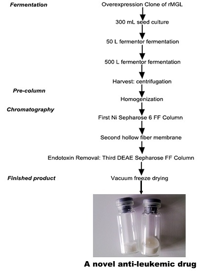
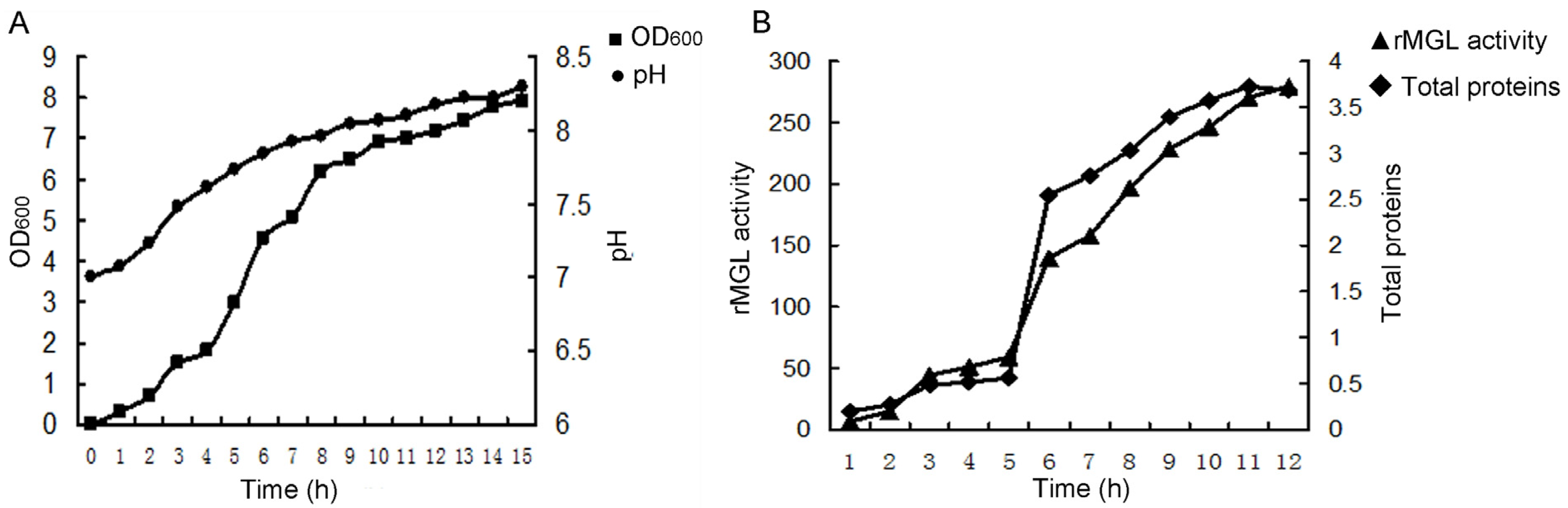
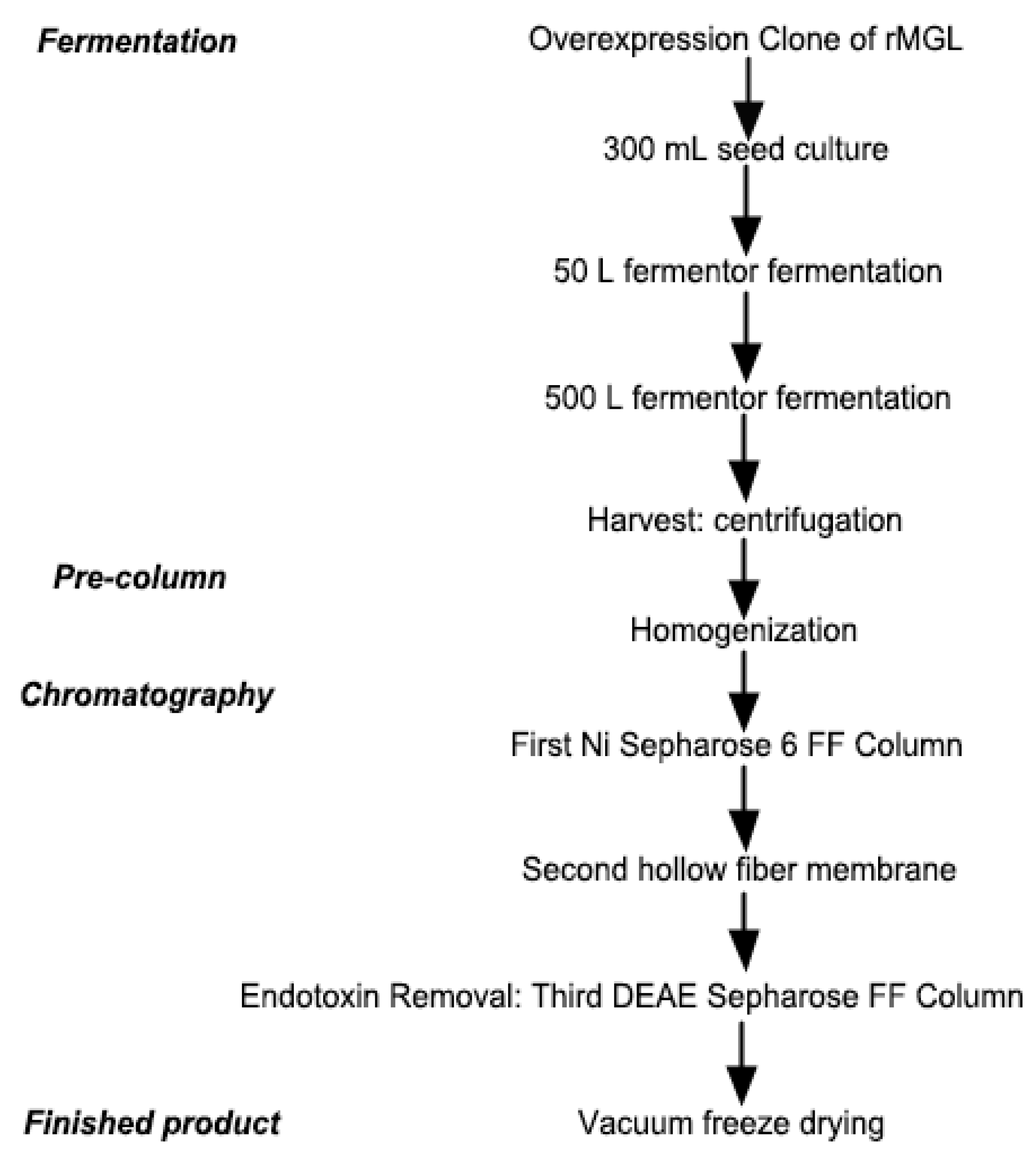
2.1. Large-Scale Fermentation for rMGL Production

Fermentation was conducted in a 500 L fermentor using 300 L of production medium. Cell growth increased linearly during 4–10 h of cultivation with the specific growth rate during 0.85–1 h at logarithmic growth phase. The pH of the medium increased from 7.0 to 8.3 during the fermentation phase (Figure 1A). As shown in Figure 1B, the rMGL activity increased up to 279.5 U/mL at 12 h. The total protein weight reached 3.68 mg/mL at the end of the fermentation. Cell growth increased linearly during 4–10 h of cultivation with the specific growth rate during 0.85–1 h at the logarithmic growth phase.
Figure 1. The time course of E. coli BL21 (DE3) fermentation in a 300 L production culture using a 500 L fermentor. (A) The cell growth (OD600) and the pH values during the fermentation process; (B) The rMGL activity (U/mL) and total proteins (mg/mL) at different times.
Marinedrugs 13 05492 g001

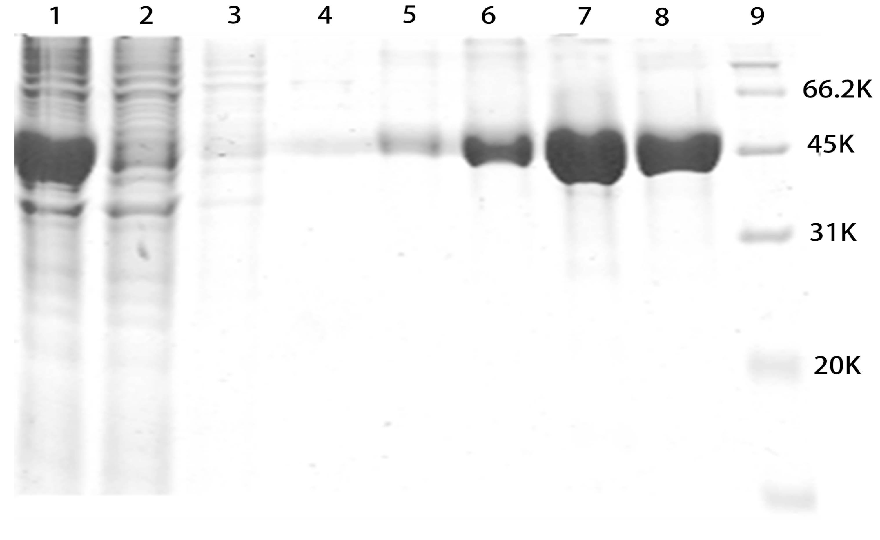
2.2. SDS-PAGE Analysis of Purified rMGL

After fermentation in the production culture, the cells were harvested by centrifugation and disrupted using a homogenizer. The cellular debris was removed by centrifugation. Crude enzyme was collected and applied to a Ni Sepharose 6 FF column. Unwanted proteins were washed away with 80 mM imidazole buffer. rMGL was eluted stepwise with buffers containing 160, 200, 300, and 400 mM imidazole. The elution fractions were collected and tested by sodium dodecyl sulfate polyacrylamide gel electrophoresis (SDS-PAGE). As shown in Figure 2, all of the elution fractions corresponded to 46 kDa on the SDS-PAGE.

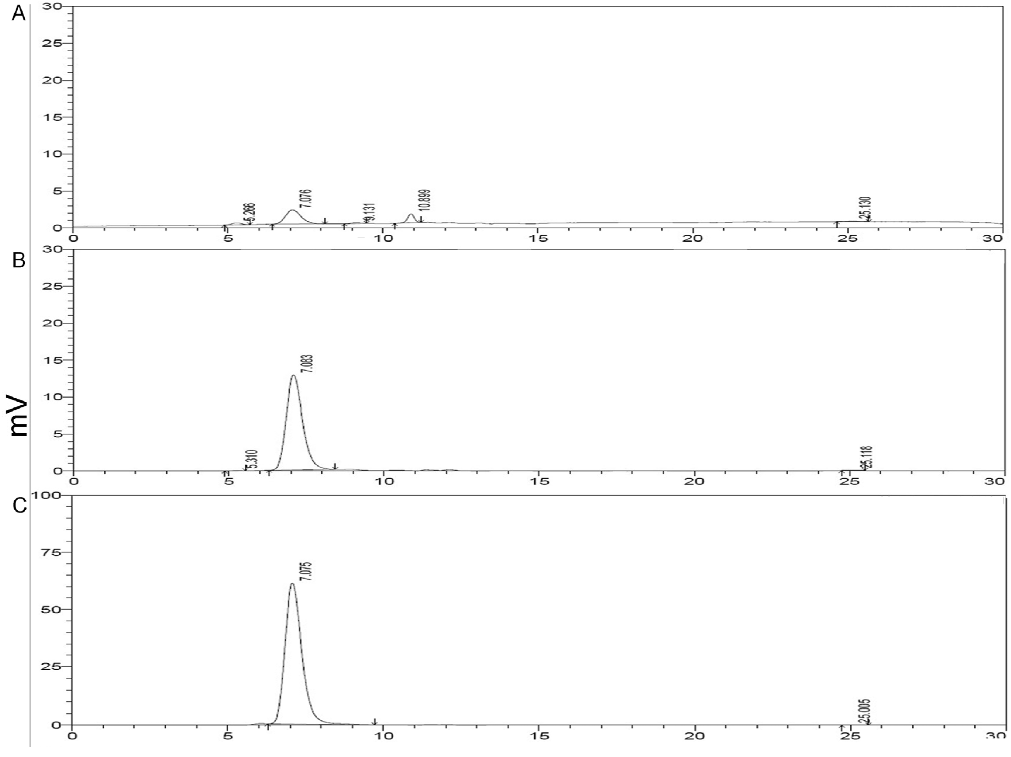
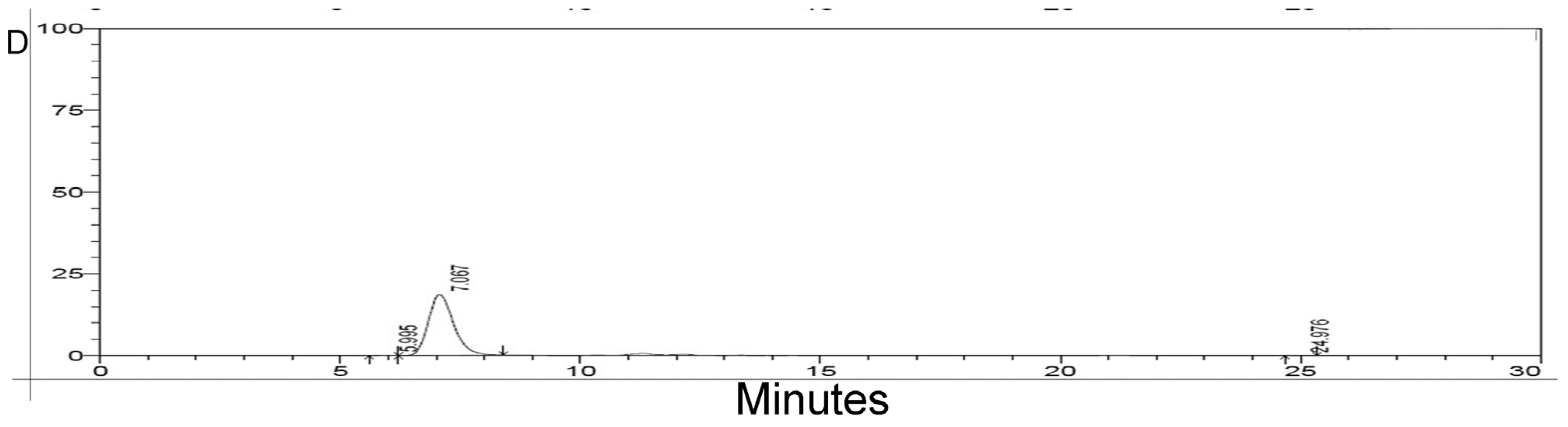
2.3. HPLC Analysis of Purified rMGL

As shown in Figure 3, the HPLC analysis demonstrated a major peak with a retention time of 7.08 min. The purity calculation indicated that the purity of rMGL eluted with 160 mM imidazole was 69.07%, while the purities of rMGL eluted with 200, 300, and 400 mM imidazole buffer were 99.34%, 99.76%, and 99.92%, respectively.
Figure 2. SDS-PAGE analysis of the samples. Lane 1, crude enzyme; Lane 2, flow through; Lane 3, 40 mM imidazole elution; Lane 4, 80 mM imidazole elution; Lane 5, 160 mM imidazole elution; Lane 6, 200 mM imidazole elution; Lane 7, 300 mM imidazole elution; Lane 8, 400 mM imidazole elution; Lane 9, marker.
Marinedrugs 13 05492 g002
Figure 3. HPLC analysis of purified rMGL. (A) rMGL eluted with 160 mM imidazole buffer; (B) rMGL eluted with 200 mM imidazole buffer; (C) rMGL eluted with 300 mM imidazole buffer; (D) rMGL eluted with 400 mM imidazole buffer.
Marinedrugs 13 05492 g003aMarinedrugs 13 05492 g003b

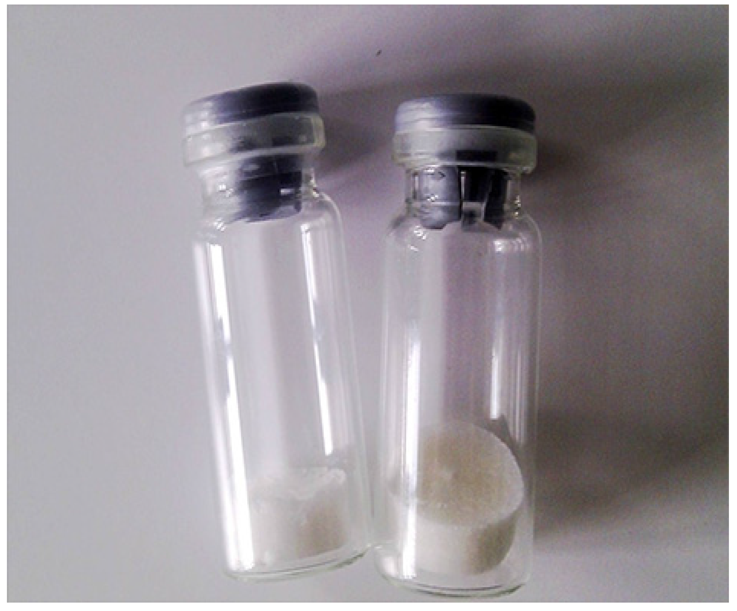
2.4. Lyophilization of rMGL

As shown in Figure A1, purified rMGL was lyophilized after desalinization and after the endotoxin was eliminated. The enzyme products are white powder. As shown in Table 1, rMGL purification is summarized. The specific activity of rMGL in lyophilized powder was 107.95 U/mg.
Table 1. rMGL purification.
StepVolume (L)Activity (U)Protein (g)SA * (U/mg)Yield (%)
HG *3075,600,000110468.4790
Ni-FF *1071,820,000567.57126.5485.50
DS * and CO *266,792,600544.86122.5979.52
EN *260,113,340506.72118.6371.56
Drying *--54,703,139506.72107.9557.25
* SA indicates specific activity; HG indicates homogenization; Ni-FF represents Ni Sepharose 6 Fast Flow; DS indicates desalination; CO indicates concentration; EN represents endotoxin removal; Drying represents vacuum freeze drying.

2.5. Comparison of rMGL Production from Various Strains

As shown in Table 2, rMGL production from various strains is summarized.
Table 2. Summary of rMGL production from various strains.
E. coli StrainPlasmidF * (L)rMGL (mg/mL)ContentActivity (U/mg)
MV1184 [4]pYH103----3.73.7
BL21(DE3) [5]pAC-10.81143
JM109 [6]pMGLTrc033002.14357
BL21(DE3) *pET-mgl5001.9553107.95
* F indicates fermentor; BL21(DE3) was used in this study.

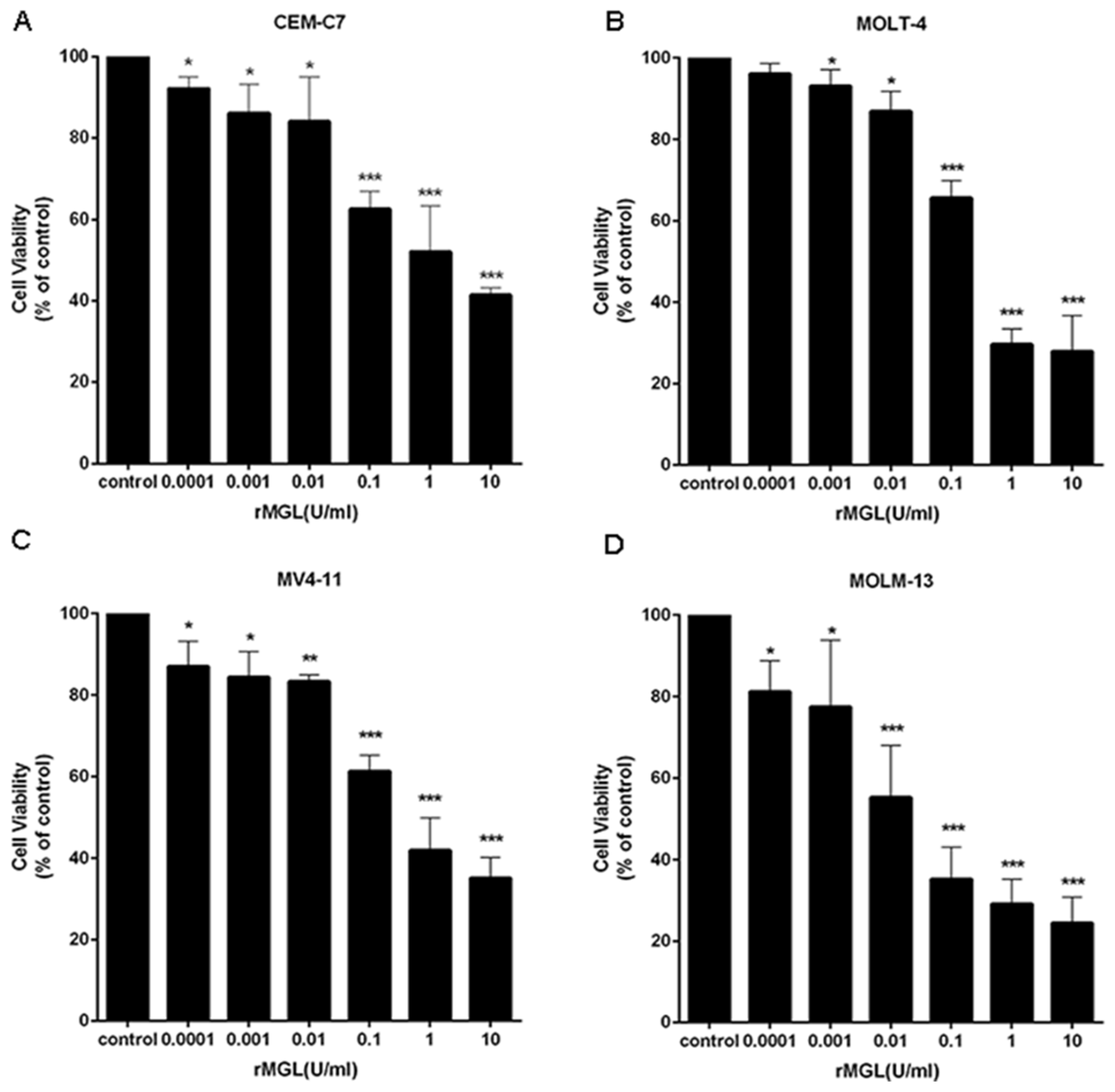
2.6. rMGL Inhibits the Proliferation of Leukemia Cell Lines

To determine the effects of rMGL on the proliferation of human leukemia cells, we tested the viability of leukemia cell lines exposed to various concentrations of rMGL for 48 h using the MTS assay. As shown in Figure 4, rMGL significantly suppressed the proliferation of leukemia cell lines in a dose-dependent manner.
Figure 4. rMGL suppresses the viability of leukemia cells CEM-C7 (A), MOLT-4 (B), MV4-11 (C) and MOLM-13 (D). The cell lines were exposed to different concentrations of rMGL for 48 h, and cell viability was evaluated using the MTS assay. The data are presented as the means ± s.d. of three independent experiments.
Marinedrugs 13 05492 g004

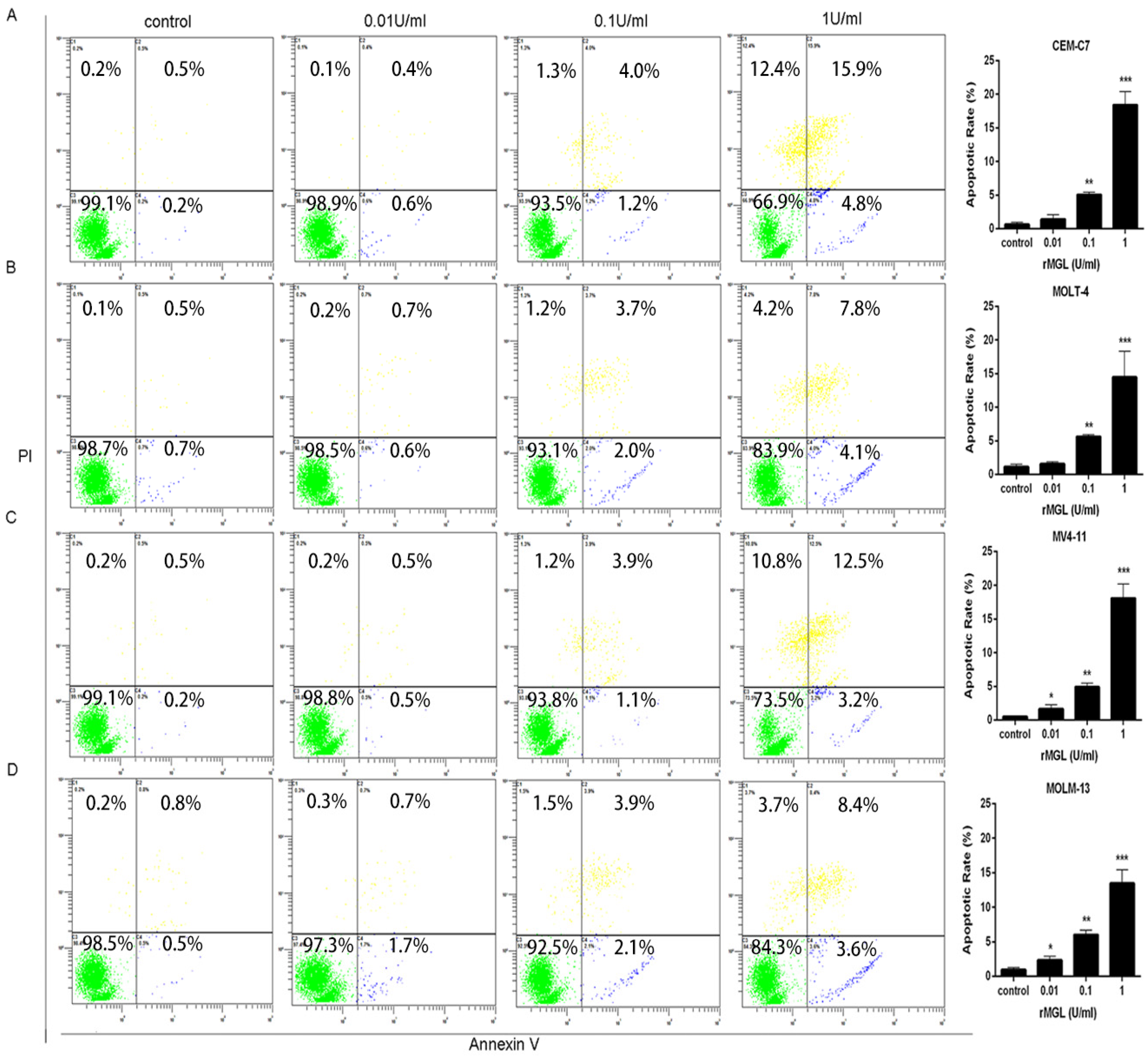
2.7. rMGL Induces Apoptosis of Leukemia Cell Lines

To examine the effects of rMGL on the apoptosis of leukemia cells, we tested the apoptotic rates of leukemia cells exposed to 0.01, 0.1, and 1 U/mL rMGL for 48 h by flow cytometry. rMGL treatment resulted in obvious increases in both early and late apoptotic populations in leukemia cells compared with the control group (Figure 5).
Figure 5. rMGL promotes cell apoptosis. The apoptotic rate was tested using Annexin V-FITC/PI staining and flow cytometry in the leukemia cell lines CEM-C7 (A), MOLT-4 (B), MV4-11 (C) and MOLM-13 (D) exposed to 0.01, 0.1, and 1 U/mL rMGL for 48 h. The results are presented as the means ± s.d. of three independent experiments.
Marinedrugs 13 05492 g005

3. Discussion

Methionine is necessary for cancer cells compared to normal cells both in vitro and in vivo [17,18,19,20,21,22]. Based on previous reports suggesting that methioninase has anti-carcinogenic properties on some types of cancers, methioninase may be used to target some malignancies with minimal toxicity [23,24,25]. Therefore, the large-scale production of recombinant methioninase was necessary. In this study, we showed an industrial manufacturing process of rMGL by high-level gene expression, large-scale fermentation, purification, and lyophilization (Figure A2). The results revealed that cell growth markedly increased during large-scale fermentation. Additionall, rMGL activity reached a maximum at 279.5 U/mL (Figure 1). After fermentation in the production culture, the analysis of purified rMGL tested by SDS-PAGE and HPLC showed that the purities of rMGL eluted with 200, 300, and 400 mM imidazole buffer were all more than 99% (Figure 3). After the purified rMGL was desalinated and the endotoxin eliminated, the final product was lyophilized as a white powder (Figure A1). High-level expression and the purification process of the enzyme provided a cost-effective manufacturing process. Table 2 summarizes the comparison of rMGL expressed in various transformants. We were able to obtain 1.95 g/L of rMGL as a soluble form using a 500 L fermentor. The content reached 53% of the total proteins in the soluble fraction in this research. In this study, the specific activity of rMGL was 107.95 U/mg, which was much greater than values reported by Tan et al. [5] and Takakura et al. [6].
The majority of AML patients who received intensive chemotherapy did not achieve complete remission. The relapse rate was high, and the overall five-year survival rate was almost 25%. Patients who were older than 60 years had a poorer prognosis [26,27]. Therefore, it was very important to discover and identify new therapeutic methods for the majority of AML patients. ALL is the most common childhood malignancy and comprises nearly one-third of all pediatric malignancies [28]. Although modern treatment strategies achieved improvement, nearly 20% of the children with ALL relapsed or died [29,30,31,32]. The capacity of leukemic cells to proliferate quickly and survive has been associated with disease progression [33]. Therefore, acting on anti-proliferative signals or pro-apoptotic signals may constitute novel therapies for acute leukemia [34,35,36]. Evidence suggests that methioninase has anti-tumor properties in some types of cancers [37,38]; we hypothesized that rMGL might have an anti-leukemic effect on leukemia cells.
In this study, we investigated the anti-leukemic activities of rMGL in ALL and AML cell lines. The proliferation test was conducted, and we observed that rMGL significantly inhibited leukemia cell growth (Figure 4). The results showed that rMGL significantly promoted leukemia cell apoptosis in a dose-dependent manner (Figure 5). Several reports have described the association between cellular apoptosis and cancer [39,40], and apoptosis induced by rMGL could be developed for the management of acute leukemia.

4. Materials and Methods

4.1. Materials

Nickel (Ni) Sepharose 6 FF and DEAE Sepharose FF were obtained from Pharmacia Biotech Inc. (Piscataway, NJ, USA). Phenylmethanesulfonyl fluoride (PMSF) and imidazole were purchased from Sigma (St. Louis, MO, USA). Peptone was purchased from OXOID (Basingstoke, Hampshire, UK), and yeast extract was purchased from Angel Yeast (Angel Yeast Co., Ltd, Yichang, China). Other reagents used in this research were analytical reagent grade or the highest grade commercially available.

4.2. Bacteria Strains and Media

The MGL gene from the deep-sea sediment metagenomic library was cloned and expressed in E. coli with vector PET-28a (+) containing the T7 promoter (Merck Millipore Corporation, Darmstadt, Germany). The expression conditions of the rMGL protein have been optimized. The seed medium included peptone (10 g/L), yeast extract (5 g/L), and NaCl (5 g/L). The production medium contained glycerol (4 g/L), peptone (8 g/L), yeast extract (8 g/L), and NaCl (6 g/L).

4.3. Large-Scale Production of rMGL

A flowchart for large-scale production of rMGL is shown in Figure A2.

4.3.1. Fermentation

(1) First preculture: The rMGL strain was maintained as a frozen vegetative stock at −80 °C in 20% glycerol. A total of 30 μL of bacteria from a vial was inoculated into 300 mL of fresh seed medium containing kanamycin and was shaken at 37 °C overnight.
(2) Second preculture: The first preculture broth was transferred to a 30 L seed medium containing 25 μg/mL kanamycin in a 50 L fermentor and was cultivated at 37 °C under agitation at 200 rpm with aeration at a rate of 25 L/min for 4 h.
(3) Production culture: The second preculture broth was transferred to 300 L of medium containing 25 μg/mL kanamycin in a 500 L fermentor under agitation at 250 rpm with aeration at a rate of 25 L/min for 3 h. Then, the isopropyl-β-d-thiogalactopyranoside (IPTG) was added at a final concentration of 0.1 mM and incubated for another 3 h at 25 °C before harvesting.

4.3.2. Preparation of the Crude Enzyme

After fermentation for 6 h in the production culture, the bacterial cells were harvested by centrifugation at 8000 rpm at 4 °C for 15 min and concentrated as a cell suspension. The cell suspensions were diluted with ice-cold extraction solution (100 mM potassium phosphate (pH 7.4) containing 40 mM imidazole, 0.5 M NaCl, 0.01% dithiothreitol, 0.1 mM PMSF, and 10 μM pyridoxal phosphate (PLP)). Then, the cell suspensions were disrupted twice with passages through a high-pressure mechanical homogenizer (Invensys Systems APS, Albertslund, Denmark) at 10,000 psi. The cellular debris was removed by centrifugation at 18,000 rpm at 4 °C for 15 min. The recombinant enzyme that was produced intracellularly in a soluble form was accumulated in the clarified supernatant. The pH of the concentrated crude enzyme was adjusted to 7.4 with 10 M NaOH.

4.3.3. Chromatographic Conditions

(1) First column: Ni Sepharose 6 FF (Pharmacia Biotech Inc., Piscataway, NJ, USA). The first column was 100 mm in diameter and 30 cm in height with a volume of 2000 mL of Ni Sepharose 6 FF. The column was washed with equilibration buffer (100 mM sodium phosphate, 80 mM imidazole, 0.5 M NaCl, pH 7.4). A total of 30 L of crude enzyme supernatant was applied to the column. After loading, the column was prewashed with equilibration buffer for approximately five volumes until the OD280 dropped below 0.01. The protein was then eluted stepwise with equilibration buffer containing 80, 160, 200, 300, and 400 mM imidazole. The elution fractions containing the enzyme were collected in 10-L volumes.
(2) Desalination and concentration: A hollow fiber membrane (molecular weight cut-off of 20 kDa, Xubang Membrane Equipment Co. Ltd, Beijing, China) was used for desalination. After 10 L of the solution containing enzyme was applied to the hollow fiber column, which had been equilibrated with 5 mM sodium phosphate buffer (pH 7.4), the enzyme was eluted with the same buffer at a flow rate of 30 L/h. The enzyme fractions were desalted and concentrated to 2 L. Then, 10 mM PLP was added to the solution at a final concentration of 10 μm.
(3) Endotoxin removal: To eliminate endotoxin, 2 L of the concentrated enzyme solution was applied on a 1000 mL DEAE Sepharose FF column (Pharmacia Biotech Inc., Piscataway, NJ, USA). The enzyme was eluted with 5 mM sodium phosphate buffer (pH 7.4) at a flow rate of 10 mL/min. The enzyme fractions, identified by their yellow color, were collected until the OD280 dropped below 0.1.

4.3.4. Finished Product

Following the above step, the enzyme solution was sub-packaged into a 10 mL Cillin glass vial (Jiangsu Huayue Pharmaceutical Glass Co., Ltd. Danyang, China) at 2 mL per vial. All of the samples were then put into a lyophilizer to be frozen at a speed of 1 °C/min until reaching −35 °C. After they were fully frozen, the samples were dried at −28 °C under vacuum conditions, followed by desorption drying at 25 °C.

4.4. Analysis of rMGL

4.4.1. Biomass

Cell growth during fermentation was monitored by measuring the OD600 with a spectrophotometer (UV-1600; Shimadzu, Kyoto, Japan). The biomass was determined with dry cell weight according to the method of Takakura et al. [6]. A total of 5 mL of the culture broth was centrifuged (8000× g, 10 min) in a pre-weighted glass test tube. Then, the cell pellet was dried at 90 °C until a constant weight was achieved.

4.4.2. Protein Concentration

The protein concentration was determined using a Bradford Assay Method with a Bradford Protein Assay Kit (Sangon, Shanghai, China) with bovine serum albumin as the standard protein.

4.4.3. Activity Assay

The rMGL activity was determined by the method of Tanaka et al. [3]. One unit of rMGL was defined as the amount of enzyme that produced 1 μmol of α-ketobutyrate per minute at an infinite concentration of l-Methionine. The specific activity was represented as the enzyme activity in units per milligram of protein.

4.4.4. HPLC

An Agilent high-performance liquid chromatography (HPLC) 1200 system (Agilent Technologies, Palo Alto, CA, USA) with a Supelco Progel TSK G3000SW column (30 cm × 7.5 mm) (Supelco, Bellefonte, PA, USA) was used for this HPLC experiment. A total of 20 μL of sample was loaded and eluted with elution solution (0.1 M sodium phosphate buffer, 0.1 M sodium chloride, and pH 7.0) at a flow rate of 1.0 mL/min.

4.5. Cell Lines and Cell Culture

CEM-C7, MOLT-4, MV4-11, and MOLM-13 cell lines were purchased from the American Type Culture Collection (ATCC, Manassas, VA, USA). All of the cells were routinely grown in a humidified atmosphere with 5% CO2 at 37 °C and were maintained in Roswell Park Memorial Institute (RPMI) 1640 medium supplemented with 10% fetal bovine serum (HyClone, Logan, UT, USA). The characteristics of the cell lines are detailed in Table 3.
Table 3. Characteristics of the cell lines used in this study.
Cell Line
CharacteristicsCEM-C7MOLT-4MV4-11MOLM-13
Cell typeT-ALLT-ALLAMLAML
Primary sitePB *PBPBPB
IC50 * (U/mL)1.700.300.640.03
* PB indicates peripheral blood; IC50 represents half-inhibitory concentration and was calculated using GraphPad Prism 6 software with triplicate values from MTS (3-(4,5-dimethylthiazol-2-yl)-5-(3-carboxymethoxyphenyl)-2-(4-sulfophenyl)-2H-tetrazolium) assays.

4.6. Cell Proliferation Assay

The anti-leukemic activities were tested using an MTS assay (Promega, Madison, WI, USA). A total of 1 × 10⁴ leukemia cells per well were treated with different concentrations of rMGL. After 48 h of incubation, the cell viability was determined using an MTS assay according to the manufacturer’s instructions.

4.7. Apoptosis Assay

The apoptotic rate was tested using the Annexin V-Fluorescein Isothiocyanate (FITC) Apoptosis Detection Kit (Nanjing Keygen, Nanjing, China). The cells were cultured at 1 × 10⁶ cells/mL and treated with rMGL (0.01, 0.1, and 1.0 U/mL). After 48 h of incubation, the cells were collected and stained with Annexin V-FITC and propidium iodide (PI) according to the manufacturer’s instructions. The untreated cells were used as the control. The rate of apoptosis was assessed using flow cytometry (Cytomics FC500 Flow Cytometer, Beckman Coulter Inc., Brea, CA, USA).

4.8. Statistical Analysis

The statistical analysis was performed using SPSS 13.0 software (Chicago, IL, USA). The data are presented as the means ± s.d. The differences between two groups were analyzed using the Student’s t-test when the data were normally distributed. If the data were not normally distributed, the differences were determined using the Wilcoxon rank sum test. The differences between multiple groups were analyzed using one-way analysis of variance (ANOVA) when the data were from normal populations and showed homogeneity of variance; alternatively, the Kruskal-Wallis test was performed. Multiple comparisons were analyzed using the Bonferroni test. All of the tests were two-sided. The differences were considered statistically significant at * p < 0.05, ** p < 0.01, and *** p < 0.001.

5. Conclusions

In this report, comprehensive optimization, including high-level expression, large-scale production, effective purification, desalination, and lyophilization, resulted in the cost-effective production of rMGL for therapeutic use. We investigated the anti-leukemic activities of rMGL in leukemia cell lines. The results showed that rMGL significantly inhibited the proliferation and induced apoptosis of leukemia cells in a dose-dependent manner. The data suggest that rMGL, used alone or in combination with other therapeutic methods, might be a promising drug for the treatment of patients with acute leukemia.

Acknowledgments

This work was supported by Guangzhou Science & Technology Plan (2008J1-C241) and Guangzhou Key Laboratory of Construction Project (2014SY000003).

Author Contributions

K.-Y.H., H.-Y.H., Y.-L.T., F.-G.X., X.-Q.L. and J.-Z.L. designed the study and interpreted the results. K.-Y.H., H.-Y.H. and Y.-L.T. drafted and revised the manuscript. K.-Y.H., H.-Y.H., Y.-L.T., F.-G.X., X.-Q.L. and J.-Z.L. carried out experiments and participated in data analysis. All authors read and approved the final manuscript.

Conflicts of Interest

The authors declare no conflicts of interest.

Appendix

Figure A1. rMGL powder after vacuum freeze drying.
Marinedrugs 13 05492 g006
Figure A2. The flowchart for large-scale production of rMGL.
Marinedrugs 13 05492 g007

References

  1. Tanaka, H.; Esaki, N.; Soda, K. Bacterial l-Methionine gamma-lyase: Characterization and application. Prog. Clin. Biol. Res. 1983, 125, 365–377. [Google Scholar] [PubMed]
  2. Tanaka, H.; Esaki, N.; Yamamoto, T.; Soda, K. Purification and properties of methioninase from Pseudomonas ovalis. FEBS Lett. 1976, 66, 307–311. [Google Scholar] [CrossRef]
  3. Tanaka, H.; Esaki, N.; Soda, K. Properties of l-Methionine gamma-lyase from Pseudomonas ovalis. Biochemistry 1977, 16, 100–106. [Google Scholar] [CrossRef] [PubMed]
  4. Inoue, H.; Inagaki, K.; Sugimoto, M.; Esaki, N.; Soda, K.; Tanaka, H. Structural analysis of the l-Methionine gamma-lyase gene from Pseudomonas putida. J. Biochem. 1995, 117, 1120–1125. [Google Scholar] [PubMed]
  5. Tan, Y.; Xu, M.; Tan, X.; Tan, X.; Wang, X.; Saikawa, Y.; Nagahama, T.; Sun, X.; Lenz, M.; Hoffman, R.M. Overexpression and large-scale production of recombinant l-Methionine-alpha-deamino-gamma-mercaptomethane-lyase for novel anticancer therapy. Protein Expr. Purif. 1997, 9, 233–245. [Google Scholar] [CrossRef] [PubMed]
  6. Takakura, T.; Ito, T.; Yagi, S.; Notsu, Y.; Itakura, T.; Nakamura, T.; Inagaki, K.; Esaki, N.; Hoffman, R.M.; Takimoto, A. High-level expression and bulk crystallization of recombinant l-Methionine gamma-lyase, an anticancer agent. Appl. Microbiol. Biotechnol. 2006, 70, 183–192. [Google Scholar] [CrossRef] [PubMed]
  7. Li, H.; Huang, Y.; Zhang, J.; Du, J.; Tan, H.; Lu, Y.; Zhou, S. Identification and characterization of a novel methionine γ-lyase gene from deep-sea sediment metagenomic library. World J. Microbiol. Biotechnol. 2011, 27, 2729–2736. [Google Scholar] [CrossRef]
  8. Hoffman, R.M. Methionine dependence in cancer cells—A review. in Vitro 1982, 18, 421–428. [Google Scholar] [CrossRef] [PubMed]
  9. Guo, H.; Lishko, V.K.; Herrera, H.; Groce, A.; Kubota, T.; Hoffman, R.M. Therapeutic tumor-specific cell cycle block induced by methionine starvation in vivo. Cancer Res. 1993, 53, 5676–5679. [Google Scholar] [PubMed]
  10. Mecham, J.O.; Rowitch, D.; Wallace, C.D.; Stern, P.H.; Hoffman, R.M. The metabolic defect of methionine dependence occurs frequently in human tumor cell lines. Biochem. Biophys. Res. Commun. 1983, 117, 429–434. [Google Scholar] [CrossRef]
  11. Cellarier, E.; Durando, X.; Vasson, M.P.; Farges, M.C.; Demiden, A.; Maurizis, J.C.; Madelmont, J.C.; Chollet, P. Methionine dependency and cancer treatment. Cancer Treat. Rev. 2003, 29, 489–499. [Google Scholar] [CrossRef]
  12. Hoffman, R.M. Development of recombinant methioninase to target the general cancer-specific metabolic defect of methionine dependence: A 40-year odyssey. Expert Opin. Biol. Ther. 2015, 15, 21–31. [Google Scholar] [CrossRef] [PubMed]
  13. Sharma, B.; Singh, S.; Kanwar, S.S. l-methionase: A therapeutic enzyme to treat malignancies. Biomed Res. Int. 2014, 2014, 506287. [Google Scholar] [CrossRef] [PubMed]
  14. Yano, S.; Li, S.; Han, Q.; Tan, Y.; Bouvet, M.; Fujiwara, T.; Hoffman, R.M. Selective methioninase-induced trap of cancer cells in S/G(2) phase visualized by FUCCI imaging confers chemosensitivity. Oncotarget 2014, 5, 8729–8736. [Google Scholar] [PubMed]
  15. Takakura, T.; Takimoto, A.; Notsu, Y.; Yoshida, H.; Ito, T.; Nagatome, F.; Ohno, M.; Kobayashi, Y.; Yoshioka, T.; Inagaki, K.; et al. Physicochemical and pharmacokinetic characterization of highly potent recombinant l-Methionine gamma-lyase conjugated with polyethylene glycol as an antitumor agent. Cancer Res. 2006, 66, 2807–2814. [Google Scholar] [CrossRef] [PubMed]
  16. Bhojwani, D.; Pui, C.-H. Relapsed childhood acute lymphoblastic leukaemia. Lancet Oncol. 2013, 14, E205–E217. [Google Scholar] [CrossRef]
  17. Strekalova, E.; Malin, D.; Good, D.M.; Cryns, V.L. Methionine deprivation induces a targetable vulnerability in triple-negative breast cancer cells by enhancing trail receptor-2 expression. Clin. Cancer Res. 2015, 21, 2780–2791. [Google Scholar] [CrossRef] [PubMed]
  18. Sinha, R.; Cooper, T.K.; Rogers, C.J.; Sinha, I.; Turbitt, W.J.; Calcagnotto, A.; Perrone, C.E.; Richie, J.P., Jr. Dietary methionine restriction inhibits prostatic intraepithelial neoplasia in TRAMP mice. Prostate 2014, 74, 1663–1673. [Google Scholar] [CrossRef] [PubMed]
  19. Kreis, W.; Goodenow, M. Methionine requirement and replacement by homocysteine in tissue cultures of selected rodent and human malignant and normal cells. Cancer Res. 1978, 38, 2259–2262. [Google Scholar] [PubMed]
  20. Poirier, L.A. The role of methionine in carcinogenesis in vivo. Adv. Exp. Med. Biol. 1986, 206, 269–282. [Google Scholar] [PubMed]
  21. Ghoshal, A.K.; Sarma, D.S.; Farber, E. Ethionine in the analysis of the possible separate roles of methionine and choline deficiencies in carcinogenesis. Adv. Exp. Med. Biol. 1986, 206, 283–292. [Google Scholar] [PubMed]
  22. Howarth, G.S.; Wang, H. Role of endogenous microbiota, probiotics and their biological products in human health. Nutrients 2013, 5, 58–81. [Google Scholar] [CrossRef] [PubMed]
  23. Tan, Y.; Xu, M.; Guo, H.; Sun, X.; Kubota, T.; Hoffman, R.M. Anticancer efficacy of methioninase in vivo. Anticancer Res. 1996, 16, 3931–3936. [Google Scholar] [PubMed]
  24. Zhao, R.; Domann, F.E.; Zhong, W. Apoptosis induced by selenomethionine and methioninase is superoxide mediated and p53 dependent in human prostate cancer cells. Mol. Cancer Ther. 2006, 5, 3275–3284. [Google Scholar] [CrossRef] [PubMed]
  25. Tan, Y.; Zavala, J., Sr.; Han, Q.; Xu, M.; Sun, X.; Tan, X.; Tan, X.; Magana, R.; Geller, J.; Hoffman, R.M. Recombinant methioninase infusion reduces the biochemical endpoint of serum methionine with minimal toxicity in high-stage cancer patients. Anticancer Res. 1997, 17, 3857–3860. [Google Scholar] [PubMed]
  26. Smith, M.; Barnett, M.; Bassan, R.; Gatta, G.; Tondini, C.; Kern, W. Adult acute myeloid leukaemia. Crit. Rev. Oncol. Hematol. 2004, 50, 197–221. [Google Scholar] [CrossRef] [PubMed]
  27. Kindler, T.; Lipka, D.B.; Fischer, T. FLT3 as a therapeutic target in AML: Still challenging after all these years. Blood 2010, 116, 5089–5102. [Google Scholar] [CrossRef] [PubMed]
  28. Stary, J.; Zimmermann, M.; Campbell, M.; Castillo, L.; Dibar, E.; Donska, S.; Gonzalez, A.; Izraeli, S.; Janic, D.; Jazbec, J.; et al. Intensive chemotherapy for childhood acute lymphoblastic leukemia: Results of the randomized intercontinental trial ALL IC-BFM 2002. J. Clin. Oncol. 2013, 31. [Google Scholar] [CrossRef] [PubMed]
  29. Inukai, T.; Kiyokawa, N.; Campana, D.; Coustan-Smith, E.; Kikuchi, A.; Kobayashi, M.; Takahashi, H.; Koh, K.; Manabe, A.; Kumagai, M.; et al. Clinical significance of early T-cell precursor acute lymphoblastic leukaemia: Results of the Tokyo Children’s Cancer Study Group Study L99-15. Br. J. Haematol. 2012, 156, 358–365. [Google Scholar] [CrossRef] [PubMed]
  30. Luo, X.-Q.; Ke, Z.-Y.; Huang, L.-B.; Guan, X.-Q.; Zhang, Y.-C.; Zhang, X.-L. High-risk childhood acute lymphoblastic leukemia in China: Factors influencing the treatment and outcome. Pediatr. Blood Cancer 2009, 52, 191–195. [Google Scholar] [CrossRef] [PubMed]
  31. Gao, C.; Zhao, X.-X.; Li, W.-J.; Cui, L.; Zhao, W.; Liu, S.-G.; Yue, Z.-X.; Jiao, Y.; Wu, M.-Y.; Li, Z.-G. Clinical features, early treatment responses, and outcomes of pediatric acute lymphoblastic leukemia in China with or without specific fusion transcripts: A single institutional study of 1004 patients. Am. J. Hematol. 2012, 87, 1022–1027. [Google Scholar] [CrossRef] [PubMed]
  32. Widjajanto, P.H.; Sutaryo, S.; Purwanto, I.; Ven, P.M.V.; Veerman, A.J.P. Early response to dexamethasone as prognostic factor: Result from Indonesian Childhood WK-ALL protocol in Yogyakarta. J. Oncol. 2012, 2012, 417941. [Google Scholar] [CrossRef] [PubMed]
  33. Obrador-Hevia, A.; Serra-Sitjar, M.; Rodriguez, J.; Villalonga, P.; Fernandez de Mattos, S. The tumour suppressor FOXO3 is a key regulator of mantle cell lymphoma proliferation and survival. Br. J. Haematol. 2012, 156, 334–345. [Google Scholar] [CrossRef] [PubMed]
  34. Essafi, M.; Baudot, A.D.; Mouska, X.; Cassuto, J.-P.; Ticchioni, M.; Deckert, M. Cell-penetrating TAT-FOXO3 fusion proteins induce apoptotic cell death in leukemic cells. Mol. Cancer Ther. 2011, 10, 37–46. [Google Scholar] [CrossRef] [PubMed]
  35. Ausserlechner, M.J.; Salvador, C.; Deutschmann, A.; Bodner, M.; Viola, G.; Bortolozzi, R.; Basso, G.; Hagenbuchner, J.; Obexer, P. Therapy-resistant acute lymphoblastic leukemia (ALL) cells inactivate FOXO3 to escape apoptosis induction by trail and noxa. Oncotarget 2013, 4, 995–1007. [Google Scholar] [PubMed]
  36. Hussain, A.R.; Al-Rasheed, M.; Manogaran, P.S.; Al-Hussein, K.A.; Platanias, L.C.; Al Kuraya, K.; Uddin, S. Curcumin induces apoptosis via inhibition of PI3'-kinase/AKT pathway in Acute T cell Leukemias. Apoptosis 2006, 11, 245–254. [Google Scholar] [CrossRef] [PubMed]
  37. Kudou, D.; Misaki, S.; Yamashita, M.; Tamura, T.; Takakura, T.; Yoshioka, T.; Yagi, S.; Hoffman, R.M.; Takimoto, A.; Esaki, N.; et al. Structure of the antitumour enzyme l-Methionine gamma-lyase from pseudomonas putida at 1.8 angstrom resolution. J. Biochem. 2007, 141, 535–544. [Google Scholar] [CrossRef] [PubMed]
  38. Cavuoto, P.; Fenech, M.F. A review of methionine dependency and the role of methionine restriction in cancer growth control and life-span extension. Cancer Treat. Rev. 2012, 38, 726–736. [Google Scholar] [CrossRef] [PubMed]
  39. Janssens, V.; Rebollo, A. The role and therapeutic potential of Ser/Thr phosphatase PP2A in apoptotic signalling networks in human cancer cells. Curr. Mol. Med. 2012, 12, 268–287. [Google Scholar] [CrossRef] [PubMed]
  40. He, L.; Yang, X.; Cao, X.; Liu, F.; Quan, M.; Cao, J. Casticin induces growth suppression and cell cycle arrest through activation of FOXO3a in hepatocellular carcinoma. Oncol. Rep. 2013, 29, 103–108. [Google Scholar] [PubMed]

