Effects of Lissoclibadins and Lissoclinotoxins, Isolated from a Tropical Ascidian Lissoclinum cf. badium, on IL-8 production in a PMA-stimulated Promyelocytic Leukemia Cell Line

Abstract
:1. Introduction
2. Materials and Methods
2.1. Materials
2.2. Cell lines and culture conditions
2.4. Detection of human IL-8 by ELISA
2.5. Determination of cell proliferation
2.6. Determination of cytotoxicity
3. Results
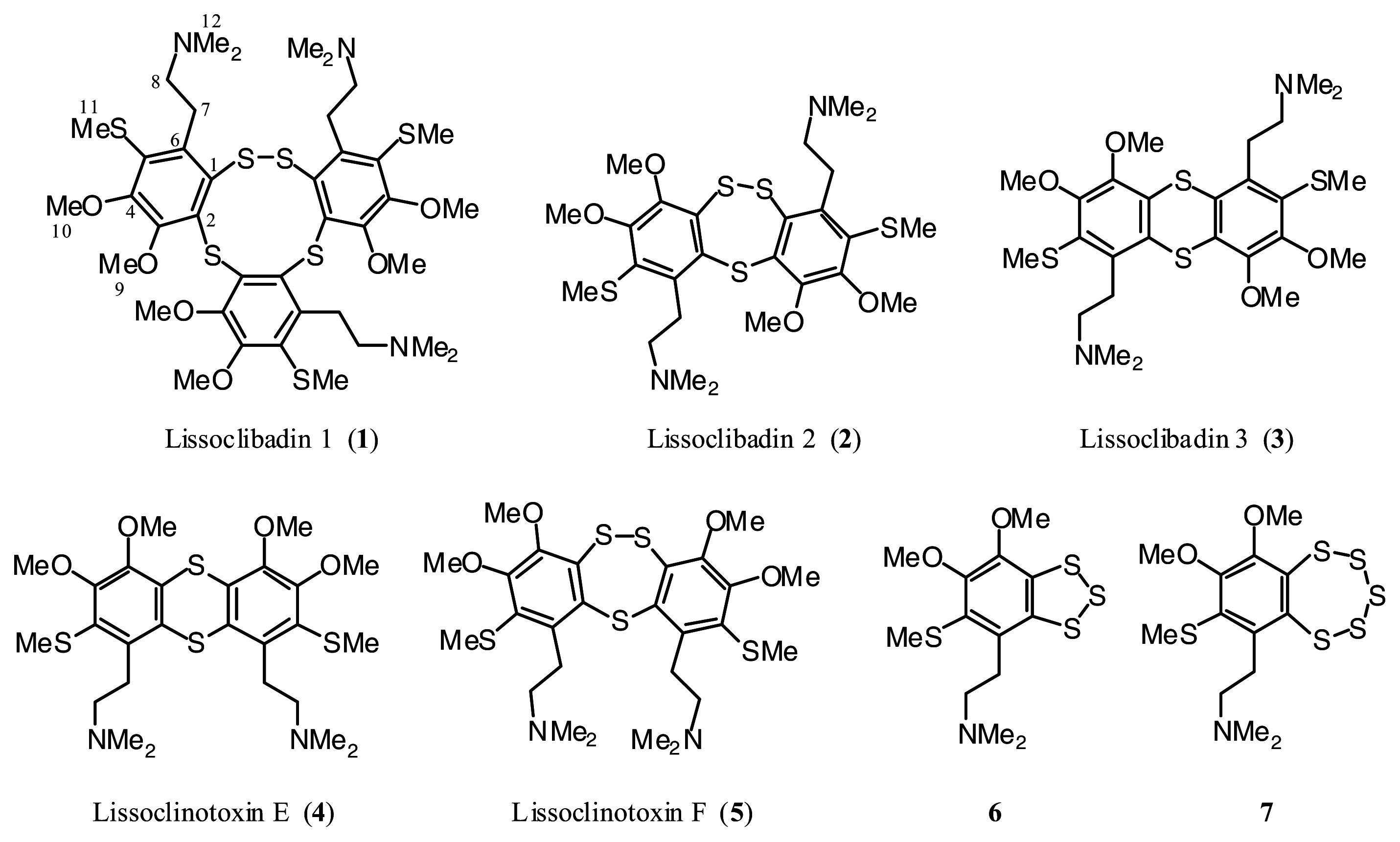
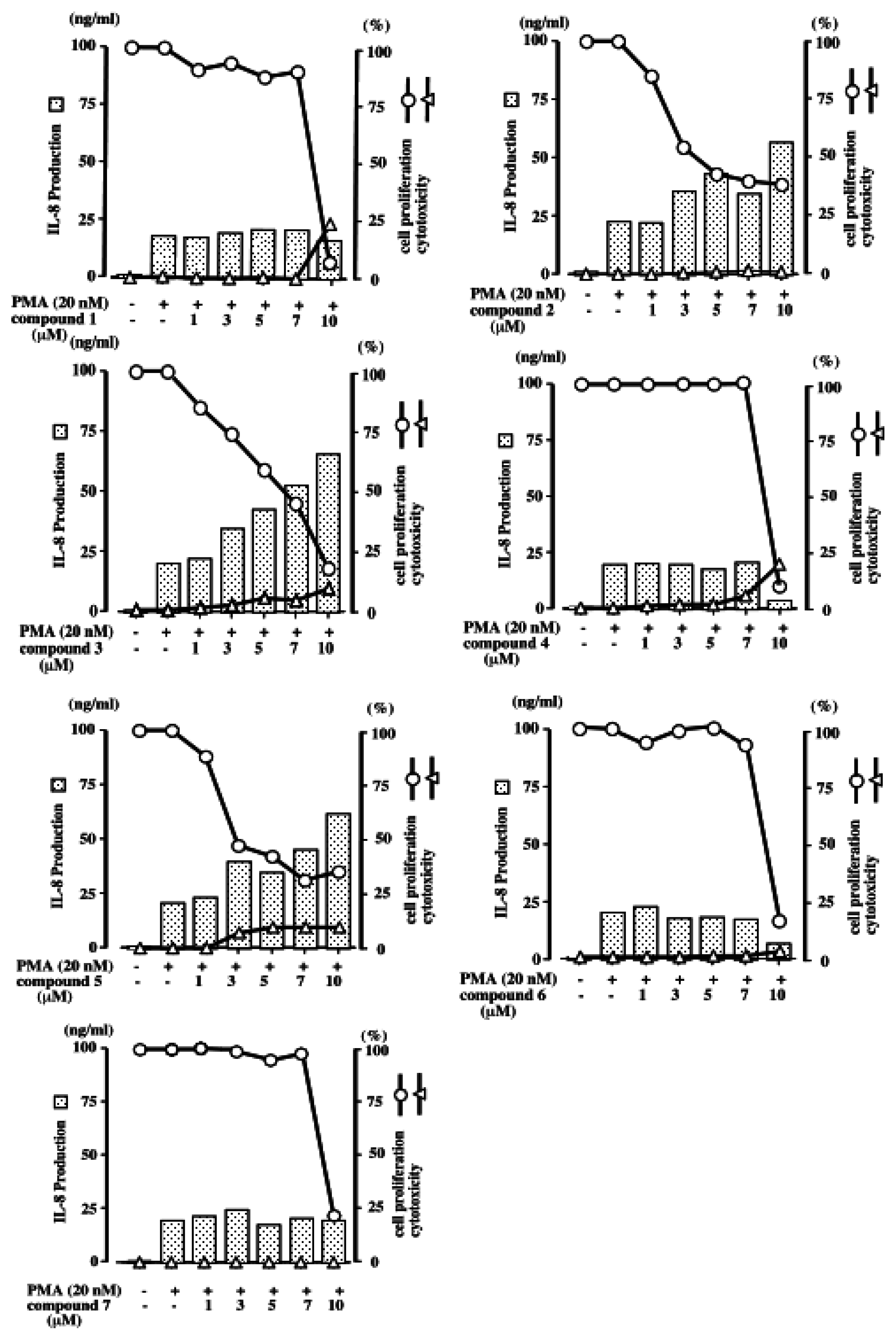
3.1. Effects of compounds 1–7 on IL-8 production by PMA-stimulated HL-60 cells
3.2. Inhibition of cell proliferation and cytotoxicity of compounds 1–7 against HL-60 cells
4. Discussion
Acknowledgements
References
- Blunt, J. W.; Copp, B. R.; Munro, M. H. G.; Northcote, P. T.; Prinsep, M. R. Marine natural products. Nat. Prod. Rep 2005, 22, 15–61. [Google Scholar]2004, 21, 1–49.2003, 20, 1–48.
- Faulkner, D. J. Marine natural products. Nat. Prod. Rep 2002, 19, 1–48. [Google Scholar]2001, 18, 1–49.2000, 17, 7–55.1999, 16, 155–198.1998, 15, 113–158.1997, 14, 259–302.1996, 13, 75–125.1995, 12, 223–269.1994, 11, 355–394.1993, 10, 497–539.1992, 9, 323–364.1991, 8, 97–147.1990, 7, 269–309.1988, 5, 613–576.1987, 4, 539–576.1986, 3, 1–33.1984, 1.
- Liu, H.; Pratasik, S. B.; Nishikawa, T.; Shida, T.; Tachibana, K.; Fujiwara, T.; Nagai, H.; Kobayashi, H.; Namikoshi, M. Lissoclibadin 1, a Novel Trimeric Sulfur-Bridged Dopamine Derivative, from the Tropical Ascidian Lissoclinum cf. badiu. Tetrahedron Lett 2004, 45, 7015– 7017. [Google Scholar]
- Liu, H.; Fujiwara, T.; Nishikawa, T.; Mishima, Y.; Nagai, N.; Shida, T.; Tachibana, K.; Kobayashi, H.; Mangindaan, R. E. P.; Namikoshi, M. Lissoclibadins 1–3, three new polysulfur alkaloids, from the ascidian Lissoclinum cf. badium. Tetrahedron 2005, 61, 8611–8615. [Google Scholar]
- Oda, T.; Sakakibara, Y.; Sato, Y.; Hanzawa, H.; Hata, T. Effects of (+)-, (−)- and (+−)-Indenestrols A and B on Microtubule Polymerization. Chem. Pharm. Bull 1992, 40, 588–592. [Google Scholar]
- Graves, D. T.; Jiang, Y. Molecular cloning and functional analysis of the promoter of the human squalene synthase gene. Crit. Rev. Oral Biol. Med 1995, 6, 109–118. [Google Scholar]
- Di Celle, P. F.; Carbone, A.; Marchis, D.; Zhou, D.; Sozzani, S.; Zupo, S.; Pini, M.; Mantovani, A.; Foa, R. Cytokine gene expression in B-cell chronic lymphocytic leukemia: evidence of constitutive interleukin-8 (IL-8) mRNA expression and secretion of biologically active IL-8 protein. Blood 1994, 84, 220–228. [Google Scholar]
- Green, A. R.; Green, V. L.; White, M. C.; Speirs, V. Expression of Cytokine messenger RNA in normal and neoplastic human breast tissue: identification of interleukin-8 as a potential regulatory factor in breast tumours. Int. J. Cancer 1997, 72, 937–941. [Google Scholar]
- Konig, B.; Steinbach, F.; Janocha, B.; Drynda, A.; Stumm, M.; Philipp, C.; Allhoff, E. P.; Konig, W. The differential expression of proinflammatory cytokines IL-6, IL-8 and TNF-alpha in renal cell carcinoma. Anticancer Res 1999, 19, 1519–1524. [Google Scholar]
- Galffy, G.; Mohammed, K. A.; Dowling, P. A.; Nasreen, N.; Ward, M. J.; Antony, V. B. Interleukin 8: an autocrine growth factor for malignant mesothelioma. Cancer Res 1999, 59, 367–371. [Google Scholar]
- Kasahara, T.; Oda, T.; Hatake, K.; Akiyama, M.; Mukaida, N.; Matsushima, K. Interleukin-8 and monocyte chemotactic protein-1 production by a human glioblastoma cell line, T98G in coculture with monocytes: involvement of monocyte-derived interleukin-1alpha. Eur. Cytokine Netw 1998, 9, 47–55. [Google Scholar]
- Carmichael, J.; DeGraff, W. G.; Gazdar, A. F.; Minna, J. D.; Mitchell, J. B. Evaluation of a tetrazolium-based semiautomated colorimetric assay: assessment of chemosensitivity testing. Cancer Res 1987, 47, 939–942. [Google Scholar]
- Shimizu, S.; Nomoto, M.; Naito, S.; Yamamoto, T.; Momose, K. Stimulation of nitric oxide synthase during oxidative endothelial cell injury. Biochem. Pharmacol. 1998, 55, 77–83. [Google Scholar]
- Patil, A. D.; Freyer, A. J.; Killmer, L.; Zuber, G.; Carte, B.; Jurewicz, A. J.; Johnson, R. K. Lissoclin disulfoxide, a novel dimeric alkaloid from the ascidian Lissoclinum sp.: inhibitor of interleukin-8 receptors. Nat. Prod. Lett 1997, 10, 225–229. [Google Scholar]
- Sample availability: not available.


© 2006 by MDPI Reproduction is permitted for noncommercial purposes.
Share and Cite
Oda, T.; Fujiwara, T.; Liu, H.; Ukai, K.; Mangindaan, R.E.P.; Mochizuki, M.; Namikoshi, M. Effects of Lissoclibadins and Lissoclinotoxins, Isolated from a Tropical Ascidian Lissoclinum cf. badium, on IL-8 production in a PMA-stimulated Promyelocytic Leukemia Cell Line. Mar. Drugs 2006, 4, 15-21. https://doi.org/10.3390/md401015
Oda T, Fujiwara T, Liu H, Ukai K, Mangindaan REP, Mochizuki M, Namikoshi M. Effects of Lissoclibadins and Lissoclinotoxins, Isolated from a Tropical Ascidian Lissoclinum cf. badium, on IL-8 production in a PMA-stimulated Promyelocytic Leukemia Cell Line. Marine Drugs. 2006; 4(1):15-21. https://doi.org/10.3390/md401015
Chicago/Turabian StyleOda, Taiko, Takeshi Fujiwara, Hongwei Liu, Kazuyo Ukai, Remy E. P. Mangindaan, Masataka Mochizuki, and Michio Namikoshi. 2006. "Effects of Lissoclibadins and Lissoclinotoxins, Isolated from a Tropical Ascidian Lissoclinum cf. badium, on IL-8 production in a PMA-stimulated Promyelocytic Leukemia Cell Line" Marine Drugs 4, no. 1: 15-21. https://doi.org/10.3390/md401015