Cnidarian Toxins Acting on Voltage-Gated Ion Channels

Marine Biological Laboratory, 7 MBL Street, Woods Hole, MA 02543, USA
*
Author to whom correspondence should be addressed.
Mar. Drugs 2006, 4(3), 70-81; https://doi.org/10.3390/md403070
Submission received: 21 February 2006
/
Accepted: 27 February 2006
/
Published: 6 April 2006
(This article belongs to the Special Issue Marine Drugs and Ion Channels)
Abstract
:Voltage-gated ion channels generate electrical activity in excitable cells. As such, they are essential components of neuromuscular and neuronal systems, and are targeted by toxins from a wide variety of phyla, including the cnidarians. Here, we review cnidarian toxins known to target voltage-gated ion channels, the specific channel types targeted, and, where known, the sites of action of cnidarian toxins on different channels.
Introduction
Voltage-gated ion channels underlie electrical excitability in cells, and they also play important roles in non-excitable cells [1]. Voltage-gated channels open in response to changes in membrane potential, allowing ions to flow down the electrochemical gradient across the cell membrane. They are thus gated (by voltage), and they form an ion-selective pore. Voltage-gated channels represent a protein superfamily that comprises more than 140 members, and is part of the even larger ion channel superfamily [1,2].
The main, pore-forming protein of voltage-gated ion channels is the α (or α1) subunit. It consists of four homologous domains (I–IV), each containing 6 predicted transmembrane regions (S1–S6), with a loop between S5 and S6 that reenters the membrane. These four domains surround a central pore, with this S5–S6 loop thought to serve as the ion selectivity filter of the pore. Voltage-gated potassium (KV) channels consist of single domains that form a tetramer of α subunits in the membrane [3]. In contrast, voltage-gated sodium (NaV) and calcium (CaV) α (or α1) subunits contain all four homologous domains in a monomeric structure [1,4,5].
The simplest ion channels consist of a tetrameric structure forming a central pore and selectivity filter [1,2,6]. These channels contain equivalents of the S5–S6 transmembrane regions along with the S5–S6 loop. The voltage-gated channel family is thought to have arisen through addition of an N-terminal voltage-gating domain (S1–S4) to this basic structure, with the four-domain NaV and CaV α subunits arising presumably via two rounds of gene duplication [1,7,8]. Incorporation of regulatory domains at the intracellular, C-terminus of the basic channel structure yields sites for gating and modulation of voltage-gated and voltage-gated-like channels by intracellular factors.
Any particular ion channel family (e.g., NaV channels) typically comprises a variety of different subtypes with particular physiological, pharmacological, and structural characteristics. For example, there are ten members of the mammalian CaV channel gene family, each of which has distinct roles in cellular signaling. The potential for expression of α1 splicing variants adds to this diversity, as does modulation of channel function by auxiliary subunits and second messenger pathways [9–14].
Voltage-gated channels are critical to normal neuromuscular transmission. Disruption of their normal function can lead to rapid paralysis. As such, they represent excellent targets for toxins from a variety of organisms including snakes [15], arthropods [16], molluscs [17,18,19], and, the subject of this review, cnidarians [20]. These toxins can serve as remarkably powerful molecular probes of channel structure-function relationships, as well as models for studying molecular evolution of toxins and targets in predators and prey [21]. Some ion channel toxins may also prove to have therapeutic value, as is the case for toxins from cone snails that target a particular Ca2+ channel subtype and are currently being investigated as candidate drugs for their antinociceptive and other potential beneficial effects [18,19,22].
Cnidarian Ion Channel Toxins
The cnidarians are the earliest extant organisms with a neuromuscular system. They are simple carnivores that are radially symmetrical, with a mouth surrounded by tentacles. These tentacles contain high concentrations of stinging cells called nematocysts (cnidocysts), which are specialized epidermal cells that produce, store, and inject toxins used for protection and predation. Venoms contain a bouquet of substances, including peptides, proteins, phospholipids, phospholipases, glycoproteins, sterols, bioactive amines, and carbohydrates [23]. Thus, the cnidarians represent a rich source of venoms and toxins. The phylum comprises four extant classes: the hydrozoans; the cubozoids, or box jellies; the scyphozoans, or true jellyfish; and the anthozoa, the sea anemones and corals.
Sea anemone toxins dominate in terms of number identified and they are also the best characterized in terms of mechanism of action [20]. To date, many cnidarian toxins that act on NaV and various KV channels have been described in detail; toxins from cnidarians that clearly act on CaV channels have thus far not been characterized.
NaV Channel Toxins
Some of the most thoroughly studied cnidarian toxins are polypeptide toxins from sea anemones that target NaV channels [20,24,25,26]. These toxins are polypeptides of ~5 kDa, and more than 50 different sea anemone NaV channel toxins have been isolated or cloned from a large variety of species. Particular toxins, including ATX II from Anemonia sulcata[27], and Anthopleurin A (ApA) [28] and Anthopleurin B (ApB) [29] from Anthopleura xanthogrammica, have proven especially useful as probes of NaV channel structure and function. Furthermore, recent studies have provided evidence for in vivo epileptogenic effects of toxins (e.g., cangitoxin from Bunodosoma cangicum) [30].
Norton [25] described the classification of anemone NaV channel toxins into different types based on their amino acid sequences (Fig. 1). Type 1 and Type 2 toxins are polypeptides of 46–49 amino acids; Type 3 peptides, from the genus Entacmaea (family Actiniidae), are shorter, containing 27–32 amino acids. Calitoxins I and II from Calliactis parasitica[31,32] are long peptides that do not fit clearly into Types 1 or 2, and may constitute a fourth class. Members of the Type 1 and Type 2 classes show extensive sequence similarity within each class (≥60%), but sequence similarity between members of the two classes is significantly lower (~30%), and there is no immunological cross-reactivity between the two groups. Members of both classes contain six conserved cysteine residues that are cross-linked by three disulfide bridges, and both have basic C-terminal sequences, though more markedly for Type 2 toxins (Fig. 1). The Type 1 and 2 toxins are largely distributed according to taxonomy. Toxins from the Actiniidae family are Type 1, while both types of toxins can be found in the family Stichodactylidae [20,25]. A single species can produce toxins from both classes, as is seen with Gigantoxin II and Gigantoxin III from Stichodactyla gigantea[33].
Anemone NaV channel toxins were initially characterized as cardiac stimulants [34] and neurotoxins [35], with their predominant effect based on relative affinities for cardiac or neuronal NaV isoforms. They were subsequently shown to interact with neurotoxin receptor site 3 of NaV channels, with their action being the delay of channel inactivation [36–40]. The open state of the channel is thus prolonged during depolarization. Neurotoxin receptor site 3 is one of at least six neurotoxin receptor sites on NaV channels. It is also a receptor for α-scorpion toxins, which do not share sequence homology with the anemone toxins. Receptor site 3 is a complex receptor site that includes the extracellular loop between transmembrane segments S3 and S4 in domain IV of the channel [41]. The toxins bind to this site via electrostatic interactions, predominantly with the negatively charged E1613 residue in rat brain NaV channels [42] and D1612 in cardiac NaV channels [43]. Contributions from other, nearby residues [44] and unidentified contacts in the IS5–S6 and IVS5–S6 loops [45,46] are also likely involved in interaction with these toxins. Interestingly, unlike toxins that interact with receptor site 4, ApB and other site 3 toxins do not bind phospholipids [47].
Work on the ApB toxin has defined a region critical for interaction with NaV channels. ApB, like other Type 1 toxins, has a conformation that is primarily a β-structure, with a four-stranded antiparallel β sheet linked by β-turns and loops with no α-helix [48]. A disordered region corresponding to residues 8–17 of ApB and found in all active sea anemone toxins is referred to as the Arg-14 loop [49]. This region appears to be critical for interaction with receptor site 3 of the NaV channel. Furthermore, the relative non-selectivity of ApB for cardiac and neuronal channels versus the relative selectivity of ApA for cardiac channels is associated with two cationic residues (R12, K49) found in ApB but not in ApA [50].
KV Channel Toxins
There have been at least 11 KV channel toxins that have been identified in various sea anemones to date. These peptide toxins fall into three classes based on structural and functional differences (Table 1, Fig. 2).
Type 1 toxins potently block KV1 (Shaker) channels. They contain 35–37 amino acids that form two nearly perpendicular stretches of helices and include three disulfide bridges [58, 59]. A conserved dyad of lysine (K22 in ShK) and tyrosine (Y23 in ShK) appear to be essential residues required for interaction with rat brain KV channels [60]. A similar result is found for the BgK toxin [56, 58, 59]. Recently, use of ShK and analogs to selectively block KV1.3, a lymphocyte KV channel that is important for the activation of terminally differentiated effector memory (TEM) T cells, is being investigated as a candidate treatment for multiple sclerosis and other autoimmune diseases [63].
The Type 2 KV channel toxins, represented by the AsKCs, are made up of 58 or 59 amino acid residues, and also block KV1 channels, though with less potency than Type 1 toxins [64]. They have sequence homology to dendrotoxins, KV channel toxins from snake venom; they also show homology to Kunitz-type protease inhibitors, and exhibit protease inhibitory activity.
Type 3 KV channel toxins include BDS-I and BDS-II from A. sulcata[65], and APETx1 from Anthopleura elegantissima[66]. BDS-I and II are selective blockers of KV3 (Shaw) channels, predominantly the fast-inactivating KV3.4 channel. APETx1 is highly similar (54%) to the BDS toxins. However, unlike the BDS toxins, it selectively inhibits ether-a-go-go-related gene (ERG) KV channels such as the human ERG (HERG; KV11.1) channel, an essential component of cardiac cells that controls the duration of the plateau phase of the action potential. APETx1 acts by modifying the voltage-dependence of HERG gating [66].
Concluding remarks
Although sea anemone toxins have provided a wealth of pharmacological probes, toxins from only a relatively few cnidarian species have thus far been characterized. Novel venoms from other anemone species as well as other cnidarians have the potential to provide even more powerful tools, as well as possible pharmaceutical applications. For example, CaV channels are critical components of the neuromuscular junction and of neurosecretory activity, and are targeted by toxins from a wide variety of organisms. Interestingly, however, purified cnidarian toxins with clear selectivity for CaV channels have not yet been described. A potential candidate CaV toxin may be the Bainh toxin isolated from Bunodosoma granulifera[72]. This toxin increases the force of contraction in mammalian ventricular muscle preparations, an effect that appears to be mediated by a voltage-independent, toxin-induced increase in L-type CaV currents.
Several non-anemone venoms produce perturbations in ion transport across excitable membranes [73], and potential CaV toxins may be present in the crude venoms of the hydrocoral Millepora complanata[74] and the sea nettle Chrysaora quinquecirrha[75]. SNTX, a toxin also from C. quinquecirrha, creates large cation-selective channels that open and close spontaneously and show voltage-dependence of open probability [76]. It is thus likely that as more venoms from different cnidarian species are examined in greater detail, a wider diversity of ion channel toxins will emerge.
Acknowledgments
We thank William Morgan, Peter A.V. Anderson, and Joseph Consiglio for helpful discussions. S.M.M. and R.M.G. were supported by NIH grant AI 40522 and NIH/NSF Woods Hole Center for Oceans and Human Health grant WHOI-A100354/A100360. R.M.G. was also supported by the MBL Neal Cornell Research Fund fellowship.
Abbreviations
| KV channel | voltage-gated potassium channel |
| NaV | voltage-gated sodium channel |
| CaV | voltage-gated calcium channel |
| ApA | Anthopleurin A |
| ApB | Anthopleurin B |
| ATX II | Anemone sulcata toxin II |
| Bg II | Bunodosoma granulifera toxin II |
| Sh I | peptide neurotoxin I from Stichodactyla helianthus |
| RP II | polypeptide toxin II from Radianthus paumotensis |
| RP III | polypeptide toxin III from Radianthus paumotensis |
| RTX I | neurotoxin I from Radianthus macrodactylus |
| PaTX | toxin from Paracicyonis actinostoloides |
| Er I | peptide toxin I from Entacmaea ramsayi |
| Da I | peptide toxin I from Dofleinia armata |
| ATX III | Anemone sulcata toxin III |
| ShK | potassium channel toxin from Stichodactyla helianthus |
| BgK | potassium channel toxin from Bunodosoma granulifera |
| AsKS | kalciceptine from Anemonia sulcata |
| HmK | potassium channel toxin from Heteractis magnifica |
| AeK | potassium channel toxin from Actinia equina |
| AsKC 1-3 | kalcicludines 1-3 from Anemonia sulcata |
| BDS-I, BDS-II | blood depressing toxins I and II from Anemonia sulcata |
| APETx1 | potassium channel toxin 1 from Anthopleura elegantissima |
| SNTX | sea nettle toxin |
| E | glutamic acid |
| D | aspartic acid |
| K | lysine |
| R | arginine |
| Y | tyrosine |
| HERG | human ether-a-go-go-related gene |
- Samples Availability: Available from the authors.
References
- Yu, F. H.; Yarov-Yarovoy, V.; Gutman, G. A.; Catterall, W. A. Overview of molecular relationships in the voltage-gated ion channel superfamily. Pharmacol. Rev 2005, 57, 387–395. [Google Scholar]
- MacKinnon, R. Potassium channels. FEBS Lett 2003, 555, 62–65. [Google Scholar]
- Gutman, G. A.; Chandy, K. G.; Grissmer, S.; Lazdunski, M.; McKinnon, D.; Pardo, L. A.; Robertson, G. A.; Rudy, B.; Sanguinetti, M. C.; Stuhmer, W.; Wang, X. International Union of Pharmacology. LIII. Nomenclature and molecular relationships of voltage-gated potassium channels. Pharmacol. Rev 2005, 57, 473–508. [Google Scholar]
- Catterall, W. A.; Goldin, A. L.; Waxman, S. G. International Union of Pharmacology. XLVII. Nomenclature and structure-function relationships of voltage-gated sodium channels. Pharmacol. Rev 2005, 57, 397–409. [Google Scholar]
- Catterall, W. A.; Perez-Reyes, E.; Snutch, T. P.; Striessnig, J. International Union of Pharmacology. XLVIII. Nomenclature and structure-function relationships of voltage-gated calcium channels. Pharmacol. Rev 2005, 57, 411–425. [Google Scholar]
- Doyle, D. A.; Morais Cabral, J.; Pfuetzner, R. A.; Kuo, A.; Gulbis, J. M.; Cohen, S. L.; Chait, B. T.; MacKinnon, R. The structure of the potassium channel: molecular basis of K+ conduction and selectivity. Science 1998, 280, 69–77. [Google Scholar]
- Strong, M.; Chandy, K. G.; Gutman, G. A. Molecular evolution of voltage-sensitive ion channel genes: on the origins of electrical excitability. Mol. Biol. Evol 1993, 10, 221–242. [Google Scholar]
- Anderson, P. A.; Greenberg, R. M. Phylogeny of ion channels: clues to structure and function. Comp. Biochem. Physiol. B Biochem. Mol. Biol 2001, 129, 17–28. [Google Scholar]
- Dolphin, A. C. A short history of voltage-gated calcium channels. Br. J. Pharmacol 2006, 147, S56–S62. [Google Scholar]
- Richards, M. W.; Butcher, A. J.; Dolphin, A. C. Ca2+ channel β-subunits: structural insights AID our understanding. Trends Pharmacol. Sci 2004, 25, 626–632. [Google Scholar]
- Dolphin, A. C. Beta subunits of voltage-gated calcium channels. J. Bioenerg. Biomembr 2003, 35, 599–620. [Google Scholar]
- Hanlon, M. R.; Wallace, B. A. Structure and function of voltage-dependent ion channel regulatory beta subunits. Biochemistry 2002, 41, 2886–2894. [Google Scholar]
- Walker, D.; De Waard, M. Subunit interaction sites in voltage-dependent Ca2+ channels: role in channel function. Trends Neurosci 1998, 21, 148–154. [Google Scholar]
- Birnbaumer, L.; Qin, N.; Olcese, R.; Tareilus, E.; Platano, D.; Costantin, J.; Stefani, E. Structures and functions of calcium channel β subunits. J. Bioenerg. Biomembr 1998, 30, 357–375. [Google Scholar]
- Joseph, R.; Pahari, S.; Hodgson, W. C.; Kini, R. M. Hypotensive agents from snake venoms. Curr. Drug Targets Cardiovasc. Haematol. Disord 2004, 4, 437–459. [Google Scholar]
- Kudo, Y.; Shibata, S. The potent excitatory effect of a novel polypeptide, anthopleurin-B, isolated from a sea anemone (Anthopleura xanthogrammica) on the frog spinal cord. J. Pharmacol. Exp. Ther 1980, 214, 443–448. [Google Scholar]
- French, R. J.; Terlau, H. Sodium channel toxins--receptor targeting and therapeutic potential. Curr. Med. Chem 2004, 11, 3053–3064. [Google Scholar]
- Terlau, H.; Olivera, B. M. Conus venoms: a rich source of novel ion channel-targeted peptides. Physiol. Rev 2004, 84, 41–68. [Google Scholar]
- Layer, R.T.; McIntosh, J.M. Conotoxins: therapeutic potential and application. Mar. Drugs 2006, 4, 82–118. [Google Scholar]
- Honma, T.; Shiomi, K. Peptide Toxins in Sea Anemones: Structural and Functional Aspects. Mar. Biotechnol 2006, 8, 1–10. [Google Scholar]
- Geffeney, S. L.; Fujimoto, E.; Brodie, E. D., 3rd; Brodie, E. D.; Ruben, P. C. Evolutionary diversification of TTX-resistant sodium channels in a predator-prey interaction. Nature 2005, 434, 759–763. [Google Scholar]
- Wen, L.; Yang, S.; Zhou, W.; Zhang, Y.; Huang, P. New conotoxin SO-3 targeting N-type voltage-sensitive calcium channels. Mar. Drugs 2006, 4, 215–227. [Google Scholar]
- Watters, M. R. Tropical marine neurotoxins: venoms to drugs. Semin. Neurol 2005, 25, 278–289. [Google Scholar]
- Kem, W. R.; Pennington, M. W.; Dunn, B. M. Sea anemone polypeptide toxins affecting sodium channels: Initial structure-activity investigations. Hall, S., Strichartz, G., Eds.; In Marine Toxins: Origin, Structure, and Molecular Pharmacology; Washington, D.C; American Chemical Society, 1990; pp. 279–289. [Google Scholar]
- Norton, R. S. Structure and structure-function relationships of sea anemone proteins that interact with the sodium channel. Toxicon 1991, 29, 1051–1084. [Google Scholar]
- Ahmed, A-S.; McArthur, J.; Ostroumov, V.; French, R.J. Marine toxins that target voltage-gated sodium channels. Mar. Drugs 2006, 4, 157–192. [Google Scholar]
- Wunderer, G.; Fritz, H.; Wachter, E.; Machleidt, W. Amino-acid sequence of a coelenterate toxin: toxin II from Anemonia sulcata. Eur. J. Biochem 1976, 68, 193–198. [Google Scholar]
- Tanaka, M.; Hainu, M.; Yasunobu, K. T.; Norton, T. R. Amino acid sequence of the Anthopleura xanthogrammica heart stimulant, anthopleurin A. Biochemistry 1977, 16, 204–208. [Google Scholar]
- Reimer, N. S.; Yasunobu, C. L.; Yasunobu, K. T.; Norton, T. R. Amino acid sequence of the Anthopleura xanthogrammica heart stimulant, anthopleurin-B. J. Biol. Chem 1985, 260, 8690– 8693. [Google Scholar]
- Cunha, R. B.; Santana, A. N.; Amaral, P. C.; Carvalho, M. D.; Carvalho, D. M.; Cavalheiro, E. A.; Maigret, B.; Ricart, C. A.; Cardi, B. A.; Sousa, M. V.; Carvalho, K. M. Primary structure, behavioral and electroencephalographic effects of an epileptogenic peptide from the sea anemone Bunodosoma cangicum. Toxicon 2005, 45, 207–217. [Google Scholar]
- Cariello, L.; de Santis, A.; Fiore, F.; Piccoli, R.; Spagnuolo, A.; Zanetti, L.; Parente, A. Calitoxin, a neurotoxic peptide from the sea anemone Calliactis parasitica: amino acid sequence and electrophysiological properties. Biochemistry 1989, 28, 2484–2489. [Google Scholar]
- Spagnuolo, A.; Zanetti, L.; Cariello, L.; Piccoli, R. Isolation and characterization of two genes encoding calitoxins, neurotoxic peptides from Calliactis parasitica (Cnidaria). Gene 1994, 138, 187–191. [Google Scholar]
- Shiomi, K.; Honma, T.; Ide, M.; Nagashima, Y.; Ishida, M.; Chino, M. An epidermal growth factor-like toxin and two sodium channel toxins from the sea anemone Stichodactyla gigantea. Toxicon 2003, 41, 229–236. [Google Scholar]
- Norton, T. R.; Shibata, S.; Kashiwagi, M.; Bentley, J. Isolation and characterization of the cardiotonic polypeptide anthopleurin-A from the sea anemone Anthopleura xanthogrammica. J. Pharm. Sci 1976, 65, 1368–1374. [Google Scholar]
- Beress, L.; Beress, R.; Wunderer, G. Isolation and characterisation of three polypeptides with neurotoxic activity from Anemonia sulcata. FEBS Lett 1975, 50, 311–314. [Google Scholar]
- Bergman, C.; Dubois, J. M.; Rojas, E.; Rathmayer, W. Decreased rate of sodium conductance inactivation in the node of Ranvier induced by a polypeptide toxin from sea anemone. Biochim. Biophys. Acta 1976, 455, 173–184. [Google Scholar]
- Catterall, W. A.; Beress, L. Sea anemone toxin and scorpion toxin share a common receptor site associated with the action potential sodium ionophore. J. Biol. Chem 1978, 253, 7393–7396. [Google Scholar]
- Vincent, J. P.; Balerna, M.; Barhanin, J.; Fosset, M.; Lazdunski, M. Binding of sea anemone toxin to receptor sites associated with gating system of sodium channel in synaptic nerve endings in vitro. Proc. Natl. Acad. Sci. U.S.A 1980, 77, 1646–1650. [Google Scholar]
- Schweitz, H.; Vincent, J. P.; Barhanin, J.; Frelin, C.; Linden, G.; Hugues, M.; Lazdunski, M. Purification and pharmacological properties of eight sea anemone toxins from Anemonia sulcata, Anthopleura xanthogrammica, Stoichactis giganteus, and Actinodendron plumosum. Biochemistry 1981, 20, 5245–5252. [Google Scholar]
- Warashina, A.; Ogura, T.; Fujita, S. Binding properties of sea anemone toxins to sodium channels in the crayfish giant axon. Comp. Biochem. Physiol. C 1988, 90, 351–359. [Google Scholar]
- Cestele, S.; Catterall, W. A. Molecular mechanisms of neurotoxin action on voltage-gated sodium channels. Biochimie 2000, 82, 883–892. [Google Scholar]
- Rogers, J. C.; Qu, Y.; Tanada, T. N.; Scheuer, T.; Catterall, W. A. Molecular determinants of high affinity binding of alpha-scorpion toxin and sea anemone toxin in the S3-S4 extracellular loop in domain IV of the Na+ channel alpha subunit. J. Biol. Chem 1996, 271, 15950–15962. [Google Scholar]
- Benzinger, G. R.; Kyle, J. W.; Blumenthal, K. M.; Hanck, D. A. A specific interaction between the cardiac sodium channel and site-3 toxin anthopleurin B. J. Biol. Chem 1998, 273, 80–84. [Google Scholar]
- Oliveira, J. S.; Redaelli, E.; Zaharenko, A. J.; Cassulini, R. R.; Konno, K.; Pimenta, D. C.; Freitas, J. C.; Clare, J. J.; Wanke, E. Binding specificity of sea anemone toxins to Nav 1.1–1.6 sodium channels: unexpected contributions from differences in the IV/S3–S4 outer loop. J. Biol. Chem 2004, 279, 33323–33335. [Google Scholar]
- Tejedor, F. J.; Catterall, W. A. Site of covalent attachment of alpha-scorpion toxin derivatives in domain I of the sodium channel alpha subunit. Proc. Natl. Acad. Sci. U.S.A 1988, 85, 8742–8746. [Google Scholar]
- Thomsen, W. J.; Catterall, W. A. Localization of the receptor site for alpha-scorpion toxins by antibody mapping: implications for sodium channel topology. Proc. Natl. Acad. Sci. U.S.A 1989, 86, 10161–10165. [Google Scholar]
- Smith, J. J.; Alphy, S.; Seibert, A. L.; Blumenthal, K. M. Differential phospholipid binding by site 3 and site 4 toxins. Implications for structural variability between voltage-sensitive sodium channel domains. J. Biol. Chem 2005, 280, 11127–11133. [Google Scholar]
- Monks, S. A.; Pallaghy, P. K.; Scanlon, M. J.; Norton, R. S. Solution structure of the cardiostimulant polypeptide anthopleurin-B and comparison with anthopleurin-A. Structure 1995, 3, 791–803. [Google Scholar]
- Seibert, A. L.; Liu, J.; Hanck, D. A.; Blumenthal, K. M. Arg-14 loop of site 3 anemone toxins: effects of glycine replacement on toxin affinity. Biochemistry 2003, 42, 14515–14521. [Google Scholar]
- Khera, P. K.; Benzinger, G. R.; Lipkind, G.; Drum, C. L.; Hanck, D. A.; Blumenthal, K. M. Multiple cationic residues of anthopleurin B that determine high affinity and channel isoform discrimination. Biochemistry 1995, 34, 8533–8541. [Google Scholar]
- Loret, E. P.; del Valle, R. M.; Mansuelle, P.; Sampieri, F.; Rochat, H. Positively charged amino acid residues located similarly in sea anemone and scorpion toxins. J. Biol. Chem 1994, 269, 16785–16788. [Google Scholar]
- Kem, W. R.; Parten, B.; Pennington, M. W.; Price, D. A.; Dunn, B. M. Isolation, characterization, and amino acid sequence of a polypeptide neurotoxin occurring in the sea anemone Stichodactyla helianthus. Biochemistry 1989, 28, 3483–3489. [Google Scholar]
- Schweitz, H.; Bidard, J. N.; Frelin, C.; Pauron, D.; Vijverberg, H. P.; Mahasneh, D. M.; Lazdunski, M.; Vilbois, F.; Tsugita, A. Purification, sequence, and pharmacological properties of sea anemone toxins from Radianthus paumotensis. A new class of sea anemone toxins acting on the sodium channel. Biochemistry 1985, 24, 3554–3561. [Google Scholar]
- Metrione, R. M.; Schweitz, H.; Walsh, K. A. The amino acid sequence of toxin RpIII from the sea anemone, Radianthus paumotensis. FEBS Lett 1987, 218, 59–62. [Google Scholar]
- Zykova, T. A.; Kozlovskaia, E. P.; Eliakov, G. B. Amino acid sequence of neurotoxin II from the sea anemone Radianthus macrodactylus. Bioorg. Khim 1988, 14, 878–882. [Google Scholar]
- Nishida, S.; Fujita, S.; Warashina, A.; Satake, M.; Tamiya, N. Amino acid sequence of a sea anemone toxin from Parasicyonis actinostoloides. Eur. J. Biochem 1985, 150, 171–173. [Google Scholar]
- Martinez, G.; Kopeyan, C. Toxin III from Anemonia sulcata: primary structure. FEBS Lett 1977, 84, 247–252. [Google Scholar]
- Tudor, J. E.; Pallaghy, P. K.; Pennington, M. W.; Norton, R. S. Solution structure of ShK toxin, a novel potassium channel inhibitor from a sea anemone. Nat. Struct. Biol 1996, 3, 317–320. [Google Scholar]
- Dauplais, M.; Lecoq, A.; Song, J.; Cotton, J.; Jamin, N.; Gilquin, B.; Roumestand, C.; Vita, C.; de Medeiros, C. L.; Rowan, E. G.; Harvey, A. L.; Menez, A. On the convergent evolution of animal toxins. Conservation of a diad of functional residues in potassium channel-blocking toxins with unrelated structures. J. Biol. Chem 1997, 272, 4302–4309. [Google Scholar]
- Pennington, M. W.; Mahnir, V. M.; Khaytin, I.; Zaydenberg, I.; Byrnes, M. E.; Kem, W. R. An essential binding surface for ShK toxin interaction with rat brain potassium channels. Biochemistry 1996, 35, 16407–16411. [Google Scholar]
- Alessandri-Haber, N.; Lecoq, A.; Gasparini, S.; Grangier-Macmath, G.; Jacquet, G.; Harvey, A. L.; de Medeiros, C.; Rowan, E. G.; Gola, M.; Menez, A.; Crest, M. Mapping the functional anatomy of BgK on Kv1.1, Kv1.2, and Kv1.3. Clues to design analogs with enhanced selectivity. J. Biol. Chem 1999, 274, 35653–35661. [Google Scholar]
- Gilquin, B.; Racape, J.; Wrisch, A.; Visan, V.; Lecoq, A.; Grissmer, S.; Menez, A.; Gasparini, S. Structure of the BgK-Kv1.1 complex based on distance restraints identified by double mutant cycles. Molecular basis for convergent evolution of Kv1 channel blockers. J. Biol. Chem 2002, 277, 37406–37413. [Google Scholar]
- Norton, R. S.; Pennington, M. W.; Wulff, H. Potassium channel blockade by the sea anemone toxin ShK for the treatment of multiple sclerosis and other autoimmune diseases. Curr. Med. Chem 2004, 11, 3041–3052. [Google Scholar]
- Schweitz, H.; Bruhn, T.; Guillemare, E.; Moinier, D.; Lancelin, J. M.; Beress, L.; Lazdunski, M. Kalicludines and kaliseptine. Two different classes of sea anemone toxins for voltage sensitive K+ channels. J. Biol. Chem 1995, 270, 25121–25126. [Google Scholar]
- Diochot, S.; Schweitz, H.; Beress, L; Lazdunski, M. Sea anemone peptides with a specific blocking activity against the fast inactivating potassium channel Kv3.4. J. Biol. Chem. 1998, 273, 6744–6749. [Google Scholar]
- Diochot, S.; Loret, E.; Bruhn, T.; Beress, L.; Lazdunski, M. APETx1, a new toxin from the sea anemone Anthopleura elegantissima, blocks voltage-gated human ether-a-go-go-related gene potassium channels. Mol. Pharmacol 2003, 64, 59–69. [Google Scholar]
- Castaneda, O.; Sotolongo, V.; Amor, A. M.; Stocklin, R.; Anderson, A. J.; Harvey, A. L.; Engstrom, A.; Wernstedt, C.; Karlsson, E. Characterization of a potassium channel toxin from the Caribbean Sea anemone Stichodactyla helianthus. Toxicon 1995, 33, 603–613. [Google Scholar]
- Aneiros, A.; Garcia, I.; Martinez, J. R.; Harvey, A. L.; Anderson, A. J.; Marshall, D. L.; Engstrom, A.; Hellman, U.; Karlsson, E. A potassium channel toxin from the secretion of the sea anemone Bunodosoma granulifera. Isolation, amino acid sequence and biological activity. Biochim. Biophys. Acta 1993, 1157, 86–92. [Google Scholar]
- Cotton, J.; Crest, M.; Bouet, F.; Alessandri, N.; Gola, M.; Forest, E.; Karlsson, E.; Castaneda, O.; Harvey, A. L.; Vita, C.; Menez, A. A potassium-channel toxin from the sea anemone Bunodosoma granulifera, an inhibitor for Kv1 channels. Revision of the amino acid sequence, disulfide-bridge assignment, chemical synthesis, and biological activity. Eur. J. Biochem 1997, 244, 192–202. [Google Scholar]
- Gendeh, G. S.; Young, L. C.; de Medeiros, C. L.; Jeyaseelan, K.; Harvey, A. L.; Chung, M. C. A new potassium channel toxin from the sea anemone Heteractis magnifica: isolation, cDNA cloning, and functional expression. Biochemistry 1997, 36, 11461–11471. [Google Scholar]
- Minagawa, S.; Ishida, M.; Nagashima, Y.; Shiomi, K. Primary structure of a potassium channel toxin from the sea anemone Actinia equina. FEBS Lett 1998, 427, 149–151. [Google Scholar]
- Salinas, E. M.; Cebada, J.; Valdes, A.; Garateix, A.; Aneiros, A.; Alvarez, J. L. Effects of a toxin from the mucus of the Caribbean sea anemone (Bunodosoma granulifera) on the ionic currents of single ventricular mammalian cardiomyocytes. Toxicon 1997, 35, 1699–1709. [Google Scholar]
- Burnett, J.W.; Weinrich, D.; Williamson, J.A.; Fenner, P.J.; Lutz, L.L.; Bloom, D.A. Autonomic neurotoxicity of jellyfish and marine animal venoms. Clin. Autonomic Res 1998, 8, 125–130. [Google Scholar]
- Rojas, A.; Torres, M.; Rojas, J.I; Feregrino, A.; Heimer-de la Cotera, E. Calcium-dependent smooth muscle excitatory effect elicited by the venom of the hydrocoral Millepora complanata. Toxicon 2002, 40, 777–785. [Google Scholar]
- Lin, W.W; Lee, C.Y.; Burnett, J.W. Effect of sea nettle (Chrysaora quinquecirrha) venom on isolated rat aorta. Toxicon 1988, 26, 1209–1212. [Google Scholar]
- Dubois, J.M.; Tanguy, J; Burnett, J.W. Ionic channels induced by sea nettle toxin in the nodal membrane. Biophys. J. 1983, 42, 199–202. [Google Scholar]
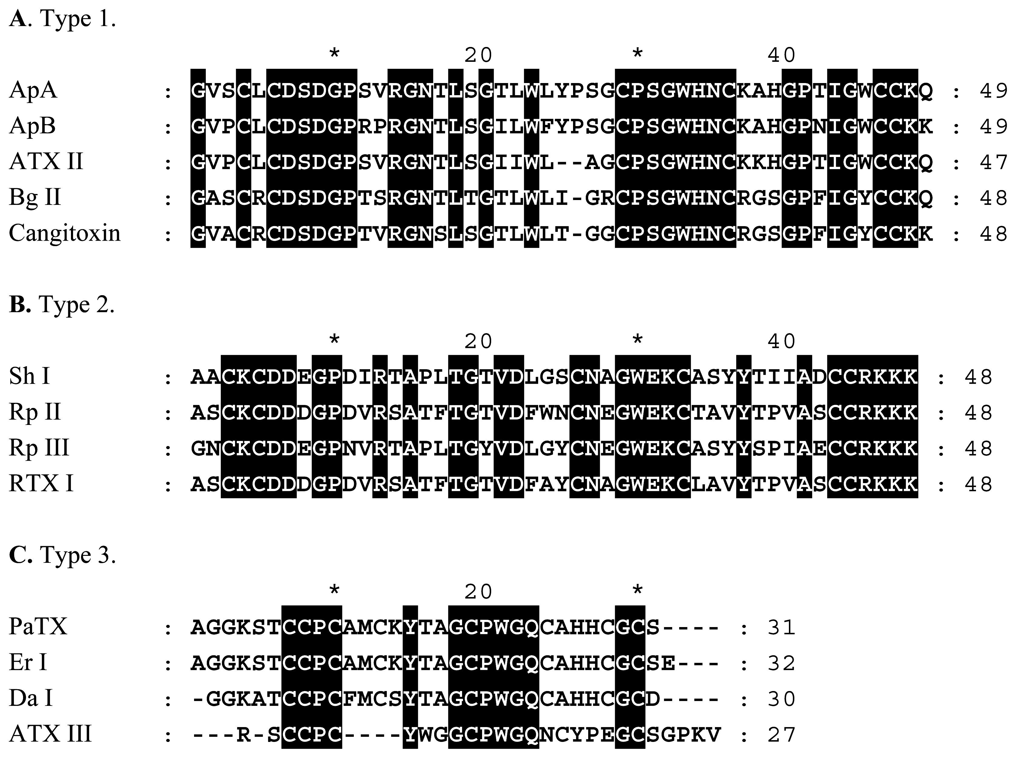
Figure 1.
Alignments of representative sea anemone NaV channel toxins.Identical residues are shaded black. NaV toxins shown are: ApA and ApB from Anthopleura xanthogrammica[28,29]; ATX II from Anemonia sulcata[27]; Bg II from Bunodosoma granulifera[51]; Cangitoxin from Bunodosoma cangicum[30]; Sh I from Stichodactyla helianthus[52]; Rp II and Rp III from Radianthus paumotensis[53,54]; RTX I from Radianthus macrodactylus[55]; PaTX from Parasicyonis actinostoloides[56]; Er I from Entacmaea ramsayi[33]; Da I from Dofleinia armata[33]; and ATX III from Anemonia sulcata[57].

Figure 2.
Alignments of sea anemone KV channel toxins.Identical residues are shaded black. KV toxins shown are: ShK from Stichodactyla helianthus[67]; BgK from Bunodosoma granulifera[69]; AsKS (kaliseptine), AsKC (kalicludines 1-3), BDS-I, and BDS-II from Anemonia sulcata[64, 65]; AeK from Actinia equina[71]; HmK from Heteractis magnifica[70]; APETx1 from Anthopleura elegantissima[66].


| Toxin | Species | Type | Reference |
|---|---|---|---|
| ShK | Stichodactyla helianthus | 1 | [67] |
| AsKS (kaliseptine) | Anemonia sulcata | 1 | [64] |
| BgK | Bunodosoma granulifera | 1 | [68,69] |
| HmK | Heteractis magnifica | 1 | [70] |
| AeK | Actinia equine | 1 | [71] |
| AsKC 1–3 (kalcicludines 1–3) | Anemonia sulcata | 2 | [64] |
| BDS-I, BDS-II | Anemonia sulcata | 3 | [65] |
| APETx1 | Anthopleura elegantissima | 3 | [66] |
© 2006 by MDPI Reproduction is permitted for noncommercial purposes.
Share and Cite
MDPI and ACS Style
Messerli, S.M.; Greenberg, R.M. Cnidarian Toxins Acting on Voltage-Gated Ion Channels. Mar. Drugs 2006, 4, 70-81. https://doi.org/10.3390/md403070
AMA Style
Messerli SM, Greenberg RM. Cnidarian Toxins Acting on Voltage-Gated Ion Channels. Marine Drugs. 2006; 4(3):70-81. https://doi.org/10.3390/md403070
Chicago/Turabian StyleMesserli, Shanta M., and Robert M. Greenberg. 2006. "Cnidarian Toxins Acting on Voltage-Gated Ion Channels" Marine Drugs 4, no. 3: 70-81. https://doi.org/10.3390/md403070