Article

Five Sesquiterpenoids from a Marine-Derived Fungus Aspergillus sp. Isolated from a Gorgonian Dichotella gemmacea

1
School of Pharmacy, Guangdong Medical College, Dongguan, 523808, China
2
Key Laboratory of Marine Drugs, Ministry of Education, School of Medicine and Pharmacy, Ocean University of China, Qingdao, 266003, China
3
School of Chemistry and Chemical Engineering, Sun Yat-sen University, Guangzhou, 510275, China
*
Authors to whom correspondence should be addressed.
Mar. Drugs 2010, 8(4), 941-949; https://doi.org/10.3390/md8040941
Submission received: 4 March 2010 / Revised: 17 March 2010 / Accepted: 26 March 2010 / Published: 29 March 2010

Abstract

:
Three new phenolic bisabolane-type sesquiterpenoids: (+)-methyl sydowate (1), 7-deoxy-7,14-didehydrosydonic acid (2), and 7-deoxy-7,8-didehydrosydonic acid (3), together with two known fungal metabolites were isolated from the fermentation broth of a marine-derived fungus Aspergillus sp., which was isolated in turn from a gorgonian Dichotella gemmacea collected from the South China Sea. Their structures were elucidated by combined spectroscopic methods, and the structure of 1 was further confirmed by single-crystal X-ray data.

Graphical Abstract

1. Introduction

Marine-derived fungi have been recognized as a potential source of structurally novel and biologically potent metabolites, and a growing number of marine fungi have been reported to produce novel bioactive secondary metabolites [13]. Especially, the genus Aspergillus has been known to be a major contributor to the secondary metabolites of marine fungal origin, for example, four sesquiterpenoids with a unique nitrobenzoyl ester from Aspergillus versicolor [4], two modified cytotoxic tripeptides from Aspergillus sp. [5], a novel pentacyclic oxindole alkaloid from Aspergillus tamari [6], four prenylated indole alkaloids from Aspergillus sp. [7] and two cyclopentapeptides from Aspergillus versicolor [8].
As part of our ongoing investigation into new bioactive natural products from marine fungi from the South China Sea, the gorgonian-derived fungus Aspergillus sp. attracted our attention because of the fact that a crude EtOAc extract of the fungal culture showed pronounced in vitro cytotoxicity against the A-549 human lung carcinoma cell line. Bioassay-guided fractionation of the extract led to the isolation of three new phenolic bisabolane-type sesquiterpenoids: (+)-methyl sydowate (1), 7-deoxy-7,14-didehydrosydonic acid (2), and 7-deoxy-7,8-didehydrosydonic acid (3), together with two known fungal metabolites: (+)-sydowic acid (4) [9,10], and (+)-sydonic acid (5) [11,12] (Figure 1). To date, this is the first report of natural products from a marine-derived fungus isolated from the fresh tissues of a gorgonian coral.
The EtOAc extract of a fermentation broth of the fungus Aspergillus sp. was subjected to silica gel column chromatography, Sephadex LH-20 and further semi-preparative HPLC, and this led to the isolation of compounds 15. Their structures were elucidated by spectroscopic data, mainly 1D and 2D NMR spectra, and the structure of 1 was further confirmed by single-crystal X-ray data.

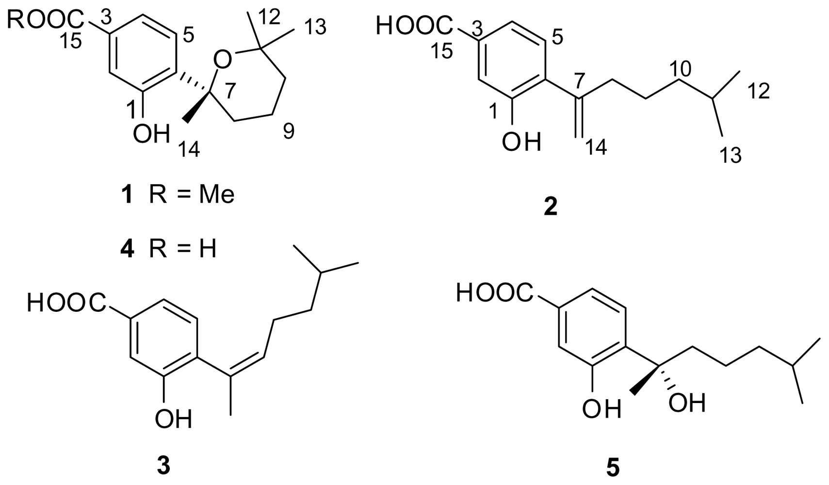
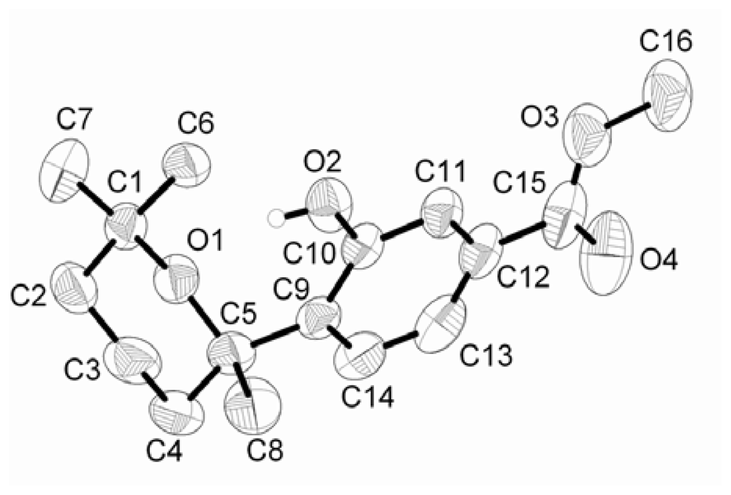
2. Results and Discussion

(+)-Methyl sydowate (1) was isolated as optically active colorless crystals ([α]25D +24.7, CHCl3). A molecular formula of C16H22O4 was confirmed by HREIMS that displayed an [M]+ m/z of 278.1500 (calcd. 278.1513). The IR absorption bands indicated the existence of hydroxyl (3,230 cm−1) and ester (1,719 cm−1) groups. The 1H-NMR spectrum of 1 showed one exchangeable proton signal at δH 9.26 (s), one ABX spin system assignable to a 1,3,4-trisubstitued benzene ring at δH 7.10 (d, J = 7.8 Hz), 7.48 (d, J = 1.8 Hz) and 7.50 (dd, J = 7.8, 1.8 Hz) (Table 1). The 13C-NMR spectrum (Table 2) displayed 16 carbon signals, including those assigned to a carboxylic group (δC 166.9) and six aromatic carbons. With six degrees of unsaturation accounted for by the molecular formula, the structure of 1 was suggested to contain another ring, in association with a benzene ring and a carboxylic group. The NMR data of 1 were closely related to those of sydowic acid (4), a bisabolane-type sesquiterpenoid previously isolated from a terrestrial fungus Aspergillus sydowi.
A comparison of NMR data showed that 1 was the methyl ester of 4. The correlations from H2-8 through H2-9 to H2-10 in the COSY spectrum revealed the CH2-CH2-CH2 subunit in 1. The C-12 and C-13 methyl singlets (δH 0.94 and 1.28) were determined to be germinal and attached to C-11 based on mutual HMBC correlations to each other and correlations from two methyl protons to C-10 and C-11. The connection between C-6 and C7 was established based on the HMBC correlations from H3-14 to C-6 and C-7, and H-5 to C-7. Crystals of 1 suitable for X-ray diffraction were obtained by slow evaporation of solution of 1 in methanol-DMF (1:1). The structure of 1 was further confirmed by a single-crystal X-ray analysis, and its ORTEP plot is depicted in Figure 2. Compound 1 exhibited a positive optical rotation similar to that of (+)-sydowic acid (4) [10], implying that its absolute configuration at C-7 was R.
7-Deoxy-7,14-didehydrosydonic acid (2) was isolated as a white powder, and its molecular formula of C15H20O3 was determined from HRESIMS data (found m/z 247.1331 [M – H], calcd. 247.1334). The molecular formula indicated 2 contained six degrees of unsaturation, which by interpretation of NMR data (Tables 1 and 2) could be attributed to four carbon-carbon double bonds, one carboxylic carbon, and one benzene ring. In the 1H-NMR spectrum, one ABX spin system assignable to a 1,3,4-trisubstitued benzene ring at δH 7.18 (d, J = 7.8 Hz), 7.66 (d, J = 1.2 Hz) and 7.65 (dd, J = 7.8, 1.2 Hz) was also observed. The 1H NMR spectrum revealed the presence of other signals including two doublet methyl groups [H3-12 (δH 0.83), and H3-13 (δH 0.83)], four methylenes [H-14a/14b (δH 5.45/5.19), H2-8 (δH 2.42), H2-9 (δH 1.39), and H2-10 (δH 1.18)], and one methine proton signal [H-11 (δH 1.51)]. From the 13C-NMR spectrum, one carboxylic carbon (δC 171.2), and eight sp2 carbons were observed. The NMR data of 2 closely resembled those of sydonic acid (5) previously isolated from a terrestrial strain of A. sydowi [11] and a marine fungus Glonium sp. [12]. The only significant differences in the 1H-NMR spectrum of 2 in comparison with 5 were two signals for H2-14 which were shifted downfield to δH 5.45 and 5.19 (instead of a methyl group at δH 1.68). The downfield shift observed for C-7 (δC 146.1 vs δC 79.3) and C-14 (δC 116.0 vs δC 21.7) in the 13C-NMR spectrum also reflected the presence of an exomethylene group rather than a methyl group connected to a quaternary carbonic carbon. Thus the gross structure of 2 was assigned as the 7,14-dehydration product of sydonic acid (5). The presence of the C-7/C-14 double bond was further supported by the HMBC correlations from H2-14 to C-6 and C-8, and from H2-8 to C-6, C-7 and C-14. These data allowed the complete structure of 2 to be assigned. Detailed assignments for carbons and protons were unambiguously accomplished by analysis of 2D NMR spectral data.
7-Deoxy-7,8-didehydrosydonic acid (3) was also obtained as a white powder with the same molecular formula, C15H20O3 (found m/z 247.1346 [M – H]?−, calcd 247.1334), as found for 2. Detailed comparison of 1H- and 13C-NMR data of 3 (Tables 1 and 2) with those of 2 illustrated the presence of an olefinic bond at C-7/C-8 rather than C-7/C-14. This double bond was easily assigned since the signals for the methylene pair H2-8 and the terminal olefinic pair H2-14 were lost and replaced by single alkene signal at δH 5.76 (1H, t, J = 7.2 Hz, H-8) and one olefinic methyl group at δH 2.00 (3H, s, H-14). In a consistent fashion, the 13C-NMR spectrum showed an olefinic carbon for C-8 (δC 132.8) and an olefinic methyl group for C-14 at δC 24.7. Thus the structure of compound 3 was assigned as the 7,8-dehydration product of sydonic acid. In addition, the Z-geometry of the double bond was determined based on the chemical shift of the methyl carbon at the trisubstituted olefinic bond, which was observed at δC 24.7 (C-14) [13].
The origin of compounds 13 is a matter needing clarification. To determine if 1 was a natural product or it merely an artifact derived from methylation of (+)-sydowic acid during the isolation process, we analyzed the crude EtOAc extract by comparison of the retention times with that of pure sample of 1 by HPLC, using CH3CN/H2O (6:4) as a mobile phase. Compound 1 was clearly detected in the crude extract which had never been exposed to methanol, thus it seems very unlikely that 1 was obtained during the work-up. Since benzyl alcohols readily dehydrate under mild conditions, compounds 2 and 3 may be transformed from 5 during their isolation process. However, no dehydrated products were detected when 5 was dissolved in MeOH and left at room temperature for three days in the presence of hydrochloric acid (0.01 mol/L). Thus, dehydration of 5 during its isolation should have not occurred and correspondingly, compounds 2 and 3 should be considered true natural products.
The structures of compounds 4 and 5 were identified as (+)-sydowic acid [9,10], and (+)-sydonic acid [11,12], respectively, by comparison of their spectroscopic data with those in the literature. (−)-Sydowic acid was previously isolated from a terrestrial strain of A. sydowi [9], but its enantiomer (+)-sydowic acid was isolated for the first time as a natural compound in the present study. Sydonic acid was previously isolated in racemic form from the same species, A. sydowi [11], and interestingly, (+)-sydonic acid was also reported in 2009 from the fungus Glonium sp. collected from the Shirakami sea area [12].
A series of phenolic bisabolane-type sesquiterpenoids have been reported from different marine invertebrates, such as the marine sponges Didiscus flavus [14], Parahigginsia sp. [15] and Epipolasis sp. [16], and gorgonians Pseudopterogorgia rigida [17], Muricia elongate [18] and Plexaurella nutans[18], indicating that there only limited chemotaxonomic significance of these compounds in marine invertebrates. This type of compounds has recently also been reported from the bacterium CNH-741 and the fungus CNC-979, isolated from marine sediment [19]. In this study, five related sesquiterpenoids were also found from the marine-derived fungus Aspergillus sp. isolated from the gorgonian coral Dichotella gemmacea collected from the South China Sea. The findings of structurally related compounds from marine invertebrates and marine microorganisms could be used as circumstantial evidence to suggest that these compounds are acquired by the invertebrates from microbial symbionts or through their diet. It should be noted that the structures of compounds 2 and 3, containing a double bond at C-7/C-14 or C-7/C-8, respectively, are unusual, since all of the previously known compounds are saturated at these positions. Recently, a strain fungus A. sydowii, isolated from healthy marine sponges Spongia obscura collected in Bahamian inshore waters, was reported as the causative agent of epidemics that affected gorgonian corals and had significantly affected their populations in the Caribbean Sea [20].
The bioactivity of compounds 1, 4 and 5 were determined against Staphylococcus aureus and methicillin resistant S. aureus by the method as Fromtling et al. [21]. All of them exhibited weak antibacterial activity, with inhibition zones of 11, 7, 5 mm in diameter, respectively, at the concentration of 100 μg/mL. No inhibition, however, was observed for methicillin resistant S. aureus (kanamycin sulfate was used as the positive control with inhibition zones of 37 and 21 mm in diameter, respectively). Sydowic acid was reported as an antioxidant before [22]. No activities were evaluated for compounds 2 and 3 because of their low yields.

3. Experimental Section

3.1. General

1H- and 13C-NMR spectra were recorded on a JEOL Eclips-600 spectrometer. ESIMS and HRESIMS were measured on a Q-TOF Ultima Global GAA076 LC mass spectrometer. HREIMS were measured on a Thermo MAT95XP High Resolution mass spectrometer and EIMS on a Thermo DSQ EI-mass spectrometer. Optical rotations were measured in chloroform using a JASCO P-1020 digital polarimeter. IR spectra were measured on a Bruker VECTOR 22 spectrophotometer. Silica gel (Qing Dao Hai Yang Chemical Group Co.; 200–300 mesh), octadecylsilyl silica gel (Unicorn; 45–60 μm) and Sephadex LH-20 (Amersham Biosciences) were used for column chromatography (CC). Precoated silica gel plates (Yan Tai Zi Fu Chemical Group Co.; G60, F-254) were used for thin layer chromatography (TLC). Semi-preparative HPLC was performed on a Waters 1525 system using a semi-preparative C18 (Kromasil 7 μm, 10 × 25 mm) column coupled with a Waters 2996 photodiode array detector.

3.2. Fungal Material

The marine-derived fungus Aspergillus sp. was isolated from a piece of tissue from the inner part of the freshly collected gorgonian coral D. gemmacea (GX-WZ-20080034), which was obtained from the Weizhou coral reef in the South China Sea in September, 2008. The strain was deposited in the Key Laboratory of Marine Drugs, the Ministry of Education of China, School of Medicine and Pharmacy, Ocean University of China, Qingdao, PR China, with the access code ZJ-2008001. The fungal strain was cultivated in 30 L liquid medium (10.0 g of glucose, 2.0 g of yeast extract, 2.0 g of peptone in 1 L of seawater, in 1 L Erlenmeyer flasks each containing 400 mL of culture broth) at 27 °C without shaking for 30 days.

3.3. Extraction and Isolation

The fungal cultures were filtered through cheesecloth, and the filtrate (30.0 L) was extracted with EtOAc (2 × 30.0 L). The organic extracts were concentrated in vacuo to yield a yellow oily residue (2.50 g). This extract was chromatographed on a silica gel column using a stepwise gradient of petroleum ether–EtOAc to afford eight fractions (Fractions 1 8). Fraction 2 (0.35 g) was isolated by column chromatography on silica gel eluted with petroleum ether–EtOAc (8:2), and then subjected to Sephadex LH-20 chromatography eluting with mixtures of petroleum ether–CHCl3–MeOH (2:1:1) to obtain compound 1 (6.0 mg). Repeated chromatography of fraction 4 (0.22 g) using Sephadex LH-20 eluted with mixtures of CHCl3–MeOH (1:1) and petroleum ether–CHCl3–MeOH (2:1:1), then by semipreparative HPLC at a flow rate of 2.0 mL/min (6:4 MeOH/H2O) yielded compounds 2 (2.2 mg), 3 (1.5 mg), 4 (2.6 mg) and 5 (30.2 mg).
(+)-Methyl sydowate (1): colorless crystals; [α]25D + 24.7 (c 0.030, CHCl3); UV (MeOH) λmax 204.0, 238.0, 293.5 nm; IR (KBr) νmax 3230, 2972, 2932, 1719, 1573, 1281, 1201 cm−1; 1H-NMR and 13C-NMR see Tables 1 and 2; EI MS m/z 278 [M]+ (59), 263 (37), 260 (46), 245 (42), 231 (29), 217 (100), 203 (52), 195 (62), 192 (51), 189 (32), 179 (37), 173 (29), 161 (23), 145 (17), 131 (14), 69 (32); HREIMS m/z [M]+ 278.1500 (calcd for C16H22O4, 278.1513). Crystallizes in triclinic, space group P-1 with a = 7.0260(13) Å, b = 8.1016(15) Å, c = 13.939(3) Å, α = 87.266(2), β = 77.823(2), γ = 76.502(2)°, C16H22O4, Mr = 278.34, V = 754.2(2) Å3, Z = 2, Dc = 1.226 g/cm3, F(000) = 300, μ = 0.087 mm−1, the final R = 0.0461 and wR = 0.1095 for 5811 observed reflections (I > 2σ (I)). The crystallographic data for 1 have been deposited at the Cambridge Crystallographic Data Centre (CCDC No.738932).
7-Deoxy-7,14-didehydrosydonic acid (2): white powder; UV (MeOH) λmax 209.8, 247.4, 300.6 nm; IR (KBr) νmax 3071, 2946, 2860, 1692, 1640, 1533, 1507, 1407, 1288, 1215, 764 cm−1; 1H-NMR and 13C-NMR see Tables 1 and 2; ESIMS m/z [M – H] 247; HRESIMS m/z [M – H] 247.1331 (calcd for C15H20O3, 247.1334).
7-Deoxy-7,8-didehydrosydonic acid (3): white powder; UV (MeOH) λmax 206.3, 253.3, 304.2 nm; IR (KBr) νmax 3104, 3065, 3005, 1699, 1540, 1514, 1447, 1215, 758 cm−1; 1H-NMR and 13C-NMR see Tables 1 and 2; ESIMS m/z [M – H] 247; HRESIMS m/z [M – H] 247.1346 (calcd for C15H20O3, 247.1334).

3.4. Antibacterial activity

The compounds were tested against S. aureus and methicillin resistant S. aureus for their inhibitory activity. Antibacterial assays were performed using a modified version of the 2-fold serial dilutions method as Fromtling et al. [21].

Acknowledgements

This work was supported by the National Natural Science Foundation of China (Nos. 40976077; 30901879; 40776073), the Research Fund for the Doctoral Program of Higher Education, Ministry of Education of China (No. 20090132110002), the Basic Research Program of Science and Technology, Ministry of Science and Technology of China (No. 2007FY210500), and the Open Research Fund Program of Key Laboratory of Marine Drugs (Ocean University of China), the Ministry of Education (No. KLMD (OUC) 200801).
  • Sample Availability: Available from the authors.

References and Notes

  1. Blunt, JW; Copp, BR; Munro, MHG; Northcote, PT; Prinsep, MR. Marine Natural Products. Nat Prod Rep 2006, 23, 26–78. [Google Scholar]
  2. Bugni, TS; Ireland, CM. Marine-Derived Fungi: A Chemically and Biologically Diverse Group of Microorganisms. Nat Prod Rep 2004, 21, 143–163. [Google Scholar]
  3. Saleem, M; Ali, MSS; Hussain, JA; Ashraf, M; Lee, YS. Marine Natural Products of Fungal Origin. Nat Prod Rep 2007, 24, 1142–1152. [Google Scholar]
  4. Belofsky, GN; Jensen, PR; Renner, MK; Fenical, W. New Cytotoxic Sesquiterpenoid Nitrobenzoyl Esters from a Marine Isolate of the Fungus Aspergillus versicolor. Tetrahedron 1998, 54, 1715–1724. [Google Scholar]
  5. Toske, SG; Jensen, PR; Kauffman, CA; Fenical, W. Aspergillamides A and B: Modified Cytotoxic Tripeptides Produced by a Marine Fungus of the Genus Aspergillus. Tetrahedron 1998, 54, 13459–13466. [Google Scholar]
  6. Suda, M; Mugishima, T; Komatsu, K; Sone, T; Tanaka, M; Mikami, Y; Shiro, M; Hirai, M; Ohizumi, Y; Kobayashi, J. Speradine A, a New Pentacyclic Oxindole Alkaloid from a Marine-Derived Fungus Aspergillus tamari. Tetrahedron 2003, 59, 3227–3230. [Google Scholar]
  7. Kato, H; Yoshida, T; Tokue, T; Nojiri, Y; Hirota, H; Ohta, T; Williams, RM; Tsukamoto, S. Notoamides A–D: Prenylated Indole Alkaloids Isolated from a Marine-Derived Fungus, Aspergillus sp. Angew Chem Int Ed 2007, 46, 2254–2256. [Google Scholar]
  8. Fremlin, LJ; Piggott, AM; Lacey, E; Capon, RJ. Cottoquinazoline A and Cotteslosins A and B, Metabolites from an Australian Marine-Derived Strain of Aspergillus versicolor. J Nat Prod 2009, 72, 666–670. [Google Scholar]
  9. Hamasaki, T; Sato, Y; Hatsuda, Y; Tanabe, M; Cary, LW. Sydowic Acid. New Metabolite from Aspergillus sydowi. Tetrahedron Lett 1975, 9, 659–660. [Google Scholar]
  10. Serra, S. Bisabolane Sesquiterpenes: Synthesis of (R)-(+)-Sydowic Acid and (R)-(+)-Curcumene ether. Syn Lett 2000, 6, 890–892. [Google Scholar]
  11. Hamasaki, T; Nagayama, K; Hatsuda, Y. Two New Metabolites, Sydonic Acid and Hydroxysydonic Acid, from Aspergillus sydowi. Agric Biol Chem 1978, 42, 37–40. [Google Scholar]
  12. Kudo, S; Murakami, T; Miyanishi, J; Tanaka, K; Takada, N; Hashimoto, M. Isolation and Absolute Stereochemistry of Optically Active Sydonic Acid from Glonium sp. (Hysteriales, Ascomycota). Biosci Biotechnol Biochem 2009, 73, 203–204. [Google Scholar]
  13. Ravi, BN; Faulkner, DJ. Cembranoid Diterpenes from a South Pacific Soft Coral. J Org Chem 1978, 43, 2127–2131. [Google Scholar]
  14. Wright, AE; Pomponi, SA; McConnell, OJ; Komoto, S; McCarthy, PJ. (+)-Curcuphenol and (+)-Curcudiol, Sesquiterpene Phenols from Shallow and Deepwater Collections of the Marine Sponge Didiscus flavus. J Nat Prod 1987, 50, 976–978. [Google Scholar]
  15. Chen, CY; Shen, YC; Chen, YJ; Sheu, JH; Duh, CY. Bioactive Sesquiterpenes from a Taiwanese Marine Sponge Parahigginsia sp. J Nat Prod 1999, 62, 573–576. [Google Scholar]
  16. Fusetani, N; Sugano, M; Matsunaga, S; Hashimoto, K. (+)-Curcuphenol and Dehydrocurcuphenol, Novel Sesquiterpenes Which Inhibit H,K-ATPase, from a Marine Sponge Epipolasis sp. Experientia 1987, 43, 1234–1235. [Google Scholar]
  17. McEnroe, FJ; Fenical, W. Structures and Synthesis of Some New Antibacterial Sesquiterpenoids from the Gorgonian Coral Pseudopterogorgia rigida. Tetrahedron 1978, 34, 1661–1664. [Google Scholar]
  18. Jeffs, PW; Lytle, LT. Isolation of (−)-α-Curcumene, (−)-β-Curcumene, and (+)-β-Bisabolene from Gorgonian Corals. Absolute Configuration of (−)-β-Curcumene. Lloydia 1974, 37, 315–317. [Google Scholar]
  19. Muelhaupt, T; Kaspar, H; Otto, S; Reichert, M; Bringmann, G; Lindel, T. Isolation, Structural Elucidation, and Synthesis of Curcutetraol. Eur J Org Chem 2005, 334–341. [Google Scholar]
  20. Ein-Gil, N; Ilan, M; Carmeli, S; Smith, GW; Pawlik, JR; Yarden, O. Presence of Aspergillus sydowii, a Pathogen of Gorgonian Sea Fans in the Marine Sponge Spongia obscura. ISME J 2009, 3, 752–755. [Google Scholar]
  21. Fromtling, RA; Galgiani, JN; Pfaller, MA; Espinel-Ingroff, A; Bartizal, KF; Bartlett, MS; Body, BA; Frey, C; Hall, G; Roberts, GD; Noltt, FB; Odds, EC; Rinaldi, MG; Suger, AM; Villareal, K. Multicenter Evaluation of a Broth Macrodilution Antifungal Susceptibility Test for Yeasts. Antimicrob Agents Chemother 1993, 37, 39–45. [Google Scholar]
  22. Ishikawa, Y; Morimoto, K; Hamasaki, T. Flavoglaucin, a Metabolite of Eurotium Chevalieri, its Antioxidation and Synergism with Tocopherol. J Am Oil Chem Soc 1984, 61, 1864–1868. [Google Scholar]
Figure 1. Structures of compounds 15.
Marinedrugs 08 00941f1
Figure 2. ORTEP drawing for methyl sydowate (1).
Marinedrugs 08 00941f2
Table 1. 1H-NMR data (CDCl3) of 13 a.
position1 δH (mult., J in Hz)2 δH (mult., J in Hz)3 δH (mult., J in Hz)
1
27.48, d (1.8)7.66, d (1.2)7.65, d (1.2)
3
47.50, dd (7.8, 1.8)7.65, dd (7.8, 1.2)7.67, dd (7.5, 1.2)
57.10, d (7.8)7.18, d (7.8)7.13, d (7.5)
6
7
82.43, ddd (13.8, 3.6, 0.6)
1.70, m
2.42, t (7.8)5.76, t (7.2)
91.74, m
1.64, m
1.39, m1.81, m
101.54, m1.18, m1.19, m
111.51, septet (6.6)1.45, m
120.94, s0.83, d (6.6)0.77, d (6.6)
131.28, s0.83, d (6.6)0.77, d (6.6)
141.49, s5.45, s
5.19, s
2.00, s
15
−OCH33.90, s
−OH9.26, s
aMeasured at 600 MHz.
Table 2. 13C-NMR data (CDCl3) of 13 a.
position123
1157.0, -C152.4, -C151.7, -C
2118.3, CH117.1, CH116.6, CH
3130.5, -C129.4, -C129.3, -C
4120.7, CH122.0, CH122.3, CH
5124.5, CH128.2, CH128.7, CH
6136.0, -C134.1, -C133.7, -C
777.6, -C146.1, -C130.2, -C
833.8, CH237.7, CH2132.8, CH
916.6, CH225.6, CH227.1, CH2
1036.7, CH238.5, CH238.6, CH2
1175.2, -C27.8, CH27.4, CH
1224.7, CH322.5, CH322.3, CH3
1331.9, CH322.5, CH322.3, CH3
1431.3, CH3116.0, CH224.7, CH3
15166.9, -C171.2, -C170.6, -C
−OCH352.0, CH3
aMeasured at 150 MHz.

