Article

Effects of Di-(2-ethylhexyl) Phthalate on the Hypothalamus–Uterus in Pubertal Female Rats

1
Department of Occupational and Environmental Health, School of Public Health, Jilin University, Changchun 130021, China
2
Scientific Research Center, China Japan Union Hospital, Jilin University, Changchun 130000, China
3
Prevention and Health Section, Jiangxi Provincial Cancer Hospital, Nanchang 330000, China
*
Author to whom correspondence should be addressed.
These authors equally contributed to this work.
Int. J. Environ. Res. Public Health 2016, 13(11), 1130; https://doi.org/10.3390/ijerph13111130
Submission received: 19 September 2016 / Revised: 26 October 2016 / Accepted: 7 November 2016 / Published: 12 November 2016
(This article belongs to the Section Environmental Health)

Abstract

:
The pollution of endocrine disruptors and its impact on human reproductive system have attracted much attention. Di-(2-ethylhexyl) phthalate (DEHP), an environmental endocrine disruptor, is widely used in food packages, containers, medical supplies and children’s toys. It can cause diseases such as infertility, sexual precocity and uterine bleeding and thus arouse concerns from the society and scholars. The effect of DEHP on pubertal female reproductive system is still not well-studied. This study was to investigate the effects of DEHP on the hypothalamus–uterus in pubertal female rats, reveal the reproductive toxicity of DEHP on pubertal female rats and its mechanism, and provide scientific evidence for the evaluation of toxicity and toxic mechanism of DEHP on reproductive system. Forty-eight pubertal female rats were randomly divided into four groups and respectively administered via oral gavage 0, 250, 500, or 1000 mg/kg/d DEHP in 0.1 mL corn oil/20 g body weight for up to four weeks. Compared with control rats, the DEHP-treated rats showed: (1) higher gonadotropin-releasing hormone (GnRH) level in the hypothalamus; (2) higher protein levels of GnRH in the hypothalamus; and (3) higher mRNA and protein levels of GnRH receptor (GnRHR) in the uterus. Our data reveal that DEHP exposure may lead to a disruption in pubertal female rats and an imbalance of hypothalamus–uterus. Meanwhile, DEHP may, through the GnRH in the hypothalamus and its receptor on the uterus, lead to diseases of the uterus. DEHP may impose a negative influence on the development and functioning of the reproductive system in pubertal female rats.

1. Introduction

Di-(2-ethylhexyl) phthalate (DEHP), an environmental endocrine disruptor, is one of the organic chemicals that is used in a large variety of industrial and consumer applications. It is the most commonly used plasticizer worldwide [1,2,3]. An endocrine disruptor alters the function of the endocrine systems and causes adverse effects in organisms [4].
DEHP has been approved for use and has also been detected in a wide range of consumer products [5,6], including personal care products [7], infant toys [8], food wraps [9], cosmetics, medical consumables, nutritional supplements, cleaning materials, lubricants, insecticides, solvents, adhesives, paints, lacquers, etc. Unfortunately, DEHP may leach slowly from these plastic products into foods, beverages, and even directly into body fluids [10,11]. It is also detected in indoor air [12,13,14], indoor dust [15,16,17,18], and air inside vehicles [19]. The general population may be exposed since DEHP is ubiquitous environmental contaminants [20,21,22]. Epidemiological investigation showed that the exposure dose of DEHP for the population was 5 mg/kg/d [23].
Studies have shown that correlating DEHP levels with negative endocrine effects in females are increasing [24]. DEHP can cause the abortion of female animals, increase ovarian weight and the follicle developmental disorders, and gonadal synthesis of estradiol and gonadotropin receptor gene expression decreased pituitary gonadotropin upregulated genes [25,26,27]. DEHP can enter the fetus through the placental barrier and gather in the fetus. This may have an impact on the fetus directly [28,29].
Until now, most of the research has focused on reproductive toxicity of adult females’ exposure to DEHP, and little is known regarding the mechanism of DEHP in pubertal females. With the development of society and economy, precocious puberty has been getting more and more attention. The rate of precocious puberty is increasing. Meanwhile, the incidents of reproductive system diseases are also rising. The average age of menarche of girls in Australia and Europe are three years earlier than before [30,31,32]. Epidemiology found that exposure to phthalates are associated with endometritis, earlier breast development, sexual precocity and earlier onset of puberty [33,34,35,36]. However, the effect of DEHP on pubertal female reproductive system is still not well studied.
Animal reproduction is regulated by a variety of factors both inside and outside the body, and the hypothalamic-pituitary-gonadal axis (HPGA) plays a major regulatory role in it. The central precocious puberty (CPP) is due to the hypothalamic-pituitary-gonadal axis function starting early, and because the level of gonadotropin-releasing hormone (GnRH) is premature increased [37]. GnRH is a hypothalamic neuronal secretory decapitate that plays an important regulatory role in the mammalian reproductive system. The present study supports the notion that GnRH-(1-5) is functionally capable of regulating the reproductive neuroendocrine system [38]. GnRH influences the reproductive processes mainly by regulating pituitary gonadotropin synthesis and release, which, in turn, modulate steroid genesis and gametogenesis [39,40]. Even if the hypothalamus is the principal source of GnRH, and the pituitary is the target site for it, several studies have reported that GnRH receptor (GnRHR) will be in many other tissues including reproductive organs, such as testes, prostates, ovaries, and uteruses [41,42,43,44].
Precocious puberty may cause polycystic ovary syndrome, and thus will cause heart disease, diabetes, endometrial cancer, infertility and other diseases [45,46]. This study investigated the effect of pubertal female rats’ exposure to DEHP on the expression of GnRH in hypothalamus and GnRHR in the uterus and to explore the role of DEHP in reproductive endocrine disruption. The DEHP doses were set from the human exposure dose (5 mg/kg/d) [23] and the LD50 (30 g/kg) in rats [47].

2. Materials and Methods

2.1. Animals and Treatment

Female Wistar rats (15 days old, 60 ± 10 g) were purchased from the Experimental Animal Center of Jilin University. They were allowed at least a 7-day acclimatization-period and observed for signs of illness before starting experimental procedures, and their body weight was measured every day. The 48 rats were randomly apportioned into 4 groups (n = 12, each). The rats were housed in polypropylene cages (6 rats per cage) with sterilized bedding, and maintained under controlled conditions of temperature (23 ± 1 °C), humidity (55% ± 5%), and a 12:12 h light-dark cycle. The experiment lasted approximately 4 weeks including the period of adaptation to the feeding regime. Rats were administered via oral gavage 0, 250 mg/kg/d DEHP (1/120 LD50), 500 mg/kg/d DEHP (1/60 LD50), 1000 mg/kg/d DEHP (1/30 LD50) (Sinopharm Chemical Reagent, purity >99%, Shanghai, China) in 0.1 mL corn oil/20 g body weight for up to 4 weeks. The experimental protocol was subject to approval by the Animal Use and Care Committee of Jilin University, and the ethical approval code of the animal experiment is 2015-03-14.

2.2. Tissue Collection

After 4 weeks, the rats were weighed and killed by decapitation. The hypothalamus and uterus of each rat were also dissected, weighed, and collected immediately after decapitation.

2.3. Quantification of GnRH

GnRH in the hypothalamus was quantified using enzyme-linked immunosorbent assay kits (R&D systems, Minneapolis, MN, USA). The total protein from the hypothalamus sample was extracted, and 0.1 mL diluted sample from each group was mixed with 10 µL GnRH antibody and 50 µL horseradish peroxidase-labeled streptavidin. This was incubated at 37 °C for 60 min. The wells were rinsed five times with distilled water and incubated with chromogen solution from the kit (50 µL A and 50 µL B) for 10 min at 37 °C in dark. The reaction was then stopped with 50 µL of stop solution, and the absorbance was read at 450 nm within 10 min.

2.4. Immunohistochemistry

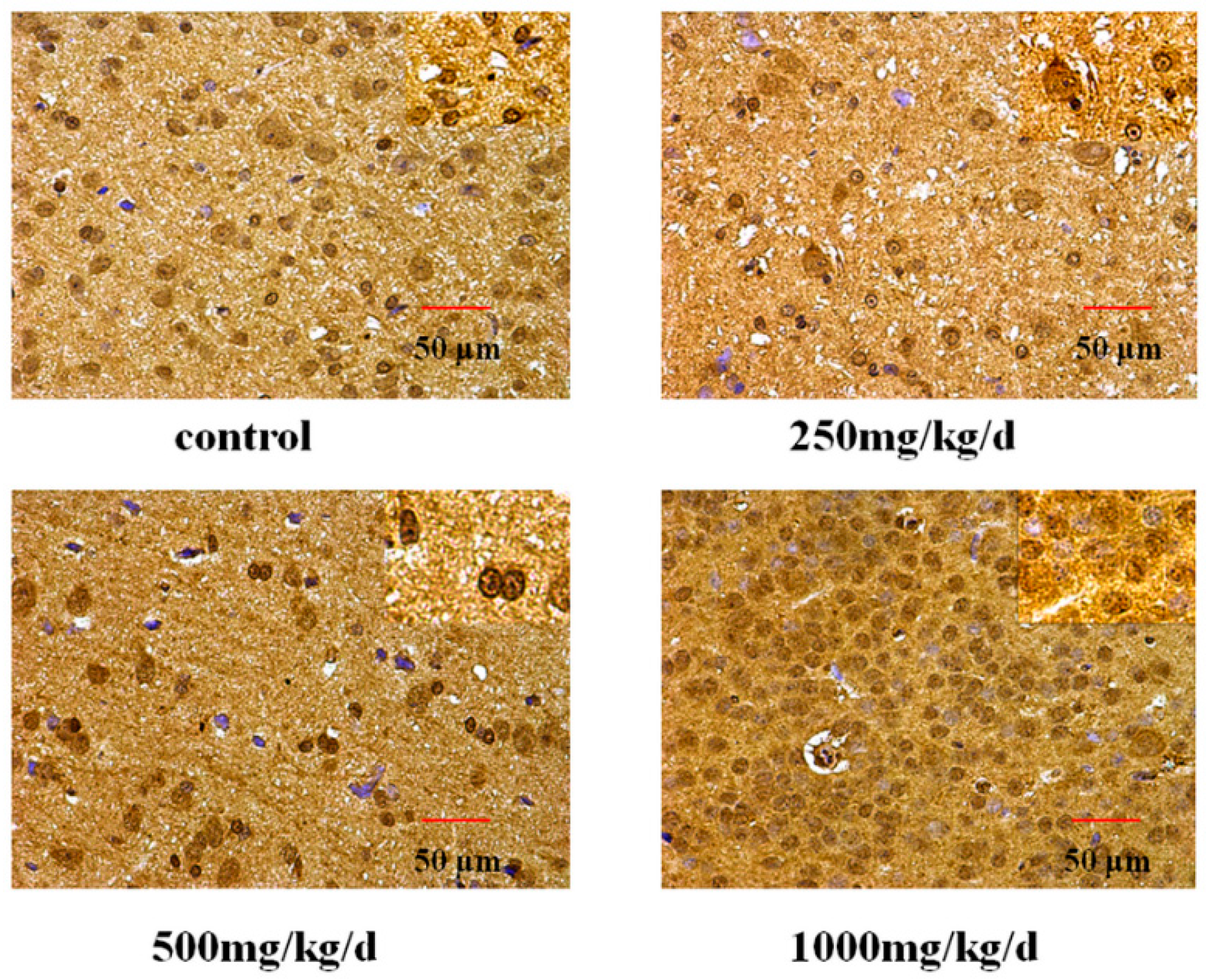
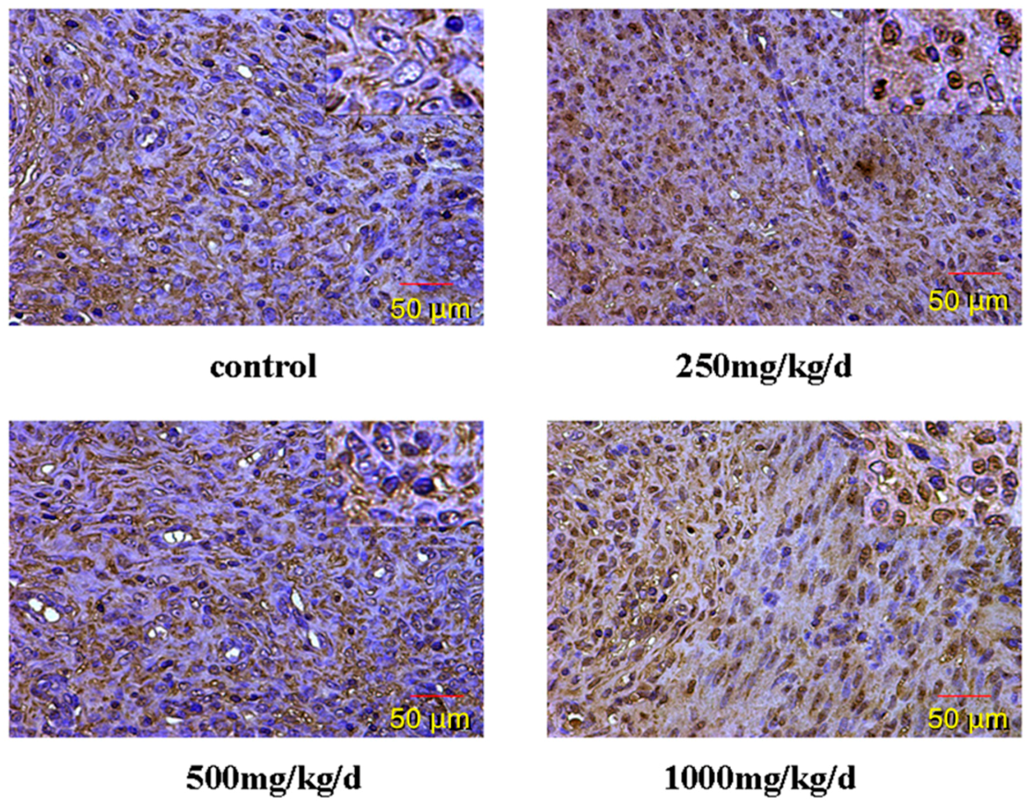
Tissue sections of hypothalamus and uterus were deparaffinized in xylene, rehydrated through graded ethanols, and finally rinsed in distilled water. The specimens were treated with antigen retrieval in 0.1 M citric acid solution by boiling for 15 min and cooling to room temperature. They were then incubated with 3% H2O2 (v/v) in methanol for 10 min to quench the endogenous peroxidase activity. The sections were blotted with normal rat serum (1:10 dilution) for 30 min at room temperature, and incubated with rabbit polyclonal primary antibody (GnRH and GnRHR, Proteintech, Rosemont, IL, USA) (1:1000 in phosphate-buffered saline (PBS) containing 0.1% bovine serum albumin) overnight at 4 °C in a moist chamber. The slides were incubated with peroxidase-conjugated anti-rabbit secondary antibody (Bioss Biotechnology Company, Shanghai, China) (1:200 dilution in PBS) for 30 min at room temperature after three washes in PBS. The staining was visualized using a 3,3′-diaminobenzidine (DAB) (Bioss Biotechnology Company, Shanghai, China) kit. The specimens were counterstained with hematoxylin (Bogoo Biotechnology Company, Shanghai, China), mounted, observed under a light microscope, and photographed [48].

2.5. RNA Extraction

Total RNA was isolated using Trizol reagent (Invitrogen, Waltham, MA, USA). The quantity and the integrity of total RNA were determined by a UV spectrophotometer and evaluated by formaldehyde denaturing gel electrophoresis.

2.6. Real-Time Reverse Transcription-PCR

Real-time reverse transcription-PCR was used to verify gene expression using Stratagene MX3000p (TaKaRa, Shiga, Japan). Reverse transcription was performed with 500 ng total RNA in a 10-ìL reaction, and 1 ìL of cDNA was then used for a 25-ìL PCR reaction mixture containing an optimal concentration of primers and SYBR-Green Supermix (SYBR premix Ex Tap II, TaKaRa, Shiga, Japan). The PCR reaction was carried out in 45 cycles of 95 °C for 20 s and 60 °C for 20 s. â-actin was used as an internal control [49]. The primers are listed in Table 1.

2.7. Western Blotting

Isolated tissues were immediately put on ice and homogenized in lysis buffer (20 mM Tris, pH 7.5, 150 mM NaCl, 1 mM EDTA, 1 mM EGTA, 1% Triton X-100, 1% deoxycholate, 1 mM sodium fluoride, 2 mM sodium orthovanadate, and complete protease inhibitor tablets). Homogenates were centrifuged at 12,000 g for 5 min at 4 °C. Protein in the supernatant was quantified using the BCA protein assay (Beyotime, Shanghai, China). Equal amounts of protein from each sample were mixed with sodium dodecyl sulfate (SDS) sample buffer (Beyotime, Shanghai, China). Samples were separated using pre-cast 10% Bis-Tris gel (Beyotime, Shanghai, China) in Tris-Glycine-SDS running buffer and transferred to nitrocellulose filter membrane (NC) (Phamacia, Goteborg, Sweden) in transfer buffer. Prior to immunoblotting, NC membranes were blocked with 5% nonfat milk in tris buffered saline tween (TBST) buffer (20 mmol/L Tris-HCl, 140 mmol/L NaCl, and 0.05% Tween 20, pH 7.5). Following incubation with the primary antibodies specific for each protein for 24 h at 4 °C—mouse anti-GnRH antibody (1:1000, 34 KDa, Abcam, Cambridge, UK), rabbit anti-GnRHR antibody (1:500, 36 KDa, Abcam, Cambridge, UK), and rabbit anti-beta Actin antibody (1:1000, 42 KDa, Abcam, Cambridge, UK)—the blots were washed with TBST and then incubated with the horseradish peroxidase-conjugated secondary antibodies (1:2000, Proteintech, Rosemont, IL, USA) for 1 h at room temperature. Immunolabeling was detected by enhanced chemiluminescence (Proteintech, Rosemont, IL, USA) according to the recommended conditions [50]. Western blot analysis was performed in duplicates for each sample and its average protein level was calculated for comparison. Digital images of the blots were created by scanning the blots on a scanner in transparency mode and optical density measurements of the bands were taken with Image-Pro software (Media Cybernetics Company, Bethesda, MD, USA). Each protein level was normalized to control samples from the same membrane and presented in percentage.

2.8. Statistical Analysis

Statistical evaluations were calculated using SPSS 22.0 statistical software (SPSS Inc., Chicago, IL, USA). All data were tested for normal distribution and independence using the Normal Plots in SPSS. Shapiro–Wilk significance was over 0.05, indicating that the assumptions were valid. Differences between the treatment and control groups were analyzed by analysis of Bonferroni’s test. Data are presented as mean ± standard error of the mean. p < 0.05 was considered significantly significant.

3. Results

3.1. General Toxicity and Reproductive Toxicity of DEHP in Pubertal Female Rats

(1)
The rats treated with DEHP appeared less active and less spirited, moved more slowly, had untidy and lusterless fur and lost hair. These changes were most obvious in the group of 1000 mg/kg/d DEHP.
(2)
Compared with the control group, the food consumption and body weight of rats treated with DEHP were significantly higher (p < 0.05, Table 2 and Table 3, while their water consumption significantly decreased (p < 0.05, Table 4).
(3)
The ovaries of the rats treated with DEHP were congestive and swelling, and the volume of the ovaries and the uterus became bigger (Figure 1).
(4)
Compared with the control group, there were no significant differences of the coefficients in uteruses of the rats treated with DEHP (p > 0.05, Figure 2).

3.2. Effect of DEHP on GnRH Level

The effect of DEHP on GnRH level in the hypothalamus was examined. We found here that GnRH level was significantly higher in the hypothalamus of rats treated with 500 mg/kg/d DEHP compared with both the control rats and the rats treated with 250 mg/kg/d DEHP (p < 0.05, Figure 1). GnRH level was significantly higher in the rats treated with 1000 mg/kg/d DEHP compared with the rats in all other groups (p < 0.05, Figure 3).

3.3. Gene and Protein Expression Levels of GnRH in the Hypothalamus

The mRNA level of GnRH in the hypothalamus was measured by real-time RT-PCR (Figure 4). Compared with the control group, there was no significant difference in the mRNA level of GnRH in the hypothalamus of the rats treated with DEHP (p > 0.05).
The protein level of GnRH in the hypothalamus was measured by Western blotting (Figure 5). Our results showed that the protein level of GnRH was significantly higher in the hypothalamus of rats treated with DEHP compared with the control rats (p < 0.05). However, the protein level of GnRH was significantly lower in rats administered 1000 mg/kg/d DEHP, compared with both of the rats treated with 250 and 500 mg/kg/d DEHP (p < 0.05).

3.4. Gene and Protein Expression Levels of GnRHR in the Uterus

The mRNA level of GnRHR in the uterus was measured by real-time RT-PCR (Figure 6). The level in the uterus was significantly higher in the rats treated with 1000 mg·kg−1·d−1 DEHP compared with the rats in all other groups (p < 0.05).
The protein level of GnRHR in the uterus was measured by Western blotting (Figure 7). The level of GnRHR in the uterus was significantly higher in the rats treated with 1000 mg/kg/d DEHP compared with the rats in all of other groups (p < 0.05). Compared with the control rats, the protein level was significantly lower in rats treated with 250 and 500 mg/kg/d DEHP (p < 0.05).

3.5. GnRH and GnRHR Immunohistochemistry

The expression of GnRH in the hypothalamus of pubertal female rats was examined by immunohistochemistry (Figure 8, Table 5). Our results showed that positive staining for GnRH was observed in the cytomembrane, cytoplasm and nucleus of glial cells and neurons, and we could see the brown yellow granules. Compared with the control rats, the level of immunohistochemical staining in the hypothalamus was significantly higher in rats treated with DEHP.
GnRHR localized in the uterus was examined by immunohistochemistry (Figure 9, Table 6). The result of this study shows that the cytomembrane, cytoplasm and nucleus of uterus epithelial cells and glandular epithelial cells are GnRHR-positive. The level of immunohistochemical staining in the uterus was significantly higher in rats treated with DEHP compared with the control rats.

4. Discussion

The puberty of female rats is one of the key stages of the development of their reproductive system and sexual differentiation. During puberty, female rats are extremely sensitive to both endogenous and exogenous hormone changes, and any small change in their sex hormone levels could exert a persistent effect on the development of the reproductive system. Pubertal development is complicated and orderly, which is affected by heredity, nutrition, systemic diseases, endocrine hormone, psycho-mental factors, etc. It begins with the releasing of GnRH by the hypothalamus. By acting on the GnRH receptors on the luteinizing hormone (LH)- and follicle-stimulating hormone (FSH) secreting cells in the anterior pituitary, GnRH promotes the secretion of LH and FSH in the pituitary, which then act on the gonad and promotes the development of it, the generation of mature gametes and the secretion of sex hormones. GnRH plays a pivotal role in regulating hypothalamic-pituitary-gonadal axis function. HPG axis is temporarily activated in the late fetal period and early infancy, suppressed during childhood, and then activated again when puberty begins. During puberty, the secretion of GnRH in the hypothalamus increases, which enters the adenohypophysis through the portal system of the hypothalamus and pituitary. With its sensitivity to GnRH gradually increasing, the adenohypophysis secretes LH and FSH when stimulated by GnRH.
DEHP may have negative impacts on the development and function of female reproductive system. According to previous studies, exposure to steroid hormones or environmental endocrine disruptors (EEDs) during pregnancy or neonatal period could affect the starting time of puberty. This proves that exposure to exogenous estrogenic chemicals in perinatal period could affect the development of the whole endocrine axis [51,52]. Exogenous estrogens, like estradiol estradiol benzoate (EB), can decrease the vaginal opening time of pubertal female rats, increase the level of GnRH in hypothalamus of rats, start the hypothalamus-pituitary-gonadal axis, and then lead to the onset of puberty [53]. Liu et al.’s study also said, compared with control rats, the DEHP-treated adult female rats showed higher GnRH levels in the hypothalamus [54].
In this study, DEHP is applied on female rats in puberty. We observed that DEHP treatment resulted in higher food consumption and body weight, and they were the same as previous results. Some studies have reported that the body weight, food intake and visceral fat content of female mice exposed to DEHP were significantly increased [
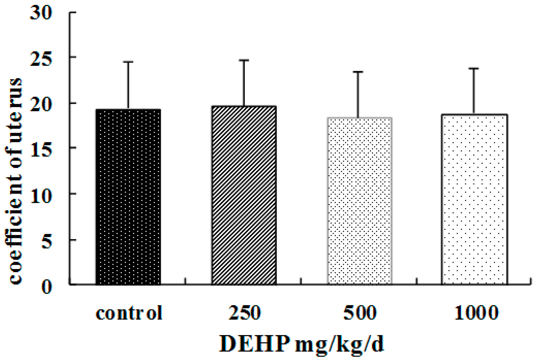
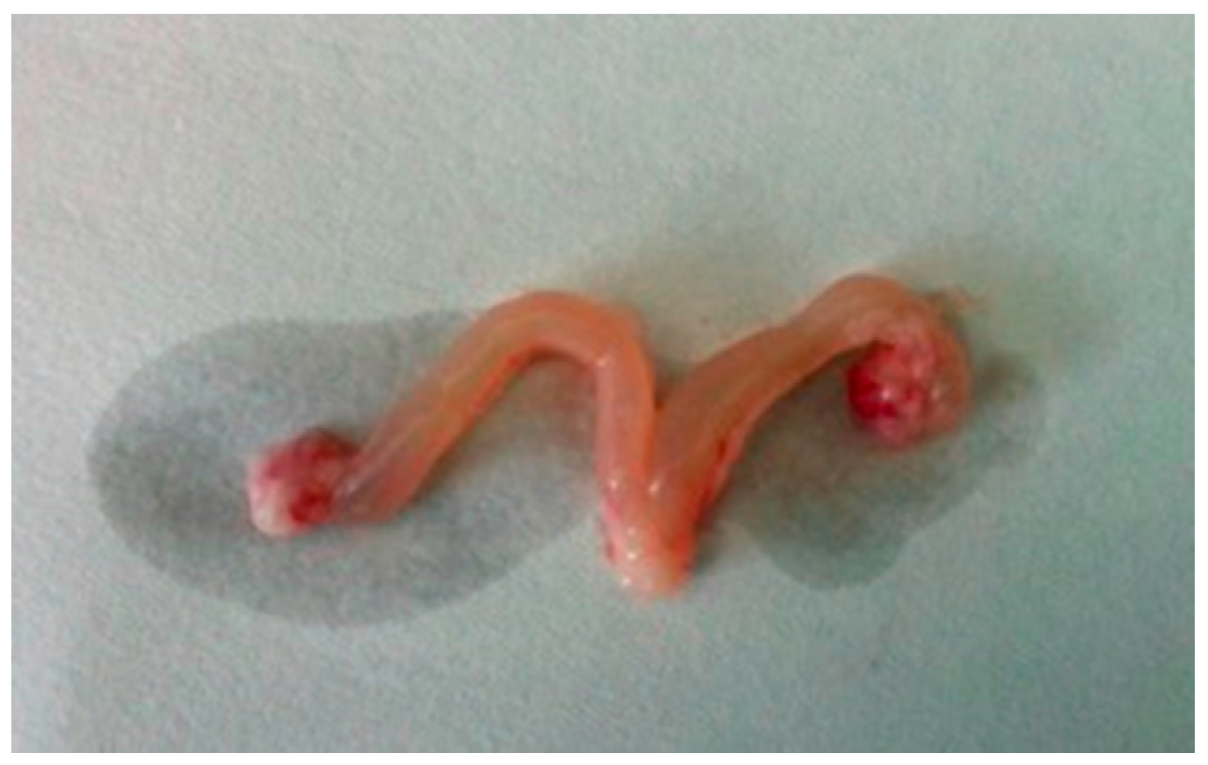
26]. The association between DEHP and higher body weight suggests its negative effects on the growth and development of pubertal female rats. In our study, we also find that the ovaries of the rats treated with DEHP were congestive and swelling, and the volume of the ovaries and uterus became bigger. It suggests ovary and uterus might be the target organ of DEHP.
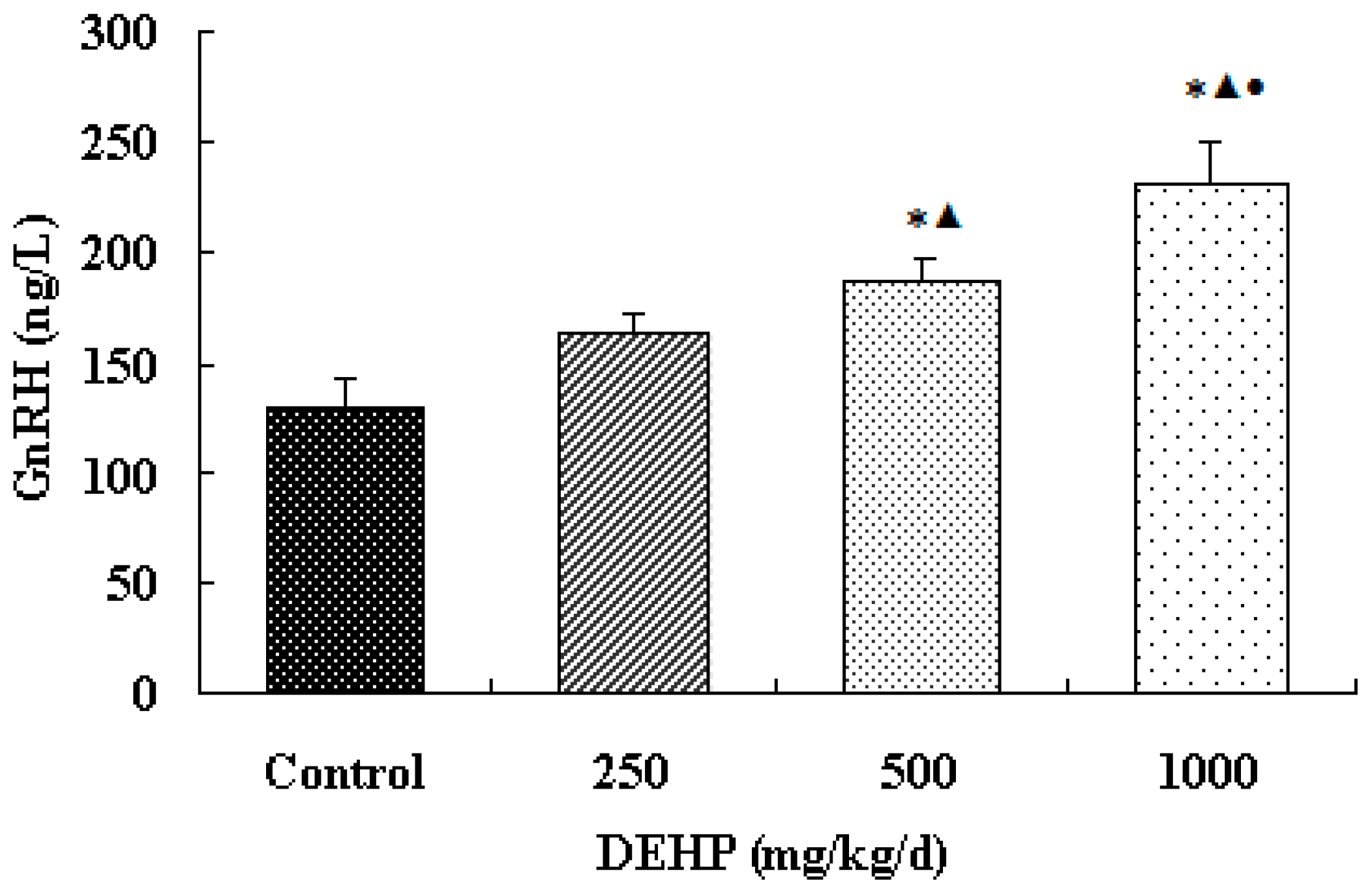
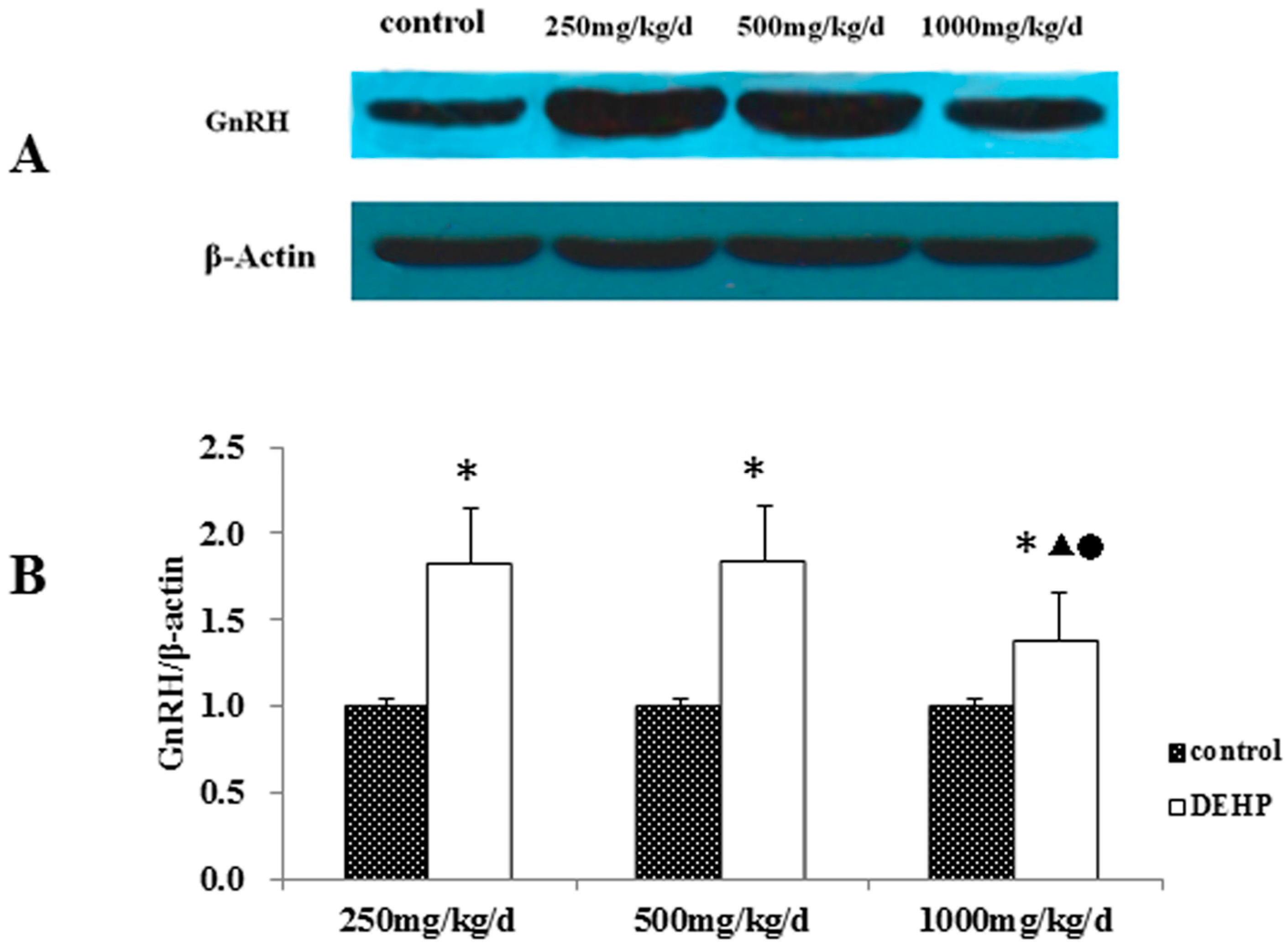
The result shows that the secretion of GnRH in the hypothalamus of the exposure group significantly increased. It indicates that the effect of DEHP is similar to that of GnRH agonists and estrogen. Sustained DEHP exposure might increase the sensitivity of GnRH in the hypothalamus and then raise the secretion of GnRH.
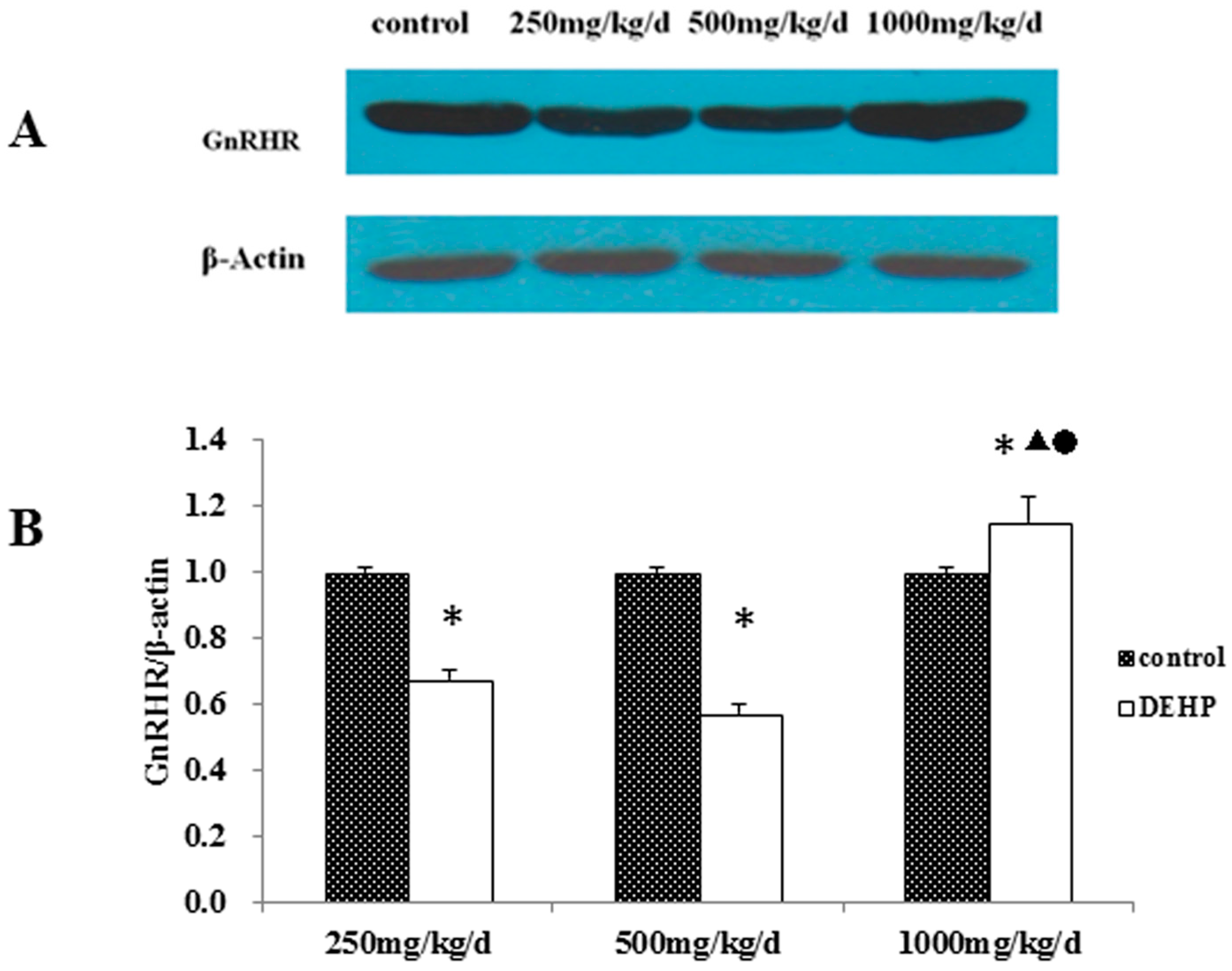
Meanwhile, however, there is no significant difference in the mRNA level of GnRH in the hypothalamus of the rats treated with DEHP. The protein level in the hypothalamus is significantly higher in the rats treated with DEHP compared with the rats in control group, and the level was significantly lower in the rats treated with 1000 mg·kg−1·d−1 DEHP compared with the rats in 250 and 500 mg/kg/d DEHP. This means that DEHP could affect the regulation of GnRH after translation, and the protein expression of GnRH is much more stable. However, 1000 mg/kg/d DEHP may inhibit the protein level of GnRH in the hypothalamus. This suggests that GnRH of hypothalamus might be an important target of DEHP.
Studies have found that EEDs, like bisphenol A and 4-nonylphenol, can have significant adverse effects on the gene regulatory network related to the function of the pituitary-ovary axis. GnRHR mRNA, FSH β-subunit, mRNA of LH β-subunit, FSH-R, mRNA of LH-R, the receptor of E and mRNA of the receptor of E all increased significantly. Those that also increased significantly included the expression levels of the mRNA of GnRH-R pathway (phosphatidylinositol pathway), E2-R receptor pathway (including E2-R receptor signal transduction pathway and crosstalk pathway between IGF, EGF and TGF) and the estrogen-metabolizing enzymes [55]. Liu et al.’s study said, compared with control rats, the DEHP-treated adult female rats showed higher mRNA and protein levels of GnRHR in the pituitary [54].
Under the influence of GnRH in the hypothalamus, the pituitary releases FSH and LH that decide the secretion of sex hormones in the ovary. The cyclic variation of the endometrium is regulated by sex hormones. Previous research suggested that GnRHR only exists in the pituitary. However, basic research on GnRH and GnRHR in recent years shows that GnRHR also exists in other organs, including the ovaries, testes, uteruses, etc. [41,42,43,44]. In addition, tissues of malignant tumors, such as breast cancer, ovarian cancer and endometrial cancer, experience high affinity of GnRHR expression as well [56,57,58].
DEHP is an environmental endocrine disruptor, and it has estrogen-like activity. Estrogen plays a role in both normal uterine physiology and uterine pathologies, such as dysfunctional uterine bleeding or breakthrough bleeding, endometriosis, infertility, leiomyoma, and endometrial cancer [59]. Previous study investigated the in vitro and in vivo effects of DEHP and also the comparison of the urinary levels of several phthalate metabolites between women with and without endometriosis. These findings suggest that exposure to phthalate may lead to establishment of endometriosis by enhancing invasive and proliferative activities of endometrial cells [60]. A report showed a 31-year old woman in Italy who had a small uterus, sterility and secondary amenorrhea. Cytogenetic analysis demonstrated the complete loss of a copy of the GnRHR gene. GnRHR may associate with gonadal function [
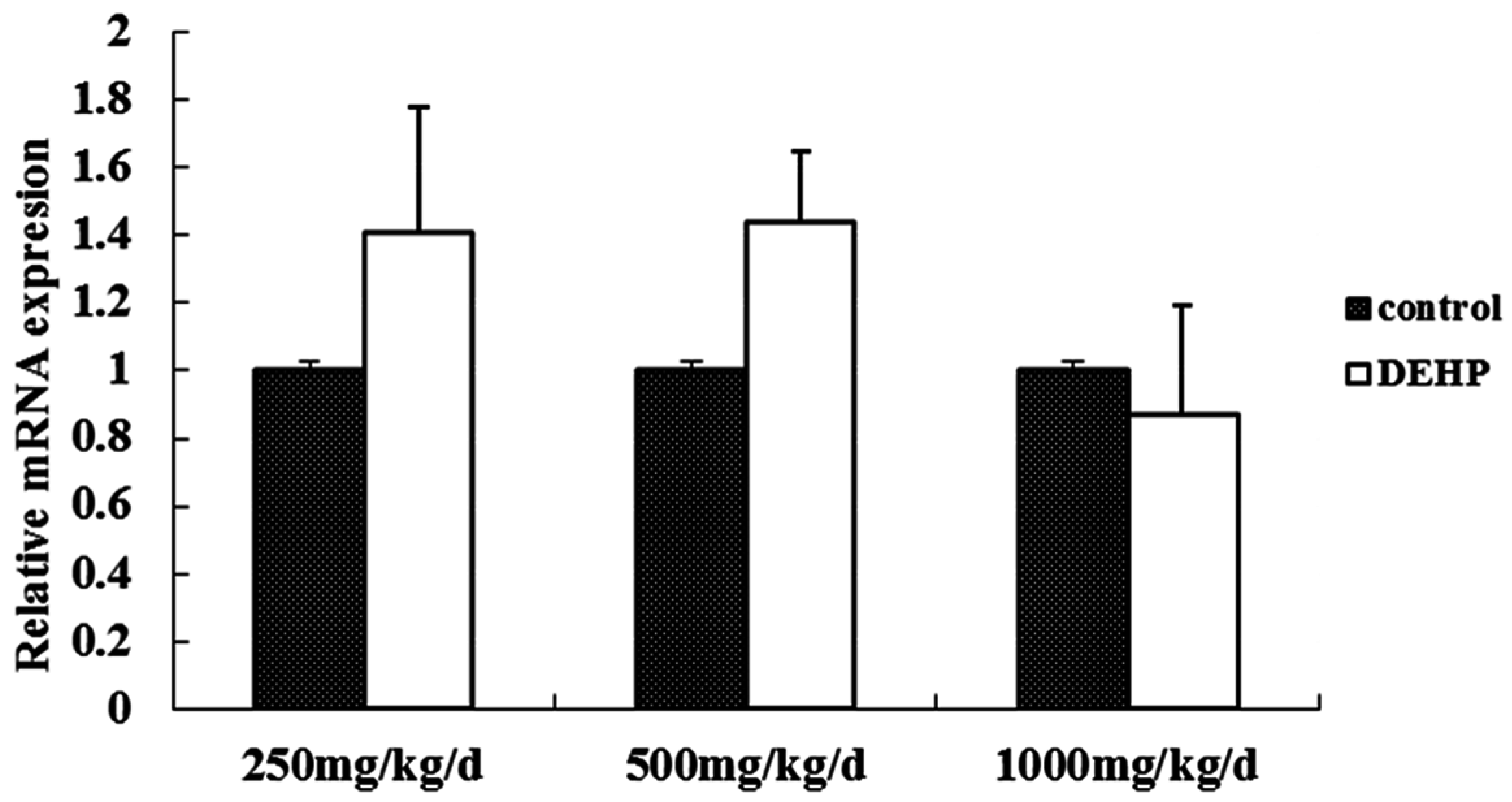
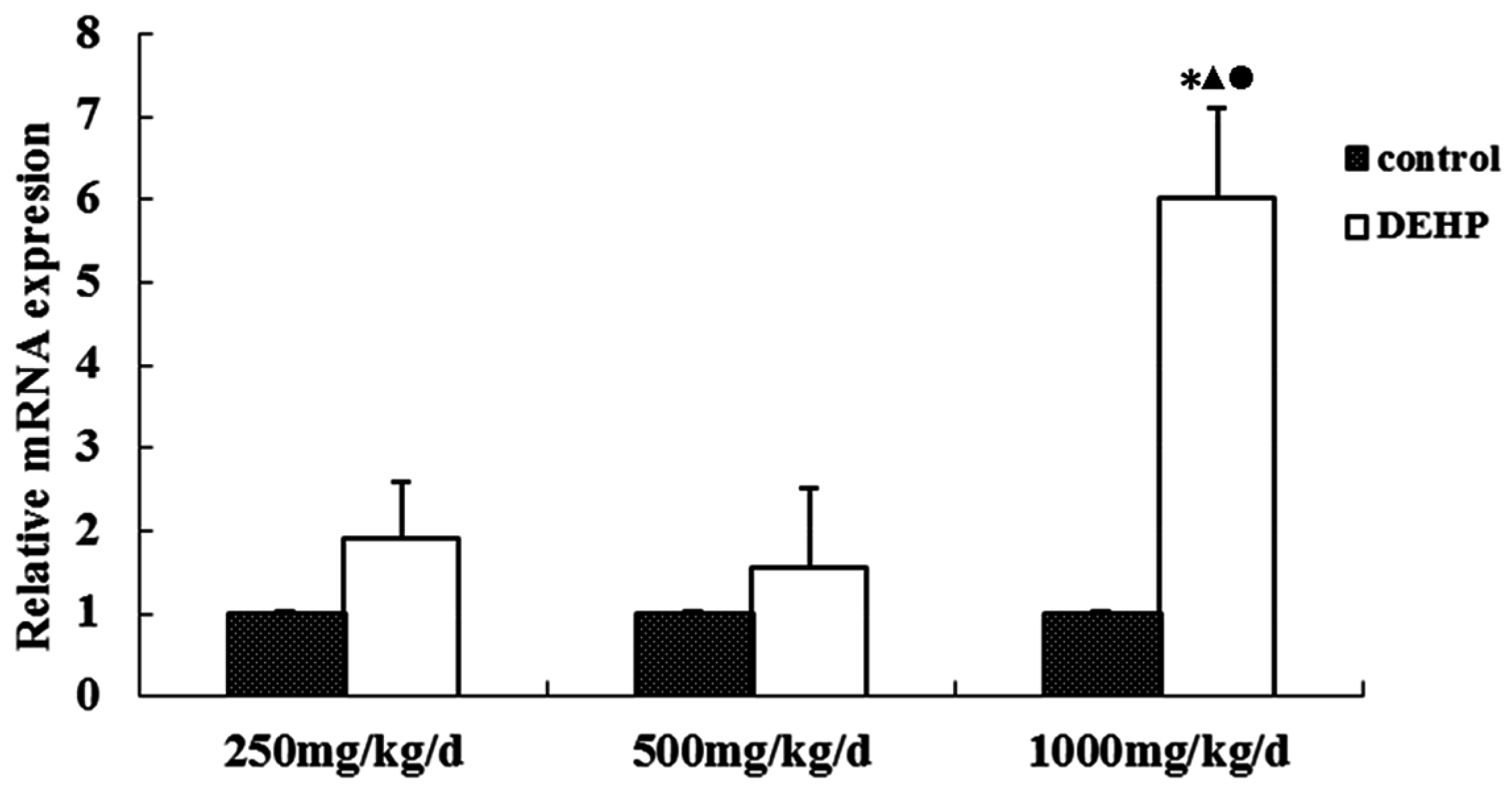
61]. The result of this study shows that the cytomembrane, cytoplasm and nucleus of uterus epithelial cells and glandular epithelial cells are GnRHR-positive. The immunoreactive substance is yellow or yellow brown. In addition, 250 and 500 mg/kg/d DEHP can decrease the protein expression level of GnRHR in uteruses, while high doses of DEHP may increase the GnRHR mRNA and protein expression levels in uteruses of pubertal female rats. This proves that DEHP may, through the toxic effect of GnRH in the hypothalamus and its receptor on the uterus, do damage to the regulatory function of autocrine or paracrine of the uterus and result in uterus related diseases.

5. Conclusions

In the present study, our data reveal that DEHP exposure may lead to a disruption in pubertal female rats and an imbalance of the hypothalamus–uterus. Meanwhile, DEHP might, through the GnRH in the hypothalamus and its receptor on the uterus, lead to diseases of the uterus. It may impose a negative influence on the development and function of the reproductive system in female rats.

Acknowledgments

This study was supported by the Graduate Innovation Fund of Jilin University (Grant No. 2015100) and the National Natural Science Foundation of China (Grant No. 81573184).

Author Contributions

Te Liu, Yiyang Jia conceived and designed the experiments; Te Liu, Lin Ye, Qi Wang, Di Sun, Jin Xu, Huaiji Chen, Feng Xu performed the experiments; Te Liu collected the data; Liting Zhou, Qi Wang, Juan Wu analyzed the data; Te Liu wrote the paper.

Conflicts of Interest

The authors declare no conflict of interest.

References

  1. Al-Saleh, I.; Shinwari, N.; Alsabbaheen, A. Phthalates residues in plastic bottled waters. J. Toxicol. Sci. 2011, 36, 469–478. [Google Scholar] [CrossRef] [PubMed]
  2. Wittassek, M.; Koch, H.M.; Angerer, J.; Bruning, T. Assessing exposure to phthalates—The human biomonitoring approach. Mol. Nutr. Food Res. 2011, 55, 7–31. [Google Scholar] [CrossRef] [PubMed]
  3. Cirillo, T.; Fasano, E.; Esposito, F.; Montuori, P.; Cocchieri, R.A. Di(2-ethylhexyl)phthalate (DEHP) and di-n-butylphthalate (DBP) exposure through diet in hospital patients. Food Chem. Toxicol. 2013, 51, 434–438. [Google Scholar] [CrossRef] [PubMed] [Green Version]
  4. Mastorakos, G.; Karoutsou, E.I.; Mizamtsidi, M.; Creatsas, G. The menace of endocrine disruptors on thyroid hormone physiology and their impact on intrauterine development. Endocrine 2007, 31, 219–237. [Google Scholar] [CrossRef] [PubMed]
  5. Heudorf, U.; Mersch-Sundermann, V.; Angerer, E. Phthalates: Toxicology and exposure. Int. J. Hyg. Environ. Health 2007, 210, 623–634. [Google Scholar] [CrossRef] [PubMed]
  6. Schettler, T. Human exposure to phthalates via consumer products. Int. J. Androl. 2006, 29, 134–139. [Google Scholar] [CrossRef] [PubMed]
  7. Koniecki, D.; Wang, R.; Moody, R.P.; Zhu, J.P. Phthalates in cosmetic and personal care products: Concentrations and possible dermal exposure. Environ. Res. 2011, 111, 329–336. [Google Scholar] [CrossRef] [PubMed]
  8. Earls, A.O.; Axford, I.P.; Braybrook, J.H. Gas chromatography-mass spectrometry determination of the migration of phthalate plasticisers from polyvinyl chloride toys and childcare articles. J. Chromatogr. A 2003, 983, 237–246. [Google Scholar] [CrossRef]
  9. Benson, R. Hazard to the developing male reproductive system from cumulative exposure to phthalate esters-dibutyl phthalate, diisobutyl phthalate, butylbenzyl phthalate, diethylhexyl phthalate, dipentyl phthalate, and diisononyl phthalate. Regul. Toxicol. Pharm. 2009, 53, 90–101. [Google Scholar] [CrossRef] [PubMed]
  10. Latini, G. Potential hazards of exposure to di-(2-ethylhexyl)-phthalate in babies: A review. Biol. Neonate 2000, 78, 269–276. [Google Scholar] [CrossRef] [PubMed]
  11. Lovekamp-Swan, T.; Davis, B.J. Mechanisms of phthalate ester toxicity in the female reproductive system. Environ. Health Perspect. 2003, 111, 139–145. [Google Scholar] [CrossRef] [PubMed]
  12. Adibi, J.J.; Whyatt, R.M.; Williams, P.L.; Calafat, A.M.; Camann, D.; Herrick, R.; Nelson, H.; Bhat, H.K.; Perera, F.A.; Silva, M.J.; et al. Characterization of phthalate exposure among pregnant women assessed by repeat air and urine samples. Environ. Health Perspect. 2008, 116, 467–473. [Google Scholar] [CrossRef] [PubMed]
  13. Xu, Y.; Hubal, E.A.C.; Little, J.C. Predicting residential exposure to phthalate plasticizer emitted from vinyl flooring: Sensitivity, uncertainty, and implications for biomonitoring. Environ. Health Perspect. 2010, 118, 253–258. [Google Scholar] [CrossRef] [PubMed]
  14. Destaillats, H.; Maddalena, R.L.; Singer, B.C.; Hodgson, A.T.; McKone, T.E. Indoor pollutants emitted by office equipment: A review of reported data and information needs. Atmos. Environ. 2008, 42, 1371–1388. [Google Scholar] [CrossRef]
  15. Bornehag, C.G.; Lundgren, B.; Weschler, C.J.; Sigsgaard, T.; Hagerhed-Engman, L.; Sundell, J. Phthalates in indoor dust and their association with building characteristics. Environ. Health Perspect. 2005, 113, 1399–1404. [Google Scholar] [CrossRef] [PubMed]
  16. Deutschle, T.; Reiter, R.; Butte, W.; Heinzow, B.; Keck, T.; Riechelmann, H. A controlled challenge study on di(2-ethylhexyl) phthalate (DEHP) in house dust and the immune response in human nasal mucosa of allergic subjects. Environ. Health Perspect. 2008, 116, 1487–1493. [Google Scholar] [CrossRef] [PubMed]
  17. Kolarik, B.; Naydenov, K.; Larsson, M.; Bornehag, C.G.; Sundell, J. The association between phthalates in dust and allergic diseases among bulgarian children. Environ. Health Perspect. 2008, 116, 98–103. [Google Scholar] [CrossRef] [PubMed] [Green Version]
  18. Schripp, T.; Fauck, C.; Salthammer, T. Chamber studies on mass-transfer of di(2-ethylhexyl)phthalate (DEHP) and di-n-butylphthalate (dnbp) from emission sources into house dust. Atmos. Environ. 2010, 44, 2840–2845. [Google Scholar] [CrossRef]
  19. Geiss, O.; Tirendi, S.; Barrero-Moreno, J.; Kotzias, D. Investigation of volatile organic compounds and phthalates present in the cabin air of used private cars. Environ. Int. 2009, 35, 1188–1195. [Google Scholar] [CrossRef] [PubMed]
  20. Wittassek, M.; Angerer, J. Phthalates: Metabolism and exposure. Int. J. Androl. 2008, 31, 131–136. [Google Scholar] [CrossRef] [PubMed]
  21. Hauser, R.; Calafat, A.M. Phthalates and human health. Occup. Environ. Med. 2005, 62, 806–818. [Google Scholar] [CrossRef] [PubMed]
  22. Wormuth, M.; Scheringer, M.; Vollenweider, M.; Hungerbuhler, K. What are the sources of exposure to eight frequently used phthalic acid esters in europeans? Risk Anal. 2006, 26, 803–824. [Google Scholar] [CrossRef] [PubMed]
  23. Kavlock, R.; Boekelheide, K.; Chapin, R.; Cunningham, M.; Faustman, E.; Foster, P.; Golub, M.; Henderson, R.; Hinberg, I.; Little, R.; et al. NTP center for the evaluation of risks to human reproduction: Phthalates expert panel report on the reproductive and developmental toxicity of di(2-ethylhexyl) phthalate. Reprod. Toxicol. 2002, 16, 529–653. [Google Scholar] [CrossRef]
  24. Martinez-Arguelles, D.B.; Campioli, E.; Culty, M.; Zirkin, B.R.; Papadopoulos, V. Fetal origin of endocrine dysfunction in the adult: The phthalate model. J. Steroid Biochem. 2013, 137, 5–17. [Google Scholar] [CrossRef] [PubMed]
  25. Pocar, P.; Fiandanese, N.; Secchi, C.; Berrini, A.; Fischer, B.; Schmidt, J.S.; Schaedlich, K.; Borromeo, V. Exposure to di(2-ethyl-hexyl) phthalate (DEHP) in utero and during lactation causes long-term pituitary-gonadal axis disruption in male and female mouse offspring. Endocrinology 2012, 153, 937–948. [Google Scholar] [CrossRef] [PubMed]
  26. Schmidt, J.S.; Schaedlich, K.; Fiandanese, N.; Pocar, P.; Fischer, B. Effects of di(2-ethylhexyl) phthalate (DEHP) on female fertility and adipogenesis in c3h/n mice. Environ. Health Perspect. 2012, 120, 1123–1129. [Google Scholar] [CrossRef] [PubMed]
  27. Gupta, R.K.; Singh, J.M.; Leslie, T.C.; Meachum, S.; Flaws, J.A.; Yao, H.H. Di-(2-ethylhexyl) phthalate and mono-(2-ethylhexyl) phthalate inhibit growth and reduce estradiol levels of antral follicles in vitro. Toxicol. Appl. Pharmacol. 2010, 242, 224–230. [Google Scholar] [CrossRef] [PubMed]
  28. Xu, Y.; Agrawal, S.; Cook, T.J.; Knipp, G.T. Di-(2-ethylhexyl)-phthalate affects lipid profiling in fetal rat brain upon maternal exposure. Arch. Toxicol. 2007, 81, 57–62. [Google Scholar] [CrossRef] [PubMed]
  29. Xu, Y.; Knipp, G.T.; Cook, T.J. Effects of di-(2-ethylhexyl)-phthalate and its metabolites on the lipid profiling in rat HRP-1 trophoblast cells. Arch. Toxicol. 2006, 80, 293–298. [Google Scholar] [CrossRef] [PubMed]
  30. Gore, A.C.; Chappell, V.A.; Fenton, S.E.; Flaws, J.A.; Nadal, A.; Prins, G.S.; Toppari, J.; Zoeller, R.T. Executive summary to EDC-2: The endocrine society’s second scientific statement on endocrine-disrupting chemicals. Endocr. Rev. 2015, 36, 593–602. [Google Scholar] [CrossRef] [PubMed]
  31. Meeker, J.D. Exposure to environmental endocrine disruptors and child development. Arch. Pediatr. Adolesc. Med. 2012, 166, E1–E7. [Google Scholar] [CrossRef] [PubMed]
  32. Behie, A.M.; O’Donnell, M.H. Prenatal smoking and age at menarche: Influence of the prenatal environment on the timing of puberty. Hum. Reprod. 2015, 30, 957–962. [Google Scholar] [CrossRef] [PubMed]
  33. Kim, Y.H.; Kim, S.H.; Lee, H.W.; Chae, H.D.; Kim, C.H.; Kang, B.M. Increased viability of endometrial cells by in vitro treatment with di-(2-ethylhexyl) phthalate. Fertil. Steril. 2010, 94, 2413–2416. [Google Scholar] [CrossRef] [PubMed]
  34. Kim, S.H.; Chun, S.; Jang, J.Y.; Chae, H.D.; Kim, C.H.; Kang, B.M. Increased plasma levels of phthalate esters in women with advanced-stage endometriosis: A prospective case-control study. Fertil. Steril. 2011, 95, 357–359. [Google Scholar] [CrossRef] [PubMed]
  35. Wolff, M.S.; Teitelbaum, S.L.; McGovern, K.; Windham, G.C.; Pinney, S.M.; Galvez, M.; Calafat, A.M.; Kushi, L.H.; Biro, F.M.; Breast, C.; et al. Phthalate exposure and pubertal development in a longitudinal study of U.S. girls. Hum. Reprod. 2014, 29, 1558–1566. [Google Scholar] [CrossRef] [PubMed]
  36. Wen, Y.; Liu, S.D.; Lei, X.; Ling, Y.S.; Luo, Y.; Liu, Q. Association of paes with precocious puberty in children: A systematic review and meta-analysis. Int. J. Environ. Res. Public Health 2015, 12, 15254–15268. [Google Scholar] [CrossRef] [PubMed]
  37. Brito, V.N.; Latronico, A.C.; Arnhold, I.J.; Mendonca, B.B. Update on the etiology, diagnosis and therapeutic management of sexual precocity. Arq. Bras. Endocrinol. Metabol. 2008, 52, 18–31. [Google Scholar] [CrossRef] [PubMed] [Green Version]
  38. Larco, D.O.; Williams, M.; Schmidt, L.; Sabel, N.; Lange, J.; Woller, M.J.; Wu, T.J. Autoshortloop feedback regulation of pulsatile gonadotropin-releasing hormone (GnRH) secretion by its metabolite, GnRH-(1-5). Endocrine 2015, 49, 470–478. [Google Scholar] [CrossRef] [PubMed]
  39. Conn, P.M.; Crowley, W.F., Jr. Gonadotropin-releasing hormone and its analogues. N. Engl. J. Med. 1991, 324, 93–103. [Google Scholar] [PubMed]
  40. Counis, R.; Laverriere, J.N.; Garrel, G.; Bleux, C.; Cohen-Tannoudji, J.; Lerrant, Y.; Kottler, M.L.; Magre, S. Gonadotropin-releasing hormone and the control of gonadotrope function. Reprod. Nutr. Dev. 2005, 45, 243–254. [Google Scholar] [CrossRef] [PubMed]
  41. Thompson, M.D.; Hendy, G.N.; Percy, M.E.; Bichet, D.G.; Cole, D.E. G protein-coupled receptor mutations and human genetic disease. Methods Mol. Biol. 2014, 1175, 153–187. [Google Scholar] [PubMed]
  42. Roch, G.J.; Busby, E.R.; Sherwood, N.M. Gnrh receptors and peptides: Skating backward. Gen. Comp. Endocrinol. 2014, 209, 118–134. [Google Scholar] [CrossRef] [PubMed]
  43. Basaran, Y.; Bolu, E.; Unal, H.U.; Sagkan, R.I.; Taslipinar, A.; Ozgurtas, T.; Musabak, U. Multiplex ligation dependent probe amplification analysis of KAL1, GnRH1, GnRHR, PROK2 and PROKR2 in male patients with idiopathic hypogonadotropic hypogonadism. Endokrynol. Pol. 2013, 64, 285–292. [Google Scholar] [CrossRef] [PubMed]
  44. Gianetti, E.; Hall, J.E.; Au, M.G.; Kaiser, U.B.; Quinton, R.; Stewart, J.A.; Metzger, D.L.; Pitteloud, N.; Mericq, V.; Merino, P.M.; et al. When genetic load does not correlate with phenotypic spectrum: Lessons from the GnRH receptor (GNRHR). J. Clin. Endocrinol. Metab. 2012, 97, E1798–E1807. [Google Scholar] [CrossRef] [PubMed]
  45. Diamanti-Kandarakis, E. PCOS in adolescents. Best Pract. Res. Clin. Obstet. Gynaecol. 2010, 24, 173–183. [Google Scholar] [CrossRef] [PubMed]
  46. Zou, C.C.; Liang, L.; Dong, G.P.; Zhao, Z.Y. Peripheral precocious puberty: A retrospective study for six years in Hangzhou, China. J. Paediatr. Child Health 2008, 44, 415–418. [Google Scholar] [CrossRef] [PubMed]
  47. Li, X.; Cai, D. Single and combined toxic effects of di-2-ethylhexyl phthalate and cypermethrin on fertility and development in the the prepubertal male rats. Wei Sheng Yan Jiu 2012, 41, 710–716. [Google Scholar] [PubMed]
  48. Kim, S.H.; Lee, H.W.; Kim, Y.H.; Koo, Y.H.; Chae, H.D.; Kim, C.H.; Lee, P.R.; Kang, B.M. Down-regulation of p21-activated kinase 1 by progestin and its increased expression in the eutopic endometrium of women with endometriosis. Hum. Reprod. 2009, 24, 1133–1141. [Google Scholar] [CrossRef] [PubMed]
  49. Schaedlich, K.; Schmidt, J.S.; Kwong, W.Y.; Sinclair, K.D.; Kurz, R.; Jahnke, H.G.; Fischer, B. Impact of di-ethylhexylphthalate exposure on metabolic programming in P19 ECC-derived cardiomyocytes. J. Appl. Toxicol. 2015, 35, 861–869. [Google Scholar] [CrossRef] [PubMed]
  50. Sukhbaatar, U.; Mijiddorj, T.; Oride, A.; Kanasaki, H. Stimulation of delta subunit-containing GABAA receptor by DS1 increases GnRH receptor expression but reduces GnRH mRNA expression in GnRH-producing GT1–7 cells. Endocrine 2015, 49, 222–230. [Google Scholar] [CrossRef] [PubMed]
  51. Ge, R.S.; Chen, G.R.; Dong, Q.; Akingbemi, B.; Sottas, C.M.; Santos, M.; Sealfon, S.C.; Bernard, D.J.; Hardy, M.P. Biphasic effects of postnatal exposure to diethylhexylphthalate on the timing of puberty in male rats. J. Androl. 2007, 28, 513–520. [Google Scholar] [CrossRef] [PubMed]
  52. Losa-Ward, S.M.; Todd, K.L.; McCaffrey, K.A.; Tsutsui, K.; Patisaul, H.B. Disrupted organization of RFamide pathways in the hypothalamus is associated with advanced puberty in female rats neonatally exposed to bisphenol A. Biol. Reprod. 2012, 87, 28. [Google Scholar] [CrossRef] [PubMed]
  53. Roth, C.; Schricker, M.; Lakomek, M.; Strege, A.; Heiden, I.; Luft, H.; Munzel, U.; Wuttke, W.; Jarry, H. Autoregulation of the gonadotropin-releasing hormone (GnRH) system during puberty: Effects of antagonistic versus agonistic GnRH analogs in a female rat model. J. Endocrinol. 2001, 169, 361–371. [Google Scholar] [CrossRef] [PubMed]
  54. Liu, T.; Li, N.; Zhu, J.; Yu, G.Y.; Guo, K.; Zhou, L.T.; Zheng, D.C.; Qu, X.F.; Huang, J.; Chen, X.; et al. Effects of di-(2-ethylhexyl) phthalate on the hypothalamus-pituitary-ovarian axis in adult female rats. Reprod. Toxicol. 2014, 46, 141–147. [Google Scholar] [CrossRef] [PubMed]
  55. Carbone, S.; Samaniego, Y.A.; Cutrera, R.; Reynoso, R.; Cardoso, N.; Scacchi, P.; Moguilevsky, J.A.; Ponzo, O.J. Different effects by sex on hypothalamic-pituitary axis of prepubertal offspring rats produced by in utero and lactational exposure to di-(2-ethylhexyl) phthalate (DEHP). Neurotoxicology 2012, 33, 78–84. [Google Scholar] [CrossRef] [PubMed]
  56. Chen, A.; Kaganovsky, E.; Rahimipour, S.; Ben-Aroya, N.; Okon, E.; Koch, Y. Two forms of gonadotropin-releasing hormone (GnRH) are expressed in human breast tissue and overexpressed in breast cancer: A putative mechanism for the antiproliferative effect of GnRH by down-regulation of acidic ribosomal phosphoproteins P1 and P2. Cancer Res. 2002, 62, 1036–1044. [Google Scholar] [PubMed]
  57. Grundker, C.; Gunthert, A.R.; Millar, R.P.; Emons, G. Expression of gonadotropin-releasing hormone II (GnRH-II) receptor in human endometrial and ovarian cancer cells and effects of GnRH-II on tumor cell proliferation. J. Clin. Endocr. Metab. 2002, 87, 1427–1430. [Google Scholar] [CrossRef] [PubMed]
  58. Chatzaki, E.; Bax, C.M.; Eidne, K.A.; Anderson, L.; Grudzinskas, J.G.; Gallagher, C.J. The expression of gonadotropin-releasing hormone and its receptor in endometrial cancer, and its relevance as an autocrine growth factor. Cancer Res. 1996, 56, 2059–2065. [Google Scholar] [PubMed]
  59. Zhang, X.; Healy, C.; Nothnick, W.B. Estrogen suppresses expression of the matrix metalloproteinase inhibitor reversion-inducing cysteine-rich protein with kazal motifs (RECK) within the mouse uterus. Endocrine 2012, 42, 97–106. [Google Scholar] [CrossRef] [PubMed]
  60. Kim, S.H.; Cho, S.; Ihm, H.J.; Oh, Y.S.; Heo, S.H.; Chun, S.; Im, H.; Chae, H.D.; Kim, C.H.; Kang, B.M. Possible role of phthalate in the pathogenesis of endometriosis: In vitro, animal, and human data. J. Clin. Endocrinol. Metab. 2015, 100, E1502–E1511. [Google Scholar] [CrossRef] [PubMed]
  61. Fiorio, P.; Rosaia De Santis, L.; Cuoco, C.; Gimelli, G.; Gastaldi, R.; Bonatti, F.; Ravazzolo, R.; Bocciardi, R. Hypogonadotropic hypogonadism in a trisomy X carrier: Phenotype description and genotype correlation. Gynecol. Endocrinol. 2015, 32, 14–17. [Google Scholar] [CrossRef] [PubMed]
Figure 1. Effects of DEHP on ovaries and the uterus in a pubertal female rat.
Ijerph 13 01130 g001
Figure 2. Effects of DEHP on coefficients of uteruses in pubertal female rats; n = 12. The coefficient of uterus was expressed as the mean value ± standard error (SE).
Ijerph 13 01130 g002
Figure 3. Effect of DEHP on gonadotropin-releasing hormone (GnRH) level in the hypothalamus (n = 12). GnRH level in the hypothalamus was expressed as the mean value ± standard error (SE). * Significant difference compared with control (p < 0.05); Significant difference compared with 250 mg/kg/d (p <0.05); Significant difference compared with 500 mg/kg/d (p < 0.05).
Ijerph 13 01130 g003
Figure 4. Effects of DEHP exposure on the level of GnRH mRNA in the hypothalamus; n = 12. The level of GnRHR mRNA was expressed as the mean value ± standard error (SE).
Ijerph 13 01130 g004
Figure 5. (A) Protein bands of GnRH expression in the hypothalamus of pubertal female rats. (B) effects of DEHP exposure on the protein level of GnRHR in the hypothalamus; n = 12. The level of GnRHR protein was expressed as the mean value ± standard error (SE). * Significant difference compared to control (p < 0.05); Significant difference compared to 250 mg/kg/d (p < 0.05); Significant difference compared to 500 mg/kg/d (p < 0.05).
Ijerph 13 01130 g005
Figure 6. Effects of DEHP exposure on the level of GnRHR mRNA in the uterus; n = 12. The level of GnRHR mRNA was expressed as the mean value ± standard error (SE). * Significant difference compared to control (p < 0.05); Significant difference compared to 250 mg/kg/d (p < 0.05); Significant difference compared to 500 mg/kg/d (p < 0.05).
Ijerph 13 01130 g006
Figure 7. (A) Protein bands of GnRHR expression in the uterus of pubertal female rats; (B) effects of DEHP exposure on the protein level of GnRHR in the uterus; n = 12. The level of GnRHR protein was expressed as the mean value ± standard error (SE). * Significant difference compared to control (p < 0.05); Significant difference compared to 250 mg/kg/d (p < 0.05); Significant difference compared to 500 mg/kg/d (p < 0.05).
Ijerph 13 01130 g007
Figure 8. Immunohistochemical staining of GnRH expression in the hypothalamus of pubertal female rats (×400).
Ijerph 13 01130 g008
Figure 9. Immunohistochemical staining of GnRHR expression in the uterus of pubertal female rats (×400).
Ijerph 13 01130 g009
Table 1. Gene-specific forward and reverse primer sequence.
GeneDirectionSequence
GNRHForwardTCCAGCCAGCACTGGTCCTA
ReverseGGGTTCTGCCATTTGATCCTC
GnRHRForwardTCACCTTCAGCTGCCTGTTCA
ReverseCTCAGCCGTGCTCTTGGGATA
β-actinForwardCCCATTGAACACGGCATTG
ReverseGGTACGACCAGAGGCATACA
Table 2. Food consumption by pubertal female rats treated with DEHP (mean ± SE, g).
GroupsnTime (Weeks)
1234
Control1213.33 ± 3.0113.66 ± 4.2014.60 ± 2.8614.53 ± 3.69
250 mg/kg/d1217.00 ± 2.44 *18.82 ± 3.10 *17.77 ± 3.3416.20 ± 3.95
500 mg/kg/d1214.25 ± 1.23 15.48 ± 3.3818.39 ± 2.9717.15 ± 2.93
1000 mg/kg/d1216.55 ± 2.22 *17.82 ± 4.3222.28 ± 2.37 *18.53 ± 1.74 *
* p < 0.05, vs. Control group; p < 0.05, vs. 250 mg/kg/d group.
Table 3. Body weights of pubertal female rats treated with DEHP during the administration period (mean ± SE, g).
GroupsnTime (Weeks)
1234
Control1276.87 ± 8.43106.73 ± 9.25134.00 ± 6.85152.12 ± 3.89
250 mg/kg/d128 85.12 ± 9.31116.58 ± 8.70 *144.27 ± 7.40 *164.69 ± 8.05 *
500 mg/kg/d1287.17 ± 9.75117.50 ± 7.46 *143.74 ± 8.89 *168.52 ± 7.59 *
1000 mg/kg/d1290.43 ± 10.91 *122.05 ± 7.88 *149.02 ± 7.35 *170.16 ± 6.46 *
* p < 0.05, vs. Control group.
Table 4. Water consumption by pubertal female rats treated with DEHP (mean ± SE, mL).
GroupsnTime (Weeks)
1234
Control1217.12 ± 3.0825.62 ± 7.2229.21 ± 7.8225.10 ± 5.87
250 mg/kg/d1218.92 ± 4.2126.04 ± 5.2026.83 ± 8.2032.85 ± 1.79 *
500 mg/kg/d1222.38 ± 4.25 *27.30 ± 3.6329.26 ± 6.6735.48 ± 5.80 *
1000 mg/kg/d1217.98 ± 1.32 17.46 ± 3.34 *,,●22.80 ± 8.9423.76 ± 3.35 ▲,
* p < 0.05, vs. Control group; p < 0.05, vs. 250 mg/kg/d group; p < 0.05, vs. 500 mg/kg/d group.
Table 5. Effects of DEHP on mean optical density (MOD) and integral optical density (IOD) of GnRH staining in the hypothalamus a.
GroupsDosage, mg/kg/dMOD (10−2)IOD (×104)
Control-15.322 ± 0.85366.195 ± 6.038
DEHP25017.081 ± 0.728 b78.000 ± 4.794 b
50016.242 ± 0.537 b72.200 ± 6.123 b
100016.492 ± 0.521 b74.340 ± 7.962 b
a n = 12, each treatment group; b Significant difference compared to control (p < 0.05).
Table 6. Effects of DEHP on MOD and IOD of GnRHR staining in the uterus a.
GroupsDosage, mg/kg/dMOD (10−2)IOD (×104)
Control-12.941 ± 1.02444.028 ± 0.868
DEHP25016.096 ± 0.711 b50.458 ± 4.719 b
50014.893 ± 0.563 b,c48.971 ± 1.825 b,c
100015.748 ± 0.160 b52.083 ± 7.565 b
a n = 12, each treatment group; b Significant difference compared to control (p < 0.05); c Significant difference compared to 250 mg/kg/d (p < 0.05).

