Article

Diesel Exhaust Particles Induce the Over expression of Tumor Necrosis Factor-α (TNF-α) Gene in Alveolar Macrophages and Failed to Induce Apoptosis through Activation of Nuclear Factor-κB (NF-κB)

by
Ramzi M. Kafoury
1,* and
Michael C. Madden
2
1
Center of Environmental Health and Molecular Toxicology Laboratory, Jackson State University, Jackson, Mississippi, USA
2
Human Exposure Section, United States Environmental Protection Agency, Chapel Hill, North Carolina
*
Author to whom correspondence should be addressed.
Int. J. Environ. Res. Public Health 2005, 2(1), 107-113; https://doi.org/10.3390/ijerph2005010107
Submission received: 15 November 2004 / Accepted: 6 February 2005 / Published: 30 April 2005

Abstract

:
Exposure to particulate matter (PM2.5–10), including diesel exhaust particles (DEP) has been reported to induce lung injury and exacerbation of asthma and chronic obstructive pulmonary disease. Alveolar macrophages play a major role in the lung’s response to inhaled particles and therefore, are a primary target for PM2.5–10 effect. The molecular and cellular events underlying DEP-induced toxicity in the lung, however, remain unclear. To determine the effect of DEP on alveolar macrophages, RAW 264.7 cells were grown in RPMI 1640 with supplements until confluency. RAW 264.7 cultures were exposed to Hank’s buffered saline solution (vehicle), vehicle containing an NF-κB inhibitor, BAY11-7082 (25μM with 11/2 hr pre-incubation), or vehicle containing DEP (250μg/ml) in the presence or absence of BAY11-7082 (25μM with 11/2 hr pre-incubation) for 4 hr and TNF-α release was determined by enzyme-linked immunosorbent assay and confirmed by western blots. RAW 264.7 apoptotic response was determined by DNA fragmentation assays. U937 cells treated with campothecin (4 μg/ml × 3 hr), an apoptosis-inducing agent, were used as positive control. We report that exposure to the carbonaceous core of DEP induces significant release of TNF-α in a concentration-dependent fashion (31 ± 4 pg/ml, n = 4, p = 0.08; 162 ± 23 pg/ml, n = 4, p < 0.05; 313 ± 31 pg/ml, n = 4, p < 0.05 at 25, 100, and 250 μg/ml, respectively). DEP exposure, however, failed to induce any apoptotic response in RAW 264.7 cells. Moreover, inhibition of NF-κB binding activity has resulted in DEP-induced apoptotic response in alveolar macrophages, as demonstrated by the NF-κB inhibitor, BAY11-7082 studies. The results of the present study indicate that DEP induce the release of TNF-α in alveolar macrophages, a primary target for inhaled particles effect. DEP-induced TNF-α gene expression is regulated at the transcriptional level by NF-κB. Furthermore, DEP-induced increase in NF-κB-DNA binding activity appears to protect against apoptosis.

Introduction

Particulate matter (PM) is released into the ambient air from the combustion of fossil products by industrial and agricultural processes, and transportation. Diesel exhaust particles generated and emitted from diesel engines are a major component of atmospheric PM. The pulmonary epithelium and resident macrophages are primary targets of inhaled particulate matter (PM2.5–10). Increased morbidity and mortality from cardiopulmonary complications have been associated with exposure to PM2.5–10. Diesel exhaust particles are of the criteria air pollutants that are implicated in inducing lung disease and injury [1]. Many studies have implicated fine particles, including DEP in airway inflammation and hyper-responsiveness [2] and in exacerbation of asthma and chronic obstructive pulmonary disease (COPD) [35]. Benzo[a]pyrene, a major aromatic hydrocarbon constituent coupled with DEP was shown to induce the release of inflammatory cytokines in human airway epithelial cells [1]. Suppression of human alveolar macrophage phagocytic activity has been correlated with exposure to DEP [6]. Exposure to PM2.5–10 has been reported to induce significant release of the inflammatory cytokine IL-8, a potent neutrophil chemoattractant in human monocytes [7]. Moreover, urban air particulates, including DEP have been reported to cause apoptosis of human alveolar macrophages [8]. Macrophage programmed cell death may provide a mechanistic approach to understanding lung inflammation and injury attributed to exposure to ambient air particulates [9].
In vitro studies have indicated that airway epithelial cells and macrophages can bind and ingest various types of PM [10]. Increased binding activity, in addition to released inflammatory cytokines will stimulate the alveolar macrophage (AM) to release increased amounts of TNFα that may induce apoptotic cell response in macrophages through the activation of DNA-binding nuclear factors, in particular, nuclear factor-κB (NF-κB). The signaling cascade of TNF-α-induced AM apoptosis may provide a mechanistic approach to the molecular mechanisms underlying PM-induced lung inflammation and injury. Therefore, the present study aimed at determining whether: 1) DEP activate alveolar macrophages and induce TNF-α gene expression, and 2) DEP induces an apoptotic response in alveolar macrophages.
Takano et al. [11] have shown that DEP (250 μg/ml) increases NF-κB-DNA binding activity in the lung of mice associated with over-expression of macrophage-activating protein-1 (MP-1) and interleukin-1β (IL-1β) genes. Hiura et al. [12] have reported that DEP induces apoptosis in RAW 264.7 macrophages. There is increased evidence that apoptosis in macrophages may be regulated by the transcription factor, NF-κB. Furthermore, Koay et al. [13] have shown that macrophage counts are associated with TNF-α release in response to LPS in the lung and are essential for initiation of NF-κB-dependent immune response.
Over-expression of pro-inflammatory genes in the lung is regulated at the transcription level [14, 15]. Many pro-inflammatory genes, for example, IL-8, IL-6, tumor necrosis factor-α (TNF-α), and granulocyte macrophage-colony stimulating factor (GM-CSF) have κB sites in their 5′-flanking regions [16]. Activation of several transcription factors, notably, NF-κB results in the expression of various proinflammatory genes, for example, TNF-α, IL-6, IL-8, and GM-CSF [17]. Increased activation of NF-κB has been demonstrated in airways and in sputum macrophages of asthmatics [18]. Glucocorticoids that inhibit NF-κB activation have been shown to reduce the survival of eosinophils, a characteristic in asthma [19].
We report here that DEP induce the release of TNF-α in the alveolar macrophage cell line, RAW 264.7. Over-expression of TNF-α gene was found to be regulated through increased NF-κB-DNA binding activity. Furthermore, DEP-induced activation of NF-κB appears to protect against apoptosis in cultured RAW 264.7 cells.

Materials and Methods

Materials

The murine alveolar macrophage cell line, RAW 264.7, RPMI 1640, antibiotics and supplements, and fetal bovine serum (FBS) were obtained from American Type Culture Collection (Rockville, MD). Hank’s buffered saline solutions (HBSS containing 30mM Hepes) were obtained from Clonetics (San Diego, CA). TNF-α antibodies and IL-1β were obtained from Santa Cruz Biotechnology (Santa Cruz, CA). Mouse recombinant sandwich immunosorbent assay (ELISA) kit specific for tumor necrosis factor-α (TNF-α) was purchased from Pierce-Endogen (Springfield, IL). Diesel-exhaust particulates (DEP, SRM 1975, treated for lipopolysaccharide, LPS and the organic fraction extracted) with a mean diameter of 0.3 μm were purchased from the National Institute of Standards and Technology (NIST, Rockville, MD).

Alveolar Macrophages (AM)

RAW 264.7 cells were cultured in RPMI 1640 with 10% FBS optimized for macrophage growth and supplemented with penicillin (100 units/ml) and streptomycin (100 μg/ml) (Gibco BRL) until confluency. RAW 264.7 cells were grown on plastic, 6-well format plates. Before exposure to DEP, the growth medium was aspirated and Hank’s Saline Solution buffered with Hepes (30 mM) (HBSS/Hepes; pH 7.4) was added.

Measurement of TNF-α Release

TNF-α is an important cytokine that plays a role in the activation of macrophages and induces macrophage apoptosis. To measure the effect of DEP exposure on TNF-α release in the alveolar macrophage, confluent monolayers of the alveolar macrophage cell line, RAW 264.7, were exposed to vehicle (HBSS/30 mM Hepes) alone, or DEP at 25, 100, or 250 μg/ml for 4 hr, and TNF-α release was determined using sandwich mouse TNF-α ELISA. Briefly, the plate was coated with a TNF-α-specific antibody by biotin. The sample is added where TNF-α will bind to its antibody. Following washing the residue of unbound protein, another specific TNF-α antibody is added to which a horse radish peroxidase (HRP) enzyme is attached by streptavidin. Methyl tert-butadiene is added as the substrate for HRP. Measurements were performed in duplicates and the assay is specific for TNF-α without any interference from other cytokines. TNF-α protein levels in the culture supernatants were calculated from corresponding absorbances measured at 450 nm using a Bio-Tek EL311 autoplate reader (Bio-Tek, Winooski, VT) and standard calibration curves. The ELISA kit has a 5 pg/ml lower limit of detection. Following ELISA determinations, western blots were performed to confirm TNF-α proteins.

Western Blot Analysis of TNF-α Protein

Denatured total proteins (30μg) of cytosolic and nuclear fractions were separated on 12% denaturing polyacrylamide gels and electrotransferred to Hybond-C nitrocellulose membranes. Following standard blocking and washing procedures, the membranes were incubated with polyclonal antibodies against TNF-α, and horseradish peroxidase-conjugated anti-goat IgG secondary antibodies. Detection of proteins was performed by enhanced chemiluminescence (ECL). Blotted membranes were exposed to Kodak hyperfilm to determine protein production.

Detection of Apoptosis

To determine whether exposure to DEP induces an apoptotic response in alveolar macrophages, 3–4 × 10⁵ of RAW 264.7 cells were grown in plastic 6-well format plates for 3 days. Monolayers were treated with vehicle (HBSS/Hepes) alone (control), vehicle containing DEP at 250μg/ml (DEP), or IL-1β at 100ng/ml (IL-1β), for 4 hr in triplicate wells. Following treatment, floating and adherent cells were pooled together and lysed with 500 μl of lysis buffer (1% SDS, 10 mM Tris, pH 7.4) for 10 min. Lysed cells were transferred to 1.5ml Eppendorf tubes and centrifuged at 1,000 × g for 10min to separate low molecular weight DNA (oligonucleosome-sized fragments derived from apoptotic cells) from high molecular DNA (derived from viable cells). A 20μl aliquot of a 1:5 dilution of the supernatant containing oligonucleosomes was used to detect apoptosis using a DNA fragmentation kit (Roche Molecular Biochemicals) according to the manufacturer’s directions. DNA fragments were separated by electrophoresis on agarose gels. U937 cells treated with campothecin, an apoptosis-inducing agent were used as positive control.
To determine the role of NF-κB in regulating the expression of TNF-α gene in alveolar macrophages, or the apoptotic response, monolayers of RAW 264.7 cells were pre-treated with BAY11-7082 (25 μg/ml × 1 1/2 hr), an NF-κB inhibitor. Following pre-treatment, RAW 264.7 cultures were treated with vehicle (HBSS/Hepes) alone, or vehicle containing DEP (250 μg/ml × 4 hr) and TNF-α release or DNA fragmentation were determined as discussed earlier.

Statistical Analysis

Experiments were replicated three times to ensure reproducibility. Comparisons of TNF-α protein levels between control and DEP-exposed groups were performed using one-way analysis of variance (ANOVA) (20). Data are expressed as mean ± standard error of the mean (SEM).

Results

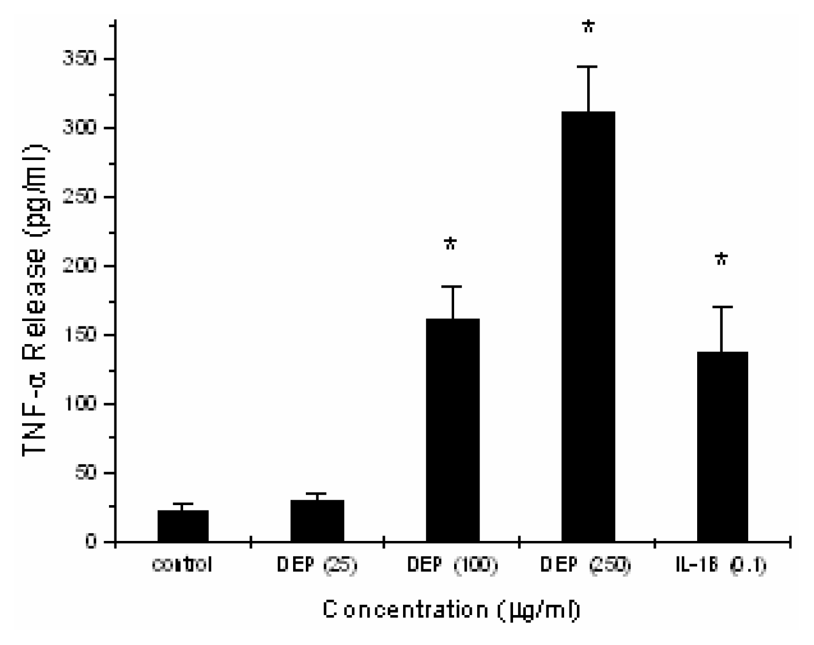
Induction of TNF-α by DEP in Alveolar Macrophages

To determine the effect of DEP exposure on TNF-α release, confluent RAW 264.7 monolayers were exposed to vehicle alone (HBSS/30mM Hepes) alone, or vehicle containing 25, 100, or 250μg/ml DEP for 4 hr, and TNF-α release was measured using mouse TNF-α ELISA. As shown in figure 1, DEP exposure induced significant release of TNF-α at the concentration levels 100 and 250 μg/ml (162 ± 23 pg/ml, n = 4, p < 0.05, and 313 ± 31 pg/ml, n = 4, p < 0.05, respectively). Exposure of RAW 264.7 cultures to a concentration level of 25 μg/ml for 4 hr did not result in any significant release of TNF-α (31 ± 4pg/ml, n = 4, p = 0.08) compared to control cultures (23 ± 4pg/ml, n = 4). The results in figure 1 show that DEP exposure induces TNF-α release in a dose-dependant fashion. In additional experiments, treatment of cultures of alveolar macrophages with IL-1β at a concentration of 100 ng/ml for 4 hr, resulted in a significant release of TNF-α (139 ± 31 pg/ml, n = 4, p < 0.05) compared to control cultures (23 ± 3 pg/ml, n =4). The effect of DEP exposure at a concentration of 100 μg/ml for 4 hr is similar to the effect induced by IL-1β, as demonstrated in figure 1.

Determination of TNF-α Protein by Western Blotting

Figure 3 demonstrates the effect of DEP exposure on TNF-α protein production as determined by western blotting techniques.

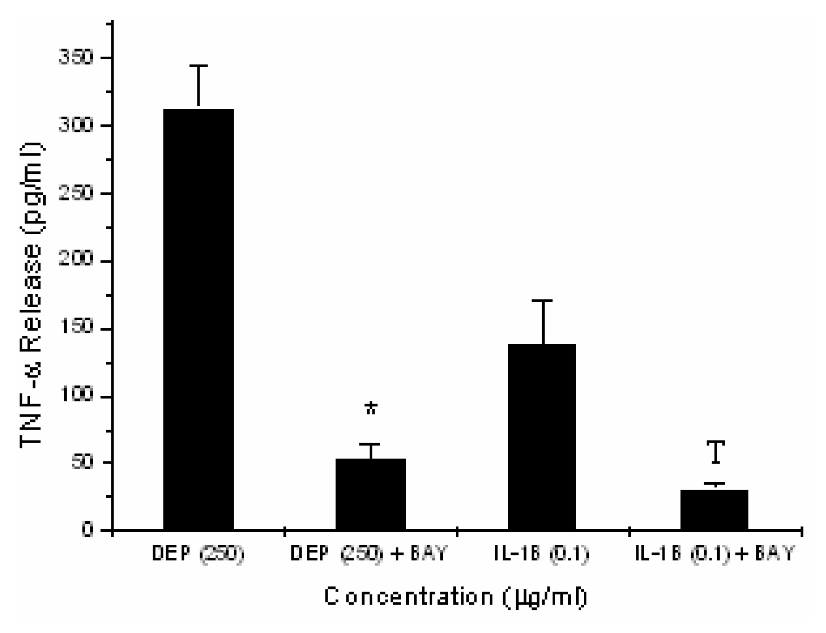
Inhibition of DEP-induced TNF-α Expression by Inhibiting NF-κB Binding Activity

Many studies have demonstrated that TNF-α gene expression is regulated at the transcriptional level by NF-κB. Inhibition of NF-κB by BAY-11 (25 μM × 1 ½ hr pre-incubation) which inhibits the phosphorylation of IκB, resulted in total abrogation of TNF-α release (83 ± 5% inhibition, n = 4, p < 0.05), as demonstrated in figure 4. Treatment of RAW 264.7 cultures with BAY-11 (25 μM with 1 ½ hr pretreatment) also significantly inhibited IL-1β-induced TNF-α release (77 ± 4% inhibition, n = 4, p<0.05), similar to the inhibitory effect on DEP-induced TNF-α release (Fig. 4).

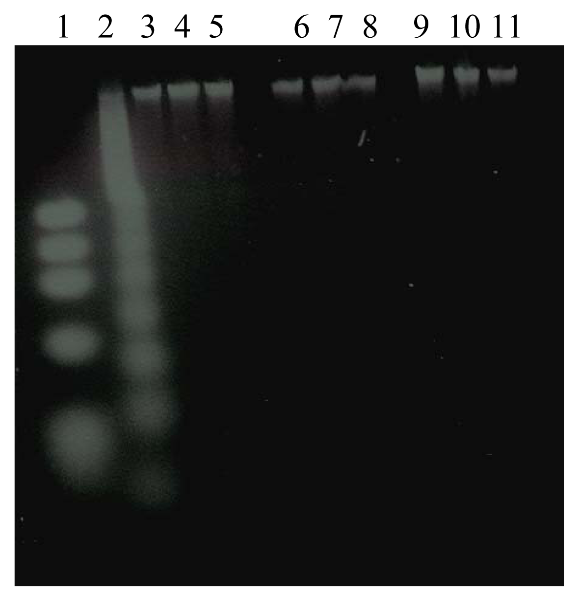
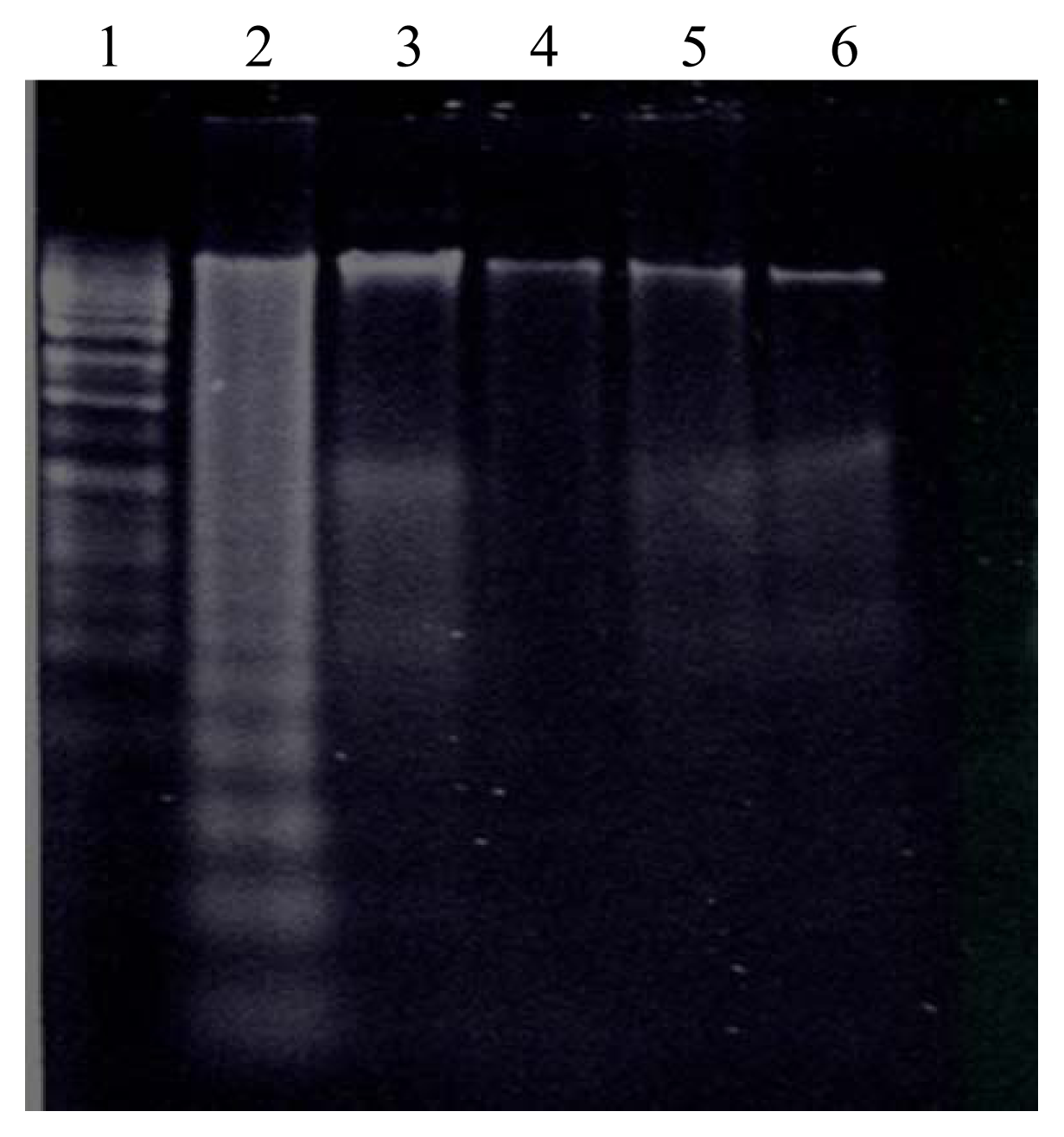
Effect of DEP Exposure on Alveolar Macrophage Apoptotic Response

Figure 5 demonstrates that treatment of cultures of RAW 264.7 cells with DEP at a concentration of 250 μg/ml for 4 hr failed to induce any apoptotic response. Apoptosis was clearly induced in U 937 cells treated with campothecin (3μM × 4 hr), an apoptosis-inducing agent, and served as positive controls (Fig. 5). However, inhibition of NF-κB binding activity by BAY-11 resulted in DEP-induced apoptotic response as demonstrated by DNA fragmentation studies (Fig 5).

Discussion

Alveolar macrophages are the lung cells responsible for ingestion and clearance of inhaled particles [21]. They play a key role in lung inflammation, asthma pathogenesis, and regulation of airway remodelling.
This view has been supported by correlation between the severity of asthma and the level of macrophage activation [22]. Activation of macrophages has been associated with increased production of the pro-inflammatory cytokines, IL-1β, TNF-α, IL-6, IL-4, platelet activating factor, and leukotrienes [22, 23]. TNF-α is a potent mediator of inflammatory and immune responses. Increased production of TNF-α by activated macrophages has been associated with pulmonary inflammation [24, 25]. TNF-α has been shown to rapidly induce NF-κB activation in cells expressing TNF-α receptors, TNFR-1 and TNFR-2 [26].
The results of the current study demonstrate that DEP exposure activates alveolar macrophages by inducing the release of the inflammatory cytokine TNF-α (Fig. 3). The release of TNF-α was found to be regulated at the transcriptional level by the nuclear factor NF-κB, as demonstrated by BAY11-70811, an inhibitor of IκB phosphorylation and its subsequent degradation (Fig. 4). Adamson et al. [27] have shown that urban particulate matter where DEP are a major component, instilled into rat lung induced significant release of TNF-α. The results of the current study are in agreement with the reported findings by Adamson and co-workers, and demonstrate that the RAW 264.7 in vitro model is useful in studying the effect of DEP in the lung.
Takano et al. [11] have shown that DEP (250μg/ml) increases NF-κB-DNA binding activity in the lung of mice with significant increase in macrophage-activating protein-1 (MP-1) and IL-1β release. In the present study, inhibition of NF-κB binding activity completely abrogated TNF-α protein expression (Fig. 4), indicating that DEP-induced TNF-α expression is regulated by NF-κB. IL-1β and TNF-α are pro-inflammatory cytokines and are both regulated at the transcriptional level by NF-κB. Moreover, TNF-α and IL-1β induce rapid nuclear translocation of NF-κB by inducing rapid phosphorylation and degradation of the inhibitory protein, IκB [15, 2830]. Our results also demonstrate that IL-1β-induced TNF-α expression is regulated by NF-κB, as demonstrated by the inhibitor BAY-11 studies (Fig. 4). NF-κB has been implicated in proapoptotic as well as antiapoptotic signaling pathways in the lung [3134]. Recently, however, many studies have suggested that translocation of NF-κB subunits to the nucleus serves to protect against apoptosis and enhance resistance to stimuli-induced cytotoxicity [3538].
The apoptotic response in macrophages may be regulated by the transcription factor NF-κB. Koay et al. [13] have shown that macrophage counts are associated with TNF-α release in response to LPS in the lung, and are essential for initiation of NF-κB-dependent immune response. Blocking of NF-κB binding activity has been shown to increase inflammatory cell apoptosis [39, 40]. Alveolar macrophages are the primary cells involved in the clearance of inhaled particles, pathogens, and apoptotic cells in the lung. Recent studies have indicated that clearance of apoptotic neutrophils by macrophages may be a determining step in the regression of COPD [41, 42]. Hiura et al. [8] have reported that DEP induce apoptosis in RAW 264.7 macrophages. However, in the same studies by Hiura and co-workers, exposure to DEP after its organic constituents have been extracted failed to induce any apoptotic response in RAW 264.7 cells. DEP-bound polycyclic aromatic hydrocarbons (PAHs), halogenated aromatic hydrocarbons (HAHs), and quinones have been implicated in the induction of apoptosis in alveolar macrophages [8], rather than extracted DEP.
In our studies, DEP were treated for LPS and the organic constituents were extracted. The failure of DEP to induce an apoptotic response in RAW 264.7 cultures in the present study may be attributed to the absence of active organic constituents bound to DEP surfaces, notably, PAHs. Alveolar macrophages are rich in enzymes active in the transformation of foreign substances, for example, cytochrome P4501A1 (CYP 1A1) that contribute to the generation of reactive oxygen species (ROS). These enzymes are induced by PAHs and other DEP-bound constituents. DEP-adsorbed organic compounds have been shown to generate ROS by activating CYP 1A1 and NADPH quinine oxidoreductase in human airway epithelial cells [43]. Inhibition of NF-κB activation by antioxidants in a Jurkat T cell line has suggested that reactive oxygen intermediates are involved in the signalling pathways of NF-κB activation [4446]. Despite the numerous studies that have addressed the cytotoxic effect of DEP on the alveolar macrophage and their role in NF-κB activation, the mechanisms underlying DEP-induced NF-κB activation remain not well defined. Earlier studies by Janssen-Heininger et al. [47] have demonstrated different signaling pathways for ROS and TNF-α in the activation of NF-κB. Using dominant negative Ras constructs, the authors were able to demonstrate the involvement of Ras in ROS-induced NF-κB activation in human airway epithelial cells. In the same studies, TNF-α-induced NF-κB activation, however, was reported to be Ras-independent. Whereas, the signaling pathways underlying TNF-α-induced NF-κB activation are well defined, oxidant-induced signaling events leading to NF-κB activation remain not well characterized. In the present study, uptake of DEP by alveolar macrophages may have resulted in a respiratory burst associated with superoxide and H2O2 generation. Released H2O2 and superoxide may have activated the mitogen-activated protein kinases/extracellular-regulated kinase kinase kinase-1 (MEKK-1) downstream from Ras. MEKK-1 has been shown to activate IκB kinase (IKK) and the c-Jun N terminal kinases (JNK), both capable of activating NF-κB downstream [48]. The activation of NF-κB and the transcription and release of TNF-α may have created an autocrine loop, where binding of released TNF-α to its receptor activates TNF receptor-associated factor (TRAF). The activation of TRAF and the subsequent activation of NF-κB-inducing kinase (NIK) would augment NF-κB activation and prevent apoptosis.
In summary, the results of our study show that exposure to DEP at physiologically relevant concentrations (25–250 μg/ml) significantly induce the release of TNF-α, a cytokine implicated in inflammatory and immune responses. This effect is induced by the carbonaceous core of DEP since DEP utilized in the present study were treated for LPS and the adsorbed organic constituents extracted. Moreover, our results indicate that TNF-α release and activation of NF-κB may protect against apoptosis in the alveolar macrophage cell line, RAW 264.7.
Figure 1. Effect of Diesel Exhaust Particles (DEP) on Tumor Necrosis Factor-α (TNF-α) Production in Alveolar Macrophages. Confluent cultures of alveolar macrophages (RAW 264.7) were exposed to vehicle (HBSS/30 mM Hepese) alone, vehicle containing DEP at 25, 100, or 250μg/ml, or IL-1β at 0.1μg/ml for 4 hours. Following exposure, TNF-α release was measured in control and exposed cultures using mouse ELISA. Data (pg/ml) are presented as mean ± SEM (n). *Significant difference (p < 0.05) from control values.
Ijerph 02 00107f1
Figure 2. Tumor Necrosis Factor-α (TNF-α) Protein Analysis by Western Blotting. Confluent monolayers of RAW 264.7 cells were treated with vehicle (HBSS/Hepes) alone, vehicle containing DEP (250 μg/ml), or IL-1β (0.1μg/ml) for 4 hours. Following the various treatments cells were harvested and lysed by sonication in lysis buffer on ice. Denatured total proteins (20 μg) of cytosolic and nuclear fractions were separated on 12% denaturing polyacrylamide gels and electrotransferred to Hybond nitrocellulose membranes. Following standard blocking and washing procedures, the membranes were incubated with polyclonal antibodies against TNF-α, and horseradish peroxidase-conjugated anti-goat IgG secondary antibodies. Detection of proteins was performed by enhanced chemiluminescence (ECL). Panel shows bands corresponding to treatment groups: 1. Control Cultures, 2. DEP-Treated Cultures, and 3. IL-1β-Treated Cultures.
Ijerph 02 00107f2
Figure 3. Effect of Diesel Exhaust Particles (DEP) on Alveolar Macrophage Apoptosis. Confluent monolayers of RAW 264.7 cells were treated with vehicle (HBSS/Hepes) alone (control), vehicle containing DEP at 250 μg/ml (DEP), or IL-1β at 0.1 μg/ml for 4 hr. Following treatment, DNA was isolated and fragmentation was assessed by DNA ladder kit on agarose according to the manufacturer’s directions. U937 cells treated with campothecin, an apoptosis-inducing agent were used as positive control. Pane shows bands corresponding to treatment groups: molecular weight marker, 1; positive control, 2; control cultures, 3, 4, 5, in triplicate; DEP-treated cultures, 6, 7, 8, in triplicate; and IL-1β-treated cultures, 9, 10, 11, in triplicate.
Ijerph 02 00107f3
Figure 4. Effect of Nuclear Factor-κB (NF-κB) Inhibition on Tumor Necrosis Factor-α (TNF-α) Production in Alveolar Macrophages. Monolayers of RAW 264.7 cells were pre-treated with BAY11-7082 (25μg/ml × 1 1/2 hr), an NF-κB inhibitor. Following pre-treatment, RAW 264.7 cultures were treated with vehicle (HBSS/Hepes) alone, or vehicle containing DEP (250 μg/ml × 4 hr) or IL-1β (0.1μg/ml × 4 hr), and TNF-α release in cellular supernatants were determined by enzyme-linked immunosorbent assays (ELISA). The data (pg/ml) are presented as mean ± SEM (n). *Significant inhibition (p < 0.05) from DEP-, or IL-1β-treated cultures.
Ijerph 02 00107f4
Figure 5. Effect of Nuclear Factor-κB (NF-κB) Inhibition on Diesel Exhaust Particles (DEP)-induced apoptosis in Alveolar Macrophages. Monolayers of RAW 264.7 cells were pre-treated with BAY11-7082 (25 μg/ml × 1 1/2 hr), an NF-κB inhibitor. Following pre-treatment, RAW 264.7 cultures were treated with vehicle (HBSS/Hepes) alone, or vehicle containing DEP (250 μg/ml × 4 hr). Following treatment, DNA was isolated and fragmentation was assessed by DNA ladder kit on agarose according to the manufacturer’s directions. U937 cells treated with campothecin (3 μM × 4 hr), an apoptosis-inducing agent were used as positive control (positive control). Lanes are as follows: molecular weight marker, 1; positive control, 2; DEP-exposed cultures pre-treated with BAY11-7082, 3; DEP-exposed cultures, 4; control cultures, 5; and control cultures pre-treated with BAY11-7082, 6.
Ijerph 02 00107f5

Acknowledgments

Funding for this study has been provided by a grant from the American Lung Association of Mississippi, and grant G12RR12359 from the NIH.

References

  1. Ohtoshi, T.; Tazikawa, H.; Okazaki, H.; Kawasaki, S.; Takeuchi, N.; Ohta, K.; Ito, K. Diesel exhaust particles (DEP) stimulate human airway epithelial cells to produce cytokines relevant to airway inflammation in vitro. J. Allergy Clin. Immunol 1998, 101, 778–785. [Google Scholar]
  2. Sagai, M.; Furuyama, A.; Ichinose, T. Biological effects of diesel exhaust particles (DEP) HI. Pathogenesis of asthma-like symptoms in mice. Free Rod. Biol. Med 1996, 21, 199. [Google Scholar]
  3. Nel, A. E.; Diaz-Sanchez, D.; Ng, D.; Hiura, J.; Saxon, A. Enhancement of allergic inflammation by the interaction between diesel exhaust particles and the immune system. J. Allergy Clin. Immunol 1998, 102, 539–554. [Google Scholar]
  4. Peterson, B. A.; Saxon, A. Global increases in allergic respiratory disease: The possible roles of diesel exhaust particles. Ann. Allergy Asthma Immunol 1996, 77(4), 263–268. [Google Scholar]
  5. Diaz-Sanchez, D. The roles of diesel exhaust particles and their associated polyaromatic hydrocarbons in the induction of allergic airway disease. Allergy 1997, 52(38s), 52–56. [Google Scholar]
  6. Jakab, G. J.; Risby, T. H.; Sehnert, S. S.; Hmieleski, R. R.; Farrington, J. E. Suppression of alveolar macrophage membrane receptor-mediated phagocytosis by model and actual particle-adsorbate complexes; initial contact with the alveolar macrophage membrane. Environ. Health Perspect 1990, 86, 33744. [Google Scholar]
  7. Monn, C.; Fendt, R.; Koller, T. Ambient pm (10) extracts inhibit phagocytosis of defined inert model particles by alveolar macrophages. Inhal. Toxicol 1990, 14(4), 369–85. [Google Scholar]
  8. Hiura, T. S.; Kaszubowski, M. P.; LI, N.; Nel, A. E. Chemicals in diesel exhaust particles generate reactive oxygen radicals and induce apoptosis in macrophages. J. Immunol 1999, 163, 5582–5591. [Google Scholar]
  9. Holian, A.; Hamilton, R. F., Jr.; Morandi, M. T.; Brown, S. D.; Li, L. Urban particule-induced apoptosis and phenotype shifts in human alveolar macrophages. Environ. Health Perspect 1998, 106, 127–132. [Google Scholar]
  10. Berry, J.; Henoc, P.; Galle, P. Phagocytosis by cells of the pulmonary alveoli. Am. J. Pathol 1978, 93, 2744. [Google Scholar]
  11. Takano, H.; Yanagisawa, R.; Ichinose, T.; Sadakane, K.; Yoshinos, S.; Yoshikawa, T.; Morita, M. Diesel exhaust particles enhance lung injury related to bacterial endotoxin through expression of proinflammatory cytokines, chemokines, and intercellular adhesion molecule-1. Am. J. Resipr. Crit. Care Med 2002, 165, 1329–1335. [Google Scholar]
  12. Hiura, T. S.; Li, N.; Kaplan, R.; Horwitz, M.; Seagraave, J. C.; Nel, A. E. The role of a mitochondrial pathway in the induction of apoptosis by chemicals extracted from diesel exhaust particles. J. Immunol 2000, 165(5), 2703–11. [Google Scholar]
  13. Koay, A. M.; Gao, X.; Washington, M. K.; et al. Macrophages are necessary for maximal nuclea factor-KP activation in response to endotoxin. Am. J. Respir. Cell. Mol. Biol 2002, 26, 572–578. [Google Scholar]
  14. Sen, R.; Baltimore, D. Multiple nuclear factors interact with the immunoglobulin enhancer sequences. Cell 1986, 46(5), 705–716. [Google Scholar]
  15. Baldwin, A. S., Jr. Series introduction: the transcription factor NF-KB and human disease. J. Clin. Invest 2001, 707(7), 3–6. [Google Scholar]
  16. Stein, B.; Baldwin, A. S. Distinct mechanisms for the regulation of the interleukin-8 gene involve synergism and cooperativity between CIEBP and NF-KB. Mol. Cell Biol 1993, 13, 7191–7198. [Google Scholar]
  17. Christman, J. W.; Sadikot, R. T.; Blackwell, T. S. The role of nuclear factor-KP in pulmonary disease. Chest 2000, 777, 1482–1487. [Google Scholar]
  18. Graziano, F. M.; Cook, E. B.; Stahl, J. L. Cytokines, chemokines, RANTES, and eotaxin. Allergy Asthma Proc 1999, 20, 141–146. [Google Scholar]
  19. Anderson, J. P. Resolution of chronic inflammation by therapeutic induction of apoptosis. Trends Pharmacol. Sci 1996, 77, 438–442. [Google Scholar]
  20. Daniel, W. W. Biostatistics: A Foundation for Analysis in Health Sciences, 5th ed; John Wiley: New York, 1991; pp. 274–366. [Google Scholar]
  21. Bousquet, J.; Jeffery, R. K.; Busse, W. W.; Johnson, M.; Vignola, A. M. Asthma: from bronchoconstriction to airways inflammation and remodeling. Am. J. Respir. Crit. Care Med 2000, 161(5), 1720–1745. [Google Scholar]
  22. Borish, L.; Mascali, J. J.; Dishuck, J.; Beam, W. R.; Martin, R. J.; Rosenwasser, L. J. Detection of alveolar macrophage-derived IL-lbeta in asthma inhibition with corticosteroids. J. Immunol 1992, 49, 3078–3082. [Google Scholar]
  23. Gosset, P.; Garcon, G.; Casset, A.; Fleurisse, L.; Hannothiaux, M. H.; Craesy, C.; Shirali, P. Benzo(a)pyrene-coated onto Fe2O3 particles-induced apoptotic events in the lungs of Sprague Dawley rats. Toxicol. Lett 2003, 143, 223–232. [Google Scholar]
  24. Driscoll, K. E.; Hassenbein, D. G.; Carter, J. M.; Kunkel, S. L.; Quinlan, T. R.; Mossman, B. T. TNF alpha and increased chemokine expression in rat lung after particle exposure. Toxicol. Lett 1995, 82–83, 483–489. [Google Scholar]
  25. Driscoll, K. E. TNF-alpha and MIP-2: role in particle-induced inflammation and regulation by oxidative stress. Toxicol. Lett 1995, 112–113, 177–183. [Google Scholar]
  26. Fiers, W. Tumor necrosis factor: characterization of the molecular, cellular, and in vivo level. FEBS Lett 1991, 285, 199–212. [Google Scholar]
  27. Adaamson, I. Y.; Prieditis, H.; Vincent, R. Soluble and insoluble air particle fractions induce differential production of tumor necrosis factor alpha in rat lung. Exp. Lung Res 2004, 30(5), 355–368. [Google Scholar]
  28. Baeuerle, P. A. IKB-NF-KB structures: at the interface of inflammation control. Cell 1998, 95(6), 729–731. [Google Scholar]
  29. Karin, M.; Ben-Neriah, Y. Phosphorylation meets ubiquitination: the control of NF-κB activity. Annu. Rev. Immunol 2000, 18, 621–663. [Google Scholar]
  30. Phelps, C. B.; Sengchanthalangsy, L. L.; et al. Mechanism of IKBOC binding to NF-κB dimmers. J. Biol. Chem 2000, 275(38), 29840–29846. [Google Scholar]
  31. Grimm, S.; Bauer, M. K. A.; Bauerle, P. A.; Schulze-Osthoff, K. Bcl-2 down-regulates the activity of transcription factor NF-KB induced apoptosis. J. Cell Biol 1996, 134, 13–23. [Google Scholar]
  32. Van Antwerp, D. J.; Martin, S. J.; Venna, I. M.; Green, D. R. Inhibition of TNF-induced apoptosis by NF-KB. Trends Cell Biol 1998, 8, 107–111. [Google Scholar]
  33. Manna, S. K.; Aggarwal, B. B. Lipopolysaccharide inhibits TNF-induced apoptosis: role of nuclear factor-kappa B activation and reactive oxygen intermediates. J. Immunol 1999, 162, 1510–1518. [Google Scholar]
  34. Kaltschmidt, B.; Kaltschmidt, C.; Hofmann, T. G.; Hehner, S. P.; Droge, W.; Schmitz, L. M. The pro- or anti-aapoptotic function of NF-KB is determined by the nature of the apoptotic stimulus. Eur. J. Biochem 2000, 267, 3828–3835. [Google Scholar]
  35. Wang, C. Y.; Mayo, M. W.; Baldwin, A. S., Jr. TNF- and cancer therapy-induced apoptosis: potentiation by inhibition of NF-kappa B. Science 1996, 274, 784–787. [Google Scholar]
  36. van Antwerp, D. J.; Martin, S. J.; Kafri, T.; Green, D. R.; Verma, I. M. Suppression of TNF-alpha- induced apoptosis by NF-kappa B. Science 1996, 274, 787–789. [Google Scholar]
  37. Beg, A. A.; Baltimore, D. An essential role for NF- kappa B in preventing TNF-a-induced cell death. Science 1996, 274, 782–784. [Google Scholar]
  38. Liu, Z.; Hsu, H.; Goeddel, D. V.; Karin, M. Dissection of TNF receptor I effector functions: JNK activation is not linked to apoptosis while NF-κB activation prevents cell death. Cell 1996, 87, 565–576. [Google Scholar]
  39. Maiuri, M. C.; Tajana, G.; Iuvone, T.; De Stefano, D.; Mele, G.; Ribecca, M. T.; Cinelli, M. P.; Romano, M. F.; Turco, M. C.; Carnuccio, R. Nuclear factor-kappa B regulates inflammatory cell apoptosis and phagocytosis in rat carrageenin-sponge implant model. Am. J. PathoL 2004, 165(1), 115–1126. [Google Scholar]
  40. D’Acquisto, F.; De Cristofaro, F.; Maiuri, M. C.; Tajana, G.; Crnuccio, R. Protective role of nuclear factor kappa B against nitric oxide-induced apoptosis in J774 macrophages. Cell Death Differ 2001, 8(2), 144–151. [Google Scholar]
  41. Rahman, I. Oxidative stress and gene transcription in asthma and chronic obstructive pulmonary disease: antioxidant therapeutic targets. Curr. Drug Targets Inflamm. Allergy 2002, 1(3), 291–315. [Google Scholar]
  42. Barnes, P. J.; Karin, M. Nuclear factor-κB: a pivotal transcription factor in chronic inflammatory diseases. N. Engl. J. Med 1997, 336(15), 1066–1071. [Google Scholar]
  43. Baulig, A.; Garlatti, M.; Bonvalott, V.; Marchand, A.; Barouki, R.; Marano, F.; Baeza-Squiban, A. Involvement of reactive oxygen species in the metabolic pathways triggered by diesel exhaust particles in human airway epithelial cells. Am. J. Physiol 2003, 285, L671–L679. [Google Scholar]
  44. Schreck, R.; Reiber, P.; Baeurlef, P. A. Reactive oxygen intermediates as apparently widely used messengers in the activation of the NF-KB transcription factor and HIV-1. EMBO J 1991, 10, 2247–2258. [Google Scholar]
  45. Staal, F. J.; Anderson, M. T.; Herzenberg, L. A. Redox regulation regulation of aactivaation of NF-κB transcription factor complex: effects of N- acetylcysteine. Methods Enzymol 1995, 252, 168–174. [Google Scholar]
  46. Kumagai, Y.; Arimoto, T.; Shinyashiki, M.; Shimojo, N.; Nakai, Y.; Yoshikawa, T.; Sagai, M. Generation of reactive oxygen species during interaction of diesel exhaust particle component with NADPH-cytochrome p450 reductase and involvement of the bioactivation in the DNA damage. Free Rad. Biol. Med 1997, 22(3), 479–487. [Google Scholar]
  47. Janssen-Heininger, Y. M. W.; Macara, I.; Mossman, B. T. Cooperatively between oxidants and tumor necrosis factor in the activation of nuclear factor (NF)-KB: requirement of Ras/mitoge-activated protein kinases in the activation of NF-κB by oxidants. Am. J. Respir. Cell Mol. Biol 1999, 20, 942–952. [Google Scholar]
  48. Malinin, N. L.; Boldin, M. P.; Kovalenka, A. V.; Wallach, D. MAP3K-related kinase involved in NF-κB induction by TNF, CD95, and IL-1. Nature 1997, 385, 540–544. [Google Scholar]

