Article

Cytokines and Adhesion Molecules Expression in the Brain in Human Cerebral Malaria

by
Henry Armah
1,
Edwin Kwame Wiredu
1,2,*,
Alfred Kofi Dodoo
3,
Andrew Anthony Adjei
1,2,
Yao Tettey
1 and
Richard Gyasi
1
1
Department of Pathology, University of Ghana Medical School
2
Department of Medical Laboratory Sciences, School of Allied Health Sciences
3
Histology & Electron Microscopy Unit, Noguchi Memorial Institute for Medical Research, University of Ghana College of Health Sciences, Accra, Ghana
*
Author to whom correspondence should be addressed.
Int. J. Environ. Res. Public Health 2005, 2(1), 123-131; https://doi.org/10.3390/ijerph2005010123
Submission received: 15 November 2004 / Accepted: 6 February 2005 / Published: 30 April 2005

Abstract

:
Although the role of systemic proinflammatory cytokines, IL-1β and TNF-α, and their up-regulation of adhesion molecules, ICAM-1, VCAM-1 and E-Selectin, in the pathogenesis of cerebral malaria (CM) is well established, the role of local cytokine release remain unclear. Immunohistochemistry (IHC) was used to compare the expression of ICAM-1, VCAM-1, E-Selectin, IL-1β, TNF-α and TGF- β at light microscopic level in cerebral, cerebellar and brainstem postmortem cryostat sections from 10 CM, 5 severe malarial anemia (SMA), 1 purulent bacterial meningitis (PBM), 2 non-central nervous system infections (NCNSI) and 3 non-infections (NI) deaths in Ghanaian children. Fatal malaria and Salmonella sepsis showed significantly higher vascular expression of all 3 adhesion molecules, with highly significant co-localization with sequestration in the malaria cases. However, there was negligible difference between CM and SMA. TGF-β showed intravascular and perivascular distribution in all cases, but expression was most intense in the PBM case and CM group. TNF-α and IL-1β showed prominent brain parenchymal staining, in addition to intravascular and perivascular staining, in only the PBM case and CM group. The maximal expression of all 6 antigens studied was in the cerebellar sections of the malaria cases. Endothelial activation is a feature of fatal malaria and Salmonella sepsis, with adhesion molecule expression being highly correlated with sequestration. IL-1β and TNF-α are upregulated in only cases with neurodegenerative lesions, whilst TGF-β is present in all cases. Both cytokines and adhesion molecules were maximally upregulated in the cerebellar sections of the malaria cases.

Introduction

Despite technological advances and global economic development, malaria is still the parasitic disease responsible for the greatest number of deaths worldwide. Malaria parasites infects between 300 to 500 million people, causing up to 2 million deaths globally per year (mostly children in sub-Saharan Africa) from complications of primarily cerebral malaria (CM) and severe malarial anaemia (SMA) [1]. CM, characterised by seizures and loss of consciousness, is an important complication of Plasmodium falciparum infection with mortality rate of 15–20% [2]. Despite this high mortality rate, the pathogenic mechanisms of CM have not been well elucidated. Little is known about the blood brain barrier (BBB) interactions in CM that result in the neurological disorder, and it is unclear how the intraerythrocytic parasite, which sequesters in the cerebral microvasculature but rarely enters the brain parenchyma, influence parenchymal function to induce coma and death. The unavailability of infected human specimens and suitable animal models has hindered a thorough understanding of the pathogenesis.
There is compelling evidence that the inflammatory response at the BBB in CM involve increased systemic production of proinflammatory cytokines, especially tumour necrosis factor (TNF)-α and interleukin (IL)-1β [26], and up-regulation of cerebral endothelial adhesion molecules, especially intercellular adhesion molecule (ICAM)-1, vascular cell adhesion molecules (VCAM)-1 and endothelial (E)-selectin, that facilitate sticking of parasitized erythrocytes (PEs) to blood vessels [710]. There is immunohistochemical (IHC) evidence for endothelial cell (EC) activation during CM in humans [8, 1012], and animal models of CM [13]. Endothelial activation occurred not only in fatal CM, but also in the other fatal malaria syndromes, indicating that systemic endothelial activation is a feature of fatal malaria [8, 10, 11]. A role for tyrosine phosphorylation has been demonstrated in EC activation for two adhesion molecules (VCAM-1 and E-Selectin) induced by TNF-α [14].
In addition to systemic proinflammatory cytokine production, local cytokine release could contribute to organ-specific pathophysiology, especially in the brain in the case of CM. Recent studies have shown the expression of proinflammatory cytokine protein, including TNF-α, in postmortem brain tissue in human CM [5, 15, 16]. TNF-α is over-expressed in cerebral microglia, astrocytes, monocytes and vascular endothelium in mice with CM, relative to controls [1520]. However, the lack of a classical inflammatory response to the presence of PEs in the brain microvasculature indicates anti-inflammatory cytokine involvement, and recent reports have suggested an anti-inflammatory and neuroprotective role for transforming growth factor (TGF)-β in the host defense mechanism against neuronal loss in neurodegenerative diseases [21], including CM.
TGF-β has been found in both haemorrhagic white-matter lesions of human CM [5], and in white-matter lesions of human immunodeficiency virus (HIV)-1 encephalitis brain samples [22]. This association of TGF-β with central nervous system (CNS) neurodegenerative lesions suggests an anti-inflammatory and neuroprotective role for TGF-β in the host defense mechanism against neuronal cell loss [21]. In mice infected with lethal or non-lethal strains of malaria parasites, a strong and sustained TGF-β response, beginning on the 5th to 6th post-infection day when the peak parasite replication has been reached, was associated with abrogation of mortality and resolution of infection [23]. Furthermore, neutralization of TGF-β leads to 100% mortality in BALB/c mice infected with normally non-lethal P. chabaudi A/J [23].
In-vitro studies have shown TNF-α mRNA in inflammatory infiltrates within the meninges of experimental rabbit pneumococcal meningitis [24], and upregulation of neuronal TNF-α expression in response to bacterial lipopolysaccharide [25]. IL-1β is not expressed in normal human brain, but induced and expressed intraparenchymally in human CM brain and in meningeal infiltrating leukocytes of human meningoencephalitides cases [5]. In in-vitro studies, IL-1β is neurotoxic and rapidly induced in response to neuronal cell death, and therefore is suggested to play a causal role in ischaemic cell death and neurodegeneration in the brain [26]. Tongren and colleagues [27] showed that the proinflammatory cytokines (IL-1β and TNF-α) and TH-1 cytokine [interferon (IFN)-γ] had the highest level of mRNA expression in the cerebellum during late P. coatneyi infection in rhesus monkeys, agreeing with histopathologic observations of the preferential sequestration of PE in the cerebellum in rhesus monkey [2729], and in human CM [30]. However, recent evidence from mice indicates that it may be overproduction of lymphotoxin-α (LT-α) rather than TNF-α that leads to CM, since mice deficient in TNF-α were found to be just as susceptible to CM as controls whereas LT-α deficient mice were resistant to CM pathology, dying from hyperparasitaemia and severe anaemia instead [31].
Although, the role of cytokines and adhesion molecules has been extensively studied in human malaria, the role of local cytokine release in the brain in human CM is unclear. Children living in sub-Saharan Africa bear the brunt of malaria mortality, yet there is a dearth of relevant postmortem studies in African children, and most of the few human CM postmortem immunohistochemical (IHC) studies are in non-African adults. Therefore, the role of local cytokine release and associated up-regulation of adhesion molecules in human CM has not been adequately confirmed and substantiated, especially in sub-Saharan African children.
The present study was aimed at ascertaining the role of cytokines (TNF-α, IL-1β and TGF-β) and adhesion molecules (ICAM-1, VCAM-1 and E-Selectin) in fatal CM, and used IHC techniques to examine and compare the distribution of these 6 antigens at light microscopical level in postmortem human cerebral, cerebellar and brainstem cryostat sections of Ghanaian children dying from CM, severe malarial anaemia (SMA), purulent bacterial meningitis (PBM), non-central nervous system infections (NCNSI) and non-infections (NI) cases.

Materials and Methods

Case Selection

During the peak malaria season, from July to September 2001, all clinically certified deaths in children admitted to the Emergency Unit at the Department of Child Health, Korle-Bu Teaching Hospital, Accra, Ghana, with detailed clinical and laboratory records and in whom duly signed written informed consent had been obtained from parents or guardians after the death of their child were included in the study. Twenty-one (21) parents or guardians agreed to and signed the consent form for the participation of their children and to donate tiny brain tissue samples at autopsy for this study. Volunteered cadavers were immediately moved from the Emergency Unit to the morgue for cold storage at 4°C and a full autopsy with removal of brain tissue samples done within 12 hours of death.
In each volunteered case, brain tissue blocks of about 0.3–0.5cm3 were surgically removed from 3 regions of the brain, namely cerebrum, cerebellum and brainstem. Brain smear cytology was done for each case at autopsy and stained with Giemsa for the demonstration of parasitized erythrocytes and/or malaria pigment in the cerebral microvasculature as previously described [32]. The full autopsy gross findings, autopsy brain smear cytologic/microscopic findings, in addition to the detailed clinical and diagnostic laboratory records were used to classify the 21 cases into five groups, namely:
(1)
Cerebral malaria [CM].
(2)
Malaria complicated by severe anaemia/severe malarial anaemia [SMA].
(3)
Purulent bacterial meningitis [PBM] (i.e. central nervous system infection other than cerebral malaria)
(4)
Non-central nervous system infection [NCNSI] (i.e. infection in an anatomic organ-system other than the central nervous system), and (5) Non-infection deaths [NI] (i.e. no focus of infection found clinically or at autopsy).
A CM death was defined as clinically fulfilling WHO definition of severe malaria [33] with Blantyre coma score ≤2, and gross autopsy findings of slaty-gray discolouration and/or white matter petechial haemorrhages of brain, and/or brain smear cytologic findings of parasitised erythrocytes and/or malaria pigment in the cerebral microvasculature. SMA deaths fulfilled WHO definition of severe malaria [33] with hemoglobin ≤ 5 g/dl, and absence of gross autopsy findings of slaty-gray discolouration and white matter petechial haemorrhages, but presence of moderate to severe pallor of all internal organs, and absence of brain smear cytologic findings of parasitised erythrocytes and malaria pigment in the cerebral microvasculature. Deaths with clinically negative P. falciparum peripheral parasitaemia, absence of gross autopsy findings of slatey gray discolouration of brain, liver and spleen, and white matter petechial haemorrhages, and absence of brain smear cytologic findings of parasitised erythrocytes and malaria pigment in the cerebral microvasculature, were included as non-malartia controls.
Based on above criteria, the 21 volunteered cases were made up of 10 CM, 5 SMA, 1 PBM, 2 NCNSI, and 3 NI deaths. The study was approved by both the Ethical and Protocol Review Committee of the University of Ghana Medical School, and the Institutional Review Board (IRB) of the Noguchi Memorial Institute for Medical Research, Accra, Ghana.

Brain Tissue Preparation and Immunohistochemistry

All the sixty-three (63) brain tissue samples obtained at autopsy within 12 hours of death were immediately snap-frozen in liquid nitrogen, and stored at −80°C until used. Fourteen (14) cryostat sections of 5–7 μm thickness were cut from each of the 63 brain tissue samples, mounted unto SuperFrost® Plus (Menzel-Glazer, Germany) light microscope slides and 2 of each stained for the localization of the six antigens studied and two for negative controls. Two independent experiments, using 6 sections for the 6 antigens and 1 negative control section in each, were performed by identical methods for each of the 63 samples to ensure reproducibility.
Cryostat sections were air-dried overnight at room temperature, fixed in 100% acetone for 10 minutes, and air-dried for 30 minutes at room temperature. Slides were immunostained immediately using a standard indirect alkaline phosphatase method. Briefly, sections were incubated with a 1 : 20 dilution of normal rabbit serum (DAKO, Denmark) in Tris-buffered saline (TBS), pH 7.4 for 30 minutes in a humidified chamber to reduce non-specific protein binding. Primary antibodies against ICAM-1 (mouse anti-human, monoclonal, Immunotec, UK; dilution 1 : 500), VCAM-1 (mouse anti-human, monoclonal, DAKO, UK; dilution 1 : 200), E-Selectin (mouse anti-human, monoclonal, DAKO, UK; dilution 1 : 200), TNF-α (mouse anti-human, monoclonal, Serotec, UK; dilution 1 : 250), IL-1β (mouse anti-human, monoclonal, Serotec, UK; dilution 1 : 250) and TGF-β (mouse anti-human, monoclonal, Serotec, UK; dilution 1 : 250) were applied for 60 minutes at room temperature. Antibodies were used as dilutions in TBS, pH 7.4.
Sections were then washed with TBS pH 7.4 for 15 minutes (3 times, each for 5 minutes). Secondary incubation and staining were performed using indirect immunostaining microscopic technique with Universal DAKO Alkaline Phosphatase Anti-Alkaline Phosphatase (APAAP) Kit™ System 40 for Monoclonal Mouse Antibody (DAKO Corporation, USA). After incubating with freshly prepared chromogenic substrate for 8–10 minutes, the sections were then washed in tap water, lightly counterstained with Meyer’s Haematoxylin for 3 minutes, washed again, and mounted with glycergel (DAKO, Denmark). Negative control slides were prepared for each of the 63 samples by substituting the primary antibody with normal non-immune mouse IgG (DAKO, Denmark). Appropriate concentrations of primary antibodies were determined using optimization on malaria and non-malaria tissues.

Evaluation of Immunostaining

Slides were examined and graded blindly by two independent authors [HA & EKW], who were not involved in the immunostaining of the sections and had no access to the clinical, laboratory and autopsy details of volunteers until after the results had been collated. Ten fields per section, immunostained with ICAM-1, VCAM-1 and E-Selectin, were examined under a magnification of X400 and the degree of staining and the number of vessels showing staining were assessed using a five-tiered semi-quantitative scale: −, negative (no endothelial cell staining); +/−, positive endothelial cell staining on < 25% of vessels; +, positive endothelial cell staining on > 25% and < 50% of vessels; ++, positive endothelial cell staining on > 50% and < 75% of vessels; and +++, positive endothelial cell staining on > 75% of vessels.
Similarly, ten fields per section, immunostained for TNF-α, TGF-β and IL-1β, were examined under a magnification of X100 and the location and intensity of staining were assessed using a five-tiered semi-quantitative scale: −, no red reaction product/colour; +/−, faint/mild staining in some fields; +, moderate staining in some fields; ++, moderate consistent staining in all fields; and +++, deep/strong consistent staining in all fields.
Furthermore, ten fields per section, immunostained with ICAM-1, VCAM-1 and E-Selectin, were examined under a magnification of X400 for each of the 90 brain tissue sections, 2 each from the 3 regions of the brain of the 15 malaria cases (10 CM & 5 SMA), and the following four categories of vessels were counted, namely the number of vessels showing both receptor expression and sequestration (R+S+), the number of vessels showing receptor expression but no sequestration (R+S), the number of vessels showing no receptor expression but sequestration (RS+), and the number of vessels showing neither receptor expression nor sequestration (RS).

Data Analysis

For each of the six antigens studied, the percentage of immunostained cases and the intensity of staining were compared between the five groups of cases and the three regions of the brain from which samples were obtained for this study. The co-localization of sequestration and receptor expression was analyzed using a chi-square (X2) test on the 2 × 2 contingency tables constructed for each of the three adhesion molecules by comparing the expected and observed association in cerebral vessels for the malaria cases (both CM and SMA groups). The level of significance was five percent and all analyses were performed using Epi Info 2002 statistical software (CDC).

Results

Clinical and Diagnostic Details

The clinical and diagnostic details of the 21 studied cases are summarized in Table 1. Ten died of CM, five of SMA, one of PBM, two of NCNSI and three of NI. The two NCNSI cases were severe bronchopneumonia (SBP), and typhoid perforation and septicaemia (TP&S), whilst the three NI cases were nephrotic syndrome (NPS), bleeding duodenal ulcer (BDU) and haemolytic sickle cell (SC) crisis (HSC). There were 10 males and 11 females. The mean age of the CM deaths of 69.1 months (SD = 32.1) was significantly higher than that of the SMA deaths of 20.2 months (SD = 16.25) [X2 = 7.33, df = 1, P value = 0.0068]. Eighty percent (12 out of 15) of the malaria deaths occurred within 24 hours of admission. The mean peak parasitemia of the CM deaths of 45,107/μL (SD = 27,091) was significantly lower than that of the SMA deaths of 198,453 /μL (SD = 146,913) [X2 = 14.68, df = 1, P value = 0.0001].

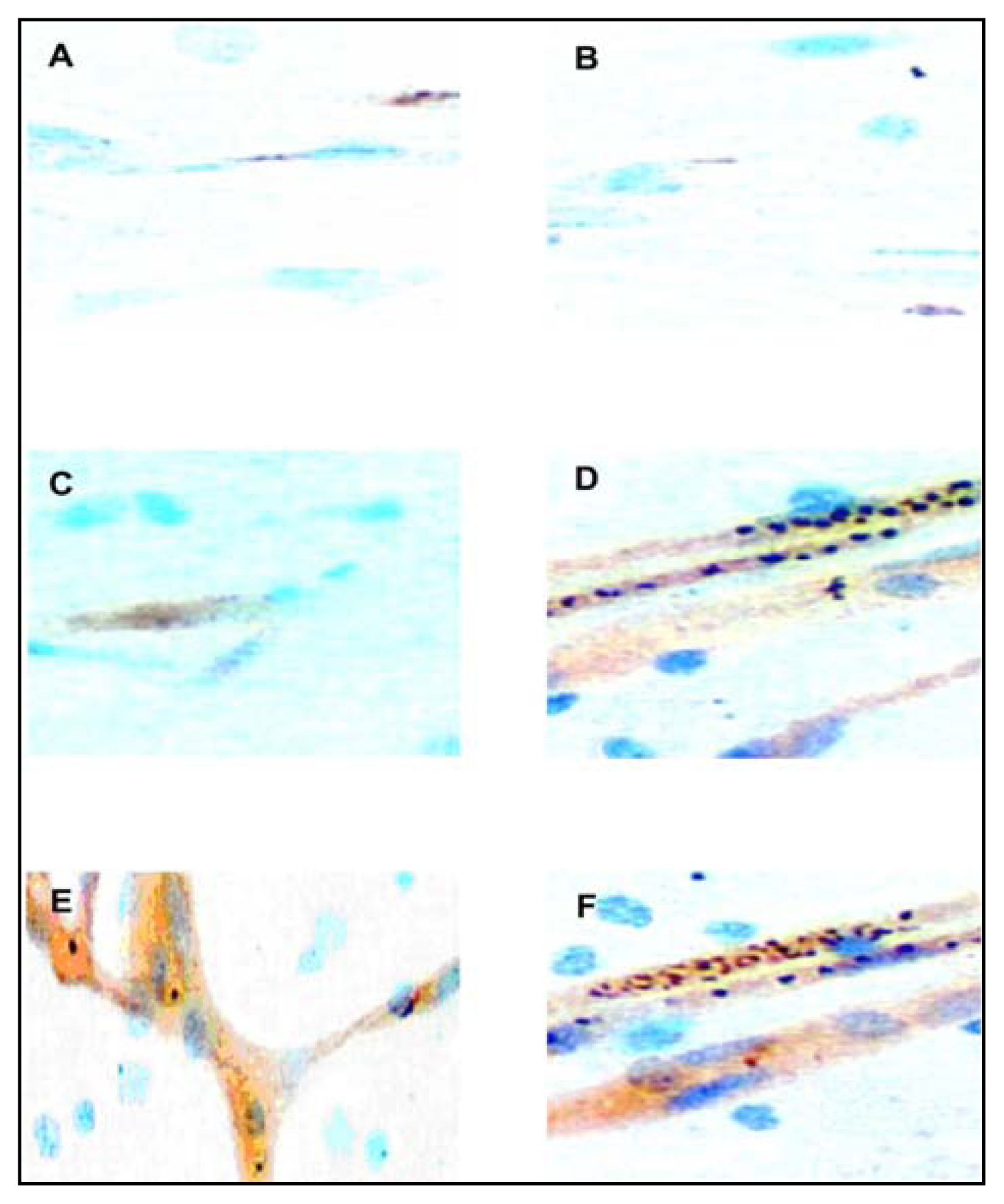
Adhesion Molecule Immunostaining

Immunostaining for the 3 adhesion molecules showed only vascular labeling (Figure 1A–F). There was no one particular receptor whose expression on the endothelial cell surface was consistently related to the presence of sequestered PEs in the cerebral microvessels of the malaria cases. Generally, if a vessel showed positive staining for a marker of endothelial activation, this was present throughout its length, rather than being related to the presence of PEs in one segment or over one endothelial cell. Within vessels from the same patient and same region of the brain of the fatal malaria cases, there was heterogeneity between parasite sequestration and the presence of receptor staining. Hence, not uncommonly, there were vessels engorged with PEs without any evidence of adhesion molecule expression and vice versa.
ICAM-1 showed positive staining of the endothelia of < 25% of the brain microvessels (average score of +/−) in all the non-malaria deaths (Figure 1A), whilst VCAM-1 showed positive staining of the endothelia of < 25% of the brain microvessels (average score of +/−) in some (highest being in the cerebellar sections of 4 out of 5) of the non-malaria deaths (Figure 1B), except the case of typhoid perforation and septicaemia. E-Selectin staining was consistently and uniformly negative in all the cerebral microvessels (score of −) in all the non-malaria controls, with the exception of the case of typhoid perforation and septicaemia. ICAM-1 (Figure 1C), VCAM-1 and E-Selectin showed positive staining in 25 to 50% of the microvessels (average score of +) in all the brain sections of the case of typhoid perforation and septicaemia.
The expression of these three sequestration receptors on the cerebral endothelium of the malaria cases showed an increase in both the proportion of cases showing expression and the intensity of staining. ICAM-1 showed positive staining in nearly all (> 75%) vessels (average score of +++) in all malaria cases (Figure 1D). VCAM-1 showed positive staining in most (50 to 75%) of the microvessels (average score of ++) in all the malaria cases, whilst E-selectin showed positive staining in 25 to 50% of the microvessels (average score of +) in all the malaria cases. There was no significant differences in endothelial staining between the 10 cases of fatal CM and the 5 cases of fatal SMA, since both groups had a similar increased intensity of ICAM-1, VCAM-1 and E-selectin expression of +++, ++ and + respectively. However, the increased adhesion molecule expression was more often not associated with sequestered PEs in the SMA cases, than for the CM cases. Thus, though heterogeneity between sequestration and receptor expression was observed in all the malaria cases, it was more extreme in the SMA cases (Figures 1D & 1E). The staining patterns of all the 3 adhesion molecules in brain sections from the case of typhoid perforation and septicaemia differed significantly from that of the other 5 non-malaria cases and the 15 malaria cases. The intensity of staining of all 3 adhesion molecules were significantly higher than that of the other 5 non-malaria cases, but not as intense as that of the 15 fatal malaria cases. Generally, the maximal expression (in terms of increased percentage of cases showing expression and intensity of staining) of all the 3 adhesion molecules in the malaria cases was evident in the cerebellar sections (Figures 1D & 1F).
We found that the presence of sequestered PEs was highly significantly associated with the expression of ICAM-1 (P = 3.1 X 10⁻¹⁶), VCAM-1 (P = 1.2 X 10⁻¹²) and E-selection (P = 6.1 X 10⁻¹⁶) [degrees of freedom (df) = 1] in the fatal malaria cerebral vessels (Table 2). The relative risk of cerebral vessels expressing ICAM-1, VCAM-1 and E-Selectin showing sequestered PEs was 1.73, 1.53 and 1.67 respectively.

Cytokine Immunostaining

Positive immunostaining for TGF-β showed intravascular and perivascular distribution (Figures 2A & 2B), whilst there was intravascular, perivascular and prominent brain parenchymal staining for IL-1β (Figures 2C–E) and TNF-α (Figures 2F). TGF-β was detected in intravascular and perivascular distribution in brain tissue from all 5 groups studied, but expression was more intense in PBM and CM groups (Figures 2A & 2B). TGF-β showed moderate intravascular and perivascular immunostaining in some fields (average score of +) of all the NCNSI sections (Figure 2A), whilst it’s staining was strong in intravascular and perivascular locations in all fields (average score of +++) of all the CM and PBM sections (Figure 2B).
IL-1β staining was limited to only some (none of the brainstem sections) of the PBM brain sections (Figure 2C) and all of the CM brain sections (Figure 2D) in a predominantly intravascular and perivascular pattern, but in none of the sections of the other three groups (Figure 2E). Additionally, it was more intense in CM group than the PBM case (Figures 2C & D). IL-1β showed moderate intravascular and perivascular immunostaining in some fields (average score of +) of some of the PBM sections (none of the PBM brainstem sections showed any IL-1β staining) [Figure 2C]. IL-1β showed strong intravascular and perivascular immunostaining in all fields (average score of +++) of the CM sections (Figure 2D), whilst it showed no staining (score of −) in none of the fields of all the NCNSI sections (Figure 2E).
TNF-α was expressed in all the PBM and CM (Figure 2F) brain sections in intravascular, perivascular and intraparenchymal pattern, but none of the sections of the other three groups. The intensity of staining was, generally, more intense in the CM group compared to the PBM case. TNF-α showed moderate intravascular, perivascular and parenchymal immunostaining in some fields (average score of +) of the PBM sections. TNF-α showed strong intravascular, perivascular and parenchymal staining in all fields (average score of +++) of all the CM sections (Figure 2F), whilst it showed no immunostaining in none of the fields (score of −) of all the NCNSI sections. Generally, the expression of all the three cytokines studied was highest in the cerebellar sections of the studied cases.

Discussion

The pathology of fatal P. falcipaium malaria has been extensively investigated, but many areas of controversy and inadequate knowledge still remain. Several hypotheses have been developed to explain the pathogenesis of CM. The release of Plasmodium GPI toxin, production of pro-inflammatory cytokines (both systematically and locally), up-regulation of cerebral endothelial adhesion molecule expression and associated sequestration of PEs and their downstream consequences, such as mechanical blockage, ischaemia, acidosis, haemorrhage, and nitric oxide production have been implicated in the pathogenesis [3]. Most studies, however, have focused on plasma and CSF levels of cytokines in clinical studies or have used animal models in tissue studies. There is a dearth of direct evidence for local cytokines release in human CM brains [5, 15, 16], mainly because of difficulties in obtaining human post-mortem tissue from malaria cases.
Malaria-induced brain inflammation is known to be mediated partly by complex cellular and immunomodulator interactions involving co-regulators such as cytokines and adhesion molecules, resulting in the sequestration of Plasmodium-infected erythrocytes. However, the role of sequestered platelets and leucocytes, chemokines and chemokine receptors in malaria brain immunopathogenesis still remain unclear. Apart from the sequestration of Plasmodium-infected erythrocytes, recent studies [3437] have revealed significant accumulation of platelets and leukocytes in the distal microvasculature of the brains of human cases of CM, suggesting a role for platelet and leukocyte sequestration in human CM pathology. Sarfo et al (2004) recently reported up-regulated expression of RANTES and its receptors (CCR3 and CCR5) in the cerebellar and cerebral regions of post-mortem human CM brains [38]. Additionally, others [39, 40] have reported increased migration of CCR5+ leukocytes into the brain in experimental murine CM models. These studies support the hypothesis that leukocyte recruitment by chemokines may play a role in the pathogenesis of human CM.
In this study, we identified and localized the induced expressions of ICAM-1, VCAM-1, E-Selectin, TNF-α, TGF-β and IL-1β in 3 different regions of the brain during human CM, non-CM and non-malaria deaths to ensure a more extensive and exhaustic comparison in ascertaining the role of local production of cytokines and adhesion molecule expression in the brain in human CM. IHC analysis revealed differential expression patterns of the 6 antigens studied in the 3 brain regions and 5 groups of diseases. Fatal malaria (both CM and SMA) and Salmonella septicaemia were associated with induction of endothelial activation markers, with significantly higher levels of ICAM-1, VCAM-1 and E-Selectin expression on vessels in the brain compared to non-malaria controls. All the non-malaria (NM) controls, except the case of salmonella septicaemia, showed a low-level of ICAM-1 and VCAM-1 expression, but no E-Selectin expression consistently. ICAM-1 was most widely expressed and intense in the malaria cases, and hence may mediate the bulk of PE sequestration.
Furthermore, we observed no significant differences between the endothelial receptor immunostaining in the CM and SMA cases, as previously reported by others [8, 10, 11], and indicate that systemic endothelial activation is a feature of fatal malaria and systemic sepsis. There was highly significant co-localization of sequestration with the expression of ICAM-1, VCAM-1 and E-Selectin in cerebral vessels of the malaria cases, as previously observed [10], and further supports a role for these receptors in sequestration in-vivo. The observation of vessels engorged with sequestered PEs without any evidence of adhesion molecule expression, and vice versa, was not a rarity. The heterogeneity observed in the distribution of sequestered PEs and receptor expression in the fatal malaria sections may suggest that other factors like sequestered platelets and leucocytes, in addition to the sequestered PEs, may play a role in the pathogenesis of CM. The heterogeneity between PE sequestration and adhesion molecule expression in our study may indeed reflect the degree of sequestration of chemokine-releasing leucocytes and the consequent endothelial cell activation at the site.
TGF-β was detected in an intravascular and perivascular distribution, but not intraparenchymal, in the brain sections from all the 5 groups studied, but expression was most intense in the meningitis and CM groups, thus the cases with neurodegenerative lesions. As previously suggested [5], serum leakage may be the most probable principal source of the low-level expression in the 3 disease groups with no neurodegenerative lesions (SMA, NCNSI & NI), whilst the more intense expression in the 2 disease groups with neurodegenerative lesions (CM & PBM) may be the result of additional production by reactive glial responding to local tissue damage. This finding is similar to that of a previous report [5], and further supports the suggested anti-inflammatory and neuroprotective role of TGF-β in host defense mechanism against neuronal cell loss [21], since recently TGF-β has been associated with neurodegenerative lesions [5, 21, 22].
TNF-α and IL-1β were detected within the brain parenchyma in only the PBM and CM groups, suggesting neuronal and/or glial up-regulation of TNF-α and IL-1β expression in response to local bacterial and malarial antigens, respectively. The brain parenchymal expression of TNF-α in the CM brain sections of the Ghanaian children we studied, collaborates a similar recent report in CM deaths in Malawian children [5]. Others have shown the up-regulation of neuronal TNF-α expression [25] and TNF-α expression in infiltrating meningeal leukocytes [24] in experimental animal models of meningitis, but we observed brain parenchymal TNF-α expression in the human PBM case studied. IL-1β, though present in CM, was not as high in intensity as TNF-α expression. Our observed parenchymal IL-1β protein expression in both the PBM and CM brain sections contrasts a previous report [5] that showed no staining for IL-1β in brains without CM infection, but IL-1β was only expressed using immunofluorescence on infiltrating leukocytes in PBM cases and not in the brain parenchyma [5]. We propose that this local proinflammatory cytokine release may be neurotoxic and contribute to ischaemic cell death in the brain, as previously suggested [26].
Significantly, the endothelial activation in the case of Salmonella septicaemia was not as intense as that in the fatal malaria sections, and unlike the CM sections was not associated with local proinflammatory cytokine release. We, therefore, propose that the local presence of malarial antigens by way of sequestered PEs in the fatal malaria sections contributes to the more intense endothelial activation compared to systemic sepsis, and that the quantitatively more sequestered PEs in the cases of CM than SMA may account for the observed local release of proinflammatory cytokines in CM but not in SMA. Similarly, the local presence of bacterial antigens in the PBM case may account for the observed local release of proinflammatory cytokines in the PBM case. The lack of parasite adherence ligands (PfEMP-1) in the absence of malaria infection explains why sequestered PEs were not found in the Salmonella sepsis brain sections, despite the up-regulation of adhesion molecules. In the meningitis case, the observed increase in the expression of these proinflammatory cytokine expressions was not associated with an increased receptor expression.
Generally, the expression of all the 6 antigens studied was maximal in the CM cerebellar sections. Others have observed maximal cerebellar cytokine [27] and adhesion molecules [28] in the P. coatneyi infected Rhesus monkeys. Additionally, our observed maximal expression of cytokines and adhesion molecules in the cerebellum in human CM correlates well with histopathologic observations of maximal sequestration of PEs in this region of the brain in both human CM [30] and Rhesus monkey model of CM [2729]. The maximal up-regulation of adhesion molecules and cytokines in human CM cerebellar sections, though the other brain regions in all probability should be equally exposed to the elevated circulating proinflammatory cytokines in plasma and CSF, suggests that the maximal sequestration of PEs in this region of the brain is the trigger event for the local IL-1β and TNF-α expression. Most probably, parasite-derived factors resulting from sequestered PEs may have induced the local proinflammatory cytokine release. Our observed maximal cytokine and adhesion molecule induction and the previously reported maximal PE sequestration [30] and maximal up-regulation of RANTES and its corresponding receptors [38] in human CM cerebellar sections, correlates well with the documented cognitive impairment in Kenyan and Senegalese children surviving CM [41], since the cerebellum controls co-ordinated movement and some forms of cognitive learning [42].
The current study, though limited by the absence of the assessment of peripheral blood for the 6 selected immunomodulator markers (sICAM, sVCAM, sE-Selectin, TNFα, IL-1β and TGFβ), suggests that local proinflammatory cytokine release may be a major immune mediator during human CM pathogenesis, particularly in the cerebellum. The mechanisms by which the induced proinflammatory cytokines in the brain in human CM mediate immunopathology is unclear. In conclusion, this study provides evidence to suggest that the induction of IL-1β and TNF-α in the brain in human CM is involved in further up-regulation of adhesion molecules, and may exacerbate the observed immunopathology associated with the disease, particularly in the cerebellum. Further studies currently in progress will reveal the specific role of these molecules in the exacerbation of CM. To our knowledge, this is the first report indicating maximal expression of adhesion molecules and cytokines in human CM cerebellar sections.
Figure 1A–F. (original magnification, X400): Immunohistology for ICAM-1, VCAM-1 & E-selectin. (A–B) Sections of non-malaria cases: (A) Staining for ICAM-1: positive in < 25% of brain microvessel (average score +/−). (B) Staining for VCAM-1: positive in < 25% of brain microvessels (average score +/−). (C) Section of typhoid perforation and septicaemia case: Staining for ICAM-1: positive in 25 to 50% of microvessels (average score +). (D–F) Sections of malaria cases: (D) Staining for ICAM-1: positive in > 75% of microvessels (average score +++). (E) Staining for ICAM-1: positive in > 75% of microvessels (average score +++). Note the more extreme heterogeneity between sequestration and receptor expression in the SMA case in 1E than the CM case in 1D. (F) Staining for ICAM-1: Positive in > 75% of microvessels (average score +++). Note the more intense staining of the CM cerebellar section in 1F compared to the CM cerebral section in 1D.
Ijerph 02 00123f1
Figure 2A–F. (original magnification, X100): Immunohistology for TGFβ, IL-1β & TNFα. (A–B) TGFβ staining (intravascular & perivascular). (A) NCNSI section: moderate in some fields (average score +). (A) CM/PBM section: strong in all fields (average score +++). (C–E) IL-1β staining (intravascular, perivascular & parenchymal). (C) PBM section: moderate in some fields (average score +). (D) CM section: strong in all fields (average score +++). (E) NCNSI section: no staining in all fields (average score −). TNFα staining (intravascular, perivascular & parenchymal). CM/PBM section: strong in all fields (average score +++).
Ijerph 02 00123f2
Table 1. Clinical and Diagnostic Details of the 21 Studied Cases.
CaseAge (months)SexDuration of Admission (hours)Hb (g/dl)Blantyre Coma ScorePeripheral Parasitaemia on Admission (μl)Brain Smear#Final Diagnosis
156M28.58.1214,654PosCM
296M1312.2254,392PosCM
350F46.0021,413PosCM
448F3.54.7488,796NegSMA
5108F127.4214,486PosCM
6132M319.2178,624PosCM
748F14.56.9274,092PosCM
818M88.150NegSBP*
984M64.150NegHSC**
1039F36.4254,867PosCM
119F13.44440,262NegSMA
12144M1.52.550NegBDU
1372M58.530NegPBM
148F2.54.04140,251NegSMA
1590M488.750NegTP&S
1618M23.04231,175NegSMA
1718F3.52.8391,782NegSMA
1848F318.0280,640PosCM
1972F27.2217,789PosCM
2042F16.6140,115PosCM
2136M3710.450NegNPS§
#Pos: Parasitized erythrocytes adhering to cerebral microvessels; Neg: No malaria parasites or pigment in cerebral microvessels.
*SBP: Severe Bronchopneumonia with no bacterial growth after 7 days of incubation of blood culture.
**HSC: Haemolytic Sickle Cell crisis;
BDU: Bleeding Duodenal Ulcer;
PBM: Purulent Bacterial Meningitis.
TP&S: Typhoid Perforation and Septicaemia with Salmonella typhi isolated in blood culture after 48 hours of incubation.
§NPS: Nephrotic Syndrome.
Table 2. Quantitation of Cerebral Vessels and Co-localization of Sequestration with Expression of Receptors in the 15 Malaria Cases.
AntigenTotal Number of VesselsR+S+R+SRS+RSX2ValueP Value (df = 1)Relative Risk
ICAM-186035017313020766.553.1 X 10−161.73
VCAM-182130512718020950.521.2 X 10−121.53
E-Selectin84331714016022665.436.1 X 10−161.67
R+S+: Number of vessels showing both receptor expression and sequestration; R+S: Number of vessels showing receptor expression but no sequetration; RS+: Number of vessels showing no receptor expression but sequestration; RS: Number of vessels showing neither receptor expression nor sequestration.

References

  1. Tropical Disease Research (TDR), Malaria. In Tropical Research; 7th Program Report, UNDPA World Bank/WHO: Geneva, 1998; pp. 3–40.
  2. Kwiatkowski, D.; Hill, A. V.; Sambou, I.; Twumasi, P.; Castracane, J.; Manogue, K. R.; Cerami, A.; Brewster, D. R.; Greenwood, B. M. TNF concentration in fatal cerebral, non-fatal cerebral and uncomplicated Plasmodium falciparum malaria. Lancet 1990, 336, 1201–1204. [Google Scholar]
  3. Newton, C. R. J. C.; Krishna, S. Severe falciparum malaria in children: current understanding of pathophysiology and supportive treatment. Pharmacol Ther 1998, 79, 1–53. [Google Scholar]
  4. Artavanis-Tsakonas, K.; Tongren, J. E.; Riley, E. M. The war between the malaria parasite and the immune system: immunity, immunoregulation and immuno-pathology. Clin Exp Immunol 2003, 133, 145–52. [Google Scholar]
  5. Brown, H.; Turner, G.; Rogerson, S.; Tembo, M.; Mwenechanya, J.; Molyneux, M.; Taylor, T. Cytokine Expression in the Brain in Human Cerebral Malaria. J Infect Dis 1999, 180, 1742–1746. [Google Scholar]
  6. Mazier, D.; Idrissa-Boubou, M. Immunogenetics and cerebral malaria. Bull Soc. Pathol Exot 1999, 92, 249–255. [Google Scholar]
  7. Rogerson, S. J.; Tembenu, R.; Dobano, C.; Plitt, S.; Taylor, T. E.; Molyneux, M. E. Cytoadherence characteristics of Plasmodium falciparum-infected erythrocytes from Malawian children with severe and uncomplicated malaria. Am J. Trop. Med. Hyg 1999, 61, 461–412. [Google Scholar]
  8. Silamut, K.; Phu, N. R.; Whitty, C.; Turner, G. D.; Louwrier, K.; Mai, N. T.; Simpson, J. A.; Hien, T. T.; White, N. J. A quantitative analysis of the microvascular sequestration of malaria parasites in the Human Brain. Am J. Pathol 1999, 755, 395–410. [Google Scholar]
  9. Ockenhouse, C. F.; Ho, M.; Tandon, N. N.; van Seventer, G. A.; Shaw, S.; White, N. J.; Jamieson, G. A.; Chulay, J. D.; Webster, H. K. Molecular basis of sequestration in severe and uncomplicated Plasmodium falciparum malaria: differential adhesion of infected erythrocytes to CD36 and ICAM-1. J Infect Dis 1991, 164, 163–169. [Google Scholar]
  10. Turner, G. D. H.; Morrison, H.; Jones, M.; Davis, T. M. E.; Looareesuwan, S.; Buley, I. D.; Gatter, K. C.; Newbold, C. I.; Pukrittayakamee, S.; Nagachinta, B.; White, N. J.; Berendt, A. R. An immunohistochemical study of the pathology of fatal Malaria: evidence for systemic endothelial activation. Am J. Pathol 1994, 145, 1057–1069. [Google Scholar]
  11. Turner, G. D.; Ly, V. C.; Nguyen, T. H.; Tran, T. H.; Nguyen, H. P.; Bethell, D.; Wyllie, S.; Louwrier, K.; Fox, S. B.; Gatter, K. C.; Day, N. P.; Tran, T. H.; White, N. J.; Berendt, A. R. Systemic endothelial activation in both mild and severe malaria: correlating dermal microvascular endothelial cell phenotype and soluble cell adhesion molecules with disease severity. Am J. Pathol 1998, 152, 1477–1487. [Google Scholar]
  12. Ockenhouse, C.; Tegoshi, T.; Maeno, Y.; Benjamin, C.; Ho, M.; Kan, K.; Thway, Y.; Win, K.; Aikawa, M.; Lobb, R. Human vascular endothelial cell adhesion receptors for Plasmodium falciparum infected erythrocytes: roles for endothelial leukocytes adhesion molecule-1 and vascular cell adhesion molecule-1. J. Exp Med 1992, 176, 1183–1189. [Google Scholar]
  13. Aikawa, M.; Brown, A.; Smith, CD.; Tegoshi, T.; Howard, R. J.; Hasler, T. H.; Ito, Y.; Perry, G.; Collins, W. E.; Webster, K. A primate model for human cerebral malaria: Plasmodium coatneyi- infected rhesus monkeys. Am J. Trop. Med. Hyg 1992, 46, 391–397. [Google Scholar]
  14. Weber, C.; Negrescu, E.; Erl, W.; Pietsch, A.; Frankenberger, M.; Ziegler-Heitbrock, H. W.; Siess, W.; Weber, P. C. hihibitors of protein tyrosine kinase suppress TNF-stimulated induction of endothelial cell adhesion molecules. J. Immunol 1995, 755, 445–451. [Google Scholar]
  15. Udomsangpetch, R.; Chivapat, S.; Viriyavejakul, P.; Riganti, M.; Wilairatana, P.; Pongpontatn, E.; Looaresuwan, S. Involvement of cytokines in the histopathology of Malaria. Am J. Trop. Med. Hyg 1997, 57, 501–506. [Google Scholar]
  16. Porta, J.; Carota, A.; Pizzolato, G. P.; Wildi, E.; Widmer, M. C.; Margairaz, C.; Grau, G. E. Immunopathological changes in Human Cerebral Malaria. Clin Neuropathol 1993, 12, 142–146. [Google Scholar]
  17. Medana, I. M.; Hunt, N. H.; Chaudhri, G. TNF-αexpression in the brain during fatal murine cerebral malaria: evidence for production by microglia and astrocytes. Am J. Pathol 1997, 750, 1473–1483. [Google Scholar]
  18. Polder, T. W.; Eling, W. M.; Kubat, K.; Jerusalem, C. R. Histochemistry of cerebral lesions in mice infected with. Plasmodium berghei. Trop. Med. Parasitol 1988, 39, 277–283. [Google Scholar]
  19. Polder, T. W.; Eling, W. M.; Jerusalem, C. R.; Wijers-Rouw, M. J Neural Sci 1991a, 707, 24–34.
  20. Polder, T. W.; Jerusalem, C. R.; Eling, W. M. Morphological characteristics of intracerebral arterioles in clinical (Plasmodium falciparum) and experimental (Plasmodium berghei) cerebral malaria. J Neurol Sci 1991b, 707, 35–46. [Google Scholar]
  21. Flanders, K. C.; Ren, R. F.; Lippa, C. F. Transforming growth factor-betas in neurodegenerative disease. Prog Neurobiol 1998, 54, 71–85. [Google Scholar]
  22. Johnson, M. D.; Gold, L. L. Distribution of transforming growth factor-beta isoforms in HIV-1 encephalitis. Hum Pathol 1996, 27, 643–9. [Google Scholar]
  23. Omer, F. M.; Riley, E. M. Transforming growth factor beta production is inversely correlated with severity of murine malaria infection. J. Exp. Med 1998, 188, 39–48. [Google Scholar]
  24. Bitsch, A.; Trostdorf, F.; Brack, W.; Schmidt, H.; Fischer, F. R.; Nau, R. Central nervous system TNF-alpha mRNA expression during rabbit experimental pneumococcal meningitis. Neurosci. Lett 1997, 237, 105–108. [Google Scholar]
  25. Gahring, L. C.; Carlson, N. G.; Kulmar, R. A.; Rogers, S. W. Neuronal expression of tumour necrosis factor alpha in the murine brain. Neuroimmunomodulation 1996, 3, 289–303. [Google Scholar]
  26. Rothwell, N. J.; Strijbos, P. J. Cytokines in neurodegeneration and repair. Int J Dev Neurosci 1995, 13, 179–185. [Google Scholar]
  27. Tongren, J. E.; Yang, C.; Collins, W. E.; Sullivan, J. S.; Lai, A. A.; Xiao, L. Expression of proinflammatory cytokines in four regions of the brain in Macaque Mulatta ( rhesus) monkeys infected with Plasmodium coatneyi. Am J. Trop. Med. Hyg 2000, 62, 530–534. [Google Scholar]
  28. Smith, C. D.; Brown, A. E.; Nakazawa, S.; Fujioka, H.; Aikawa, M. Multi-organ erythrocyte sequestration and ligand expression in rhesus monkeys infected with Plasmodium coatneyi malaria. Am J. Trop. Med. Hyg 1996, 55, 379–383. [Google Scholar]
  29. Sein, K. K.; Maeno, Y.; Thuc, H. V.; Anh, T. K.; Aikawa, M. Differential sequestration pattern of parasitized erythrocytes in the cerebrum and cerebellum in Human Cerebral Malaria. Am J Trop Med Hyg 1993a, 48, 504–511. [Google Scholar]
  30. Sein, K. K.; Brown, A. E.; Maeno, Y.; Smith, C. D.; Corcoran, K. D.; Hansukjariya, P.; Webster, H. K.; Aikawa, M. Sequestration pattern of parasitized erythrocytes in cerebrum, mid-brain and cerebellum of Plasmodium coatneyi-iniected rhesus monkeys (Macaca mulatto). Am J Trop Med Hyg 1993b, 49, 513–519. [Google Scholar]
  31. Engwerda, C. R.; Mynott, T. L.; Sawhney, S.; de Souza, J. B.; Bickle, Q. D.; Kaye, P. M. Locally up-regulated lymphotoxin alpha, not systemic tumour necrosis factor alpha is the principal mediator of murine cerebral malaria. J. Exp Med 2002, 195, 1371–1377. [Google Scholar]
  32. Raja, R. N. Postmortem examination in cerebral malaria: a new simple method of demonstrating parasites in the capillaries of the brain. Indian Med. Gazette 1922, 57, 298–299. [Google Scholar]
  33. World Health Organization, Communicable Diseases Cluster: Severe falciparum malaria. In Trans Roy Soc Trop Med Hyg; 2000; Volume 94, pp. 81–90.
  34. Grau, G. E.; Mackenzie, C. D.; Carr, R. A.; Redard, M.; Pizzolato, G.; Allasia, C.; Cataldo, C.; Taylor, T. E.; Molyneux, M. E. Platelet accumulation in brain microvessels in fatal paediatric cerebral malaria. J. Infect Dis 2003, 187, 461–466. [Google Scholar]
  35. Wassmer, S. C.; Combes, V.; GRAU, G. E. Pathophysiology of cerebral malaria: role of host cells in the modulation of cytoadhesion. Ann NY Acad Scl 2003a, 992, 30–38. [Google Scholar]
  36. Wassmer, S. C.; Coltel, N.; Combes, V.; Grau, G. E. Pathogenesis of cerebral malaria: facts and hypotheses. Med Trop (Mars) 2003b, 63, 254–257. [Google Scholar]
  37. Wassmer, S. C.; Lepolard, C.; Traore, B.; Pouvelle, B.; Gysin, J.; Grau, G. E. Platelets reorient Plasmodium falciparum-infected erythrocyte cytoadhesion activated endothelial cells. J. Infect Dis 2004, 189, 180–189. [Google Scholar]
  38. Sarfo, B. Y.; Singh, S.; LiUard, J. W.; Quarshie, A.; Gyasi, R. K.; Armah, H.; Adjei, A. A.; Jolly, P.; Stiles, J. K. The cerebral-malaria-associated expression of RANTES, CCR3 and CCR5 in post mortem tissue samples. Ann Trop Med. Parasitol 2004, 98, 297–303. [Google Scholar]
  39. Belnoue, E.; Kayabanda, M.; Deschemin, J. C. CCR5 deficiency decreases susceptibility to experimental cerebral malaria. Blood 2003, 101, 4253–4259. [Google Scholar]
  40. Nitcheu, J.; Bonduelle, O.; Combadiere, C.; Tefit, M.; Seilhean, D.; Mazier, D.; Combadiere, B. Perforin-dependent brain-infiltrating cytotoxic CD8+ T lymphocytes mediate experimental cerebral malaria pathogenesis. J. Immunol 2003, 170, 2221–2228. [Google Scholar]
  41. Holding, P. A.; Stevenson, J.; Peshu, N.; Marsh, K. Cognitive sequelae of severe malaria with impaired consciousness. Trans Roy Soc Trop Med Hyg 1999, 93, 529–534. [Google Scholar]
  42. Schmahmann, J. D. An emerging concept: The cerebellar contribution to higher functions. Arch. Neurol 1991, 45, 1178–87. [Google Scholar]

