Article

Aqueous Vernomia amygdalina Extracts Alter MCF-7 Cell Membrane Permeability and Efflux

by
Michael M. Opata
1,3 and
Ernest B. Izevbigie
1,2,3,*
1
The Laboratory of Phytoceuticals, and Cancer Prevention and Therapies, Jackson State University, 1400 J. R. Lynch Street, P.O Box 18540, Jackson, MS 39217, USA
2
NIH-RCMI Center for Environmental Health, College of Science Engineering and Technology, Jackson State University, 1400 J. R. Lynch Street, P.O Box 18540, Jackson, MS 39217, USA
3
Department of Biology, Jackson State University, 1400 J. R. Lynch Street, P.O Box 18540, Jackson, MS 39217, USA
*
Author to whom correspondence should be addressed.
Int. J. Environ. Res. Public Health 2006, 3(2), 174-179; https://doi.org/10.3390/ijerph2006030019
Submission received: 10 November 2005 / Accepted: 6 June 2006 / Published: 30 June 2006

Abstract

:
Breast cancer is the second leading cause of cancer related deaths of women in the United States. Several treatment strategies have been developed over the past decade to reduce cancer morbidity and mortality rates. While mortality rates have declined in some ethnic populations, the overall cancer incidence continues to grow. Hence, chemotherapeutic agents are needed to improve cancer treatment outcome. Previous studies show that low concentrations (microgram/ml) of water-soluble leaf extracts of a Nigerian edible plant, V. amygdalina (VA), potently retard the proliferative activities of estrogen receptor positive (ER+) human breast cancerous cells (MCF-7) cells in vitro in a concentration-dependent fashion. The anti-proliferative activities of VA are extracellular signal-regulated kinases 1/2 (ERKs 1/2)-dependent. Cell culture and animal model studies, conducted by other investigators using other plant extracts, have also revealed that plant extract components called thionins may be responsible for their anticancer activities. These thionins are believed to interact with the cells in ways that compromise membrane potential/permeability resulting in the alteration of efflux, cytosolic activities, and subsequent cell death. Therefore, we hypothesized that VA exposure may compromise cell membrane as another mode of action to elicit its anticancer activities in MCF-7 cells. The exposure of cells to VA decreased [3H]thymidine uptake in a concentration-dependent (0, 30, and 100 μg/ml VA) manner (p < 0.05) but increased [3H]thymidine release, expressed as percent of [3H]thymidine incorporated, into the medium (p < 0.05). The amount of [3H]thymidine released into the medium was 1.7, 7.4, and 11.0 % for 0, 30, and 100 μg/ml VA respectively. Thus suggesting the membranes in VA-treated cells were compromised in a concentration-dependent fashion.

Introduction

Global cancer incidence is projected to raise from 10.3 million cases in 1996 to 14.7 million in 2020. The good news is that between 30–40 % of all cases of cancer are preventable by feasible and appropriate diets, physical activities, and maintenance of appropriate body weight according to the 2005 expert report summary commissioned by World Cancer Research Fund (WCRF) and the American Institute for Cancer Research (AICR) [1]. Specifically, breast cancer is the second leading cause of cancer-related deaths of women. One in every eight women will be diagnosed with the disease in her life time in the U.S [2]. Upon diagnosis, and confirmation of malignancy, in most cases, either by core needle biopsy or fine-needle aspiration cytology, the general conventional treatment practices include breast-conserving surgery plus radiation therapies, mastectomy, mastectomy plus reconstruction, and chemotherapies. Treatment selection is based on several factors such as: tumor stage, proliferative activity of the primary tumor, estrogen receptor (ER) and progesterone receptor (PR), and human epidermal growth factor receptor (HER) status and others. Although, these treatment strategies have provided some benefits for cancer patients, yet evidence continues to emerge that increasing number breast cancer patients are combining the use of botanicals with convention conventional therapies [3]. The premise of the health benefits of botanicals is based partly on the inverse relationship between the consumption of plant products, especially in the form of fruits and vegetables and cancer risks of many sites [416] furthermore, many cancer chemotherapies trace their origins to botanicals, and additional candidates, including the anticancer aqueous Venonia amygdalina (VA) extracts (from a Nigerian edible plant) first described by Izevbigie et al., [17], are being evaluated in various laboratories. Pre-clinical studies show that low concentrations (IC50 = ~6 μg/ml for DNA synthesis inhibition) of aqueous soluble leaf extracts of a Nigerian edible plant, (VA), potently retard the proliferative activities of human breast cancerous cells in vitro in a concentration-dependent fashion [17]. Since then, we have also shown that the anti-proliferative activities of VA are extracellular signal-regulated kinases 1/2 (ERKs 1/2)-dependent [18]. Others have reported the presence of thionins in some plant extracts [1920]. These thionins function by altering the cell membrane permeability, efflux, and cytoplasmic activities resulting in cell death [2125]. Thionins, low-molecular-weight proteins occurring in seeds, stems, roots, and leaves of a number of plant species, are the active components that elicit the cytotoxic effects against bacteria, fungi, and some eukaryotic cells [21, 26]. More than eight families of antimicrobial peptides, ranging from 2–9 kD have been identified in plants; these are thionins, defensins, so-called lipid transfer proteins, hevein-and–knottin-like peptides, MBP1, IbAMP, and Snakins [27]. Here, we sought to provide additional insights on the VA mode of action by evaluation of cell membrane permeability and efflux in VA treated MCF-7 cells. We hypothesized that VA exposure may compromise cell membrane resulting in alteration of efflux. We are reporting here that exposure of cells to VA decreased [3H] thymidine uptake (p < 0.05) but increased [3H] thymidine release into the medium (p < 0.05) in a concentration-dependent fashion. Thus suggesting alteration in membrane permeability of the VA treated cells. These findings represent yet another mode of action by which VA elicits its anticancer effects.

Materials and Methods

Human breast tumor cell line MCF-7 purchased from ATCC. Fetal Bovine Serum (FBS) and Phosphate Buffered Saline (PBS) were purchased from Gibco BRL (Grand Island, NY). [3H]thymidine (1mCi/ml) was purchased from ICN Pharmaceuticals (Irving, CA). All other chemicals were obtained from Sigma Chemical Company (St. Louis, MO)

V. amygdalina Preparation

Pesticide-free fresh VA leaves were collected in Benin City, Nigeria. Following Good Agricultural Practices (GAP), leaves were rinsed with distilled water and air-dried under a shade. The dried leaves were packed in a bag and stored 4°C for transportation to the U.S. within 3 days. Upon arrival in the U.S., the leaves were removed from the bag, placed in a container of distilled water (1:1) ratio, macerated, and kept at 4°C overnight. The mixture was then filtered through a clean white piece of cloth to remove the particulate matter before filtration through a 0.45μm filtration unit for sterilization. The resulting solution was lyophilized and stored at −20°C

Cell Culture

MCF-7 cells were grown at densities of 5 × 10⁵ and 4 × 10⁴ cells in 100 mm and 35 mm diameter tissue culture plates for the cell count and [3H]thymidine incorporation studies respectively. The cells were propagated in RPMI 1640 medium supplemented with 10% FBS and 1% pen/strep/fungisome mixture in a humidified incubator under an atmosphere of 95% air and 5% CO2 at 37°C. Fresh medium was supplied every 48 hrs.

Cell Proliferation Studies

Cells at the logarithmic growth phase were treated with different concentrations (0–1000 μg/ml) VA with the appropriate controls. Twenty four hours following treatments, duplicate 100 mm diameter plates were randomly selected for cell counts. The medium was aspirated from cultures and the cells were washed twice with PBS pH 7.4. The resulting cell monolayers were treated with 1 ml trypsin per 100 mm diameter plate and incubated briefly at 37°C. The cells were viewed microscopically to ensure a complete cell detachment. The cells were then re-suspended in RPMI 1640 medium and counted with hemacytometer.

DNA Synthesis Assay

DNA synthesis was determined by [3H]thymidine incorporation assay in the presence and absence of 10% FBS. For DNA synthesis in the presence of serum, the proliferating cells were stimulated with different concentrations (0–1000 μg/ml) VA for 18 hrs and incubated with 1μCi/ml [3H] thymidine/35 mm well for an additional 6 hrs. Parallel experiments were conducted in the absence of serum, the cells were grown to sub-confluence in medium containing 1% pen/strep/ fungisome and 10% FBS, then serum starved for 24 hrs for synchronization. Fresh serum-free medium was provided to the cells and then treated with various concentrations (0–1000 μg/ml) for 18 hrs. One μCi/ml [3H] thymidine was added to each 35 mm well. The cells were grown for an additional 6 hrs. All incubations were terminated by aspirating off the culture medium, and performing three sequential washes with ice cold PBS, pH 7.4, followed by the addition of 2 ml / 35 mm of ice-cold TCA at 4°C for 10 min to fix the cells. The cells were then washed three times with ice-cold distilled water and then solubilized with 1ml of 0.5 M NaOH at 37°C for 30 min. After solubilization, a sample of 0.5 ml per 35 mm well was transferred to scintillation vials, 5 ml of scintillation cocktail was added to each vial, and radioactivities were determined using a scintillation counter.

Membrane Permeability and Efflux Determination

Parallel experiments were conducted using cells at the logarithmic phase of growth to evaluate the effect of VA on cell permeability and efflux. The cells were treated with 0, 30, or 100 μg/ml VA for 18 hrs, followed by a tritium-challenge for another 6 hrs. The medium was aspirated and cells were washed three times with ice cold PBS (pH 7.4) and overlaid with medium. The cells were incubated at 37°C for 1 hr, and aliquot samples were taken for radioactivity determination.

Statistical Analysis

Results are expressed as the mean ± SEM of values obtained either in duplicate or triplicate from at least three independent experiments. Differences between treatments were compared by Student’s t test; P values < 0.05 were considered significant. When more than two means were compared, significance was determined by one-way ANOVA followed by the multiple comparisons using the Student-Neuman-Keul’s test.

Results

Time-Dependent Cell Growth

Figure 1 shows that cells propagated at a density of 5 × 10⁵ cells per 100 mm diameter plate attained confluence at day 8.
The doubling phase started at day 3 (3,382,500 cells) and persisted through day 5 (10,532,500) cells per 100 mm diameter plate.

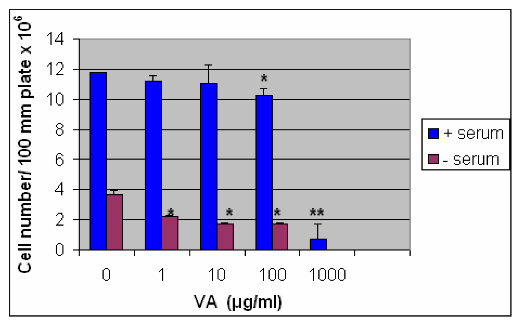
Inhibition of Cell Growth by VA in the Presence and Absence of Serum

Figure 2 shows that the exposure of cells to increasing concentrations of VA inhibited cell growth in the presence and absence of serum. One hundred and thousand microgram per milliliter (100 and 1000 μg/ml) inhibited growth about 0.2 (p < 0.05) and 15-fold [(p < 0.001) (N=6)] in the presence of serum (blue bars). This finding corroborates previous findings by Izevbigie and colleagues [17]. The cells were more VA-sensitive under serum-free milieu: 1 and 10 μg/ml VA inhibited cell growth by 40 and 54 % under serum-free conditions compared to 6 and 7 % respectively in the presence of serum. At 1000 μg/ml) no cell survived under serum-free conditions. In contrast, 6% of the cells survived in the presence of serum.

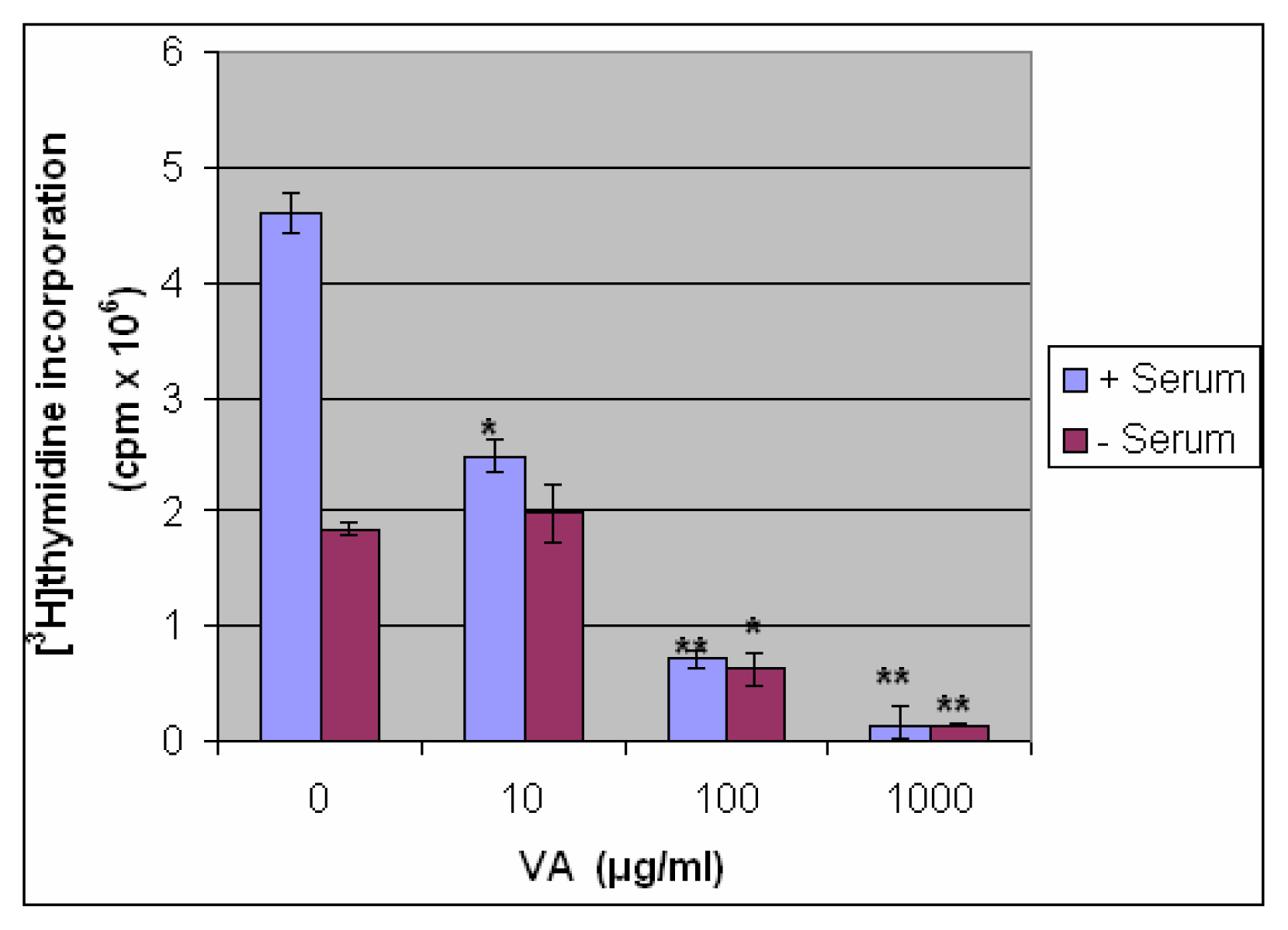
Inhibition of DNA Synthesis by VA in the Presence and Absence of Serum

As shown in Figure 3, the treatment of cells with VA inhibited DNA synthesis in a concentration-dependent (0–1000 μg/ml) fashion. Ten (10), 100, and 1000 μg/ml VA inhibited DNA synthesis 46, 85, and 97 % respectively. Similar concentrations of VA inhibited DNA synthesis 0, 66, 97 % under serum-free conditions compared to controls.

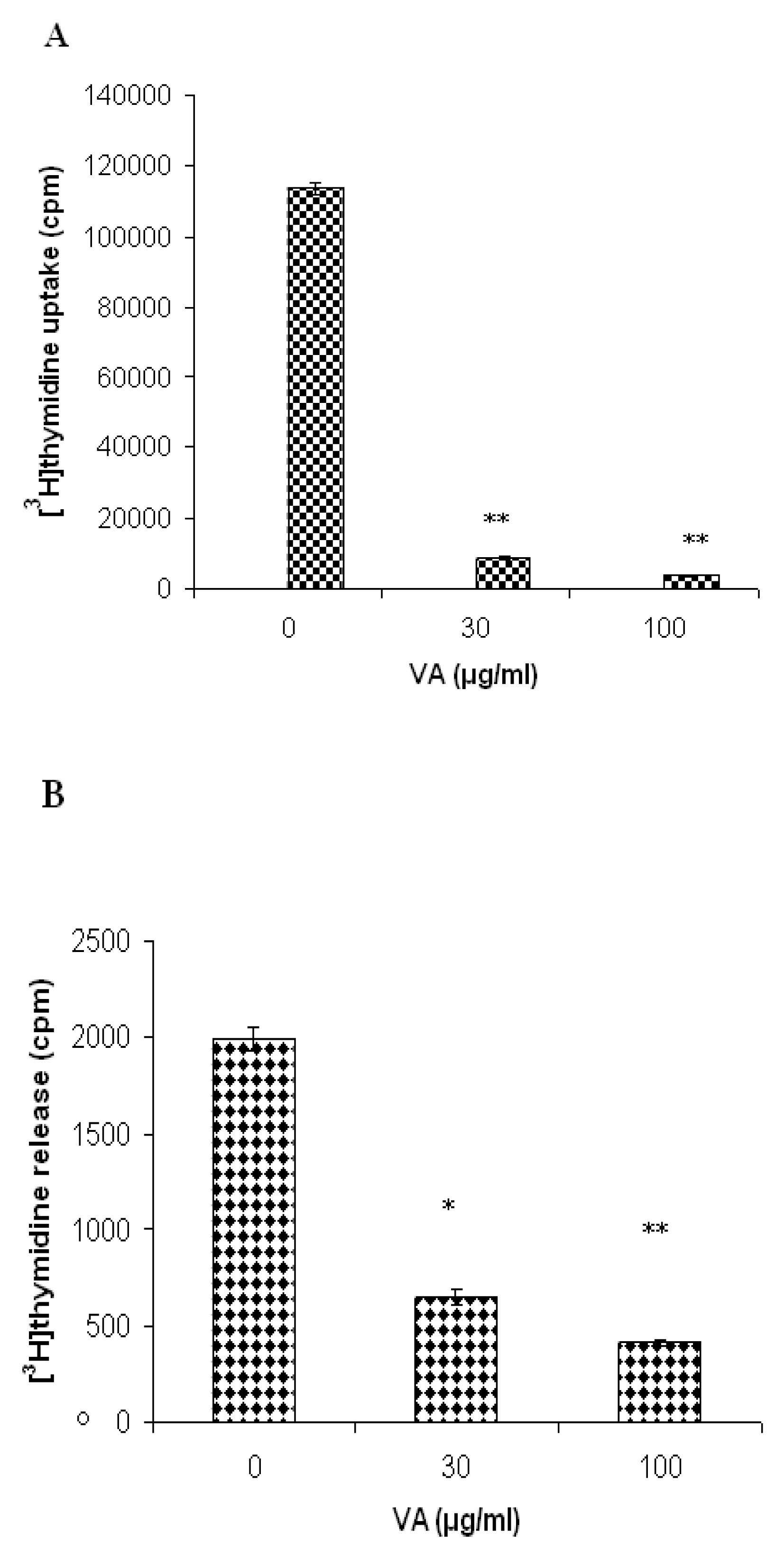
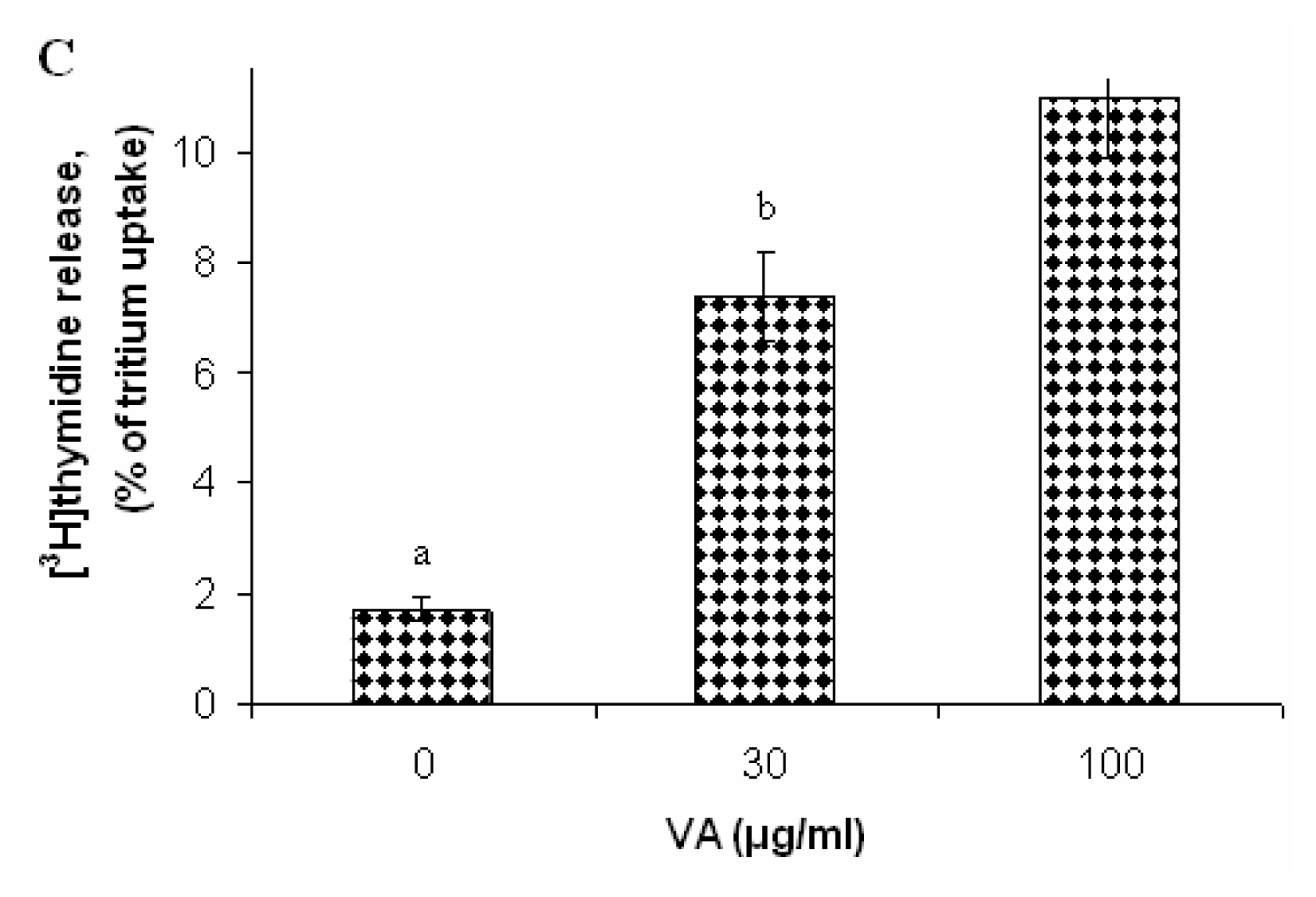
Effects of V. Amygdalina Treatment on Membrane Permeability and Efflux

The exposure of cells to VA decreased [3H]thymidine incorporation (uptake) in a concentration-dependent manner with 13-fold inhibition at 30 μg/ml (p < 0.001) and more than 30-fold (p < 0.0001) at 100 μg/ml VA (Figure 4a). [3H]thymidine uptake was approximately 113,633; 8698; and 3708 counts per minute (cpm) for 0, 30, and 100 μg/ml VA treated cells respectively (figure 4a). [3H]thymidine release was approximately 1992, 648, and 412 cpm for 0,30, and 100 μg/ml respectively (figure 4b). V. amygdalina increased [3H]thymidine release into the medium [3H]thymidine release was expressed as a percentage of [3H]thymidine uptake (1.7, 7.4, and 11.0%) for 0, 30, and 100 μg/ml VA (figure 4c); thus suggesting that the membranes of VA-treated cells were compromised. This observation is consistent with previous reports that plant extracts may contain small cystein-rich peptides that may disrupt membrane architecture of the cell and thus compromise permeability and efflux control [2125].

Discussion

V. amygdalina (VA), member of the compositae family, is a small shrub that grows in the tropical Africa. In Nigeria, VA is commonly called bitter leaf due to its bitter taste. The VA leaves are consumed either as a vegetable (macerated leaves in soup) or aqueous extracts as tonic for the treatment of various illnesses. Many herbalists and local healers in Nigeria prescribe aqueous VA for their patients. These herbalists may be right as increasing evidence suggests that the aqueous VA does possess some biologically-active components that may provide health benefits to patients suffering from the following diseases: bacterial-induced periodontal diseases [28]; liver diseases--- Babalola and colleagues [29] used serum enzymes such as alanine amino transferase (ALT), ornithine carbamoyl transferase (OCT), and aspartate amino transferase (AST) as markers of liver toxicity - these enzymes were modulated by the VA extracts; and cancer [1718,30] by modulation of biotransformation enzymes (P450) and extracellular signal-regulated kinase 1/2 (ERKs 1/2) activities. Izevbigie and colleagues [18] were the first to show one mode of action (ERKs signaling pathway) of possibly many pathways to explain VA anticancer effects mechanistically. Evidence for thionins, low-molecular-weight proteins occurring in seeds, stems, roots, and leaves of a number of plant species as active components that elicit a wide range of activities from antimicrobial to cytotoxicity in eukaryotic cells [21,26]. The thionins are believed to form channels on the cell plasma membrane surface to alter the cell efflux, depolarization and subsequent cell death. In the present studies, we confirmed the growth-inhibitory activities of VA extracts in the presence and absence of serum (Figure 2) previously reported by (Izevbigie and colleagues [18] who reported an IC50 value of 20 μg/ml VA as determined by cell counts compared to about an IC50 value of 218 μg/ml (10-fold increase) obtained in the present study. The only plausible explanation we have for the discrepancy in VA extracts potencies is batch variation--- extracts in this study came from a different batch with probably a lower activity. However, the DNA synthesis experiments showed a lower IC50 value of 12 μg/ml in cells cultured in the presence of serum compared the 5.6 ± 0.2 previously reported [18]. There was a shift in IC50 value from left to right (approx. four-fold) increase when cells were grown in the absence of serum. The reason is partly due to the fact that activity (DNA inhibition) of the VA-treated cells was compared to control whose growth was already compromised due to serum withdrawal (figure 3). Next, we hypothesized, as the literature suggests that plant extracts such as VA, may elicit part of their actions by compromising cell membrane permeability and efflux. To test this, VA-treated and control MCF-7 cells were challenged with [3H]thymidine for 6 hrs. [3H]thymidine release by VA-treated cells versus control cells were assessed. As shown in figure 4A, the exposure of cells to VA decreased [3H]thymidine uptake in a concentration-dependent fashion (0 = 113,633; 30 = 8698; and 100 μg/ml VA = 3708 cpm) at highest concentration of 100 μg/ml by 30-fold (p < 0.0001) but increased [3H]thymidine release, expressed as percent of [3H]thymidine uptake, into the medium 6-fold (p < 0.05) figure 4c. The amount of [3H]thymidine released into the medium (% of uptake) was 1.7, 7.4, and 11 % for 0, 30, and 100 μg/ml VA respectively. Taken together, these data suggest that VA exposure compromised the cell membranes in a concentration-dependent fashion which is in agreement with previous investigators that components of plant products such as roots, leaves, barks etc may interact with cell membrane, form ion channels which may depolarize cells and subsequent death [2024]. In conclusion, we have previously reported that VA inhibits the growth-promoting pathway of extracellular signal-regulated kinases (ERKs 1/2) to elicit it anticancer effects in MCF-7 cells. We are now reporting that another mode of action utilized by VA to regulate cell growth may include membrane permeability and efflux alteration.
Figure 1. Time-dependent Growth. Cells were plated at a density of 5 × 10⁵ cells/ml and propagated in RPMI 1640 medium supplemented with 10 % FBS (serum) and 1% pen/strep/fungisome mixture for eight days. Duplicate samples were randomly selected and counted using a hemacytometer every 24 h. Each data point represents the mean ± SEM of three independent experiments (N = 6).
Ijerph 03 00174f1
Figure 2. Inhibition of cell proliferation by VA extracts in the presence and absence of serum. Cells at the logarithmic growth phase were treated with various concentrations (0–000 μg/ml) VA for 24 h. Followed by cell counts as described under the materials and methods section. Each data point represents the mean of three independent experiments done in duplicates (N=6). *p < 0.05, **p <0.001.
Ijerph 03 00174f2
Figure 3. Inhibition of DNA synthesis by VA extracts in the presence and absence of serum. For DNA synthesis in the presence of serum, cells at logarithmic growth phase were treated with various (0–1000 μg/ml) VA for 18 h followed 6 h treatment of 1 μCi/ml [3H]thymidine. For DNA synthesis in the absence of serum, cells well serum-starved overnight before treatment with VA. [3H]thymidine uptake was determined as described under the materials and methods section. * p <0.05; **p <0.02; ***p <0.01.
Ijerph 03 00174f3
Figure 4. Effects of V. amygdalina treatment on membrane permeability and efflux. Cells at logarithmic growth phase were treated with various (0, 30, and 100 μg/ml) VA for 18 h followed 6 h treatment of 1 μCi/ml [3H]thymidine. The membrane permeability and efflux was determined as described under the material and method section. Each data point represents the mean ± SEM of three independent experiments. *p < 0.05; **p < 0.001.
Ijerph 03 00174f4aIjerph 03 00174f4b

Acknowledgements

Our appreciation also goes to Drs. Abdul A. Mohamed and Felix A. Okojie, Mark G. Hardy, James Perkins for their assistance. We also thank Ms. Jacqueline Humphrey (secretary to the dean of CSET) for her clerical assistance.

References

  1. American Institute for Cancer Research (AIRC), Food, nutrition and the prevention of cancer: a global perspective (summary); Washington DC, 1997; pp. 9–15.
  2. Jemal, T.; Thomas, A.; Murray, T.; Thun, M. Cancer Statistics. CA Cancer J. Clin 2002, 52, 23–47. [Google Scholar]
  3. Richardson, M. A.; Sanders, T.; Palmer, J. L.; Greisinger, A.; Singletary, S. E. Complementary/ Alternative medicine use in a comprehensive cancer center and the implication for oncology. J. Clin. Oncol 2000, 18, 2505–2514. [Google Scholar]
  4. Zeegers, M.P.; Goldbohm, R. A.; Van den Brant, P. A. Consumption of vegetables and fruits and urothelial cancer incidence: a prospective study. Cancer Epidemiol. Biomarkers Prev 2001, 10, 1121–1128. [Google Scholar]
  5. Pezzuto, J. M. Plant-derived anticancer agents. Biochem. Pharmacol 1997, 53, 121–133. [Google Scholar]
  6. Kobayashi, T.; Nakata, T.; Kuzumaki, T. Effect of flavonoids on cell cycle progression in prostate cancer cells. Cancer Lett 2000, 176, 17–23. [Google Scholar]
  7. Block, G.; Patterson, B.; Subar, A. Fruits, vegetables, and cancer prevention: a review of the epidemiological evidence. Nutr. Cancer 1992, 18, 1–29. [Google Scholar]
  8. Fahey, J. W.; Zhang, Y.; Talalay, P. Broccoli sprouts: an exceptionally rich source of inducers of enzymes that protect against chemical carcinogens. Proc Natl. Acad. Sci. USA 1997, 94, 10367–10372. [Google Scholar]
  9. Maoka, T.; Mochida, K.; Kozuka, M.; Ito, Y.; Fujiwara, Y.; Hashimoto, K.; Enjo, F.; Ogata, M.; Nobukuni, Y.; Tokuda, H.; Nishino, H. Cancer chemopreventive activity of carotenoids in the fruits of red paprika capsicum annuum L. Cancer Lett 2001, 172, 103–109. [Google Scholar]
  10. Terry, P.; Lagergren, J.; Hansen, H.; Wolk, A.; Nyren, O. Fruits and vegetables consumption in the prevention of esophageal and cardiac cancers. Eur. J. Cancer Prev 2001, 10, 365–369. [Google Scholar]
  11. Rock, C. L.; Moskowitz, A.; Huizar, B.; Saenz, C. C.; Clark, J. T.; Daly, T. L.; Chin, H.; Behling, C.; Ruffin, M. T. 4th. High vegetable and fruit diet intervention in premenopausal women with cervical intraepithelial neoplasia. J. Am. Diet Assoc 2001, 101, 1167–1174. [Google Scholar]
  12. Simopoulos, A. P. The Mediterranean diets: what is so special about the diet of Greece? The scientific evidence. J. Nutr 2001, 131 11 Suppl, 3065S–3073S. [Google Scholar]
  13. Singletary, K. Natural products and cancer chemoprevention. J. Nutr 2000, 130, 465–466. [Google Scholar]
  14. Chung, S. Y.; Sung, M. K.; Kim, N. H.; Jang, J. O. Inhibition of P-glycoprotein by natural products in human breast cancer cells. Arch. Pharm. Res 2005, 28(7), 823–828. [Google Scholar]
  15. Jakubilova, J.; Bao, Y.; Sedlac, J. Isothiocyanates induce cell cycle arrest, apoptosis and mitochondrial potential depolarization in HL-60 and multidrug-resistant cell lines. Anticancer Res 2005, 25(5), 3375–3386. [Google Scholar]
  16. Dragsted, L.; Srube, M.; Larsen, J. Cancer-preventive factors in fruits and vegetables: biochemical and biological background. Parmacol. Toxicol 1993, 72 suppl 1, 116–135. [Google Scholar]
  17. Izevbigie, E. B. Discovery of water-soluble anticancer agents (Edotides) from a vegetable found in Benin City Nigeria. Exp. Biol. Med 2003, 228, 293–298. [Google Scholar]
  18. Izevbigie, E. B.; Bryant, J. L.; Walker, A. A Novel inhibitor of extracellular signal-regulated kinases and human breast cancer cell growth. Exp. Biol. Med 2004, 229, 163–169. [Google Scholar]
  19. Jahansson, S.; Gullbo, J.; Lindholm, P.; Ek, B.; Thunberg, E.; Samuelsson, G.; Larsson, R.; Bohlin, L.; Claeson, P. Small, novel proteins from the mistletoe phoradendron tomentosum exhibit highly selective cytotoxicity to human breast cancer cells. Cell Mol. Life Sci 2003, 60, 165–75. [Google Scholar]
  20. Karkabounas, S.; Assimakopoulos, D.; Malamas, M.; Skaltsounis, A. L.; Leonce, S.; Zelovitis, J.; Stefanou, D.; Evangelou, A. Antiproliferative and anticarcinogenic effects of an aqueous preparation of Abies alba and Viscum album se abies, on a L-1210 malignant cell line and tumor-bearing Wistar rats. Anticancer Res 2000, 20(6B), 4391–4395. [Google Scholar]
  21. Florack, D. E.; Stickema, W. J. Thionins: properties, possible biological roles and mechanisms of action. Plant Mol. Biol 1994, 26, 25–37. [Google Scholar]
  22. Froy, O.; Gurevitz, M. Membrane potential modulators: a thread of scarlet from plants to animals. FASEB J 1998, 12, 1793–1796. [Google Scholar]
  23. Coulon, A.; Berkane, E.; Sautereau, A.M.; Urech, K.; Rouge, P.; Lopez, A. Modes of membrane interaction of a natural cysteine-rich peptides: viscotoxin A3. Biochim. Biophys Acta 2002, 1559(2), 145–159. [Google Scholar]
  24. Stec, B.; Markman, O.; Rao, U.; Heffron, G.; Henderson, S.; Vernon, L. P.; Brumfeld, V.; Teeter, M.M. Proposal for molecular mechanism of thionins deduced from physico-chemical studies of plant toxins. J. Pept. Res 2004, 64(6), 210–224. [Google Scholar]
  25. Carrasco, L.; Vazquez, D.; Hernandez-Lucas, C.; Carbonero, P.; Carcia-Olmedo, F. Thionins: plant peptides that modify membrane permeability in cultured mammalian cells. Eur. J. Biochem 1981, 116, 185–189. [Google Scholar]
  26. Hughes, P.; Dennist, E.; Whitecross, M.; Llewellyn, D.; Gage, P. The cytotoxic plant protein, β-purothionin, forms ion channels in lipid membrane. J. Biol. Chem 2000, 275(2), 823–827. [Google Scholar]
  27. Garcia-Olmedo, F.; Molina, A.; Alamillo, J.M.; Rodriquez-Palenzuela, P. Plant defense peptides. Biopolymers 1998, 47(6), 479–491. [Google Scholar]
  28. Taiwo, O.; Xu, H.X.; Lee, S.F. Antibacterial activities of extracts from Nigerian chewing sticks. Phytother. Res 1999, 13(8), 675–679. [Google Scholar]
  29. Babalola, O. O.; Anetor, J. I.; Adeniyi, F. A. Amelioration of carbon tetrachloride-induced hepatotoxicity by terpenoid extract from leaves of Vernonia amygdalina. African. J. Med. Med. Sci 2001, 30(1–2), 91–93. [Google Scholar]
  30. Howard, C. B.; Stevens, J.; Izevbigie, E. B.; Walker, A.; McDaniel, O. Time and dose-dependent modulation of phase 1 and phase 2 gene expression in response to treatment of MCF-7 cells with a natural anti-cancer agent. Cell. Mol. Biol 2003, 49(7), 1057–1065. [Google Scholar]

