Aberrant Protein Phosphorylation in Cancer by Using Raman Biomarkers

 ,
, 
 and
and
Abstract
1. Introduction
2. Results
3. Discussion
4. Materials and Methods
4.1. Chemicals
4.2. Patients
4.3. Cell Line
4.4. Equipment
4.5. Data Processing
4.6. Statistics
5. Conclusions
Author Contributions
Funding
Conflicts of Interest
Ethics Approval and Consent to Participate
References
- Ludwig, J.A.; Weinstein, J.N. Biomarkers in cancer staging, prognosis and treatment selection. Nat. Rev. Cancer 2005, 5, 845–856. [Google Scholar] [CrossRef] [Scilit] [PubMed]
- Jang, M.; Kim, S.S.; Lee, J. Cancer cell metabolism: Implications for therapeutic targets. Exp. Mol. Med. 2013, 45, e45. [Google Scholar] [CrossRef] [Scilit] [PubMed]
- DeBerardinis, R.J.; Lum, J.J.; Hatzivassiliou, G.; Thompson, C.B. The biology of cancer: Metabolic reprogramming fuels cell growth and proliferation. Cell Metab. 2008, 7, 11–20. [Google Scholar] [CrossRef] [Scilit] [PubMed]
- Ganapathy-Kanniappan, S.; Geschwind, J.-F.H. Tumor glycolysis as a target for cancer therapy: Progress and prospects. Mol. Cancer 2013, 12, 152. [Google Scholar] [CrossRef] [Scilit]
- Schulze, A.; Downward, J. Flicking the Warburg switch-tyrosine phosphorylation of pyruvate dehydrogenase kinase regulates mitochondrial activity in cancer cells. Mol. Cell 2011, 44, 846–848. [Google Scholar] [CrossRef] [Scilit]
- Singh, V.; Ram, M.; Kumar, R.; Prasad, R.; Roy, B.K.; Singh, K.K. Phosphorylation: Implications in Cancer. Protein J. 2017, 36, 1–6. [Google Scholar] [CrossRef] [Scilit]
- Hitosugi, T.; Kang, S.; Vander Heiden, M.G.; Chung, T.-W.; Elf, S.; Lythgoe, K.; Dong, S.; Lonial, S.; Wang, X.; Chen, G.Z.; et al. Tyrosine phosphorylation inhibits PKM2 to promote the Warburg effect and tumor growth. Sci. Signal. 2009, 2, ra73. [Google Scholar] [CrossRef] [Scilit]
- DeBerardinis, R.J.; Mancuso, A.; Daikhin, E.; Nissim, I.; Yudkoff, M.; Wehrli, S.; Thompson, C.B. Beyond aerobic glycolysis: Transformed cells can engage in glutamine metabolism that exceeds the requirement for protein and nucleotide synthesis. Proc. Natl. Acad. Sci. USA. 2007, 104, 19345–19350. [Google Scholar] [CrossRef] [Scilit]
- Martin, K.; Steinberg, T.H.; Cooley, L.A.; Gee, K.R.; Beechem, J.M.; Patton, W.F. Quantitative analysis of protein phosphorylation status and protein kinase activity on microarrays using a novel fluorescent phosphorylation sensor dye. Proteomics Int. Ed. 2003, 3, 1244–1255. [Google Scholar] [CrossRef] [Scilit]
- Olive, D.M. Quantitative methods for the analysis of protein phosphorylation in drug development. Expert Rev. Proteomics 2004, 1, 327–341. [Google Scholar] [CrossRef] [Scilit]
- Yan, J.X.; Packer, N.H.; Gooley, A.A.; Williams, K.L. Protein phosphorylation: Technologies for the identification of phosphoamino acids. J. Chromatogr. A 1998, 808, 23–41. [Google Scholar] [CrossRef] [Scilit]
- Ficarro, S.B.; McCleland, M.L.; Stukenberg, P.T.; Burke, D.J.; Ross, M.M.; Shabanowitz, J.; Hunt, D.F.; White, F.M. Phosphoproteome analysis by mass spectrometry and its application to Saccharomyces cerevisiae. Nat. Biotechnol. 2002, 20, 301–305. [Google Scholar] [CrossRef] [Scilit] [PubMed]
- Garcia, B.A.; Shabanowitz, J.; Hunt, D.F. Analysis of protein phosphorylation by mass spectrometry. Methods 2005, 35, 256–264. [Google Scholar] [CrossRef] [Scilit] [PubMed]
- Krutzik, P.O.; Irish, J.M.; Nolan, G.P.; Perez, O.D. Analysis of protein phosphorylation and cellular signaling events by flow cytometry: Techniques and clinical applications. Clin. Immunol. 2004, 110, 206–221. [Google Scholar] [CrossRef] [Scilit] [PubMed]
- Mandell, J.W. Phosphorylation state-specific antibodies: Applications in investigative and diagnostic pathology. Am. J. Pathol. 2003, 163, 1687–1698. [Google Scholar] [CrossRef] [Scilit]
- Rush, J.; Moritz, A.; Lee, K.A.; Guo, A.; Goss, V.L.; Spek, E.J.; Zhang, H.; Zha, X.-M.; Polakiewicz, R.D.; Comb, M.J. Immunoaffinity profiling of tyrosine phosphorylation in cancer cells. Nat. Biotechnol. 2005, 23, 94–101. [Google Scholar] [CrossRef] [Scilit]
- Aebersold, R.; Mann, M. Mass spectrometry-based proteomics. Nature 2003, 422, 198–207. [Google Scholar] [CrossRef] [Scilit]
- Kircher, M.F.; de la Zerda, A.; Jokerst, J.V.; Zavaleta, C.L.; Kempen, P.J.; Mittra, E.; Pitter, K.; Huang, R.; Campos, C.; Habte, F.; et al. A Brain Tumor Molecular Imaging Strategy Using A New Triple-Modality MRI-Photoacoustic-Raman Nanoparticle. Nat. Med. 2012, 18, 829–834. [Google Scholar] [CrossRef] [Scilit]
- Brozek-Pluska, B.; Kopeć, M.; Abramczyk, H. Development of a new diagnostic Raman method for monitoring epigenetic modifications in the cancer cells of human breast tissue. Anal. Methods 2016, 8, 8542–8553. [Google Scholar] [CrossRef] [Scilit]
- Abramczyk, H.; Brozek-Pluska, B. New look inside human breast ducts with Raman imaging. Raman candidates as diagnostic markers for breast cancer prognosis: Mammaglobin, palmitic acid and sphingomyelin. Anal. Chim. Acta 2016, 909, 91–100. [Google Scholar] [CrossRef] [Scilit]
- Protein Phosphorylation Analysis Tools-Tools for Success in Analyzing Phosphorylation Targets. Available online: https://www.sigmaaldrich.com/life-science/proteomics/post-translational-analysis/phosphorylation/protein-phosphorylation-analysis-tools.html (accessed on 18 October 2019).
- Huang, C.; Freter, C. Lipid metabolism, apoptosis and cancer therapy. Int. J. Mol. Sci. 2015, 16, 924–949. [Google Scholar] [CrossRef] [Scilit] [PubMed]
- Herbst, R.S. Review of epidermal growth factor receptor biology. Int. J. Radiat. Oncol. Biol. Phys. 2004, 59, 21–26. [Google Scholar] [CrossRef] [Scilit] [PubMed]
- Cho, H.-S.; Leahy, D.J. Structure of the extracellular region of HER3 reveals an interdomain tether. Science 2002, 297, 1330–1333. [Google Scholar] [CrossRef] [Scilit] [PubMed]
- Camidge, D.R.; Pao, W.; Sequist, L.V. Acquired resistance to TKIs in solid tumours: Learning from lung cancer. Nat. Rev. Clin. Oncol. 2014, 11, 473–481. [Google Scholar] [CrossRef] [Scilit] [PubMed]
- Paez, J.G.; Jänne, P.A.; Lee, J.C.; Tracy, S.; Greulich, H.; Gabriel, S.; Herman, P.; Kaye, F.J.; Lindeman, N.; Boggon, T.J.; et al. EGFR mutations in lung cancer: Correlation with clinical response to gefitinib therapy. Science 2004, 304, 1497–1500. [Google Scholar] [CrossRef] [Scilit] [PubMed]
- Okishio, N.; Fukuda, R.; Nagai, M.; Nagai, Y.; Nagatomo, S.; Kitagawa, T. Tyrosine phosphorylation-induced changes in absorption and UV resonance Raman spectra of Src-peptides. J. Raman Spectrosc. 1998, 29, 31–39. [Google Scholar] [CrossRef]
- Stone, N.; Kendall, C.; Smith, J.; Crow, P.; Barr, H. Raman spectroscopy for identification of epithelial cancers. Faraday Discuss. 2004, 126, 141–157, discussion 169–183. [Google Scholar] [CrossRef] [Scilit]
- Thomas, G.J. Raman spectroscopy of protein and nucleic acid assemblies. Annu. Rev. Biophys. Biomol. Struct. 1999, 28, 1–27. [Google Scholar] [CrossRef] [Scilit]
- Shaver, J.M.; Christensen, K.A.; Pezzuti, J.A.; Morris, M.D. Structure of Dihydrogen Phosphate Ion Aggregates by Raman-Monitored Serial Dilution. Appl. Spectrosc. 1998, 52, 259–264. [Google Scholar] [CrossRef] [Scilit]
- Xie, Y.; Zhang, D.; Jarori, G.K.; Davisson, V.J.; Ben-Amotz, D. The Raman detection of peptide tyrosine phosphorylation. Anal. Biochem. 2004, 332, 116–121. [Google Scholar] [CrossRef] [Scilit]
- Deng, H.; Bloomfield, V.A.; Benevides, J.M.; Thomas, G.J. Dependence of the Raman signature of genomic B-DNA on nucleotide base sequence. Biopolys. Orig. Res. Biomol. 1999, 50, 656–666. [Google Scholar]
- Guan, Y.; Thomas, G.J. Vibrational analysis of nucleic acids. IV. Normal modes of the DNA phosphodiester structure modeled by diethyl phosphate. Biopolymers 1996, 39, 813–835. [Google Scholar] [CrossRef]
- Guan, Y.; Choy, G.S.-C.; Glaser, R.; Thomas, G.J., Jr. Vibrational Analysis of Nucleic Acids. 2. Ab Initio Calculation of the Molecular Force Field and Normal Modes of Dimethyl Phosphate. J. Phys. Chem. 1995, 99, 12054–12062. [Google Scholar] [CrossRef] [Scilit]
- Wilkinson, G.R. R. J. H. Clark and R. E. Hester (Editors). Advances in Infrared and Raman Spectroscopy; Wiley: New York, NY, USA, 1985; Volume 12, p. 487. [Google Scholar] [CrossRef] [Scilit]
- Abramczyk, H.; Surmacki, J.; Kopeć, M.; Olejnik, A.K.; Lubecka-Pietruszewska, K.; Fabianowska-Majewska, K. The role of lipid droplets and adipocytes in cancer. Raman imaging of cell cultures: MCF10A, MCF7, and MDA-MB-231 compared to adipocytes in cancerous human breast tissue. Analyst 2015, 140, 2224–2235. [Google Scholar] [CrossRef] [Scilit]
- Carey, P.R. Biochemical Applications of Raman and Resonance Raman Spectroscopes; Elsevier: Amsterdam, The Netherland, 1982; ISBN 978-0-12-159650-7. [Google Scholar]
- Surmacki, J.; Brozek-Pluska, B.; Kordek, R.; Abramczyk, H. The lipid-reactive oxygen species phenotype of breast cancer. Raman spectroscopy and mapping, PCA and PLSDA for invasive ductal carcinoma and invasive lobular carcinoma. Molecular tumorigenic mechanisms beyond Warburg effect. Analyst 2015, 140, 2121–2133. [Google Scholar] [CrossRef] [Scilit] [PubMed]
- Abramczyk, H.; Imiela, A.; Śliwińska, A. Novel strategies of Raman imaging for exploring cancer lipid reprogramming. J. Mol. Liq. 2019, 274, 52–59. [Google Scholar] [CrossRef] [Scilit]
- Abramczyk, H.; Surmacki, J.; Kopeć, M.; Olejnik, A.K.; Kaufman-Szymczyk, A.; Fabianowska-Majewska, K. Epigenetic changes in cancer by Raman imaging, fluorescence imaging, AFM and scanning near-field optical microscopy (SNOM). Acetylation in normal and human cancer breast cells MCF10A, MCF7 and MDA-MB-231. Analyst 2016, 141, 5646–5658. [Google Scholar] [CrossRef] [Scilit] [PubMed]
- Kopeć, M.; Abramczyk, H. Angiogenesis—a crucial step in breast cancer growth, progression and dissemination by Raman imaging. Spectrochim. Acta Part A Mol. Biomol. Spectrosc. 2018, 198, 338–345. [Google Scholar] [CrossRef] [Scilit]
- Kopec, M.; Imiela, A.; Abramczyk, H. Monitoring glycosylation metabolism in brain and breast cancer by Raman imaging. Sci. Rep. 2019, 9, 166. [Google Scholar] [CrossRef] [Scilit]
- Gajjar, K.; Heppenstall, L.D.; Pang, W.; Ashton, K.M.; Trevisan, J.; Patel, I.I.; Llabjani, V.; Stringfellow, H.F.; Martin-Hirsch, P.L.; Dawson, T.; et al. Diagnostic segregation of human brain tumours using Fourier-transform infrared and/or Raman spectroscopy coupled with discriminant analysis. Anal. Methods 2012, 5, 89–102. [Google Scholar] [CrossRef] [Scilit]
- Camp Jr, C.H.; Lee, Y.J.; Heddleston, J.M.; Hartshorn, C.M.; Walker, A.R.H.; Rich, J.N.; Lathia, J.D.; Cicerone, M.T. High-speed coherent Raman fingerprint imaging of biological tissues. Nat. Photonics 2014, 8, 627–634. [Google Scholar] [CrossRef] [Scilit] [PubMed]
- Mikoliunaite, L.; Rodriguez, R.D.; Sheremet, E.; Kolchuzhin, V.; Mehner, J.; Ramanavicius, A.; Zahn, D.R.T. The substrate matters in the Raman spectroscopy analysis of cells. Sci. Rep. 2015, 5, 13150. [Google Scholar] [CrossRef] [Scilit]
- Distinguishing Different Cancerous Human Cells by Raman Spectroscopy Based on Discriminant Analysis Methods—Semantic Scholar. Available online: https://www.semanticscholar.org/paper/Distinguishing-Different-Cancerous-Human-Cells-by-Tang-Xia/1aa11eaa9ceca1435cb3d4f5cd7055e468a9ba2e (accessed on 18 October 2019).
- Lau, D.P.; Huang, Z.; Lui, H.; Man, C.S.; Berean, K.; Morrison, M.D.; Zeng, H. Raman spectroscopy for optical diagnosis in normal and cancerous tissue of the nasopharynx-preliminary findings. Lasers Surg. Med. 2003, 32, 210–214. [Google Scholar] [CrossRef] [Scilit] [PubMed]
- Austin, L.A.; Osseiran, S.; Evans, C.L. Raman technologies in cancer diagnostics. Analyst 2016, 141, 476–503. [Google Scholar] [CrossRef] [Scilit] [PubMed]
- Ghomi, M. Applications of Raman Spectroscopy to Biology: From Basic Studies to Disease Diagnosis; IOS Press: Amsterdam, The Netherland, 2012; ISBN 978-1-60750-999-8. [Google Scholar]
- Lin, K.; Zheng, W.; Lim, C.M.; Huang, Z. Real-time In vivo Diagnosis of Nasopharyngeal Carcinoma Using Rapid Fiber-Optic Raman Spectroscopy. Theranostics 2017, 7, 3517–3526. [Google Scholar] [CrossRef] [Scilit] [PubMed]
- Feng, S.; Pan, J.; Wu, Y.; Lin, D.; Chen, Y.; Xi, G.; Lin, J.; Chen, R. Study on gastric cancer blood plasma based on surface-enhanced Raman spectroscopy combined with multivariate analysis. Sci. China Life Sci. 2011, 54, 828–834. [Google Scholar] [CrossRef] [Scilit]
- Li, S.; Chen, G.; Zhang, Y.; Guo, Z.; Liu, Z.; Xu, J.; Li, X.; Lin, L. Identification and characterization of colorectal cancer using Raman spectroscopy and feature selection techniques. Opt. Express 2014, 22, 25895–25908. [Google Scholar] [CrossRef] [Scilit]
- Klisch, T.J.; Vainshtein, A.; Patel, A.J.; Zoghbi, H.Y. Jak2-mediated phosphorylation of Atoh1 is critical for medulloblastoma growth. Elife 2017, 6, e31181. [Google Scholar] [CrossRef] [Scilit]
- Tirinato, L.; Pagliari, F.; Limongi, T.; Marini, M.; Falqui, A.; Seco, J.; Candeloro, P.; Liberale, C.; Di Fabrizio, E. An Overview of Lipid Droplets in Cancer and Cancer Stem Cells. Stem Cells Int. 2017, 2017, 1656053. [Google Scholar] [CrossRef] [Scilit]
- Brasaemle, D.L.; Dolios, G.; Shapiro, L.; Wang, R. Proteomic analysis of proteins associated with lipid droplets of basal and lipolytically stimulated 3T3-L1 adipocytes. J. Biol. Chem. 2004, 279, 46835–46842. [Google Scholar] [CrossRef] [Scilit]
- Kuerschner, L.; Moessinger, C.; Thiele, C. Imaging of lipid biosynthesis: How a neutral lipid enters lipid droplets. Traffic 2008, 9, 338–352. [Google Scholar] [CrossRef] [Scilit] [PubMed]
- Recent Advances in Understanding Proteins Involved in Lipid Droplet Formation, Growth and Fusion. Available online: https://www.ncbi.nlm.nih.gov/pubmed/24894352 (accessed on 18 October 2019).
- Jägerström, S.; Polesie, S.; Wickström, Y.; Johansson, B.R.; Schröder, H.D.; Højlund, K.; Boström, P. Lipid droplets interact with mitochondria using SNAP23. Cell Biol. Int. 2009, 33, 934–940. [Google Scholar] [CrossRef] [Scilit] [PubMed]
- Yu, W.; Cassara, J.; Weller, P.F. Phosphatidylinositide 3-kinase localizes to cytoplasmic lipid bodies in human polymorphonuclear leukocytes and other myeloid-derived cells. Blood 2000, 95, 1078–1085. [Google Scholar] [CrossRef] [Scilit] [PubMed]
- Bozza, P.T.; Viola, J.P.B. Lipid droplets in inflammation and cancer. Prostaglandins Leukot. Essent. Fat. Acids 2010, 82, 243–250. [Google Scholar] [CrossRef] [Scilit] [PubMed]
- Yu, W.; Bozza, P.T.; Tzizik, D.M.; Gray, J.P.; Cassara, J.; Dvorak, A.M.; Weller, P.F. Co-compartmentalization of MAP kinases and cytosolic phospholipase A2 at cytoplasmic arachidonate-rich lipid bodies. Am. J. Pathol. 1998, 152, 759–769. [Google Scholar]
- Li, C.-W.; Xia, W.; Lim, S.-O.; Hsu, J.L.; Huo, L.; Wu, Y.; Li, L.-Y.; Lai, C.-C.; Chang, S.-S.; Hsu, Y.-H.; et al. AKT1 Inhibits Epithelial-to-Mesenchymal Transition in Breast Cancer through Phosphorylation-Dependent Twist1 Degradation. Cancer Res. 2016, 76, 1451–1462. [Google Scholar] [CrossRef] [Scilit]
- Onken, J.; Vajkoczy, P.; Torka, R.; Hempt, C.; Patsouris, V.; Heppner, F.L.; Radke, J. Phospho-AXL is widely expressed in glioblastoma and associated with significant shorter overall survival. Oncotarget 2017, 8, 50403–50414. [Google Scholar] [CrossRef] [Scilit]
- Lin, P.-C.; Yang, Y.-F.; Tyan, Y.-C.; Hsiao, E.S.L.; Chu, P.-C.; Lee, C.-T.; Lee, J.-C.; Chen, Y.-M.A.; Liao, P.-C. Identification of Phosphorylated Cyclin-Dependent Kinase 1 Associated with Colorectal Cancer Survival Using Label-Free Quantitative Analyses. PLoS ONE 2016, 11, e0158844. [Google Scholar] [CrossRef] [Scilit]
- Lemmon, M.A.; Schlessinger, J. Cell signaling by receptor tyrosine kinases. Cell 2010, 141, 1117–1134. [Google Scholar] [CrossRef] [Scilit]
- Spill, F.; Reynolds, D.S.; Kamm, R.D.; Zaman, M.H. Impact of the physical microenvironment on tumor progression and metastasis. Curr. Opin. Biotechnol. 2016, 40, 41–48. [Google Scholar] [CrossRef] [Scilit]
- Brantley-Sieders, D.; Schmidt, S.; Parker, M.; Chen, J. Eph receptor tyrosine kinases in tumor and tumor microenvironment. Curr. Pharm. Des. 2004, 10, 3431–3442. [Google Scholar] [CrossRef] [Scilit] [PubMed]
- Liberti, M.V.; Locasale, J.W. The Warburg Effect: How Does it Benefit Cancer Cells? Trends Biochem. Sci. 2016, 41, 211–218. [Google Scholar] [CrossRef] [Scilit] [PubMed]
- Baenke, F.; Peck, B.; Miess, H.; Schulze, A. Hooked on fat: The role of lipid synthesis in cancer metabolism and tumour development. Dis. Models Mech. 2013, 6, 1353–1363. [Google Scholar] [CrossRef] [Scilit] [PubMed]
- Hanahan, D.; Weinberg, R.A. Hallmarks of cancer: The next generation. Cell 2011, 144, 646–674. [Google Scholar] [CrossRef] [Scilit] [PubMed]
- Huang, W.-C.; Li, X.; Liu, J.; Lin, J.; Chung, L.W.K. Activation of androgen receptor, lipogenesis, and oxidative stress converged by SREBP-1 is responsible for regulating growth and progression of prostate cancer cells. Mol. Cancer Res. 2012, 10, 133–142. [Google Scholar] [CrossRef] [Scilit] [PubMed]
- Zaytseva, Y.Y.; Harris, J.W.; Mitov, M.I.; Kim, J.T.; Butterfield, D.A.; Lee, E.Y.; Weiss, H.L.; Gao, T.; Evers, B.M. Increased expression of fatty acid synthase provides a survival advantage to colorectal cancer cells via upregulation of cellular respiration. Oncotarget 2015, 6, 18891–18904. [Google Scholar] [CrossRef] [Scilit] [PubMed]
- Cai, Y.; Crowther, J.; Pastor, T.; Abbasi Asbagh, L.; Baietti, M.F.; De Troyer, M.; Vazquez, I.; Talebi, A.; Renzi, F.; Dehairs, J.; et al. Loss of Chromosome 8p Governs Tumor Progression and Drug Response by Altering Lipid Metabolism. Cancer Cell 2016, 29, 751–766. [Google Scholar] [CrossRef] [Scilit]
- Porstmann, T.; Santos, C.R.; Griffiths, B.; Cully, M.; Wu, M.; Leevers, S.; Griffiths, J.R.; Chung, Y.-L.; Schulze, A. SREBP activity is regulated by mTORC1 and contributes to Akt-dependent cell growth. Cell Metab. 2008, 8, 224–236. [Google Scholar] [CrossRef] [Scilit]
- Röhrig, F.; Schulze, A. The multifaceted roles of fatty acid synthesis in cancer. Nat. Rev. Cancer 2016, 16, 732–749. [Google Scholar] [CrossRef] [Scilit]
- Santarpia, L.; Lippman, S.M.; El-Naggar, A.K. Targeting the MAPK-RAS-RAF signaling pathway in cancer therapy. Expert Opin. Ther. Targets 2012, 16, 103–119. [Google Scholar] [CrossRef] [Scilit]
- Li, X.; Jiang, Y.; Meisenhelder, J.; Yang, W.; Hawke, D.H.; Zheng, Y.; Xia, Y.; Aldape, K.; He, J.; Hunter, T.; et al. Mitochondria-Translocated PGK1 Functions as a Protein Kinase to Coordinate Glycolysis and the TCA Cycle in Tumorigenesis. Mol. Cell 2016, 61, 705–719. [Google Scholar] [CrossRef] [Scilit] [PubMed]



| Ratio | Assignment | Type | Breast | Small Intestine | Brain | |||
|---|---|---|---|---|---|---|---|---|
| Mean ± SD | p | Mean ± SD | p | Mean ± SD | p | |||
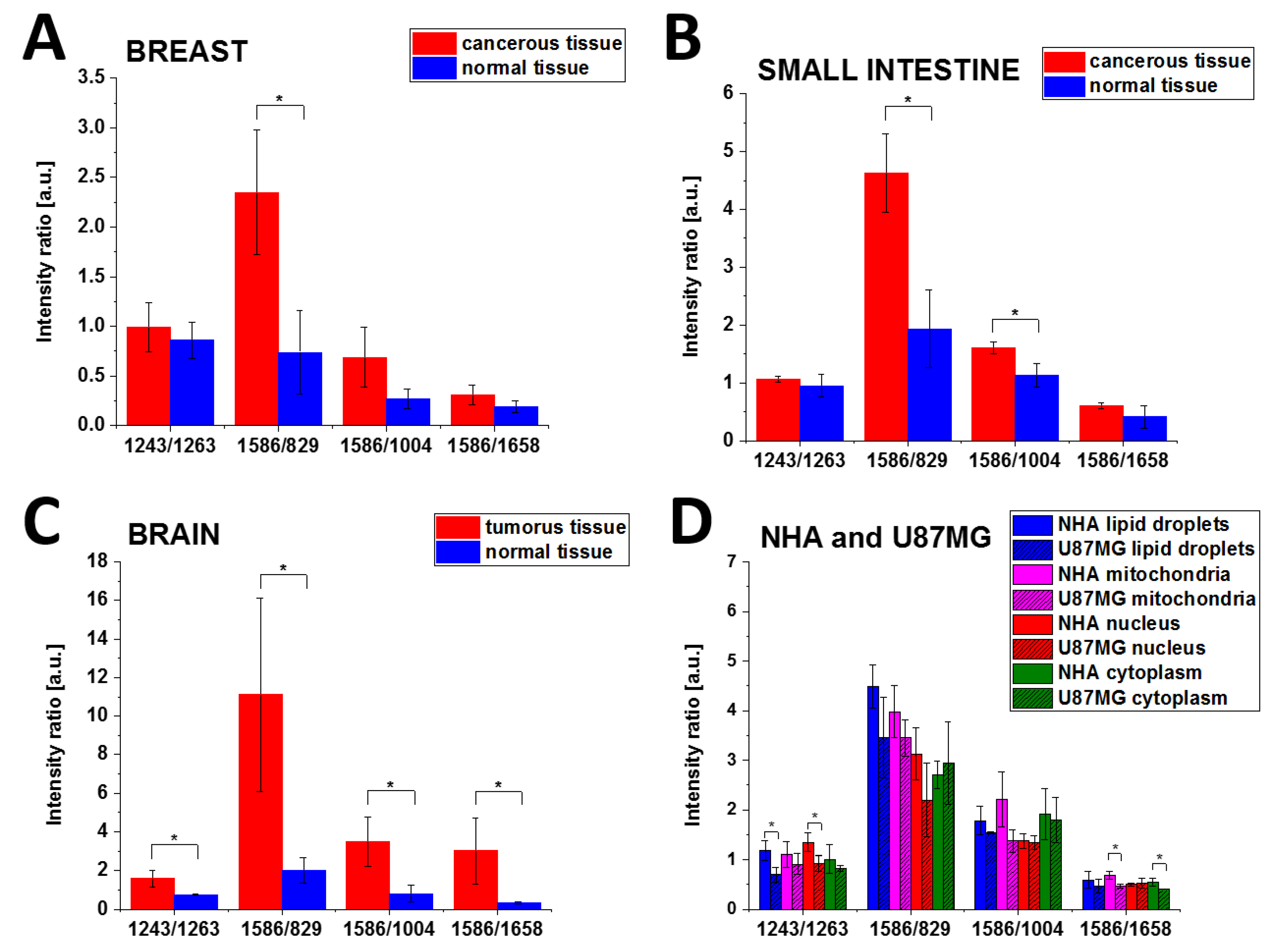
| 1243/1263 | phosphorylated proteins/non-phosphorylated proteins Amide III | N | 0.86 ± 0.18 | 0.51 | 0.95 ± 0.20 | 0.41 | 0.76 ± 0.04 | 0.03 |
| C | 0.99 ± 0.25 | 1.06 ± 0.05 | 1.59 ± 0.44 | |||||
| 1586/829 | phosphorylated proteins/tyrosine | N | 0.74 ± 0.42 | 0.02 | 1.93 ± 0.67 | 0.01 | 2.01 ± 0.68 | 0.04 |
| C | 2.35 ± 0.63 | 4.62 ± 0.68 | 11.12 ± 5.02 | |||||
| 1586/1004 | phosphorylated proteins/ phenyloalanine | N | 0.27 ± 0.10 | 0.08 | 1.13 ± 0.21 | 0.02 | 0.81 ± 0.45 | 0.03 |
| C | 0.69 ± 0.30 | 1.60 ± 0.10 | 3.49 ± 1.27 | |||||
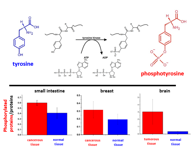
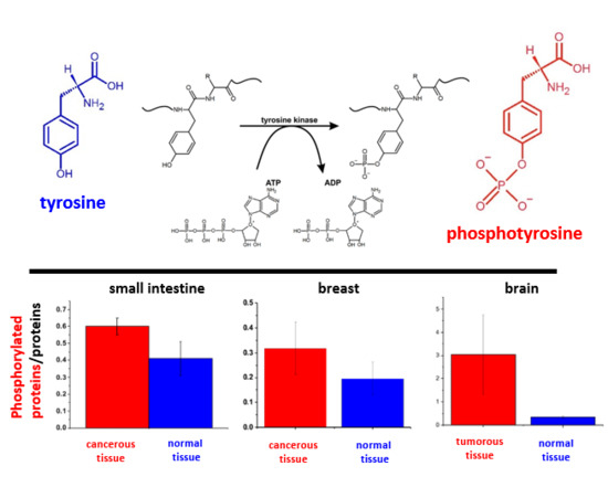
| 1586/1658 | phosphorylated proteins/proteins | N | 0.19 ± 0.06 | 0.15 | 0.41 ± 0.19 | 0.15 | 0.34 ± 0.05 | 0.05 |
| C | 0.31 ± 0.10 | 0.61 ± 0.05 | 3.03 ± 1.71 | |||||
| Parameter | Brain | Small Intestine | Breast | |||
|---|---|---|---|---|---|---|
| Tumor | Normal | Cancer | Normal | Cancer | Normal | |
| Calibration | ||||||
| Sensitivity | 100.0% | 100.0% | 100.0% | 100.0% | 66.7% | 100.0% |
| Specificity | 100.0% | 100.0% | 100.0% | 100.0% | 100.0% | 66.7% |
| AUC | 1.00 | 1.00 | 1.00 | 1.00 | 0.89 | 0.89 |
| Cross validation | ||||||
| Sensitivity | 100.0% | 100.0% | 66.7% | 66.7% | 66.7% | 66.7% |
| Specificity | 100.0% | 100.0% | 66.7% | 66.7% | 66.7% | 66.7% |
| AUC | 1.00 | 1.00 | 0.89 | 0.89 | 0.78 | 0.78 |
| Ratio | Assignment | Cell | Lipid Droplets | Mitochondria | Nucleus | Cytoplasm | ||||
|---|---|---|---|---|---|---|---|---|---|---|
| Mean ± SD | p | Mean ± SD | p | Mean ± SD | p | Mean ± SD | p | |||
| 1243/1263 | phosphorylated proteins/non-phosphorylated proteins Amide III | N | 1.18 ± 0.19 | 0.03 | 1.10 ± 0.26 | 0.38 | 1.34 ± 0.19 | 0.04 | 1.01 ± 0.28 | 0.31 |
| U | 0.70 ± 0.15 | 0.91 ± 0.21 | 0.92 ± 0.16 | 0.82 ± 0.06 | ||||||
| 1586/829 | phosphorylated proteins/tyrosine | N | 4.49 ± 0.44 | 0.12 | 3.98 ± 0.53 | 0.23 | 3.13 ± 0.53 | 0.15 | 2.71 ± 0.28 | 0.67 |
| U | 3.46 ± 0.81 | 3.45 ± 0.37 | 2.20 ± 0.75 | 2.94 ± 0.83 | ||||||
| 1586/1004 | phosphorylated proteins/phenyloalanine | N | 1.78 ± 0.29 | 0.23 | 2.21 ± 0.56 | 0.07 | 1.37 ± 0.15 | 0.81 | 1.91 ± 0.52 | 0.78 |
| U | 1.54 ± 0.02 | 1.37 ± 0.23 | 1.34 ± 0.14 | 1.79 ± 0.45 | ||||||
| 1586/1658 | phosphorylated proteins /proteins | N | 0.59 ± 0.17 | 0.40 | 0.69 ± 0.08 | 0.01 | 0.50 ± 0.03 | 0.76 | 0.54 ± 0.08 | 0.04 |
| U | 0.47 ± 0.14 | 0.46 ± 0.04 | 0.52 ± 0.10 | 0.40 ± 0.00 | ||||||
© 2019 by the authors. Licensee MDPI, Basel, Switzerland. This article is an open access article distributed under the terms and conditions of the Creative Commons Attribution (CC BY) license (http://creativecommons.org/licenses/by/4.0/).
Share and Cite
Abramczyk, H.; Imiela, A.; Brożek-Płuska, B.; Kopeć, M.; Surmacki, J.; Śliwińska, A. Aberrant Protein Phosphorylation in Cancer by Using Raman Biomarkers. Cancers 2019, 11, 2017. https://doi.org/10.3390/cancers11122017
Abramczyk H, Imiela A, Brożek-Płuska B, Kopeć M, Surmacki J, Śliwińska A. Aberrant Protein Phosphorylation in Cancer by Using Raman Biomarkers. Cancers. 2019; 11(12):2017. https://doi.org/10.3390/cancers11122017
Chicago/Turabian StyleAbramczyk, Halina, Anna Imiela, Beata Brożek-Płuska, Monika Kopeć, Jakub Surmacki, and Agnieszka Śliwińska. 2019. "Aberrant Protein Phosphorylation in Cancer by Using Raman Biomarkers" Cancers 11, no. 12: 2017. https://doi.org/10.3390/cancers11122017
APA StyleAbramczyk, H., Imiela, A., Brożek-Płuska, B., Kopeć, M., Surmacki, J., & Śliwińska, A. (2019). Aberrant Protein Phosphorylation in Cancer by Using Raman Biomarkers. Cancers, 11(12), 2017. https://doi.org/10.3390/cancers11122017