Oxidation States of Fe in Constituent Minerals of a Spinel Lherzolite Xenolith from the Tariat Depression, Mongolia: The Significance of Fe3+ in Olivine

Abstract
:1. Introduction
2. General Geology and Sampling Site
3. Experimental Methods
3.1. Sample Preparation and Purity Evaluation
3.2. Electron Microprobe Analysis
3.3. Raman Spectroscopy
3.4. Transmission Electron Microscopy (TEM)
3.5. X-ray Powder Diffraction Measurement and Rietveld Analysis
3.6. 57Fe Mössbauer Spectroscopy
4. Results
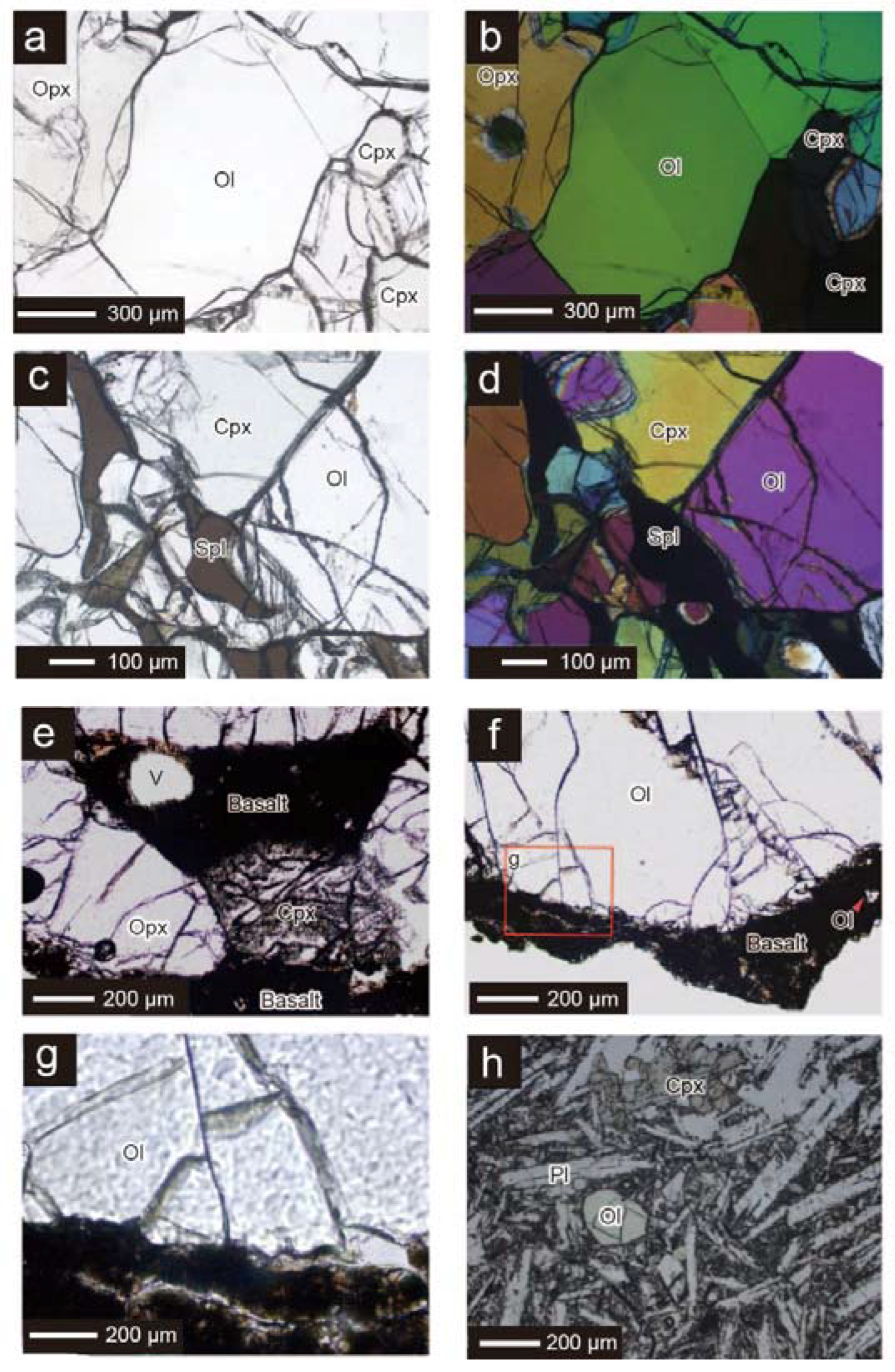
4.1. Petrography
4.2. Chemical Compositions of Constituent Minerals
4.3. Purity of the Samples Separated for Mössbauer Analysis
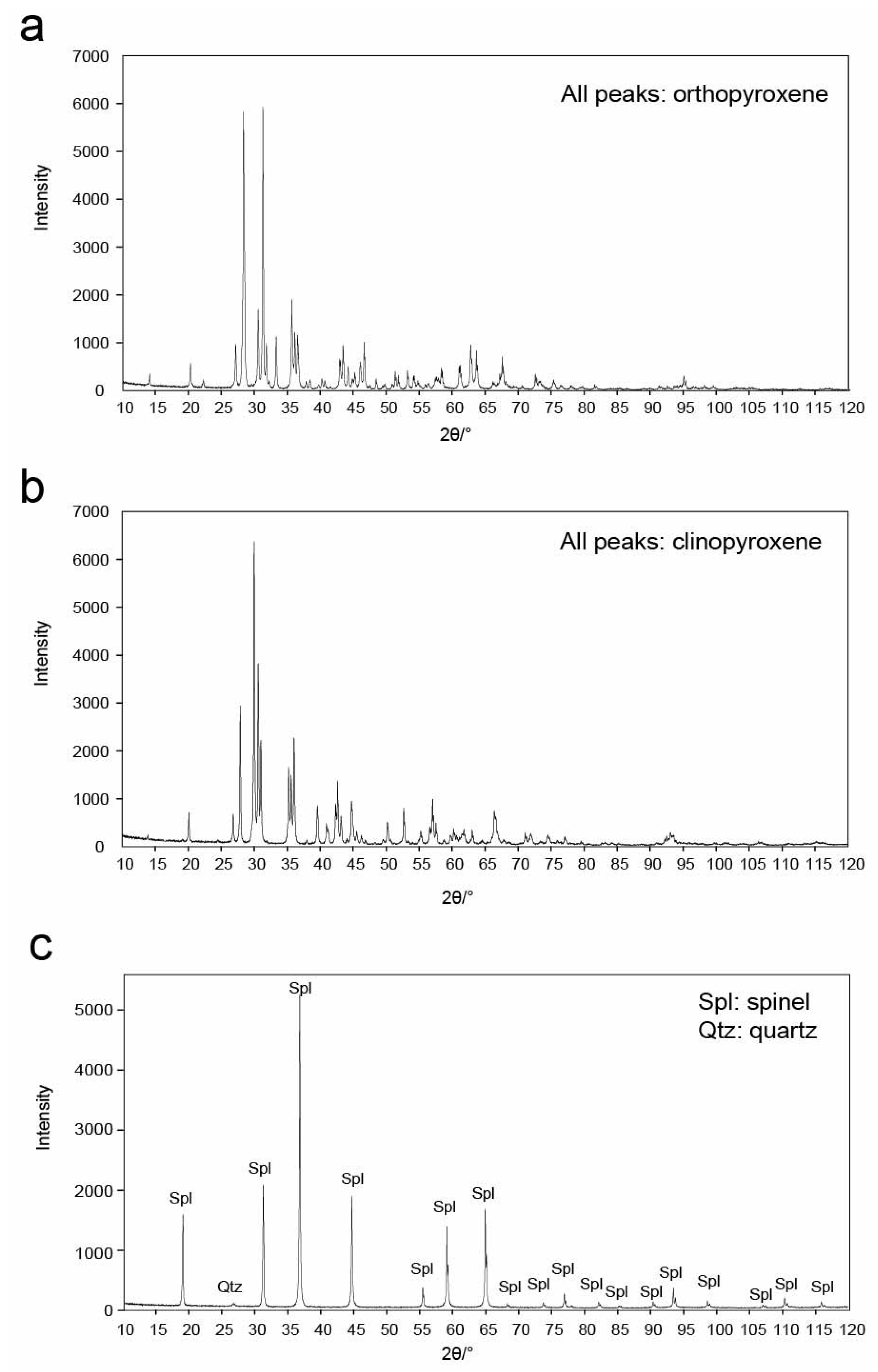
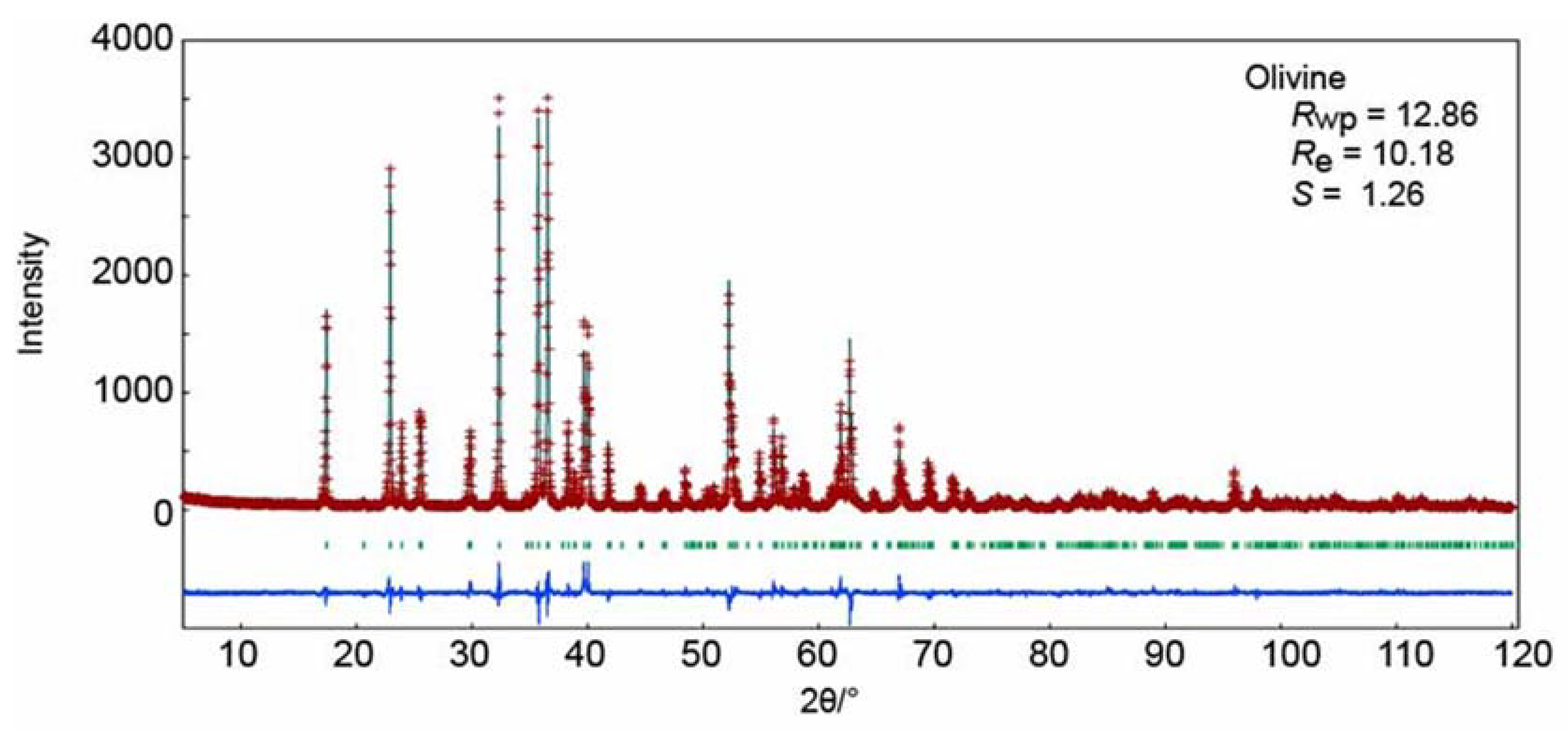
4.4. X-ray Powder Diffraction and X-ray Rietveld Analysis
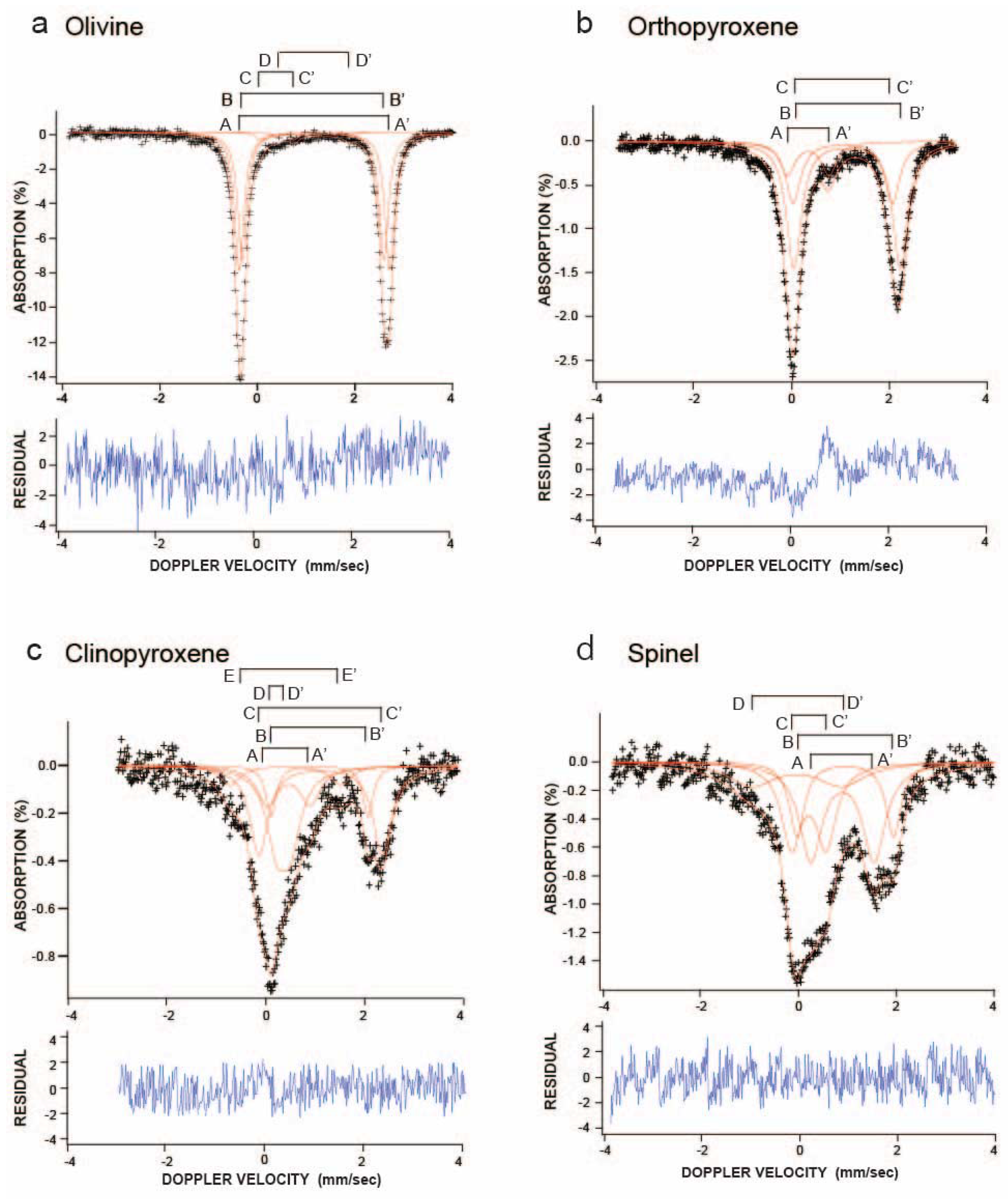
4.5. 57Fe Mössbauer Spectroscopic Analysis
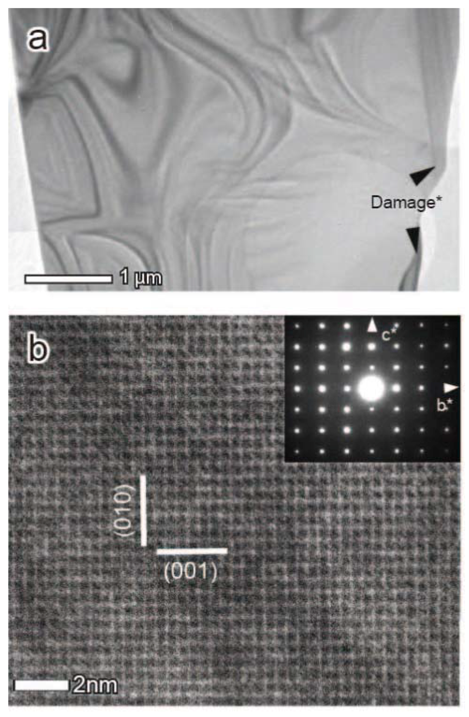
4.6. Raman Spectroscopy, Transmission Electron Microscopy, and Electron Diffraction Analysis
5. Discussion
5.1. Construction of Structural Formula of Olivine, Orthopyroxene, Clinopyroxene, and Spinel
5.2. Significance of Oxidation States of Fe in Olivine, Clinopyroxene, Orthopyroxene, and Spinel
5.2.1. Olivine
5.2.2. Clinopyroxene, Orthopyroxene, and Spinel
5.3. Evaluation of Redox Condition of the Source Area of the Tariat Spinel Lherzolite Xenoliths
Author Contributions
Acknowledgments
Conflicts of Interest
References
- Dyar, M.D.; McGuire, A.V. Redox equilibria and crystal chemistry of coexisting minerals from spinel lherzolite mantle xenoliths. Am. Mineral. 1989, 74, 969–980. [Google Scholar]
- Wood, B.J.; Virgo, D. Upper mantle oxidation state: Ferric iron contents of lherzolite spinels by 57Fe Mössbauer spectroscopy and resultant oxygen fugacities. Geochimica et Cosmochimica Acta 1989, 53, 1277–1291. [Google Scholar] [CrossRef]
- Canil, D.; Virgo, D.; Scarfe, C.M. Oxidation states of mantle xenoliths from British Columbia, Canada. Contrib. Mineral. Petrol. 1990, 104, 453–562. [Google Scholar] [CrossRef]
- Bryndzia, L.T.; Wood, B.J. Oxygen thermobarometry of abyssal spinel peridotites: The redox state and C-O-H volatile composition of the Earth’s suboceanic upper mantle. Am. J. Sci. 1990, 290, 1093–1116. [Google Scholar] [CrossRef]
- McGuire, A.V.; Dayr, M.D.; Nielson, J.E. Metasomatic oxidation of upper mantle peridotite. Contrib. Mineral. Petrol. 1991, 109, 252–264. [Google Scholar] [CrossRef]
- Banfield, J.F.; Dyar, M.; Mcguire, A.V. The defect microstructure of oxidized mantle olivine from Dish Hill, California. Am. Mineral. 1992, 77, 977–986. [Google Scholar]
- Dyar, M.D.; McGuire, A.V.; Harrell, M.D. Crystal chemistry of iron in the two styles of metasomatism in the upper mantle. Geochimica et Cosmochimica Acta 1992, 56, 2579–2586. [Google Scholar] [CrossRef]
- Canil, D.; O’Neill, H.S.C.; Pearson, D.G.; Rudnick, R.L.; McDonough, W.F.; Carswell, D.A. Ferric iron in peridotites and mantle oxidation states. Earth Planet. Sci. Lett. 1994, 123, 205–220. [Google Scholar] [CrossRef]
- Woodland, A.B.; Koch, M. Variation in oxygen fugacity with depth in the upper mantle beneath the Kaapvaal Craton, southern Africa. Earth Planet. Sci. Lett. 2003, 214, 295–310. [Google Scholar] [CrossRef]
- McCammon, C.A.; Frost, D.J.; Smyth, J.R.; Laustsen, H.M.S.; Kawamoto, T.; Ross, N.L.; van Aken, P.A. Oxidation state of iron in hydrous mantle phases: Implications for subduction and mantle oxygen fugacity. Phys. Earth Planet. Inter. 2004, 143, 157–169. [Google Scholar] [CrossRef]
- Woodland, A.B.; Kornprobst, J.; Tabit, A. Ferric iron in orogenic lherzolite massifs and controls of oxygen fugacity in the upper mantle. Lithos 2006, 89, 222–241. [Google Scholar] [CrossRef]
- McCammon, C.A. Microscopic to macroscopic behavior: The influence of iron electronic state. J. Mineral. Petrol. Sci. 2006, 101, 130–140. [Google Scholar] [CrossRef]
- Ejima, T.; Akasaka, M.; Ohfuji, H. Oxidation state of Fe in olivine in a lherzolite xenolith from Oku district, Oki-Dogo Island, Shimane Prefecture, Japan. J. Mineral. Petrol. Sci. 2011, 106, 246–254. [Google Scholar] [CrossRef]
- Hao, X.L.; Li, Y.L. 57Fe Mossbauer spectroscopy of mineral assemblage in mantle spinel lherzolites from Cenozoic alkali basalt, eastern China: Petrological applications. Lithos 2013, 156–159, 112–119. [Google Scholar] [CrossRef]
- Ballhaus, C.; Berry, R.F.; Green, D.H. High pressure experimental calibration of the olivine-orthopyroxene-spinel oxygen geobarometer: Implications for the oxidation state of the upper mantle. Contrib. Mineral. Petrol. 1991, 107, 27–40. [Google Scholar] [CrossRef]
- Forst, D.J.; McCammon, C.A. The Redox state of Earth’s Mantle. Annu. Rev. Earth Planet. Sci. 2008, 36, 389–420. [Google Scholar] [CrossRef]
- Ejima, T.; Akasaka, M.; Ohfuji, H. Ferric iron within olivine in a lherzolite xenolith from Oku district, Oki-Dogo Island, Shimane Prefecture, Japan. In Proceedings of the 21st General Meeting of the International Mineralogical Association, Johannesburg, South Africa, 1–5 September 2014. [Google Scholar]
- Stosch, H.G.; Lugmair, G.W.; Kovalenko, V.I. Spinel peridotite xenoliths from the Tariat Depression, Mongolia. II: Geochemistry and Nd and Sr isotopic composition and their implications for the evolution of the subcontinental lithosphere. Geochimica et Cosmochimica Acta 1986, 50, 2601–2614. [Google Scholar] [CrossRef]
- Preß, S.; Witt, H.; Seck, D.; Eonov, D.; Kovalenko, V.I. Spinel peridotite xenoliths from the Tariat Depression, Mongolia. I: Major element chemistry and mineralogy of a primitive mantle xenolith suite. Geochimica et Cosmochimica Acta 1986, 50, 2587–2599. [Google Scholar] [CrossRef]
- Osanai, Y.; Nakano, N.; Adachi, T.; Owada, M.; Satis-Kumar, M.; Jargalan, S.; Boldbaatar, C.; Yonemura, K.; Yoshimoto, A. Garnet, Clinopyroxene megacrysts and Garnet-bearing mantle xenoliths from the Tariat Depression, Mongolia. In Proceedings of the Abstracts with Programs for the 2011 Annual Meeting of the Mineralogical Society of Japan, the Mineralogical Society of Japan, Ibaraki, Japan, 9 September 2011; p. 102. [Google Scholar] [CrossRef]
- Irifune, T.; Isshiki, M.; Sakamoto, S. Transmission electron microscope observation of the high-pressure form of magnesite retrieved from laser heated diamond anvil cell. Earth Planet. Sci. Lett. 2005, 239, 98–105. [Google Scholar] [CrossRef]
- Izumi, F.; Momma, K. Three-dimensional visualization in powder diffraction. Solid State Phenom. 2007, 130, 15–20. [Google Scholar] [CrossRef]
- Dollase, W.A. Correction of intensities for preferred orientation in powder diffractometry: Application of the March model. J. Appl. Crystallogr. 1986, 19, 267–272. [Google Scholar] [CrossRef]
- Akasaka, M.; Shinno, I. Mössbauer spectroscopy and its recent application to silicate mineralogy. J. Mineral. Soc. Jpn. 1992, 21, 3–20, (In Japanese with English Abstract). [Google Scholar] [CrossRef]
- Yang, H.; Ghose, S. A transitional structural state and anomalous Fe-Mg order-disorder in Mg-rich orthopyroxene, (Mg0.75Fe0.25)2Si2O6. Am. Mineral. 1995, 80, 9–20. [Google Scholar] [CrossRef]
- Cameron, M.; Sueno, S.; Prewitt, C.T.; Papike, J.J. High-temperature crystal chemistry of acmite, diopside, jadeite, spodumene, and ureyite. Am. Mineral. 1973, 58, 594–618. [Google Scholar]
- Peterson, R.C.; Lager, G.A.; Hitterman, R.L. A time-of-flight neutron powder diffraction study of MgAl2O4 at temperatures up to 1273 K. Am. Mineral. 1991, 76, 1455–1458. [Google Scholar]
- Young, R.A. Introduction to the Rietveld method. In The Rietveld Method; Young, R.A., Ed.; Oxford Science Publications: Oxford, UK, 1993; pp. 1–38. ISBN 9780198559122. [Google Scholar]
- Shinno, I.; Hayashi, M.; Kuroda, Y. Mössbauer studies of olivines. Mineral. J. 1974, 7, 344–358. [Google Scholar] [CrossRef]
- Ejima, T.; Akasaka, M. Fe2+/Fe3+ ratios in olivine estimated using an Electron Microprobe Analyzer verified by 57Fe Mössbauer spectroscopy. Jpn. Mag. Mineral. Petrol. Sci. 2011, 40, 55–62. [Google Scholar] [CrossRef]
- Annersten, H.; Olesch, M.; Seifert, F.A. Ferric iron in orthopyroxene: A Mössbauer spectroscopic study. Lithos 1987, 11, 301–310. [Google Scholar] [CrossRef]
- Akasaka, M. 57Fe Mössbauer study of clinopyroxenes in the join CaFe3+AlSiO6-CaTiAl2O6. Phys. Chem. Miner. 1983, 9, 205–211. [Google Scholar] [CrossRef]
- Akasaka, M. Clinopyroxene on the join CaMgSi2O6-CaFe3+AlSiO6-CaTiAl2O6 at low oxygen fugacity. Proc. Indian Acad. Sci. (Earth Planet. Sci.) 1990, 99, 39–48. [Google Scholar] [CrossRef]
- Dowty, E.; Lindsley, D.H. Mössbauer spectra of synthetic hedenbergite-ferrosilite pyroxenes. Am. Mineral. 1973, 58, 850–868. [Google Scholar]
- Williams, P.G.L.; Bancroft, G.M.; Bown, M.G.; Turnock, A.C. Anomalous Mössbauer Spectra of C2/c clinopyroxenes. Nat. Phys. Sci. 1971, 230, 149–151. [Google Scholar] [CrossRef]
- Osborne, M.D.; Fleet, M.E.; Bancroft, G.M. Fe2+-Fe3+ ordering in chromite and Cr-bearing spinels. Contrib. Mineral. Petrol. 1981, 77, 251–255. [Google Scholar] [CrossRef]
- Malysheva, T.V.; Polyakova, N.P.; Mishin, N.E. Mössbauer spectroscopy study of lunar soil sampled by Luna 24 space probe. Geokhimiya 1978, 6, 835–841. [Google Scholar]
- Warenborgh, J.C.; Annersten, H.; Ericsson, T.; Figueiredo, M.O.; Cabral, J.M.P.A. A Mössbauer study of natural gahnite spinels showing strongly temperature-dependent quadrupole splitting distributions. Eur. J. Mineral. 1990, 2, 267–271. [Google Scholar] [CrossRef]
- Ottonello, G.; Princifalle, F.; Della Giusta, A. Temperature, composition, and fO2 effects on intersite distribution of Mg and Fe2+ in olivines. Phys. Chem. Miner. 1990, 17, 301–312. [Google Scholar] [CrossRef]
- Nord, A.G.; Annersten, H.; Filippidis, A. The cation distribution in synthetic Mg-Fe-Ni olivines. Am. Mineral. 1982, 67, 1206–1211. [Google Scholar]
- Nestola, F.; Ballaran, T.B.; Balic-Zunic, T.; Secco, L.; Dal Negro, A. High-pressure behavior of an Al- and Fe-rich natural orthopyroxene. Am. Mineral. 2008, 93, 644–652. [Google Scholar] [CrossRef]
- Banfield, J.F.; Veblen, D.R.; Jones, B.F. Transmission electron microscopy of subsolidus oxidation and weathering of olivine. Contrib. Mineral. Petrol. 1990, 106, 110–123. [Google Scholar] [CrossRef]
- Ashworth, J.R.; Chambers, A.D. Symplectic reaction in olivine and the controls of intergrowth spacing in symplectites. J. Petrol. 2000, 41, 285–304. [Google Scholar] [CrossRef]
- Morioka, M.; Nagasawa, H. Ionic diffusion in olivine. In Diffusion, Atomic Ordering and Mass Transport: Selected Topics in Geochemistry; Ganguly, J., Ed.; Springer: Berlin, Germany; New York, NY, USA, 1991; Volume 8, pp. 176–197. ISBN 978-1-4613-9021-3. [Google Scholar]
- Mackwell, S.J. Oxidation kinetics of fayalite (Fe2SiO4). Phys. Chem. Miner. 1992, 19, 220–228. [Google Scholar] [CrossRef]
- Ejima, T.; Akasaka, M.; Nagao, T.; Ohfuji, H. Oxidation state of Fe in olivine in andesitic scoria from Kasayama volcano, Hagi, Yamaguchi Prefecture, Japan. J. Mineral. Petrol. Sci. 2012, 107, 215–225. [Google Scholar] [CrossRef]
- Ejima, T.; Akasaka, M.; Nagao, T.; Ohfuji, H. Oxidation states of Fe and precipitates within olivine from orthopyroxene-olivine-clinopyroxene andesite lava from Kasayama volcano, Hagi, Yamaguchi, Japan. J. Mineral. Petrol. Sci. 2013, 108, 25–36. [Google Scholar] [CrossRef]
- Ejima, T.; Akasaka, M.; Nagao, T.; Ohfuji, H. Occurrence of Fe3+ and formation process of precipitates within oxidized olivine phenocrysts in basalt lava from Kuroshima volcano, Goto Islands, Nagasaki, Japan. Mineral. Mag. 2015, 79, 1833–1848. [Google Scholar] [CrossRef]
- Ejima, T. Oxidation state of Fe within olivine phenocryst in Kami-Kometsuka scoria, Kami-Kometsuka, Aso, Kumamoto Prefecture, Japan: Effect of high temperature oxidation on the scoria. Jpn. Mag. Mineral. Petrol. Sci. 2015, 44, 323–328, (In Japanese with English Abstract). [Google Scholar] [CrossRef]
- Hwang, S.L.; Yui, T.F.; Chu, H.T.; Shen, P.; Lizuka, Y.; Yang, H.Y.; Yang, J.; Xu, Z. Hematite and magnetite precipitates in olivine from the Sulu peridotite: A result of dehydrogenation-oxidation reaction of mantle olivine? Am. Mineral. 2008, 93, 1051–1060. [Google Scholar] [CrossRef]
- Ingrin, J.; Skogby, H. Hydrogen in nominally anhydrous upper-mantle minerals: Concentration levels and implications. Eur. J. Mineral. 2000, 12, 543–570. [Google Scholar] [CrossRef]
- Perinelli, C.; Andreozzi, G.B.; Conte, A.M.; Oberti, R.; Armienti, P. Redox state of subcontinental lithospheric mantle and relationships with metasomatism: Insights from spinel peridotites from northern Victoria Land (Antarctica). Contrib. Mineral. Petrol. 2012, 164, 1053–1067. [Google Scholar] [CrossRef]
- Harris, N.; Hunt, A.; Parkinson, I.; Tindle, A.; Yondon, M.; Hammond, S. Tectonic implications of garnet-bearing mantle xenoliths exhumed by Quaternary magmatism in the Hangay dome, central Mongolia. Contrib. Mineral. Petrol. 2010, 160, 67–81. [Google Scholar] [CrossRef]


| Xenolith | Basalt | |||||||||||
|---|---|---|---|---|---|---|---|---|---|---|---|---|
| Olivine | Spinel | Orthopyroxene | Clinopyroxene | Olivine | Clinopyroxene | |||||||
| Av. (n = 48) * | s.d. * | Av. (n = 36) * | s.d. * | Av. (n = 22) * | s.d. * | Av. (n = 18) * | s.d. * | Av. (n = 18) * | s.d. * | Av. (n = 23) * | s.d. * | |
| SiO2 | 40.84 | 0.20 | - | - | 55.06 | 0.33 | 52.02 | 0.38 | 38.38 | 0.59 | 50.81 | 1.17 |
| TiO2 | - | - | 0.13 | 0.02 | 0.12 | 0.02 | 0.61 | 0.05 | - | - | 1.75 | 0.57 |
| Al2O3 | - | - | 57.54 | 0.29 | 4.67 | 0.08 | 7.32 | 0.20 | - | - | 3.23 | 1.14 |
| Cr2O3 | - | - | 11.52 | 0.20 | 0.40 | 0.03 | 0.97 | 0.07 | - | - | - | - |
| FeO ** | 9.53 | 0.14 | 9.83 | 0.12 | 5.97 | 0.09 | 2.69 | 0.13 | 23.76 | 2.80 | 7.37 | 0.75 |
| MnO | 0.14 | 0.03 | - | - | 0.16 | 0.05 | 0.09 | 0.04 | 0.32 | 0.06 | 0.14 | 0.04 |
| NiO | 0.19 | 0.02 | 0.19 | 0.02 | 0.05 | 0.01 | - | - | 0.05 | 0.03 | - | - |
| MgO | 49.30 | 0.28 | 20.79 | 0.48 | 32.56 | 0.54 | 14.77 | 0.14 | 37.62 | 2.37 | 14.40 | 0.85 |
| CaO | 0.05 | 0.01 | - | - | 0.66 | 0.04 | 18.51 | 0.61 | 0.30 | 0.05 | 21.67 | 0.56 |
| Na2O | - | - | - | - | 0.19 | 0.03 | 2.53 | 0.20 | - | - | 0.51 | 0.06 |
| Total | 100.03 | 100.00 | 99.83 | 99.50 | 100.54 | 99.98 | ||||||
| Number of Cations on the Basis of 4 Oxygens | Number of Cations on the Basis of 6 Oxygens | O = 4 | O = 6 | |||||||||
| Si | 0.999 | 0.004 | - | - | 1.902 | 0.012 | 1.884 | 0.010 | 1.000 | 0.005 | 1.887 | 0.042 |
| Ti | - | - | 0.003 | 0.000 | 0.003 | 0.001 | 0.017 | 0.001 | - | 0.049 | 0.016 | |
| Al | - | - | 1.751 | 0.006 | 0.190 | 0.003 | 0.312 | 0.009 | - | - | 0.142 | 0.050 |
| Cr | - | - | 0.235 | 0.005 | 0.011 | 0.001 | 0.028 | 0.002 | - | - | - | - |
| Fe2+ | 0.195 | 0.003 | 0.212 | 0.003 | 0.172 | 0.003 | 0.082 | 0.004 | 0.519 | 0.068 | 0.229 | 0.025 |
| Mn | 0.003 | 0.001 | - | - | 0.005 | 0.001 | 0.003 | 0.001 | 0.007 | 0.001 | 0.004 | 0.001 |
| Ni | 0.004 | 0.000 | 0.004 | 0.000 | 0.001 | 0.000 | - | - | 0.001 | 0.001 | - | - |
| Mg | 1.798 | 0.007 | 0.800 | 0.014 | 1.677 | 0.024 | 0.797 | 0.007 | 1.460 | 0.071 | 0.796 | 0.041 |
| Ca | 0.001 | 0.000 | - | - | 0.024 | 0.002 | 0.718 | 0.025 | 0.009 | 0.002 | 0.863 | 0.026 |
| Na | - | - | - | - | 0.013 | 0.002 | 0.178 | 0.014 | - | - | 0.037 | 0.004 |
| Total | 2.999 | 3.005 | 4.000 | 4.018 | 2.999 | 4.011 | ||||||
| Fo mol % | 90 | |||||||||||
| Olivine | |
|---|---|
| Radiation | CuKα (λ = 1.541769 Å) |
| Monochromator | Graphite |
| 2θ scan range (°) | 5.00–120 |
| Step interval (°2θ) | 0.02 |
| Max. intensity (counts) | 5750 |
| No. of phases refined | 1 |
| Space groupe | Pbnm |
| a (Å) | 4.7631(1) |
| b (Å) | 10.2284(1) |
| c (Å) | 5.9941(1) |
| V (Å3) | 292.022(7) |
| Z | 4 |
| Dcalc (g/cm3) | 3.32 |
| Rp (%) * | 9.55 |
| Rwp (%) * | 12.86 |
| Re (%) * | 10.18 |
| S * | 1.26 |
| Olivine | ||||||
|---|---|---|---|---|---|---|
| Site | n ** | g | x | y | z | Beq |
| M1 | 4 | Mg0.902(4)Fe0.098 | 0 | 0 | 0 | 1.58(5) |
| M2 | 4 | Mg0.926(4)Fe0.074 | 0.9860(4) | 0.2784(1) | 1/4 | 1.58(6) |
| T | 4 | 1.0Si | 0.4288(4) | 0.0949(1) | 1/4 | 1.77(5) |
| O1 | 4 | 1 | 0.7636(7) | 0.0927(3) | 1/4 | 1.8(1) |
| O2 | 4 | 1 | 0.2159(7) | 0.4482(4) | 1/4 | 1.12(9) |
| O3 | 8 | 1 | 0.2767(5) | 0.1627(2) | 0.0325(5) | 1.81(8) |
| Mineral | Doublets | I.S. * (mm/s) | Q.S * (mm/s) | FWHH * (mm/s) | Area Ratio (%) | Assignments | χ2/Freedom | Fe2+:Fe3+ |
|---|---|---|---|---|---|---|---|---|
| Olivine | AA′ | 1.165(2) | 3.081(3) | 0.228(3) | 49.8(14) | M2Fe2+ in olivine | 1.65 | 97.3(14):2.7(2) ** |
| BB′ | 1.141(2) | 2.899(4) | 0.228(3) | 46.1(14) | M1Fe2+ in olivine | |||
| CC′ | 0.40(2) | 0.69(3) | 0.228(3) | 2.7(2) | M2Fe3+ in olivine | |||
| DD′ | 1.13(3) | 1.48(6) | 0.228(3) | 1.4(2) | Fe2+ in spinel | |||
| Orthopyroxene | AA′ | 0.39(2) | 0.86(3) | 0.38(1) | 15(1) | M1Fe3+ | 1.14 | 85(8):15(1) |
| BB′ | 1.19(3) | 2.21(5) | 0.38(1) | 27(6) | M1Fe2+ | |||
| CC′ | 1.10(5) | 2.05(10) | 0.38(1) | 58(6) | M2Fe2+ | |||
| Clinopyroxene | AA′ | 0.43(1) | 0.88(2) | 0.51(4) | 16(2) | M1Fe3+ | 1.21 | 74(4):26(3) *** |
| BB′ | 1.06(1) | 2.00(2) | 0.25(4) | 11(2) | M2Fe2+ | |||
| CC′ | 1.09(1) | 2.54(2) | 0.45(3) | 34(3) | M1Fe2+ | |||
| DD′ | 0.32(1) | 0.28(2) | 0.51(4) | 30(3) | Unidentified | |||
| EE′ | 0.36(1) | 2.28(2) | 0.51(4) | 8(1) | Unidentified | |||
| Spinel | AA′ | 0.88(2) | 1.30(3) | 0.57(9) | 36(3) | Fe2+ | 1.41 | 66(6):34(5) *** |
| BB′ | 0.94(2) | 1.99(4) | 0.37(5) | 18(7) | Fe2+ | |||
| CC′ | 0.19(1) | 0.71(3) | 0.50(4) | 28(5) | Fe3+ | |||
| DD′ | −0.1(2) | 1.9(3) | 1.2(2) | 18(4) | Unidentified |
| Reference | Occarrence | Rock Type | Fe3+/ΣFe | |||
|---|---|---|---|---|---|---|
| Olivine | Clinopyroxene | Orthopyroxene | Spinel | |||
| Wood and Virgo [2] | Kilbourme Hole, New Mexico | Spinel-lherzolite xenolith | - | - | - | 0.18–0.25 |
| Wood and Virgo [2] | Massif Central, France | Spinel-lherzolite xenolith | - | - | - | 0.25–0.28 |
| Wood and Virgo [2] | Ichinomegata, Japan | Spinel-lherzolite xenolith | - | - | - | 0.26–0.28 |
| Wood and Virgo [2] | Tariat, Mongolia | Spinel-lherzolite xenolith | - | - | - | 0.16–0.24 |
| Wood and Virgo [2] | Vitim Plateau, Mongolia | Spinel-lherzolite xenolith | - | - | - | 0.15–0.27 |
| Canil et al. [3] | Southeastern Australia | Spinel-lherzolite xenolith | 0 | 0.16–0.19 | 0.06 | 0.15–0.22 |
| Canil et al. [3] | Massif Central, France | Spinel-lherzolite xenolith | 0 | 0.19 | 0.05 | 0.26 |
| Woodland et al. [11] | Beni Bousera, Morocco | Orogenic lherzolite massifs | 0 | 0.03–0.14 | 0.02–0.06 | 0.02–0.13 |
| Woodland et al. [14] | Ronda, Spain | Orogenic lherzolite massifs | 0 | 0.06–0.19 | 0.04–0.08 | 0.08–0.26 |
| Woodland et al. [11] | Pyrenees, France | Orogenic lherzolite massifs | 0 | 0.19–0.32 | 0.05–0.07 | 0.10–0.27 |
| Perinelli et al. [52] | Northern Victoria Land, Antarctica | Spinel lherzolite xenolith | - | - | - | 0.17–0.23 |
| Hao and Li [14] | Eastern China | Spinel lherzolite xenolith | 0 | 0.22–0.31 | 0.05–0.13 | 0.14–0.23 |
| This study | Tariat, Mongolia | Spinel lherzolite xenolith | 0.03 | 0.26 | 0.15 | 0.34 |
© 2018 by the authors. Licensee MDPI, Basel, Switzerland. This article is an open access article distributed under the terms and conditions of the Creative Commons Attribution (CC BY) license (http://creativecommons.org/licenses/by/4.0/).
Share and Cite
Ejima, T.; Osanai, Y.; Akasaka, M.; Adachi, T.; Nakano, N.; Kon, Y.; Ohfuji, H.; Sereenen, J. Oxidation States of Fe in Constituent Minerals of a Spinel Lherzolite Xenolith from the Tariat Depression, Mongolia: The Significance of Fe3+ in Olivine. Minerals 2018, 8, 204. https://doi.org/10.3390/min8050204
Ejima T, Osanai Y, Akasaka M, Adachi T, Nakano N, Kon Y, Ohfuji H, Sereenen J. Oxidation States of Fe in Constituent Minerals of a Spinel Lherzolite Xenolith from the Tariat Depression, Mongolia: The Significance of Fe3+ in Olivine. Minerals. 2018; 8(5):204. https://doi.org/10.3390/min8050204
Chicago/Turabian StyleEjima, Terumi, Yasuhito Osanai, Masahide Akasaka, Tatsuro Adachi, Nobuhiko Nakano, Yoshiaki Kon, Hiroaki Ohfuji, and Jargalan Sereenen. 2018. "Oxidation States of Fe in Constituent Minerals of a Spinel Lherzolite Xenolith from the Tariat Depression, Mongolia: The Significance of Fe3+ in Olivine" Minerals 8, no. 5: 204. https://doi.org/10.3390/min8050204