
The Influence of Blood and Serum Microenvironment on Spin-Labeled Magnetic Nanoparticles


Abstract
1. Introduction
2. Materials and Methods
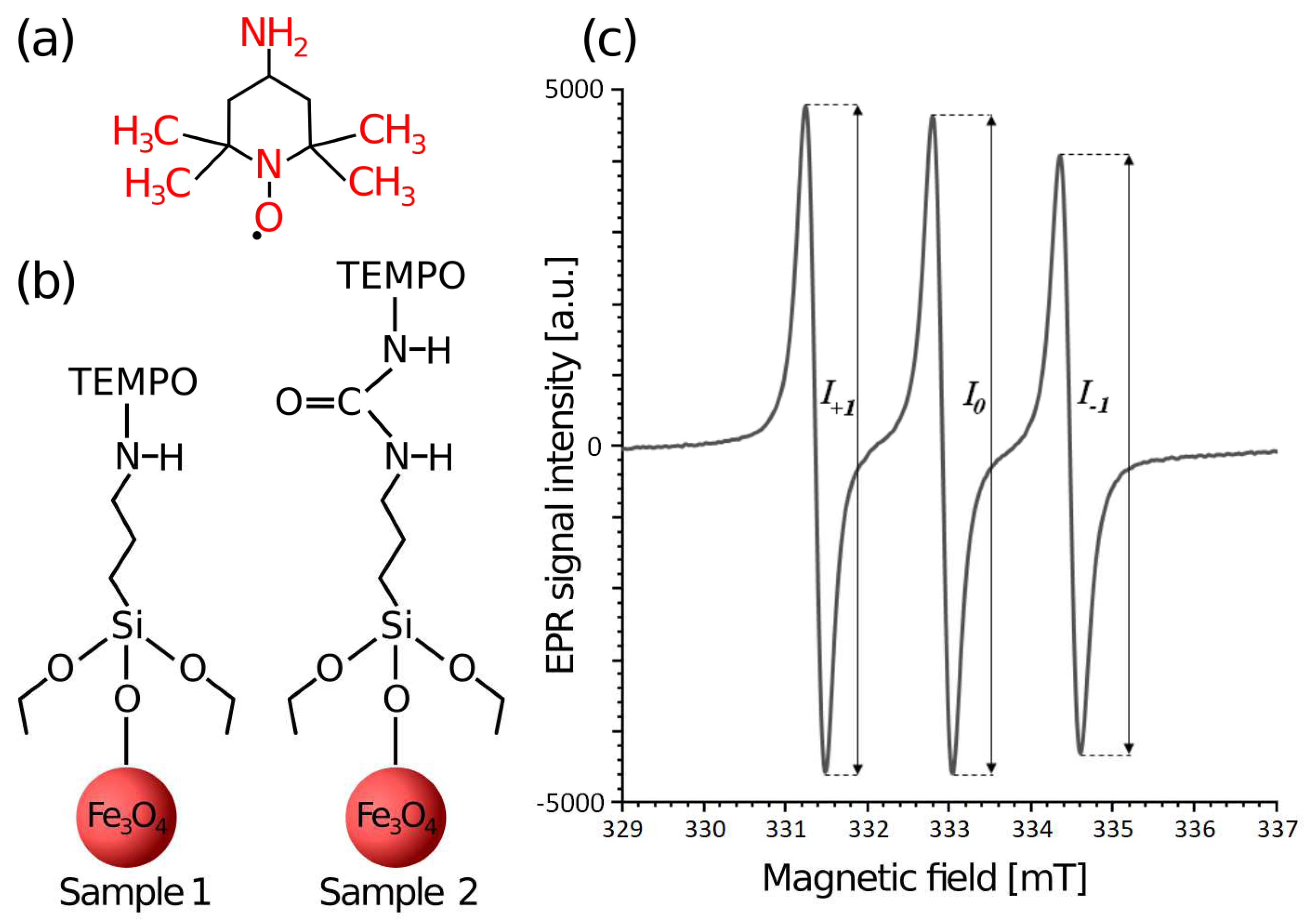
2.1. Experimental Section
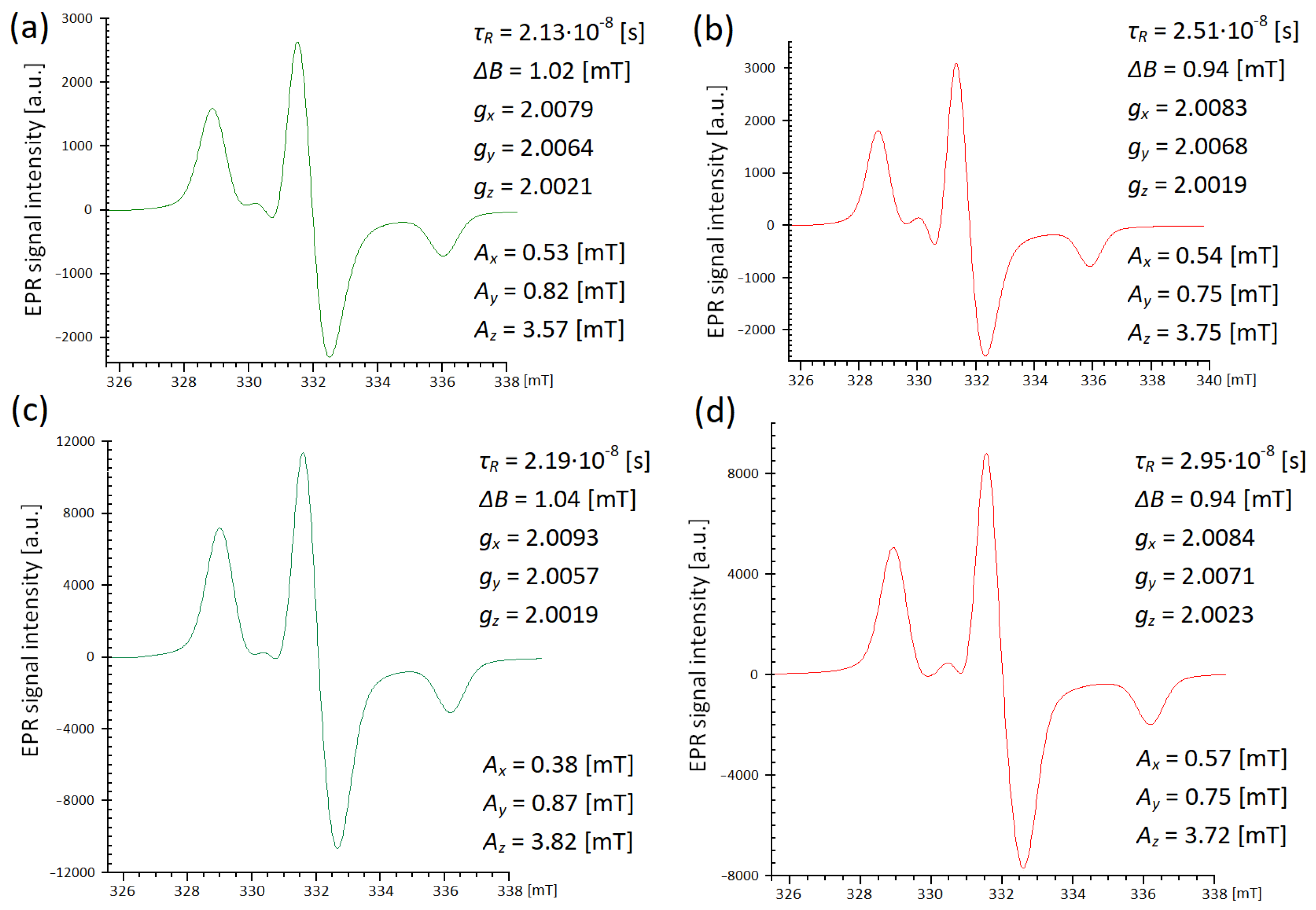
2.2. Analysis of Spectra and Simulations
3. Results
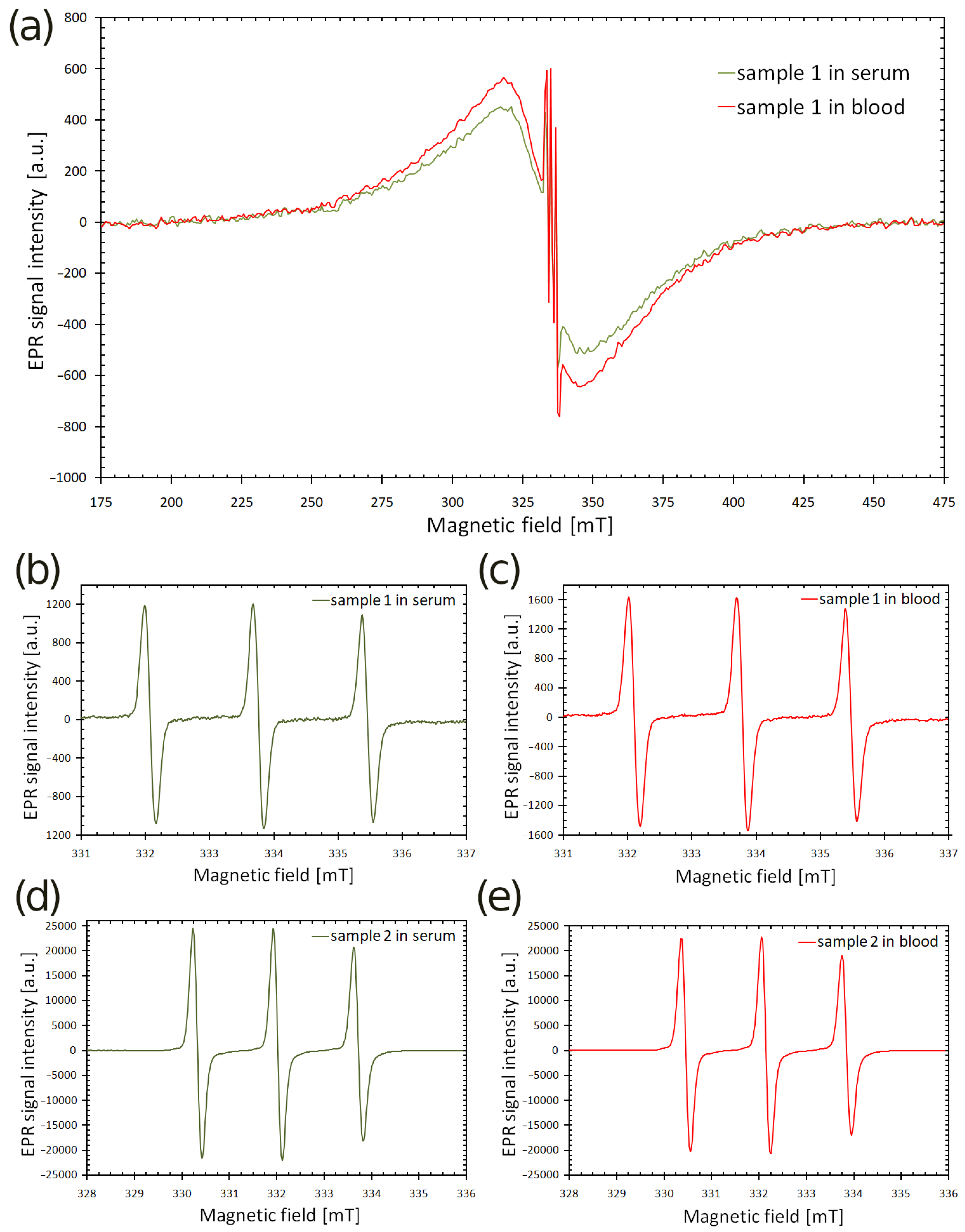
3.1. EPR Spectra at Room Temperature
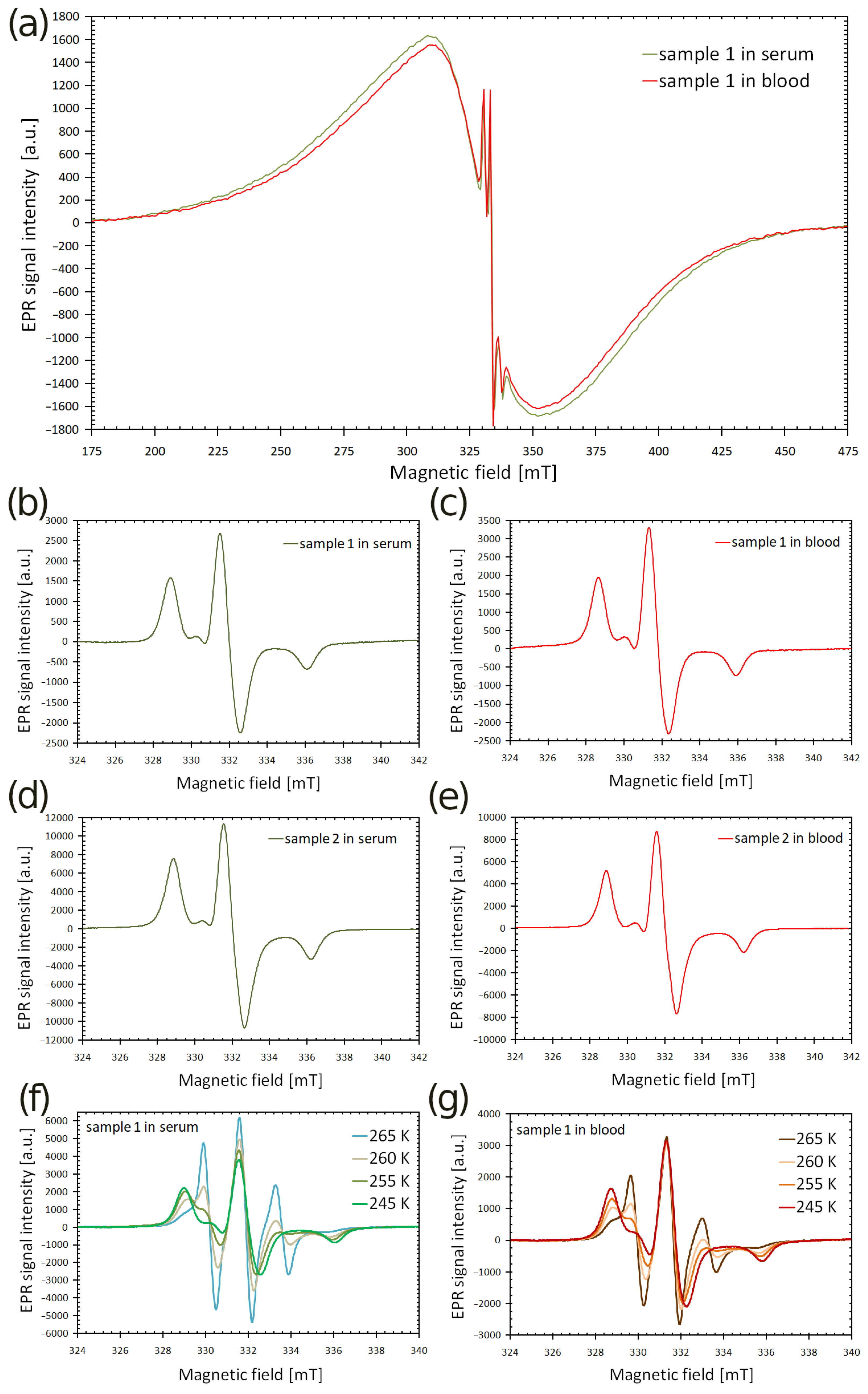
3.2. EPR Spectra of Frozen Samples
4. Discussion
5. Conclusions
Funding
Institutional Review Board Statement
Informed Consent Statement
Data Availability Statement
Acknowledgments
Conflicts of Interest
References
- Oberdick, S.D.; Jordanova, K.V.; Lundstrom, J.T.; Parigi, G.; Poorman, M.E.; Zabow, G.; Keenan, K.E. Iron oxide nanoparticles as positive T1 contrast agents for low-field magnetic resonance imaging at 64 mT. Sci. Rep. 2023, 13, 11520. [Google Scholar] [CrossRef] [Scilit]
- Palzer, J.; Eckstein, L.; Slabu, I.; Reisen, O.; Neumann, U.P.; Roeth, A.A. Iron Oxide Nanoparticle-Based Hyperthermia as a Treatment Option in Various Gastrointestinal Malignancies. Nanomaterials 2021, 11, 3013. [Google Scholar] [CrossRef] [Scilit]
- Gupta, A.K.; Gupta, M. Synthesis and surface engineering of iron oxide nanoparticles for biomedical applications. Biomaterials 2005, 26, 3995–4021. [Google Scholar] [CrossRef] [Scilit] [PubMed]
- Xu, H.; Aguilar, Z.P.; Yang, L.; Kuang, M.; Duan, H.; Xiong, Y.; Wei, H.; Wang, A. Antibody conjugated magnetic iron oxide nanoparticles for cancer cell separation in fresh whole blood. Biomaterials 2011, 32, 9758–9765. [Google Scholar] [CrossRef] [Scilit]
- Bielas, R.; Kubiak, T.; Kopčanský, P.; Šafařík, I.; Józefczak, A. Tunable particle shells of thermo-responsive liquid marbles under alternating magnetic field. J. Mol. Liq. 2023, 391, 123283. [Google Scholar] [CrossRef] [Scilit]
- Kubiak, T. Polymeric capsules and micelles as promising carriers of anticancer drugs. Polim. Med. 2022, 52, 37–50. [Google Scholar] [CrossRef] [Scilit] [PubMed]
- Mykhaylyk, O.; Dudchenko, N.; Dudchenko, A. Doxorubicin magnetic conjugate targeting upon intravenous injection into mice: High gradient magnetic field inhibits the clearance of nanoparticles from the blood. J. Magn. Magn. Mater. 2005, 293, 473–482. [Google Scholar] [CrossRef] [Scilit]
- Predoi, S.-A.; Iconaru, S.L.; Predoi, D. In Vitro and In Vivo Biological Assays of Dextran Coated Iron Oxide Aqueous Magnetic Fluids. Pharmaceutics 2023, 15, 177. [Google Scholar] [CrossRef] [Scilit] [PubMed]
- Fischer, H.; Luster, J.; Gehring, A.U. EPR evidence for maghemitization of magnetite in a tropical soil. Geophys. J. Int. 2007, 169, 909–916. [Google Scholar] [CrossRef] [Scilit]
- Dobosz, B.; Krzyminiewski, R.; Kurczewska, J.; Schroeder, G. The influence of surface modification, coating agents and pH value of aqueous solutions on physical properties of magnetite nanoparticles investigated by ESR method. J. Magn. Magn. Mater. 2017, 429, 203–210. [Google Scholar] [CrossRef] [Scilit]
- Kubiak, T.; Krzyminiewski, R.; Dobosz, B.; Schroeder, G.; Kurczewska, J.; Hałupka-Bryl, M. A study of magnetite nanoparticles in whole human blood by means of electron paramagnetic resonance. Acta Bio-Opt. Inf. Med. Biomed. Eng. 2015, 21, 9–15. [Google Scholar]
- Krzyminiewski, R.; Kubiak, T.; Dobosz, B.; Schroeder, G.; Kurczewska, J. EPR spectroscopy and imaging of TEMPO-labeled magnetite nanoparticles. Curr. Appl. Phys. 2014, 14, 798–804. [Google Scholar] [CrossRef] [Scilit]
- Bielas, R.; Kubiak, T.; Molcan, M.; Dobosz, B.; Rajnak, M.; Józefczak, A. Biocompatible Hydrogel-Based Liquid Marbles with Magnetosomes. Materials 2024, 17, 99. [Google Scholar] [CrossRef] [Scilit] [PubMed]
- Tudose, M.; Constantinescu, T.; Balaban, A.T.; Ionita, P. Synthesis and electron paramagnetic resonance study of a nitroxide free radical covalently bonded on aminopropyl-silica gel. Appl. Surf. Sci. 2008, 254, 1904–1908. [Google Scholar] [CrossRef] [Scilit]
- Torricella, F.; Pierro, A.; Mileo, E.; Belle, V.; Bonucci, A. Nitroxide spin labels and EPR spectroscopy: A powerful association for protein dynamics studies. Biochim. Biophys. ActaProteins Proteom. 2021, 1869, 140653. [Google Scholar] [CrossRef] [Scilit] [PubMed]
- Kovarski, A.L.; Sorokina, O.N. Study of local magnetic fields and magnetic ordering in fluid and solid matrices containing magnetite nanoparticles using TEMPOL stable radical. J. Magn. Magn. Mater. 2007, 311, 155–161. [Google Scholar] [CrossRef] [Scilit]
- Morais, P.C.; Alonso, A.; Silva, O.; Buske, N. Electron paramagnetic resonance of nitroxide-doped magnetic fluids. J. Magn. Magn. Mater. 2002, 252, 53–55. [Google Scholar] [CrossRef] [Scilit]
- Zhao, H.; Zhang, Z.; Zhao, Z.; Yu, R.; Wan, Y.; Lan, M. Preparation and characterization of novel spin labeled magnetic nanoparticles. Adv. Mat. Lett. 2011, 2, 172–175. [Google Scholar] [CrossRef] [Scilit]
- Chechik, V.; Wellsted, H.J.; Korte, A.; Gilbert, B.C.; Caldararu, H.; Ionita, P.; Caragheorgheopol, A. Spin-labelled Au nanoparticles. Farad. Disc. 2004, 125, 279–291. [Google Scholar]
- Krzyminiewski, R.; Dobosz, B.; Schroeder, G.; Kurczewska, J. ESR as a monitoring method of the interactions between TEMPO-functionalized magnetic nanoparticles and yeast cells. Sci. Rep. 2019, 9, 18733. [Google Scholar]
- Wu, W.; He, Q.; Jiang, C. Magnetic Iron Oxide Nanoparticles: Synthesis and Surface Functionalization Strategies. Nanoscale Res. Lett. 2008, 3, 397–415. [Google Scholar] [CrossRef] [Scilit] [PubMed]
- Stoll, S.; Schweiger, A. Easy spin: Simulating cw ESR spectra. In ESR Spectroscopy in Membrane Biophysics; Hemminga, M.A., Berliner, L.J., Eds.; Springer: New York, NY, USA, 2007; Volume 27, pp. 299–321. [Google Scholar]
- Pucci, C.; Degl’Innocenti, A.; BelenliGümüş, M.; Ciofani, G. Superparamagnetic iron oxide nanoparticles for magnetic hyperthermia: Recent advancements, molecular effects, and future directions in the omics era. Biomater. Sci. 2022, 10, 2103–2121. [Google Scholar] [CrossRef] [Scilit] [PubMed]
- Kliava, J.; Berger, R. Size and shape distribution of magnetic nanoparticles in disordered systems: Computer simulations of superparamagnetic resonance spectra. J. Magn. Magn. Mater. 1999, 205, 328–342. [Google Scholar] [CrossRef] [Scilit]
- Vázquez-Victorio, G.; Acevedo-Salas, U.; Valenzuela, R. Microwave Absorption in Nanostructured Spinel Ferrites. In Ferromagnetic Resonance—Theory and Applications; Yalcina, O., Ed.; InTech: Rijeka, Croatia, 2013; pp. 169–194. [Google Scholar]
- Flores-Arias, Y.; Vazquez-Victorio, G.; Ortega-Zempoalteca, R.; Acevedo-Salas, U.; Ammar, S.; Valenzuela, R. Magnetic phase transitions in ferrite nanoparticles characterized by electron spin resonance. J. Appl. Phys. 2015, 117, 17A503. [Google Scholar]
- Tran, H.B.; Matsushita, Y. Temperature and size dependence of energy barrier for magnetic flips in L10FePt nanoparticles: A theoretical study. Scr. Mater. 2024, 242, 115947. [Google Scholar]
- García-Acevedo, P.; González-Gómez, M.A.; Arnosa-Prieto, Á.; de Castro-Alves, L.; Piñeiro, Y.; Rivas, J. Role of Dipolar Interactions on the Determination of the Effective Magnetic Anisotropy in Iron Oxide Nanoparticles. Adv. Sci. 2023, 10, 2203397. [Google Scholar] [CrossRef] [Scilit] [PubMed]
- Sadat, M.E.; Bud’ko, S.L.; Ewing, R.C.; Xu, H.; Pauletti, G.M.; Mast, D.B.; Shi, D. Effect of Dipole Interactions on Blocking Temperature and Relaxation Dynamics of Superparamagnetic Iron-Oxide (Fe3O4) Nanoparticle Systems. Materials 2023, 16, 496. [Google Scholar] [CrossRef] [Scilit] [PubMed]
- Nader, E.; Skinner, S.; Romana, M.; Fort, R.; Lemonne, N.; Guillot, N.; Gauthier, A.; Antoine-Jonville, S.; Renoux, C.; Hardy-Dessources, M.D.; et al. Blood Rheology: Key Parameters, Impact on Blood Flow, Role in Sickle Cell Disease and Effects of Exercise. Front. Physiol. 2019, 10, 1329. [Google Scholar] [CrossRef] [Scilit]
- Santangelo, M.G.; Levantino, M.; Cupane, A.; Jeschke, G. Solvation of a Probe molecule by fluid supercooled water in a hydrogel at 200 K. J. Phys. Chem. B 2008, 112, 15546–15553. [Google Scholar] [CrossRef] [Scilit]



| Sample | g | Aizo (mT) | τR (s) | ε |
|---|---|---|---|---|
| sample 1 in serum | 2.0058 (±0.0002) | 1.691 (±0.014) | 5.2·10−11 (±0.5·10−11) | 0.32 (±0.03) |
| sample 1 in blood | 2.0059 (±0.0002) | 1.681 (±0.014) | 6.3·10−11 (±0.6·10−11) | 0.18 (±0.02) |
| sample 2 in serum | 2.0059 (±0.0002) | 1.681 (±0.014) | 1.13·10−10 (±0.18·10−10) | 0.057 (±0.007) |
| sample 2 in blood | 2.0058 (±0.0002) | 1.701 (±0.014) | 1.35·10−10 (±0.20·10−10) | 0.068 (±0.008) |
Disclaimer/Publisher’s Note: The statements, opinions and data contained in all publications are solely those of the individual author(s) and contributor(s) and not of MDPI and/or the editor(s). MDPI and/or the editor(s) disclaim responsibility for any injury to people or property resulting from any ideas, methods, instructions or products referred to in the content. |
© 2024 by the author. Licensee MDPI, Basel, Switzerland. This article is an open access article distributed under the terms and conditions of the Creative Commons Attribution (CC BY) license (https://creativecommons.org/licenses/by/4.0/).
Share and Cite
Kubiak, T. The Influence of Blood and Serum Microenvironment on Spin-Labeled Magnetic Nanoparticles. Magnetism 2024, 4, 114-124. https://doi.org/10.3390/magnetism4020009
Kubiak T. The Influence of Blood and Serum Microenvironment on Spin-Labeled Magnetic Nanoparticles. Magnetism. 2024; 4(2):114-124. https://doi.org/10.3390/magnetism4020009
Chicago/Turabian StyleKubiak, Tomasz. 2024. "The Influence of Blood and Serum Microenvironment on Spin-Labeled Magnetic Nanoparticles" Magnetism 4, no. 2: 114-124. https://doi.org/10.3390/magnetism4020009
APA StyleKubiak, T. (2024). The Influence of Blood and Serum Microenvironment on Spin-Labeled Magnetic Nanoparticles. Magnetism, 4(2), 114-124. https://doi.org/10.3390/magnetism4020009