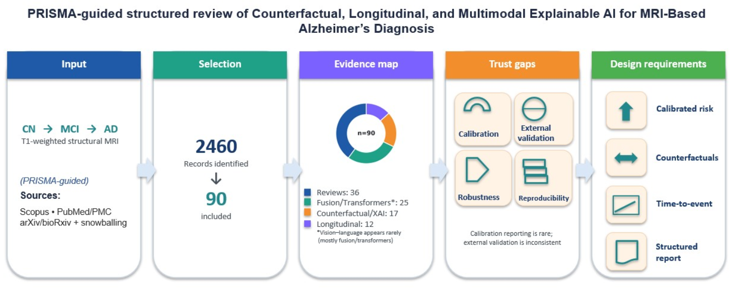


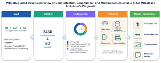
Counterfactual, Longitudinal, and Multimodal Explainable AI for MRI-Based Alzheimer’s Diagnosis: A Structured Review




Abstract
1. Introduction
2. Methods
- Disease: (Alzheimer* OR “Alzheimer’s disease” OR AD OR “mild cognitive impairment” OR MCI)
- Modality: (MRI OR “magnetic resonance imaging” OR “structural MRI” OR sMRI)
- AI methods: (“deep learning” OR “machine learning” OR “artificial intelligence” OR “neural network” OR CNN OR transformer* OR ViT OR “vision-language” OR multimodal OR fusion OR “survival analysis” OR longitudinal OR calibration OR counterfactual* OR explainab*)
- Primary Scopus query (as executed; all document types):
- TITLE-ABS-KEY ( alzheimer* OR "alzheimer’s disease" OR "mild cognitive impairment" OR mci )
- AND TITLE-ABS-KEY ( mri OR "magnetic resonance imaging" OR "structural mri" OR smri )
- AND TITLE-ABS-KEY ( "deep learning" OR "machine learning" OR "artificial intelligence"
- OR "neural network" OR cnn OR transformer* OR vit OR "vision-language"
- OR multimodal OR fusion OR "survival analysis" OR longitudinal
- OR calibration OR counterfactual* OR explainab* )
- AND PUBYEAR > 2013 AND PUBYEAR < 2027
- AND LANGUAGE ( english )
- AND ( LIMIT-TO ( SUBJAREA, "NEUR" ) OR LIMIT-TO ( SUBJAREA, "MEDI" )
- OR LIMIT-TO ( SUBJAREA, "COMP" ) OR LIMIT-TO ( SUBJAREA, "ENGI" ) )
- AND ( LIMIT-TO ( EXACTKEYWORD, "Alzheimer Disease" )
- OR LIMIT-TO ( EXACTKEYWORD, "Mild Cognitive Impairment" )
- OR LIMIT-TO ( EXACTKEYWORD, "Neuroimaging" )
- OR LIMIT-TO ( EXACTKEYWORD, "Magnetic Resonance Imaging" )
- OR LIMIT-TO ( EXACTKEYWORD, "Artificial Intelligence" )
- OR LIMIT-TO ( EXACTKEYWORD, "Deep Learning" )
- OR LIMIT-TO ( EXACTKEYWORD, "Machine Learning" )
- OR LIMIT-TO ( EXACTKEYWORD, "Dementia" )
- OR LIMIT-TO ( EXACTKEYWORD, "Neurodegenerative Diseases" )
- OR LIMIT-TO ( EXACTKEYWORD, "Multimodal Imaging" )
- OR LIMIT-TO ( EXACTKEYWORD, "Image Processing" )
- OR LIMIT-TO ( EXACTKEYWORD, "Image Analysis" ) )
- Targeted primary-study enrichment rule (transparent selection). In addition to the broad primary search above, we included primary modeling papers using the following pre-specified rule: a primary paper was eligible if it (a) used MRI for AD/MCI diagnosis, prognosis, or related tasks, and (b) introduced or empirically evaluated at least one of the review’s target clinical-translation themes such as calibration (ECE, Brier score, reliability analysis), explainability/counterfactuals (counterfactual generation, causal/attribution reasoning beyond saliency-only reporting), longitudinal/trajectory or survival-style modeling, or vision–language/reporting (captioning, report generation, retrieval-augmented reporting, VQA). Candidate primary papers were obtained through (i) snowballing from included reviews and seminal primary works, and (ii) a focused keyword pass described below.
- TITLE-ABS-KEY ( alzheimer* OR "alzheimer’s disease" OR mci )
- AND TITLE-ABS-KEY ( mri OR "magnetic resonance imaging" OR "structural mri" )
- AND TITLE-ABS-KEY ( calibration OR "expected calibration error" OR ece OR brier
- OR counterfactual* OR "causal" OR explainab* OR interpretab*
- OR "vision-language" OR caption* OR report* OR "retrieval augmented" OR rag
- OR longitudinal OR survival OR "time-to-event" )
- AND PUBYEAR > 2013 AND PUBYEAR < 2027
- AND LANGUAGE ( english )
- PubMed/PMC and arXiv/bioRxiv searches were aligned to the same concept families, adapting field tags and filters to each platform (for instance, MeSH terms in PubMed and category filters in arXiv/bioRxiv). Additionally, we performed backward/forward snowballing to capture relevant papers not indexed uniformly or not surfaced by database keywording.
- (i)
- Papers focused on AD, MCI, or the AD continuum that include MRI; multimodal works were eligible if MRI was included.
- (ii)
- Secondary evidence: systematic reviews, surveys, scoping reviews, and meta-analyses related to AI/ML/DL for MRI-based AD diagnosis/prognosis, including calibration, interpretability, multimodal fusion, longitudinal modeling, and vision–language/reporting.
- (iii)
- Primary studies: empirical modeling papers that evaluate MRI-based AD/MCI tasks and contribute to at least one target clinical-translation theme (calibration, explainability/counterfactuals, longitudinal/survival modeling, or vision–language/reporting), as specified in the targeted enrichment rule.
- (iv)
- Sufficient methodological detail to support structured extraction (task, model family, modalities, cohort/dataset, and evaluation setup).
- (i)
- Non-MRI domains; PET-only, genetics-only, or biomarker-only work without MRI.
- (ii)
- Editorials, opinion pieces, short news items, or records without extractable methods/results.
- (iii)
- General-purpose computer-vision methods not applied to AD/dementia neuroimaging.
- (iv)
- Full text inaccessible after reasonable effort, near-duplicates, or substantial overlap with an already-included entry, retaining the most complete version.
- (i)
- Bibliographic data (authors, year, venue).
- (ii)
- Study type (secondary evidence vs. primary modeling study).
- (iii)
- Cohort and dataset coverage when reported including ADNI, OASIS/OASIS-3, hospital cohorts.
- (iv)
- Modalities and preprocessing (such as bias correction, skull stripping, registration) when discussed.
- (v)
- Tasks (diagnosis, prognosis/progression, segmentation, report/caption generation, retrieval/VQA, etc.).
- (vi)
- Model family (CNN, transformer, survival model, graph-based, fusion architecture).
- (vii)
- Evaluation reporting, including external validation and calibration when available such as ECE, Brier score, reliability curves.
- (viii)
- Explainability evidence (saliency, counterfactuals, exemplar retrieval, robustness or stress-testing).
- (ix)
- Fairness/subgroup analysis (sex, age, genotype) when reported.
- (x)
- Code and data availability statements when provided.
3. Results: Included Evidence Base and Thematic Synthesis
3.1. Included Studies Overview (n = 90)
3.1.1. Modalities
3.1.2. Tasks
- Diagnostic classification (CN vs. AD, CN/MCI/AD, and finer-grained staging).
- Prognosis and longitudinal prediction (conversion risk, time-to-progression, and trajectory modeling across CN→MCI→AD).
- Quantification/segmentation of WMH and related lesion patterns, as well as ROI-based atrophy quantification.
- Explainability analysis, including saliency/feature-importance approaches and counterfactual reasoning where available.
- Vision–language tasks for neuroimaging reporting (captioning/report generation, retrieval-augmented summarization, and report simplification), noting that this direction is substantially more mature in general radiology than in AD MRI.
3.1.3. Datasets
3.1.4. Metrics and Evaluation
3.2. Study Characteristics
3.3. Thematic Synthesis by Approach Type
3.3.1. Scope of the Comparative Review
3.3.2. Method Families and Representative Approaches
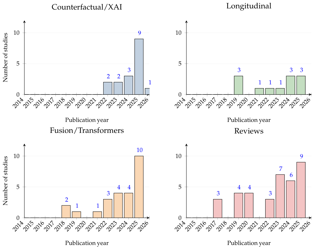
Review, Survey, and Meta-Analysis Synthesis
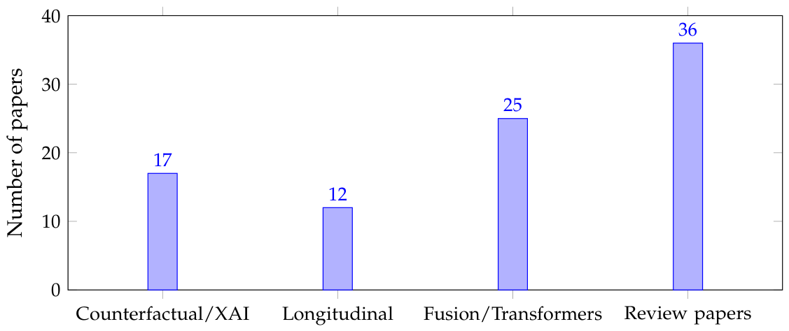
Counterfactual Reasoning and XAI (17/90)
Longitudinal Modeling and Progression Forecasting (12/90)
Fusion/Transformers and Other Deep-Learning Baselines (25/90)
3.3.3. Structured Synthesis of Findings
- Primary research concentrates on stronger fusion/transformer baselines, actionable interpretability, and progression-aware modeling. The dominant primary efforts emphasize multimodal fusion and global/transformer operators [30,36,38,44,49,50,52,53,54,55,56,57,60,63,75,85,86], counterfactual and interpretable explanation mechanisms beyond saliency [20,21,22,23,27,32,33,34,35,37,65], and longitudinal/progression modeling targeting conversion risk and biomarker dynamics [39,40,41,42,43,45,46,47,48,59,61,94].
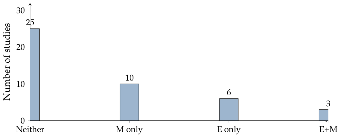
- Calibration, leakage-resistant evaluation, and robust external validation are still under-reported. Even when papers report high accuracy, many do not clearly document subject-level splitting, site-held-out testing, or calibration (reliability curves/ECE). This limitation is repeatedly highlighted across systematic reviews and meta-analyses [8,9,14,16,19]. As a practical consequence, “headline” metrics, especially extremely high accuracies reported in small or non-subject-split settings, should be interpreted cautiously in comparative synthesis [7,9,62].
- Clinical translation demands more than labels and heatmaps. Across both primary and secondary studies, there remains a gap between high-performing predictors and clinically grounded decision support: standardized evaluation, clinician-centered interpretability, reproducibility artifacts, and evidence-linked reporting remain uneven [11,12,14,19].
4. Comparative Analysis
4.1. Theme Distributions
4.2. Model Families vs. Typical Outcomes
- (i)
- Transformers vs. CNNs (3D sMRI staging and diagnosis). Transformer-era approaches treat a 3D volume as a sequence of tokens or frames, enabling explicit modeling of nonlocal dependencies across slices and long-range structure [49,50]. Variants that incorporate anatomical priors into attention attempt to improve both interpretability and clinical plausibility of highlighted evidence such as atlas-informed relevance bias [37]. Global-operator baselines provide an alternative route to nonlocal modeling by operating in frequency space, often reporting competitive discrimination while also enabling coarse localization [56]. However, many transformer-era papers report very high accuracy under limited or unclear external-validation settings, so comparative conclusions depend strongly on whether subject-level splitting, preprocessing standardization, and cohort shift are appropriately handled [9,19].In contrast, CNN families remain strong and widely used, particularly when architectural inductive bias is exploited for neuroimaging particularly in multiview slice attention fused with volumetric context [44]. Lightweight attention-driven CNN variants also continue to report high AUC/accuracy on standard datasets [75]. Transfer-learning pipelines on curated MRI datasets frequently report extremely high multi-class performance [85,86], but these results require careful interpretation because reported metrics can be sensitive to data curation choices and evaluation protocol details [9,19].
- (ii)
- Fusion vs. MRI-only (multimodal gains and common omissions). Across primary studies, multimodal fusion typically improves discrimination over MRI-only models when non-imaging signals (cognitive scores, demographics, genetics) or complementary imaging (PET) are integrated [30,36,52,53,57,63]. Modern fusion designs include cross-attention between MRI and metadata [30], patch-level cross-enhanced fusion for sMRI+FDG-PET [52], and modality-complete ensembling under missing/noisy modalities via structured representation learning [36]. Beyond PET, multimodal fusion also extends to electrophysiology: cross-attention fusion of sMRI with rsMEG demonstrates complementary structural–functional information for early classification [54].Despite these gains, the comparative literature remains limited by inconsistent reporting of missing-modality handling, harmonization steps, and site-held-out evaluation, even though these factors largely determine whether fusion models translate beyond a single cohort [15,19,78]. Multicohort benchmarking is present in a subset of stronger clinical-assessment systems [30,63], but remains the exception rather than the norm [9,19].
- (iii)
- Explainability families (from saliency to counterfactual structure). The XAI segment shows a clear qualitative progression: saliency-first explanations are increasingly supplemented or replaced by counterfactual, prototype-based, and decomposition-based explanations intended to be more actionable. Counterfactual frameworks generate explicit “change maps” or morphology edits that flip the predicted class, supporting a stronger clinical narrative than gradient heatmaps [20,21,22]. Disentangled pseudo-healthy synthesis provides individualized residual maps that isolate abnormal components from identity-preserving structure [33]. Latent causal counterfactual modeling further formalizes interventions (abduction–action–prediction) in compressed representations to generate plausible counterfactual MRIs efficiently [34]. In parallel, prototype-based 3D networks support transparent sparse voting, enabling inspection of prototypical evidence and pruning of clinically implausible prototypes [27].Nevertheless, much applied work still uses post hoc toolkits (Grad-CAM/LIME/SHAP) layered on top of strong predictors [24,26,28,31,65], including federated settings that keep raw data decentralized while still producing feature-attribution explanations [29]. Reviews consistently emphasize that explanation validity is not established by visualization alone, and that stability under perturbation/domain shift and clinician-centered evaluation remain uneven [11,12,13,14,92].
- (iv)
- Longitudinal modeling (progression-aware evidence vs. operational barriers). Longitudinal studies consistently show that temporal context supports more clinically meaningful risk stratification than snapshot staging. Structural MRI trajectory work quantifies hippocampal/subfield decline across stages and links regional atrophy to cognitive measures [39], while multi-site analysis associates atrophy acceleration with amyloid/tau positivity under distinct aging dynamics [40]. Region-wise mixed-effects modeling further connects atrophic trajectories to time-to-conversion and covariates such as APOE and age [41]. Mechanistic biomarker cascade modeling supports individualized forecasting and counterfactual “what-if” exploration [42].Predictive ML contributions highlight horizon-specific feature importance such as hippocampal vs. entorhinal dominance across different conversion tasks, cautioning against universal biomarker rankings [43]. Joint modeling that predicts future cognitive scores while synthesizing future MRI uses auxiliary generation to improve forecasting [45]. Progression prediction also benefits from multimodal temporal signals, including dynamic functional connectivity fused with deep structural representations [94]. At the same time, reviews emphasize that missingness, irregular follow-up schedules, and cohort heterogeneity create major barriers to fair comparison and robust deployment [17,18,70,80].
4.3. Evaluation and Reporting Gaps
5. Cross-Study Comparison
5.1. Modalities and Domains
5.2. Algorithms and Analytical Frameworks
5.3. Datasets and Evaluation Rigor
5.4. What Works and Where It Struggles
6. Discussion
- Evidence from secondary synthesis: comparability and generalizability are persistent bottlenecks. Across systematic reviews and meta-analyses, reported performance is repeatedly judged difficult to compare across studies because cohorts, preprocessing, class definitions, and evaluation splits vary substantially [5,6,7,8,9]. Reproducibility-focused evaluation further shows that reimplemented open-source pipelines may generalize substantially worse across cohorts than originally reported, reinforcing the need to interpret single-cohort results cautiously and to prioritize protocol transparency [19]. Meta-analytic evidence also underscores between-study heterogeneity in staging schemes and population characteristics, implying that pooled outcomes can conceal clinically meaningful variability [8,16,78].
- Representation learning: architectural progress is clear, but deployment-grade evidence lags. Primary studies reflect an evolution from CNN-centric pipelines toward transformer-era sequence modeling and nonlocal operators for 3D MRI [49,50,56]. In parallel, CNN and transfer-learning approaches remain widely used and often competitive, particularly when neuroimaging structure is exploited via multiview slice attention, attention-augmented designs, and tailored preprocessing [44,58,75,85,86]. However, across both primary and secondary studies, strong discrimination is not consistently accompanied by robust uncertainty characterization, site-held-out evaluation, or cross-cohort validation, limiting confidence in behavior under dataset shift [8,9,14,19].
- Multimodal fusion: consistent gains, uneven handling of missingness and heterogeneity. Multimodal pipelines support the view that AD is multifactorial: integrating MRI with PET, clinical/neuropsychological measures, and genetics can improve discrimination and staging [15,30,52,53,57,77,78,89]. Primary work addresses fusion through cross-attention between MRI and metadata [30], cross-enhanced PET–MRI interaction modeling [52], cascaded multimodality CNN aggregation without heavy manual preprocessing [53], and structured low-rank learning designed to tolerate missing/noisy modalities [36]. A subset of studies moves toward more clinically grounded evaluation by benchmarking across cohorts and, in some settings, against clinician performance [30,63]. Nonetheless, review papers repeatedly emphasize that missing-modality handling, harmonization across centers/scanners, and multicenter external validation are not uniformly enforced, which remains a central barrier to clinical translation [15,19,73,78,81].
- Interpretability: movement toward actionable explanations, limited reliability assessment. Interpretability-focused primary work increasingly shifts from post hoc saliency toward counterfactual and structurally grounded explanations that better align with clinical reasoning. Counterfactual generators and case-based editors provide explicit morphology changes or change maps intended to support actionable interpretation [20,21,22], while discrepancy-based attribution frames evidence localization as abnormal→normal translation [23]. Complementary families pursue built-in transparency via prototype-based voting [27], pseudo-healthy disentanglement with individualized residual atrophy mapping [33], latent causal counterfactual modeling enabling intervention in compressed representations [34], patch-level evidence selection [35], and atlas-prior attention to promote anatomically coherent transformer focus [37]. However, interpretability reviews consistently argue that explanation validity is not established by visually plausible maps alone: stability under perturbations and domain shift, clinician-centered evaluation, and coupling explanations with calibrated risk thresholds remain inconsistent and rarely standardized across studies [11,12,13,14,92].
- Longitudinal modeling: stronger biological grounding, continued protocol and reporting gaps. Longitudinal studies reinforce that temporal context is essential for forecasting and trial-relevant endpoints. Hippocampal/subfield trajectory analyses link regional decline to cognition and staging [39], multi-site modeling connects atrophy acceleration to amyloid/tau positivity [40], and mixed-effects trajectory modeling relates regional decline to time-to-conversion [41]. Mechanistic biomarker cascade modeling further motivates individualized forecasting and “what-if” exploration [42]. Predictive ML evidence indicates horizon-specific feature dominance, suggesting that static biomarker rankings may not generalize across conversion horizons [43]. Clinically relevant risk layers also emerge through vascular-marker quantification (PVS/WMH) and lesion burden modeling [46,48], and a complementary direction explores longitudinal retinal OCTA microvascular measures as non-brain biomarkers of preclinical signatures [47]. Despite these advances, missing data, irregular follow-up schedules, heterogeneous measurements, and inconsistent validation are repeatedly flagged as major barriers, and horizon-specific calibration or subgroup diagnostics remain infrequently reported [9,17,18,19,70,80]. Primary unresolved axis: calibration, robustness under shift, and clinical communication standards. A cross-cutting limitation across themes is the under-reporting of calibration and uncertainty quantification, despite repeated emphasis in systematic syntheses [8,9,14,16,19]. This gap is consequential because clinical decision support depends on reliable probability estimates, stable operating points, and transparent failure modes under cohort shift [14,19]. Fairness and subgroup auditing for instance, age, sex, genotype, and site/scanner are also rarely systematic in the primary set, even though cohort shift and demographic heterogeneity are central deployment risks [14,19]. Finally, MRI-oriented vision–language work remains sparse: while retrieval/captioning/VQA capability has been demonstrated [38], standardized, guideline-aligned reporting with explicit evidence provenance and calibrated statement confidence is not yet a common outcome target in AD MRI pipelines [14,19].
- Implications and practical priorities. Overall, the included studies suggests that the next translation step is less about incremental architecture changes and more about standardizing evaluation and reporting: subject-level leakage-resistant splits; multicenter and site-held-out validation; explicit calibration reporting (reliability curves and Brier/ECE); uncertainty-aware decision thresholds; subgroup auditing; and reproducibility artifacts that enable independent re-evaluation [9,14,19]. Progress on these dimensions would substantially improve cross-study comparability and clinical credibility, enabling more meaningful synthesis across the rapidly expanding methodological landscape [5,8,14,19].
- Limitations in the literature. Across the included studies, several recurring limitations emerge that directly map to the focus of this review on calibration, explainability, and clinically relevant MRI-based AD diagnosis across counterfactual, vision–language, longitudinal, and multimodal paradigms:
- Calibration is rarely treated as a first-class objective. Despite frequent reporting of high discrimination, explicit calibration and uncertainty quantification (reliability diagrams, ECE, Brier score, confidence intervals, or threshold-selection rationale) remain under-reported across MRI-only, multimodal, and progression-oriented pipelines [8,9,14,16,19]. This limits the interpretability of predicted probabilities as risk estimates and complicates deployment in settings where operating points and clinical decision thresholds matter [14,19]. Reported near-ceiling accuracies in some studies further motivate careful scrutiny of subject-level splitting and leakage-resistant evaluation when calibration is absent [7,9,62].
- Explainability is often visual or local, not validated for reliability. The shift from saliency maps toward counterfactual and structurally grounded explanations is a clear trend [20,21,22,23,33,34,37], and interpretable architectures such as prototypes, atlas-prior attention, patch-level evidence selection provide more inspectable rationales [27,35,37]. However, explanation fidelity and stability under perturbations, preprocessing variation, and cohort shift are rarely evaluated with standardized protocols, and clinician-centered validation remains uneven [11,12,13,14,92].
- Counterfactual modeling lacks common benchmarks and outcome-grounded validation. Counterfactual methods produce actionable artifacts such as minimal morphologic edits, pseudo-healthy synthesis, ROI delta summaries that can align with clinical reasoning [20,21,22,23,33]. Yet, counterfactual plausibility constraints, sensitivity to confounding factors, and validation against longitudinal outcomes or independent cohorts are inconsistently established, making cross-paper comparison difficult [14,21,42,43].
- Vision–language for brain MRI remains sparse and not reporting-oriented. Brain MRI vision–language capabilities (retrieval/captioning/VQA) appear only sparsely in the primary set [38]. Consequently, structured reporting-style outputs with explicit evidence provenance (linking statements to imaging/biomarker signals), calibrated statement confidence, and failure-mode characterization are not yet common endpoints in AD MRI pipelines [14,19].
- Longitudinal modeling is clinically motivated but methodologically constrained. Longitudinal studies show the value of repeated measures for progression forecasting and conversion-risk modeling [39,40,41,42,43,45,94], and vascular-marker quantification adds clinically relevant risk context [46,48]. Nevertheless, irregular follow-up schedules, missing data, and heterogeneous measurement intervals remain persistent obstacles [17,18,70,80], while horizon-specific evaluation and calibration over prediction horizons are not consistently reported [8,18,19,41,43].
- Multimodal fusion improves discrimination, but missingness and harmonization are unevenly addressed. Multimodal methods commonly report gains by integrating MRI with PET, CSF, clinical measures, and genetics [15,30,52,53,57,63,77,78,89], and some explicitly tackle missing/noisy modalities via structured representation learning [36]. However, harmonization reporting, multicenter external validation, and robustness to site/scanner shift remain inconsistent, limiting confidence in real-world generalization [15,19,73,78,81].
- Dataset concentration and protocol heterogeneity limit reproducibility and comparability. Heavy reliance on a small number of public cohorts (especially ADNI) persists across themes [5,9,19,68], while preprocessing and evaluation choices vary substantially across studies [5,6,7,8,9]. Reproducibility-focused work indicates that reimplemented pipelines may generalize substantially worse than originally reported, reinforcing the need for transparent protocols and robust cross-cohort evaluation [19].
- Subgroup robustness and fairness auditing are rarely systematic. Detailed subgroup performance and calibration by age, sex, APOE status, site/scanner strata are not consistently reported, despite clear demographic and acquisition heterogeneity in clinical practice [14,19,90]. Federated and hospital-oriented multimodal settings could enable stronger equity-aware evaluation, but fairness metrics and subgroup diagnostics remain uncommon [19,29,63].
7. Study Limitations
8. Conclusions
Author Contributions
Funding
Data Availability Statement
Acknowledgments
Conflicts of Interest
Abbreviations
References
- World Health Organization. Dementia. Fact Sheet. 2025. Available online: https://www.who.int/news-room/fact-sheets/detail/dementia (accessed on 31 December 2025).
- Alzheimer’s Association. 2025 Alzheimer’s disease facts and figures. Alzheimer’s Dement. 2025, 21, e70235. [Google Scholar] [CrossRef]
- Page, M.J.; McKenzie, J.E.; Bossuyt, P.M.; Boutron, I.; Hoffmann, T.C.; Mulrow, C.D.; Shamseer, L.; Tetzlaff, J.M.; Akl, E.A.; Brennan, S.E.; et al. The PRISMA 2020 statement: An updated guideline for reporting systematic reviews. BMJ 2021, 372, n71. [Google Scholar] [CrossRef]
- Tricco, A.C.; Lillie, E.; Zarin, W.; O’Brien, K.K.; Colquhoun, H.; Levac, D.; Moher, D.; Peters, M.D.J.; Horsley, T.; Weeks, L.; et al. PRISMA Extension for Scoping Reviews (PRISMA-ScR): Checklist and Explanation. Ann. Intern. Med. 2018, 169, 467–473. [Google Scholar] [CrossRef] [PubMed]
- Saikia, P.; Kalita, S.K. Alzheimer Disease Detection Using MRI: Deep Learning Review. SN Comput. Sci. 2024, 5, 507. [Google Scholar] [CrossRef]
- Alsubaie, M.G.; Luo, S.; Shaukat, K. Alzheimer’s Disease Detection Using Deep Learning on Neuroimaging: A Systematic Review. Mach. Learn. Knowl. Extr. 2024, 6, 464–505. [Google Scholar] [CrossRef]
- Kaur, A.; Mittal, M.; Bhatti, J.S.; Thareja, S.; Singh, S. A systematic literature review on the significance of deep learning and machine learning in predicting Alzheimer’s disease. Artif. Intell. Med. 2024, 154, 102928. [Google Scholar] [CrossRef]
- Battineni, G.; Chintalapudi, N.; Amenta, F. Machine Learning Driven by Magnetic Resonance Imaging for the Classification of Alzheimer Disease Progression: Systematic Review and Meta-Analysis. JMIR Aging 2024, 7, e59370. [Google Scholar] [CrossRef] [PubMed]
- Basanta-Torres, S.; Rivas-Fernández, M.Á.; Galdo-Alvarez, S. Artificial Intelligence for Alzheimer’s disease diagnosis through T1-weighted MRI: A systematic review. Comput. Biol. Med. 2025, 197, 111028. [Google Scholar] [CrossRef]
- Ebrahimighahnavieh, A.; Luo, S.; Chiong, R. Deep learning to detect Alzheimer’s disease from neuroimaging: A systematic literature review. Comput. Methods Programs Biomed. 2020, 187, 105242. [Google Scholar] [CrossRef]
- Khosroshahi, M.T.; Morsali, S.; Gharakhanlou, S.; Motamedi, A.; Hassanbaghlou, S.; Vahedi, H.; Pedrammehr, S.; Kabir, H.M.D.; Jafarizadeh, A. Explainable Artificial Intelligence in Neuroimaging of Alzheimer’s Disease. Diagnostics 2025, 15, 612. [Google Scholar] [CrossRef]
- Saif, F.H.; Al-Andoli, M.N.; Wan Bejuri, W.M.Y. Explainable AI for Alzheimer Detection: A Review of Current Methods and Applications. Appl. Sci. 2024, 14, 10121. [Google Scholar] [CrossRef]
- Vimbi, V.; Shaffi, N.; Mahmud, M. Interpreting artificial intelligence models: A systematic review on the application of LIME and SHAP in Alzheimer’s disease detection. Brain Inform. 2024, 11, 10. [Google Scholar] [CrossRef] [PubMed]
- Martin, S.A.; Townend, F.J.; Barkhof, F.; Cole, J.H. Interpretable machine learning for dementia: A systematic review. Alzheimer’s Dement. 2023, 19, 2135–2149. [Google Scholar] [CrossRef] [PubMed]
- Zhang, R.; Sheng, J.; Zhang, Q.; Wang, J.; Wang, B. A review of multimodal fusion-based deep learning for Alzheimer’s disease. Neuroscience 2025, 576, 80–95. [Google Scholar] [CrossRef]
- Guo, H.; Yang, Z.; Zhang, G.; Lv, L.; Zhao, X. Meta analysis of the diagnostic efficacy of transformer-based multimodal fusion deep learning models in early Alzheimer’s disease. Front. Neurol. 2025, 16, 1641548. [Google Scholar] [CrossRef] [PubMed]
- Aberathne, I.; Kulasiri, D.; Samarasinghe, S. Detection of Alzheimer’s disease onset using MRI and PET neuroimaging: Longitudinal data analysis and machine learning. Neural Regen. Res. 2023, 18, 2134–2140. [Google Scholar] [CrossRef]
- Martí-Juan, G.; Sanroma-Guell, G.; Piella, G. A survey on machine and statistical learning for longitudinal analysis of neuroimaging data in Alzheimer’s disease. Comput. Methods Programs Biomed. 2020, 189, 105348. [Google Scholar] [CrossRef]
- Akhavan Aghdam, M.; Bozdag, S.; Saeed, F.; Alzheimer’s Disease Neuroimaging Initiative. Machine-learning models for Alzheimer’s disease diagnosis using neuroimaging data: Survey, reproducibility, and generalizability evaluation. Brain Inform. 2025, 12, 8. [Google Scholar] [CrossRef]
- Oh, K.; Yoon, J.S.; Suk, H.I. Learn-Explain-Reinforce: Counterfactual Reasoning and its Guidance to Reinforce an Alzheimer’s Disease Diagnosis Model. IEEE Trans. Pattern Anal. Mach. Intell. 2023, 45, 4843–4857. [Google Scholar] [CrossRef]
- Oh, K.; Heo, D.W.; Mulyadi, A.W.; Jung, W.; Kang, E.; Lee, K.H.; Suk, H.I. A quantitatively interpretable model for Alzheimer’s disease prediction using deep counterfactuals. NeuroImage 2025, 309, 121077. [Google Scholar] [CrossRef]
- Valoor, A.; Gangadharan, G.R. Unveiling the decision making process in Alzheimer’s disease diagnosis: A case-based counterfactual methodology for explainable deep learning. J. Neurosci. Methods 2025, 413, 110318. [Google Scholar] [CrossRef] [PubMed]
- Zia, T.; Murtaza, S.; Bashir Bhatti, N.; Windridge, D.; Nisar, Z. VANT-GAN: Adversarial Learning for Discrepancy-Based Visual Attribution in Medical Imaging. Pattern Recognit. Lett. 2022, 156, 112–118. [Google Scholar] [CrossRef]
- Junior, K.J.; Kouayep, S.C.; Tagne Poupi, T.A.; Kim, H.C.; Alzheimer’s Disease Neuroimaging Initiative. Alzheimer’s Multiclassification Using Explainable AI Techniques. Appl. Sci. 2024, 14, 8287. [Google Scholar] [CrossRef]
- Zhang, X.; Han, L.; Zhu, W.; Sun, L.; Zhang, D. An explainable 3D residual self-attention deep neural network for joint atrophy localization and Alzheimer’s disease diagnosis using structural MRI. IEEE J. Biomed. Health Inform. 2022, 26, 5289–5297. [Google Scholar] [CrossRef]
- Mohanraj, S.; Sujatha, R. A novel CNN–fuzzy–XAI approach for Alzheimer’s disease severity classification using brain MRI scans. Cogent Eng. 2025, 12, 2575105. [Google Scholar] [CrossRef]
- De Santi, L.A.; Schlötterer, J.; Scheschenja, M.; Wessendorf, J.; Nauta, M.; Positano, V.; Seifert, C. PIPNet3D: Interpretable Detection of Alzheimer in MRI Scans. arXiv 2024, arXiv:2403.18328. [Google Scholar] [CrossRef]
- Jahan, S.; Abu Taher, K.; Kaiser, M.S.; Mahmud, M.; Rahman, M.S.; Hosen, A.S.M.S.; Ra, I.H. Explainable AI-based Alzheimer’s prediction and management using multimodal data. PLoS ONE 2023, 18, e0294253. [Google Scholar] [CrossRef]
- Jahan, S.; Adib, M.R.S.; Huda, S.M.; Rahman, M.S.; Kaiser, M.S.; Hosen, A.S.M.S.; Ghimire, D.; Park, M.J. Federated Explainable AI-Based Alzheimer’s Disease Prediction with Multimodal Data. IEEE Access 2025, 13, 43435–43454. [Google Scholar] [CrossRef]
- Rahman, S.; Rahman, M.M.; Bhatt, S.; Sundararajan, R.; Faezipour, M. NeuroNet-AD: A Multimodal Deep Learning Framework for Multiclass Alzheimer’s Disease Diagnosis. Bioengineering 2025, 12, 1107. [Google Scholar] [CrossRef] [PubMed]
- Ibrahim, N.; Elrefaei, L.; Abdel-Ghaffar, E.A. An interpretable model for the diagnosis of Alzheimer’s disease using deep learning and machine learning. In Proceedings of the 2025 15th International Conference on Electrical Engineering (ICEENG), Cairo, Egypt, 12–15 May 2025; pp. 1–8. [Google Scholar] [CrossRef]
- Parvin, S.; Nimmy, S.F.; Kamal, M.S. Convolutional neural network based data interpretable framework for Alzheimer’s treatment planning. Vis. Comput. Ind. Biomed. Art 2024, 7, 3. [Google Scholar] [CrossRef]
- Li, Z.; Zhao, K.; Chen, P.; Wang, D.; Yao, H.; Zhou, B.; Lu, J.; Wang, P.; Zhang, X.; Han, Y.; et al. Disentangled Representation Learning for Capturing Individualized Brain Atrophy via Pseudo-Healthy Synthesis. IEEE J. Biomed. Health Inform. 2025, 29, 5056–5068. [Google Scholar] [CrossRef]
- Peng, W.; Xia, T.; De Sousa Ribeiro, F.; Bosschieter, T.M.; Adeli, E.; Zhao, Q.; Glocker, B.; Pohl, K.M. Latent Causal Modeling for 3D Brain MRI Counterfactuals. In Deep Generative Models; Mukhopadhyay, A., Oksuz, I., Engelhardt, S., Mehrof, D., Yuan, Y., Eds.; Lecture Notes in Computer Science; Springer: Cham, Switzerland, 2026; Volume 16128, pp. 192–201. [Google Scholar] [CrossRef]
- Chitrakala, S.; Bharathi, U. ARE-PaLED: Augmented Reality-Enhanced Patch-Level Explainable Deep Learning System for Alzheimer’s Disease Diagnosis from 3D Brain sMRI. Symmetry 2025, 17, 1108. [Google Scholar] [CrossRef]
- Dong, A.; Li, Z.; Wang, M.; Shen, D.; Liu, M. High-Order Laplacian Regularized Low-Rank Representation for Multimodal Dementia Diagnosis. Front. Neurosci. 2021, 15, 634124. [Google Scholar] [CrossRef] [PubMed]
- Madhumitha, V.; Padhye, S.; Madarkar, S.S.; Agrawal, S.; Mopuri, K.R. Rel-SA: Alzheimer’s Disease Detection Using Relevance-augmented Self Attention by Inducing Domain Priors in Vision Transformers. In Proceedings of the 2025 IEEE/CVF Conference on Computer Vision and Pattern Recognition Workshops (CVPRW), Nashville, TN, USA, 11–12 June 2025. [Google Scholar] [CrossRef]
- Dhinagar, N.J.; Thomopoulos, S.I.; Thompson, P.M. Leveraging a Vision-Language Model with Natural Text Supervision for MRI Retrieval, Captioning, Classification, and Visual Question Answering. bioRxiv 2025. [Google Scholar] [CrossRef]
- Zhao, W.; Wang, X.; Yin, C.; He, M.; Li, S.; Han, Y. Trajectories of the Hippocampal Subfields Atrophy in the Alzheimer’s Disease: A Structural Imaging Study. Front. Neuroinform. 2019, 13, 13. [Google Scholar] [CrossRef] [PubMed]
- Somu, S.; Zhu, A.H.; Narula, S.; Jahanshad, N.; Nir, T.M. Longitudinal Multi-Site Modeling of Brain Atrophy Trajectories Associated with Amyloid and Tau. In Proceedings of the 2024 20th International Symposium on Medical Information Processing and Analysis (SIPAIM), Antigua, Guatemala, 13–15 November 2024. [Google Scholar] [CrossRef]
- Wei, X.; Du, X.; Xie, Y.; Suo, X.; He, X.; Ding, H.; Zhang, Y.; Ji, Y.; Chai, C.; Liang, M.; et al. Mapping cerebral atrophic trajectory from amnestic mild cognitive impairment to Alzheimer’s disease. Cereb. Cortex 2023, 33, 1310–1327. [Google Scholar] [CrossRef]
- Petrella, J.R.; Jiang, J.; Sreeram, K.; Dalziel, S.; Doraiswamy, P.M.; Hao, W.; Alzheimer’s Disease Neuroimaging Initiative. Personalized Computational Causal Modeling of the Alzheimer Disease Biomarker Cascade. J. Prev. Alzheimer’s Dis. 2024, 11, 435–444. [Google Scholar] [CrossRef]
- Mieling, M.; Yousuf, M.; Bunzeck, N.; Alzheimer’s Disease Neuroimaging Initiative. Predicting the progression of MCI and Alzheimer’s disease on structural brain integrity and other features with machine learning. GeroScience 2025, 48, 463–487. [Google Scholar] [CrossRef] [PubMed]
- Chen, L.; Qiao, H.; Zhu, F. Alzheimer’s Disease Diagnosis with Brain Structural MRI Using Multiview-Slice Attention and 3D Convolution Neural Network. Front. Aging Neurosci. 2022, 14, 871706. [Google Scholar] [CrossRef]
- Zhao, Y.; Ma, B.; Che, T.; Li, Q.; Zeng, D.; Wang, X.; Li, S. Multi-view prediction of Alzheimer’s disease progression with end-to-end integrated framework. J. Biomed. Inform. 2022, 125, 103978. [Google Scholar] [CrossRef]
- Barisano, G.; Iv, M.; Choupan, J.; Hayden-Gephart, M.; the Alzheimer’s Disease Neuroimaging Initiative. Robust, fully-automated assessment of cerebral perivascular spaces and white matter lesions: A multicentre MRI longitudinal study of their evolution and association with risk of dementia and accelerated brain atrophy. eBioMedicine 2024, 111, 105523. [Google Scholar] [CrossRef] [PubMed]
- Sheikh-Bahaei, N.; Sepehrband, F.; Barisano, G.; Acharya, J.; Rajamohan, A.G.; Law, M.; Toga, A.W.; Chui, C.H. Microvascular signature of preclinical Alzheimer’s disease in a longitudinal cohort. Alzheimer’s Dement. 2019, 15, P886. [Google Scholar] [CrossRef]
- Wu, Y.; Dong, Z.; Li, H.B.; Chong, Y.F.; Ji, F.; Chong, J.S.X.; Tang, N.R.J.; Hilal, S.; Fu, H.; Chen, C.L.H.; et al. WMH-DualTasker: A Weakly Supervised Deep Learning Model for Automated White Matter Hyperintensities Segmentation and Visual Rating Prediction. Hum. Brain Mapp. 2025, 46, e70212. [Google Scholar] [CrossRef]
- Akan, T.; Alp, S.; Bhuiyan, M.A.N. Vision Transformers and Bi-LSTM for Alzheimer’s Disease Diagnosis from 3D MRI. arXiv 2024, arXiv:2401.03132. [Google Scholar] [CrossRef]
- Akan, T.; Alp, S.; Bhuiyan, M.S.; Disbrow, E.A.; Conrad, S.A.; Vanchiere, J.A.; Kevil, C.G.; Bhuiyan, M.A.N. Leveraging Video Vision Transformer for Alzheimer’s Disease Diagnosis from 3D Brain MRI. arXiv 2025, arXiv:2501.15733. [Google Scholar] [CrossRef]
- Mandal, P.K.; Shukla, D. Brain Metabolic, Structural, and Behavioral Pattern Learning for Early Predictive Diagnosis of Alzheimer’s Disease. J. Alzheimer’s Dis. 2018, 63, 935–939. [Google Scholar] [CrossRef]
- Leng, Y.; Cui, W.; Peng, Y.; Yan, C.; Cao, Y.; Yan, Z.; Chen, S.; Jiang, X.; Zheng, J.; Alzheimer’s Disease Neuroimaging Initiative. Multimodal cross enhanced fusion network for diagnosis of Alzheimer’s disease and subjective memory complaints. Comput. Biol. Med. 2023, 157, 106788. [Google Scholar] [CrossRef]
- Liu, M.; Cheng, D.; Wang, K.; Wang, Y.; Alzheimer’s Disease Neuroimaging Initiative. Multi-Modality Cascaded Convolutional Neural Networks for Alzheimer’s Disease Diagnosis. Neuroinformatics 2018, 16, 295–308. [Google Scholar] [CrossRef]
- Liu, Y.; Wang, L.; Ning, X.; Gao, Y.; Wang, D. Enhancing early Alzheimer’s disease classification accuracy through the fusion of sMRI and rsMEG data: A deep learning approach. Front. Neurosci. 2024, 18, 1480871. [Google Scholar] [CrossRef]
- Jin, Y.; Xiao, H.; Chu, J.; Lv, F.; Li, Y.; Li, T. Cross-modal Causal Intervention for Alzheimer’s Disease Prediction. arXiv 2025, arXiv:2507.13956v1. [Google Scholar] [CrossRef]
- Zhang, S.; Chen, X.; Ren, B.; Yang, H.; Yu, Z.; Zhang, X.Y.; Zhou, Y. 3D Global Fourier Network for Alzheimer’s Disease Diagnosis Using Structural MRI. In Proceedings of the Medical Image Computing and Computer Assisted Intervention–MICCAI 2022; Wang, L., Dou, Q., Fletcher, P.T., Speidel, S., Li, S., Eds.; Lecture Notes in Computer Science; Springer: Cham, Switzerland, 2022; Volume 13431, pp. 34–43. [Google Scholar] [CrossRef]
- Qiang, Y.R.; Zhang, S.W.; Li, J.N.; Li, Y.; Zhou, Q.Y.; Alzheimer’s Disease Neuroimaging Initiative. Diagnosis of Alzheimer’s disease by joining dual attention CNN and MLP based on structural MRIs, clinical and genetic data. Artif. Intell. Med. 2023, 145, 102678. [Google Scholar] [CrossRef]
- Hazarika, R.A.; Maji, A.K.; Kandar, D.; Jasinska, E.; Krejci, P.; Leonowicz, Z.; Jasinski, M. An Approach for Classification of Alzheimer’s Disease Using Deep Neural Network and Brain Magnetic Resonance Imaging (MRI). Electronics 2023, 12, 676. [Google Scholar] [CrossRef]
- Hojjati, S.H.; Ebrahimzadeh, A.; Babajani-Feremi, A.; Alzheimer’s Disease Neuroimaging Initiative. Identification of the Early Stage of Alzheimer’s Disease Using Structural MRI and Resting-State fMRI. Front. Neurol. 2019, 10, 904. [Google Scholar] [CrossRef]
- Ding, T.; Xiang, D.; Schubert, K.E.; Dong, L. GKAN: Explainable Diagnosis of Alzheimer’s Disease Using Graph Neural Network with Kolmogorov-Arnold Networks. arXiv 2025, arXiv:2504.00946v1. [Google Scholar] [CrossRef]
- Gu, S.K.; Purushothaman, A.; Pradeep, R.; Sreeni, K. AlzFusionFormer: Integrating multiple transformers for early Alzheimer’s disease detection from multi-modal data. Biomed. Signal Process. Control 2025, 112, 108601. [Google Scholar] [CrossRef]
- Mmadumbu, A.C.; Saeed, F.; Ghaleb, F.; Qasem, S.N. Early detection of Alzheimer’s disease using deep learning methods. Alzheimer’s Dement. 2025, 21, e70175. [Google Scholar] [CrossRef] [PubMed]
- Qiu, S.; Miller, M.I.; Joshi, P.S.; Lee, J.C.; Xue, C.; Ni, Y.; Wang, Y.; De Anda-Duran, I.; Hwang, P.H.; Cramer, J.A.; et al. Multimodal deep learning for Alzheimer’s disease dementia assessment. Nat. Commun. 2022, 13, 3404. [Google Scholar] [CrossRef] [PubMed]
- Jumaili, M.L.F.; Sonuç, E. ML-Driven Alzheimer’s disease prediction: A deep ensemble modeling approach. SLAS Technol. 2025, 32, 100298. [Google Scholar] [CrossRef]
- Sheikh, F.; Al Marouf, A.; Rokne, J.G.; Alhajj, R. Lightweight Deep Learning Models with Explainable AI for Early Alzheimer’s Detection from Standard MRI Scans. Diagnostics 2025, 15, 2709. [Google Scholar] [CrossRef]
- Ali, S.; Piana, M.; Pardini, M.; Garbarino, S. Graph neural networks in Alzheimer’s disease diagnosis: A review of unimodal and multimodal advances. Front. Neurosci. 2025, 19, 1623141. [Google Scholar] [CrossRef]
- Pan, D.; Huang, Y.; Zeng, A.; Jia, L.; Song, X.; Alzheimer’s Disease Neuroimaging Initiative (ADNI). Early Diagnosis of Alzheimer’s Disease Based on Deep Learning and GWAS. In Human Brain and Artificial Intelligence; Zeng, A., Pan, D., Hao, T., Zhang, D., Shi, Y., Song, X., Eds.; Communications in Computer and Information Science; Springer: Singapore, 2019; Volume 1072, pp. 52–68. [Google Scholar] [CrossRef]
- Agarwal, D.; Marques, G.; de la Torre-Díez, I.; Franco Martin, M.A.; García Zapiraín, B.; Martín Rodríguez, F. Transfer Learning for Alzheimer’s Disease through Neuroimaging Biomarkers: A Systematic Review. Sensors 2021, 21, 7259. [Google Scholar] [CrossRef]
- Shukla, A.; Tiwari, R.; Tiwari, S. Review on Alzheimer Disease Detection Methods: Automatic Pipelines and Machine Learning Techniques. Sci 2023, 5, 13. [Google Scholar] [CrossRef]
- Franciotti, R.; Nardini, D.; Russo, M.; Onofrj, M.; Sensi, S.L.; Alzheimer’s Disease Neuroimaging Initiative; Alzheimer’s Disease Metabolomics Consortium ADMC. Comparison of Machine Learning-based Approaches to Predict the Conversion to Alzheimer’s Disease from Mild Cognitive Impairment. Neuroscience 2023, 514, 143–152. [Google Scholar] [CrossRef]
- Muydinov, A. Advances in Deep Learning Techniques for Alzheimer’s Disease Detection Using MRI Images Review. In Proceedings of the 8th International Conference on Future Networks & Distributed Systems (ICFNDS ’24), Marrakesh, Morocco, 11–12 December 2024; pp. 266–269. [Google Scholar] [CrossRef]
- Jo, T.; Nho, K.; Saykin, A.J. Deep Learning in Alzheimer’s Disease: Diagnostic Classification and Prognostic Prediction Using Neuroimaging Data. Front. Aging Neurosci. 2019, 11, 220. [Google Scholar] [CrossRef] [PubMed]
- Raza, M.L.; Hassan, S.T.; Jamil, S.; Hyder, N.; Batool, K.; Walji, S.; Abbas, M.K. Advancements in deep learning for early diagnosis of Alzheimer’s disease using multimodal neuroimaging: Challenges and future directions. Front. Neuroinform. 2025, 19, 1557177. [Google Scholar] [CrossRef]
- Arya, A.D.; Verma, S.S.; Chakarabarti, P.; Chakrabarti, T.; Elngar, A.A.; Kamali, A.M.; Nami, M. A systematic review on machine learning and deep learning techniques in the effective diagnosis of Alzheimer’s disease. Brain Inform. 2023, 10, 17. [Google Scholar] [CrossRef]
- Alsubaie, M.G.; Luo, S.; Shaukat, K.; Zhang, W.; Li, J. A Novel Deep Learning Approach for Alzheimer’s Disease Detection: Attention-Driven Convolutional Neural Networks with Multi-Activation Fusion. AI 2025, 6, 324. [Google Scholar] [CrossRef]
- Wang, R.; Peng, S.; Zhu, J.; Xu, Y.; Wang, M.; Zhang, L.; Qiu, Y.; Hou, D.; Wang, Q.; Liu, R. Innovations in Alzheimer’s disease diagnostic technologies: Clinical prospects of novel biomarkers, multimodal integration, and non-invasive detection. Front. Neurol. 2025, 16, 1651708. [Google Scholar] [CrossRef]
- Deshpande, P.; Kulkarni, S. Exploring Integration of Multimodal Deep Learning Approaches for Enhanced Alzheimer’s Disease Diagnosis: A Review of Recent Literature. SN Comput. Sci. 2024, 5, 852. [Google Scholar] [CrossRef]
- Odusami, M.; Maskeliūnas, R.; Damaševičius, R.; Misra, S. Machine learning with multimodal neuroimaging data to classify stages of Alzheimer’s disease: A systematic review and meta-analysis. Cogn. Neurodyn. 2024, 18, 775–794. [Google Scholar] [CrossRef] [PubMed]
- Thulasimani, V.; Shanmugavadivel, K.; Cho, J.; Veerappampalayam Easwaramoorthy, S. A Review of Datasets, Optimization Strategies, and Learning Algorithms for Analyzing Alzheimer’s Dementia Detection. Neuropsychiatr. Dis. Treat. 2024, 20, 2203–2225. [Google Scholar] [CrossRef] [PubMed]
- Zhou, Y.; Song, Z.; Han, X.; Li, H.; Tang, X. Prediction of Alzheimer’s Disease Progression Based on Magnetic Resonance Imaging. ACS Chem. Neurosci. 2021, 12, 4209–4223. [Google Scholar] [CrossRef] [PubMed]
- Naik, B.; Mehta, A.; Shah, M. Denouements of machine learning and multimodal diagnostic classification of Alzheimer’s disease. Vis. Comput. Ind. Biomed. Art 2020, 3, 26. [Google Scholar] [CrossRef]
- Forouzannezhad, P.; Abbaspour, A.; Fang, C.; Cabrerizo, M.; Loewenstein, D.; Duara, R.; Adjouadi, M. A survey on applications and analysis methods of functional magnetic resonance imaging for Alzheimer’s disease. J. Neurosci. Methods 2019, 317, 121–140. [Google Scholar] [CrossRef]
- Sarica, A.; Cerasa, A.; Quattrone, A. Random Forest Algorithm for the Classification of Neuroimaging Data in Alzheimer’s Disease: A Systematic Review. Front. Aging Neurosci. 2017, 9, 329. [Google Scholar] [CrossRef] [PubMed]
- Rathore, S.; Habes, M.; Iftikhar, M.A.; Shacklett, A.; Davatzikos, C. A review on neuroimaging-based classification studies and associated feature extraction methods for Alzheimer’s disease and its prodromal stages. NeuroImage 2017, 155, 530–548. [Google Scholar] [CrossRef]
- Altwijri, O.; Alanazi, R.; Aleid, A.; Alhussaini, K.; Aloqalaa, Z.; Almijalli, M.; Saad, A. Novel Deep-Learning Approach for Automatic Diagnosis of Alzheimer’s Disease from MRI. Appl. Sci. 2023, 13, 13051. [Google Scholar] [CrossRef]
- Zhao, Z.; Yeoh, P.S.Q.; Zuo, X.; Chuah, J.H.; Chow, C.O.; Wu, X.; Lai, K.W. Vision transformer-equipped Convolutional Neural Networks for automated Alzheimer’s disease diagnosis using 3D MRI scans. Front. Neurol. 2024, 15, 1490829. [Google Scholar] [CrossRef]
- Khojaste-Sarakhsi, M.; Haghighi, S.S.; Fatemi Ghomi, S.M.T.; Marchiori, E. Deep learning for Alzheimer’s disease diagnosis: A survey. Artif. Intell. Med. 2022, 130, 102332. [Google Scholar] [CrossRef]
- Illakiya, T.; Karthik, R. Automatic Detection of Alzheimer’s Disease using Deep Learning Models and Neuro-Imaging: Current Trends and Future Perspectives. Neuroinformatics 2023, 21, 339–364. [Google Scholar] [CrossRef]
- Sharma, S.; Mandal, P.K. A Comprehensive Report on Machine Learning-based Early Detection of Alzheimer’s Disease using Multi-modal Neuroimaging Data. ACM Comput. Surv. 2022, 55, 43:1–43:44. [Google Scholar] [CrossRef]
- Ottaviani, S.; Monacelli, F. Rethinking Dementia Risk Prediction: A Critical Evaluation of a Multimodal Machine Learning Predictive Model. J. Alzheimer’s Dis. 2024, 97, 1097–1100. [Google Scholar] [CrossRef]
- Weiner, M.W.; Veitch, D.P.; Aisen, P.S.; Beckett, L.A.; Cairns, N.J.; Green, R.C.; Harvey, D.; Jack, C.R.; Jagust, W.; Morris, J.C.; et al. Recent publications from the Alzheimer’s Disease Neuroimaging Initiative: Reviewing progress toward improved AD clinical trials. Alzheimer’s Dement. 2017, 13, e1–e85. [Google Scholar] [CrossRef]
- Bibi, N.; Courtney, J.; McGuinness, K. Enhancing Brain Disease Diagnosis with XAI: A Review of Recent Studies. ACM Trans. Comput. Healthc. 2025, 6, 1–35. [Google Scholar] [CrossRef]
- Wang, R.; Bashyam, V.; Yang, Z.; Yu, F.; Tassopoulou, V.; Chintapalli, S.S.; Skampardoni, I.; Sreepada, L.P.; Sahoo, D.; Nikita, K.; et al. Applications of generative adversarial networks in neuroimaging and clinical neuroscience. NeuroImage 2023, 269, 119898. [Google Scholar] [CrossRef]
- Abrol, A.; Fu, Z.; Du, Y.; Calhoun, V.D. Multimodal Data Fusion of Deep Learning and Dynamic Functional Connectivity Features to Predict Alzheimer’s Disease Progression. In Proceedings of the 41st Annual International Conference of the IEEE Engineering in Medicine and Biology Society (EMBC), Berlin, Germany, 23–27 July 2019; pp. 4409–4413. [Google Scholar] [CrossRef]



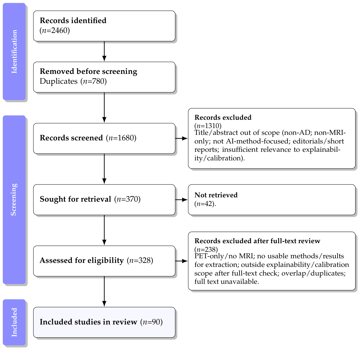
| Stage | Count(s) | Notes |
|---|---|---|
| Identification | 2460 | Records identified across Scopus, PubMed/PMC, arXiv/bioRxiv, and snowballing. |
| Deduplication | 780 removed; 1680 remaining | Duplicates removed to obtain unique records for screening. |
| Screening | 1310 excluded | Title/abstract screening excluded records due to scope mismatch (non-AD, non-MRI, not AI-method-focused), non-retrievable short formats, or insufficient relevance. |
| Retrieval | 370 sought; 42 not retrieved | Reports sought for full-text retrieval. |
| Eligibility | 328 assessed; 238 excluded | Full texts (or detailed preprints) assessed; exclusions included PET-only/no MRI, insufficient extractable methods/results, out-of-scope focus after full-text check, overlap/duplicates, or unavailable full text. |
| Inclusion | 90 included | Papers retained for the final synthesis. |
| Validation setting | Calibration reporting |
| Internal split/cross-validation vs. external site-held-out evaluation or multicenter testing. | Whether calibration was assessed, such as ECE/Brier/reliability curves, and whether post hoc calibration, such as temperature scaling, was used. |
| Domain shift handling | Reproducibility signals |
| Use of harmonization, scanner/site adjustment, domain adaptation, or robustness testing under distribution shift. | Availability of code, trained weights, detailed preprocessing, and sufficient implementation details to enable replication. |
| Study | Type Family | Method (Brief) | Task | Modality | Dataset | Trust |
|---|---|---|---|---|---|---|
| Oh et al. (2023) [20] | Prim/XAI–CF | LEAR: CF maps + explanation-guided attention | Staging | T1 sMRI | ADNI | E0C0 |
| Oh et al. (2025) [21] | Prim/XAI–CF | Deep CF “progression” MRI; ROI quantification | Risk/XAI | T1 sMRI | NR | E0C0 |
| Valoor & Gangadharan (2025) [22] | Prim/XAI–CF | Case-based CF maps (U-Net + GAN) | XAI | T1 sMRI | ADNI (rep.) | E0C0 |
| Khosroshahi et al. (2025) [11] | Review/XAI | Survey: SHAP/LIME/Grad-CAM/LRP, etc. | Review | MRI/PET | N/A | NA |
| Zia et al. (2022) [23] | Prim/Gen–XAI | VANT-GAN: abnormal→normal; discrepancy map | XAI | Med. img. | ADNI+ (rep.) | E0C0 |
| Junior et al. (2024) [24] | Prim/XAI (saliency) | ResNet-50 + attention; LIME/Grad-CAM | Classif. | T1 sMRI | ADNI (rep.) | E0C0 |
| Zhang et al. (2022) [25] | Prim/XAI (attn) | 3D ResNet + self-attention; atrophy localization | Classif./Loc. | 3D sMRI | NR | E0C0 |
| Mohanraj & Sujatha (2025) [26] | Prim/Hybrid–XAI | CNN + fuzzy decision; Grad-CAM/SHAP | Severity | T1 sMRI | OASIS/ADNI (rep.) | E0C0 |
| De Santi et al. (2024) [27] | Preprint/Proto–XAI | PIPNet3D: prototype “scoring sheet” | Classif. | 3D sMRI | ADNI-1 (rep.) | E0C0 |
| Jahan et al. (2023) [28] | Prim/Tabular + XAI | Multimodal + RF; SHAP explanations | Classif. | Multimodal | OASIS-3 (rep.) | E0C0 |
| Jahan et al. (2025) [29] | Prim/Fed + XAI | Federated RF; SHAP; privacy-preserving | Classif. | Multimodal | OASIS-3 (rep.) | E0C0 |
| Rahman et al. (2025) [30] | Prim/Fusion | CNN (MRI) + clinical fusion (NeuroNet-AD) | Classif. | Multimodal | ADNI + OASIS-3 | E1C0 |
| Ibrahim et al. (2025) [31] | Prim/TL + XAI | TL ensembles + saliency/Grad-CAM | Classif. | T1 sMRI | NR | E0C0 |
| Parvin et al. (2024) [32] | Prim/Multimodal + XAI | Tabular + MRI + gene; SP-LIME/LRP | Planning | Multimodal | OASIS + gene (rep.) | E0C0 |
| Li et al. (2025) [33] | Prim/Gen–CF | Pseudo-healthy synthesis; residual atrophy map | Atrophy CF | T1 sMRI | Multi-site (2) | E1C0 |
| Peng et al. (2026) [34] | Prim/Gen–CF | VQ-VAE + SCM; abduction→action CFs | CF synth. | 3D sMRI | ADNI+ NCANDA (rep.) | E1C0 |
| Chitrakala & Bharathi (2025) [35] | Prim/XAI pipeline | Patch-level explainable 3D sMRI pipeline | Classif. | 3D sMRI | ADNI + AIBL (rep.) | E1C0 |
| Dong et al. (2021) [36] | Prim/Fusion | Low-rank multimodal + Laplacian regularization | Classif. | MRI + PET + CSF | ADNI (rep.) | E0C0 |
| Madhumitha et al. (2025) [37] | Prim/Transformer–XAI | Relevance-augmented self-attention (domain priors) | Classif. | 3D sMRI | NR | E0C0 |
| Dhinagar et al. (2025) [38] | Preprint/VLM | VLM: retrieval + captioning + VQA | Multi-task | MRI + text | Mixed corpora | E0C0 |
| Zhao et al. (2019) [39] | Prim/Trajectory | Hippocampal subfield trajectories (FreeSurfer) | Trajectory | T1 sMRI | NR | E0CNA |
| Somu et al. (2024) [40] | Prim/Longitudinal | Multi-site atrophy trajectory vs. amyloid/tau | Trajectory | T1 sMRI | Multi-site (rep.) | E1CNA |
| Wei et al. (2023) [41] | Prim/Longitudinal | Mixed-effects + survival for aMCI→AD | Trajectory | T1 sMRI | ADNI (rep.) | E0CNA |
| Petrella et al. (2024) [42] | Prim/Causal modeling | Personalized causal biomarker cascade forecasting | Forecast | Multimodal | ADNI (NR) | E0CNA |
| Mieling et al. (2025) [43] | Prim/Tabular + XAI | XGBoost staging/conversion + SHAP | Conv./Cls. | sMRI-der. + tab. | ADNI (rep.) | E0C0 |
| Chen et al. (2022) [44] | Prim/CNN + Attn | Multiview-slice attention + 3D CNN | Classif. | T1 sMRI | ADNI-1/2 (rep.) | E0C0 |
| Zhao et al. (2022) [45] | Prim/Gen + Prog | Score prediction + future MRI synthesis (GAN) | Progression | T1 sMRI | ADNI (GO/2) | E0C0 |
| Barisano et al. (2024) [46] | Prim/Quant biomarker | Automated PVS/WMH; longitudinal risk association | Assoc. | sMRI + FLAIR | Multicentre | E1CNA |
| Sheikh-Bahaei et al. (2019) [47] | Prim/Biomarker | Microvascular signature (longitudinal) | Biomarker | OCTA(+PET) | NR | E0CNA |
| Wu et al. (2025) [48] | Prim/Segmentation | WMH weak supervision + rating prediction | Segm. | FLAIR | Multi-cohort (rep.) | E1CNA |
| Akan et al. (2024) [49] | Preprint/Transformer | ViT embeddings + Bi-LSTM over slices | Classif. | 3D sMRI | ADNI (rep.) | E0C0 |
| Akan et al. (2025) [50] | Preprint/ViViT | ViViT-style transformer over slice sequence | Classif. | 3D sMRI | ADNI (rep.) | E0C0 |
| Mandal & Shukla (2018) [51] | Position/Perspective | Argument for metabolic + structural + behavior fusion | Position | Multimodal | N/A | NA |
| Leng et al. (2023) [52] | Prim/Fusion | Cross-enhanced fusion (sMRI + FDG-PET) | Classif. | sMRI + FDG-PET | ADNI (rep.) | E0C0 |
| Liu et al. (2018) [53] | Prim/Fusion | Cascaded CNNs (local patches → aggregator) | Classif. | sMRI + PET | ADNI (rep.) | E0C0 |
| Liu et al. (2024) [54] | Prim/Fusion | sMRI + rsMEG cross-attention fusion | Classif. | sMRI + MEG | BioFIND (rep.) | E0C0 |
| Jin et al. (2025) [55] | Preprint/Causal + LLM | Causal intervention + LLM summaries (front-door) | Classif. | Imaging text | ADNI + NACC (rep.) | E1C0 |
| Zhang et al. (2022) [56] | Prim/Transformer | Fourier global network for 3D MRI | Classif. | T1 sMRI | ADNI + AIBL (rep.) | E1C0 |
| Qiang et al. (2023) [57] | Prim/Fusion | Dual-attention CNN + clinical/genetic MLP | Classif. | Multimodal | ADNI (rep.) | E0C0 |
| Hazarika et al. (2023) [58] | Prim/CNN | DNN classifier on MRI (baseline-style) | Classif. | T1 sMRI | ADNI (rep.) | E0C0 |
| Hojjati et al. (2019) [59] | Prim/Graph features | sMRI + rs-fMRI graph features + SVM | Classif. | sMRI + rs-fMRI | ADNI (rep.) | E0C0 |
| Ding et al. (2025) [60] | Preprint/GNN | GNN + Kolmogorov–Arnold networks (explainable) | Classif. | sMRI (graph) | ADNI (rep.) | E0C0 |
| Gu et al. (2025) [61] | Prim/FusionFormer | Multi-transformer fusion for conversion; attn/SHAP | Conversion | Multimodal | ADNI-1/2 (rep.) | E0C0 |
| Mmadumbu et al. (2025) [62] | Prim/DL pipeline | Early detection pipeline; cohort transfer reported | Classif. | Multimodal | ADNI + NACC (rep.) | E1C0 |
| Qiu et al. (2022) [63] | Prim/Multimodal DL | Multimodal dementia assessment; multi-cohort testing | Assess. | Multimodal | NACC + OASIS (rep.) | E1C0 |
| Jumaili et al. (2025) [64] | Prim/Ensemble | Deep ensemble; external tests on OASIS/ ADNI | Classif. | T1 sMRI | Private + OASIS + ADNI | E1C0 |
| Sheikh et al. (2025) [65] | Prim/Lightweight + XAI | MobileNet/EfficientNet + Grad-CAM++ | Classif. | T1 sMRI | ADNI (rep.) | E0C0 |
| Ali et al. (2025) [66] | Review/GNN | Review of GNNs for AD (uni/multimodal) | Review | Neuro imaging | N/A | NA |
| Saikia & Kalita (2024) [5] | Review/DL (MRI) | Review: MRI-based DL for AD detection | Review | MRI | N/A | NA |
| Alsubaie et al. (2024) [6] | Sys. rev./DL + ML | Systematic review of DL/ML for AD | Review | Neuro imaging | N/A | NA |
| Kaur et al. (2024) [7] | Sys. rev./DL + ML | PRISMA SLR: DL/ML for AD prediction | Review | Neuro imaging | N/A | NA |
| Pan et al. (2019) [67] | Prim/sMRI + Genetics | CNN ensemble + GWAS-guided biomarkers | Early DX | sMRI + gen | NR | E0C0 |
| Agarwal et al. (2021) [68] | Sys. rev./Transfer | Review: transfer learning for AD neuroimaging | Review | Neuro imaging | N/A | NA |
| Shukla et al. (2023) [69] | Review/Pipelines | Review: AD detection pipelines and gaps | Review | MRI | N/A | NA |
| Franciotti et al. (2023) [70] | Prim/Tabular–ML | RF vs. GB vs. XGB (voting); multimodal biomarkers + feature selection | Conversion | Multimodal | ADNI (rep.) | E0C0 |
| Muydinov (2025) [71] | Review/DL (MRI) | Review: DL for MRI-based AD detection | Review | MRI | N/A | NA |
| Battineni et al. (2024) [8] | Meta-anal./MRI | Systematic review + meta-analysis (MRI) | Review | MRI | N/A | NA |
| Jo et al. (2019) [72] | Review/DL | Review: DL for diagnosis + prognosis | Review | Neuro imaging | N/A | NA |
| Raza et al. (2025) [73] | Review/Multimodal DL | Review: multimodal DL challenges/future | Review | Multimodal | N/A | NA |
| Arya et al. (2023) [74] | Sys. rev./ML + DL | Systematic review: ML/DL for AD diagnosis | Review | Neuro imaging | N/A | NA |
| Alsubaie et al. (2025) [75] | Prim/CNN | Attention-driven CNN + multi-activation fusion | Classif. | T1 sMRI | ADNI (rep.) | E0C0 |
| Wang et al. (2025) [76] | Review/Biomarkers | Review: biomarkers + multimodal integration | Review | Multimodal | N/A | NA |
| Zhang et al. (2025) [15] | Review/Fusion | Review: multimodal fusion DL (MRI/PET, etc.) | Review | Multimodal | N/A | NA |
| Guo et al. (2025) [16] | Meta-anal./ Transformers | Meta-analysis: transformer multimodal fusion | Review | Multimodal | N/A | NA |
| Deshpande et al. (2024) [77] | Review/Multimodal DL | Review: integration of multimodal DL | Review | Multimodal | N/A | NA |
| Odusami et al. (2024) [78] | Meta-anal./Multimodal | Meta-analysis: multimodal neuroimaging ML | Review | Multimodal | N/A | NA |
| Thulasimani et al. (2024) [79] | Review/Datasets + Opt | Review: datasets/ optimization/ algorithms | Review | Neuro imaging | N/A | NA |
| Aberathne et al. (2023) [17] | Review/Longitudinal | Review: longitudinal MRI/PET ML | Review | MRI + PET | N/A | NA |
| Zhou et al. (2021) [80] | Review/Progression | Review: MRI biomarkers for progression prediction | Review | MRI | N/A | NA |
| Naik et al. (2020) [81] | Review/ML trends | Review: ML classifiers + multimodal trends | Review | Multimodal | N/A | NA |
| Martí-Juan et al. (2020) [18] | Survey/Longitudinal | Survey: longitudinal learning challenges | Review | Neuro imaging | N/A | NA |
| Forouzannezhad et al. (2019) [82] | Survey/fMRI | Survey: fMRI analysis methods for AD | Review | fMRI | N/A | NA |
| Sarica et al. (2017) [83] | Sys. rev./RF | Systematic review: RF on neuroimaging | Review | Neuro imaging | N/A | NA |
| Rathore et al. (2017) [84] | Review/Features | Review: feature extraction + pipelines | Review | Neuro imaging | N/A | NA |
| Basanta-Torres et al. (2025) [9] | Sys. rev./T1 MRI | Systematic review: T1 MRI AD classification | Review | T1 sMRI | N/A | NA |
| Altwijri et al. (2023) [85] | Prim/CNN | EfficientNet dementia staging; heavy preprocessing | Classif. | T1 sMRI | Kaggle (rep.) | E0C0 |
| Zhao et al. (2024) [86] | Prim/ViT + CNN | ViT-equipped CNN for 3D MRI diagnosis | Classif. | 3D sMRI | ADNI (rep.) | E0C0 |
| Ebrahimighahnavieh et al. (2020) [10] | Sys. rev./DL | Systematic review: DL across modalities | Review | Neuro imaging | N/A | NA |
| Khojaste-Sarakhsi et al. (2022) [87] | Survey/DL | Survey: architectures/inputs for AD DL | Review | Neuro imaging | N/A | NA |
| Illakiya & Karthik (2023) [88] | Review/DL trends | Review: DL trends + future perspectives | Review | Neuro imaging | N/A | NA |
| Sharma et al. (2022) [89] | Survey/DL | Survey: DL for AD (CNN/RNN/generative) | Review | Neuro imaging | N/A | NA |
| Akhavan Aghdam et al. (2025) [19] | Survey/Repro + Gen. | Survey: reproducibility/generalizability evaluation | Review | Neuro imaging | N/A | NA |
| Ottaviani & Monacelli (2024) [90] | Commentary | Commentary: critique of risk prediction model | Commentary | Multimodal | N/A | NA |
| Weiner et al. (2017) [91] | Review/ADNI program | Program review: ADNI publications and progress | Review | Multimodal | ADNI | NA |
| Saif et al. (2024) [12] | Review/XAI | Review: XAI methods for Alzheimer detection | Review | Neuro imaging | N/A | NA |
| Vimbi et al. (2024) [13] | Sys. rev./LIME + SHAP | Systematic review: LIME/SHAP in AD | Review | Neuro imaging | N/A | NA |
| Bibi et al. (2025) [92] | Review/XAI (broad) | Review: XAI for brain disease diagnosis | Review | Neuro imaging | N/A | NA |
| Wang et al. (2023) [93] | Review/GANs | Review: GANs in neuroimaging and clinical neuroscience | Review | Neuro imaging | N/A | NA |
| Abrol et al. (2019) [94] | Prim/Fusion | sMRI + dynamic FC fusion for MCI→AD | Conversion | sMRI + fMRI | NR | E0C0 |
| Martin et al. (2023) [14] | Sys. rev./Interp. ML | Systematic review: interpretable ML for dementia | Review | Multimodal | N/A | NA |
| Theme (Primary) | Included Works | Typical Data | Strengths/Limitations |
|---|---|---|---|
| Counterfactual & XAI (17/90) | [20,21,22,23,24,25,26,27,28,29,31,32,33,34,35,37,65] | Mostly T1 sMRI (ADNI/OASIS-like); some multimodal (tabular/genetic); occasional federated settings | Moves beyond saliency toward actionable evidence (counterfactual deltas, pseudo-healthy synthesis, prototypes, atlas-prior attention, AR patch selection). Gaps: explanation reliability under domain shift; calibration and clinician-centered evaluation often missing. |
| Longitudinal modeling (12/90) | [39,40,41,42,43,45,46,47,48,59,61,94] | Longitudinal sMRI; vascular markers; multimodal time series; conversion labels; occasional non-brain imaging biomarkers | Captures disease dynamics and conversion risk; supports biologically plausible biomarkers and “what-if” forecasting. Gaps: missingness handling and harmonization reporting vary; horizon-specific calibration and site-held-out robustness remain uneven. |
| Fusion/transformers & baselines (25/90) | [30,36,38,44,49,50,51,52,53,54,55,56,57,58,60,62,63,64,67,75,76,85,86,90,91] | MRI ± PET/CSF ± clinical/genetic; public + clinical cohorts; transformer/global operators; graph models; vision–language directions | Stronger representation learning and fusion; global operators and transformer-style sequence modeling; some multi-cohort benchmarking. Gaps: reproducibility controls, fairness/subgroup checks, and calibrated decision support under-reported; very high reported metrics often require careful split scrutiny. |
| Theme (Reviews) | Included Works | Typical Data | Strengths/Limitations |
|---|---|---|---|
| MRI/Neuroimaging DL reviews & SLRs | [5,6,7,9,10,68,69,71,72,74,83,84,87,88,89] | MRI-heavy; often ADNI/OASIS; sometimes multimodal | Broad synthesis of datasets/model families/metrics; documents protocol heterogeneity and common preprocessing choices. Gaps: uneven bias assessment; inconsistent standards for external validation and calibration. |
| Fusion/transformers/ GNN/longitudinal specialized reviews | [8,15,16,17,18,19,66,70,73,77,78,79,80,81,82] | Multimodal neuroimaging; GNNs; fMRI connectivity; longitudinal designs; biomarker integration | Deep dives by modality/model family; emphasizes translation barriers and cross-cohort concerns. Gaps: harmonization and robust external validation remain uneven; reproducibility often not enforced. |
| XAI/interpretability reviews | [11,12,13,14,92] | Mixed neuroimaging + clinical ML literature | Strong taxonomies and clinical framing for interpretability; highlights evaluation and trust bottlenecks. Gaps: limited evaluation of explanation reliability under shift; weak integration with calibrated decision support. |
| Generative neuroimaging reviews | [93] | Neuroimaging synthesis/augmentation/ harmonization | Maps the generative landscape and risks/utility for clinical neuroscience. Gaps: weak clinical validity constraints and downstream utility standards; leakage concerns. |
Disclaimer/Publisher’s Note: The statements, opinions and data contained in all publications are solely those of the individual author(s) and contributor(s) and not of MDPI and/or the editor(s). MDPI and/or the editor(s) disclaim responsibility for any injury to people or property resulting from any ideas, methods, instructions or products referred to in the content. |
© 2026 by the authors. Licensee MDPI, Basel, Switzerland. This article is an open access article distributed under the terms and conditions of the Creative Commons Attribution (CC BY) license.
Share and Cite
Farha, R.; Ojeme, B.; Khalifa, F.; Rahman, M.M. Counterfactual, Longitudinal, and Multimodal Explainable AI for MRI-Based Alzheimer’s Diagnosis: A Structured Review. J. Dement. Alzheimer's Dis. 2026, 3, 26. https://doi.org/10.3390/jdad3020026
Farha R, Ojeme B, Khalifa F, Rahman MM. Counterfactual, Longitudinal, and Multimodal Explainable AI for MRI-Based Alzheimer’s Diagnosis: A Structured Review. Journal of Dementia and Alzheimer's Disease. 2026; 3(2):26. https://doi.org/10.3390/jdad3020026
Chicago/Turabian StyleFarha, Ramisa, Blessing Ojeme, Fahmi Khalifa, and Md Mahmudur Rahman. 2026. "Counterfactual, Longitudinal, and Multimodal Explainable AI for MRI-Based Alzheimer’s Diagnosis: A Structured Review" Journal of Dementia and Alzheimer's Disease 3, no. 2: 26. https://doi.org/10.3390/jdad3020026
APA StyleFarha, R., Ojeme, B., Khalifa, F., & Rahman, M. M. (2026). Counterfactual, Longitudinal, and Multimodal Explainable AI for MRI-Based Alzheimer’s Diagnosis: A Structured Review. Journal of Dementia and Alzheimer's Disease, 3(2), 26. https://doi.org/10.3390/jdad3020026