Hyperelastic Ex Vivo Cervical Tissue Mechanical Characterization


Abstract
1. Introduction
2. Materials and Methods
2.1. Theory of Hyperelastic Models
2.1.1. Proposed Fourth Order Elastic Constants Nonlinear Model
2.1.2. Mooney-Rivlin Model
2.1.3. Ogden Model
2.2. Hysterectomy Specimens
2.3. Mechanical Tests
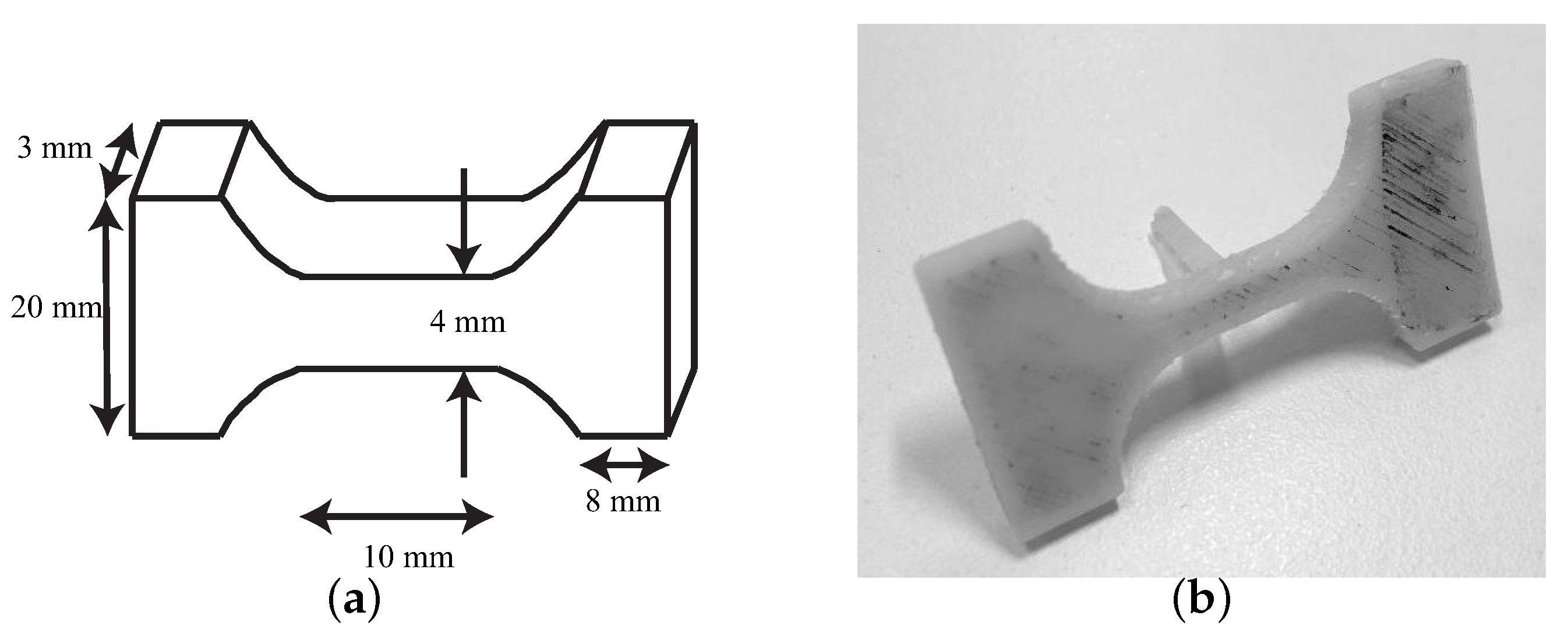
- All the seven cervical tissues were excised from the women and placed in phosphate buffered saline (PBS) to avoid loss of hydration after surgery. The connective layer was cut below the epithelial layer, and at a sufficient distance from the cervical canal to ensure that the preferred direction of the collagen fibers corresponds to the direction of the uniaxial tensile test [29,53]. The samples were tested in the Ultrasonics Laboratory at the University of Granada. Two slices were cut manually from each cervical sample, one from the epithelial layer and another one from the connective layer. The epithelial layer was cut carefully to obtain a thickness between 0.5 and 1 mm. The connective layer was obtained below the epithelial layer. All the samples were cut with the same mold (see Figure 4) to maintain the same geometry, which is necessary to locate the most unfavorable section.
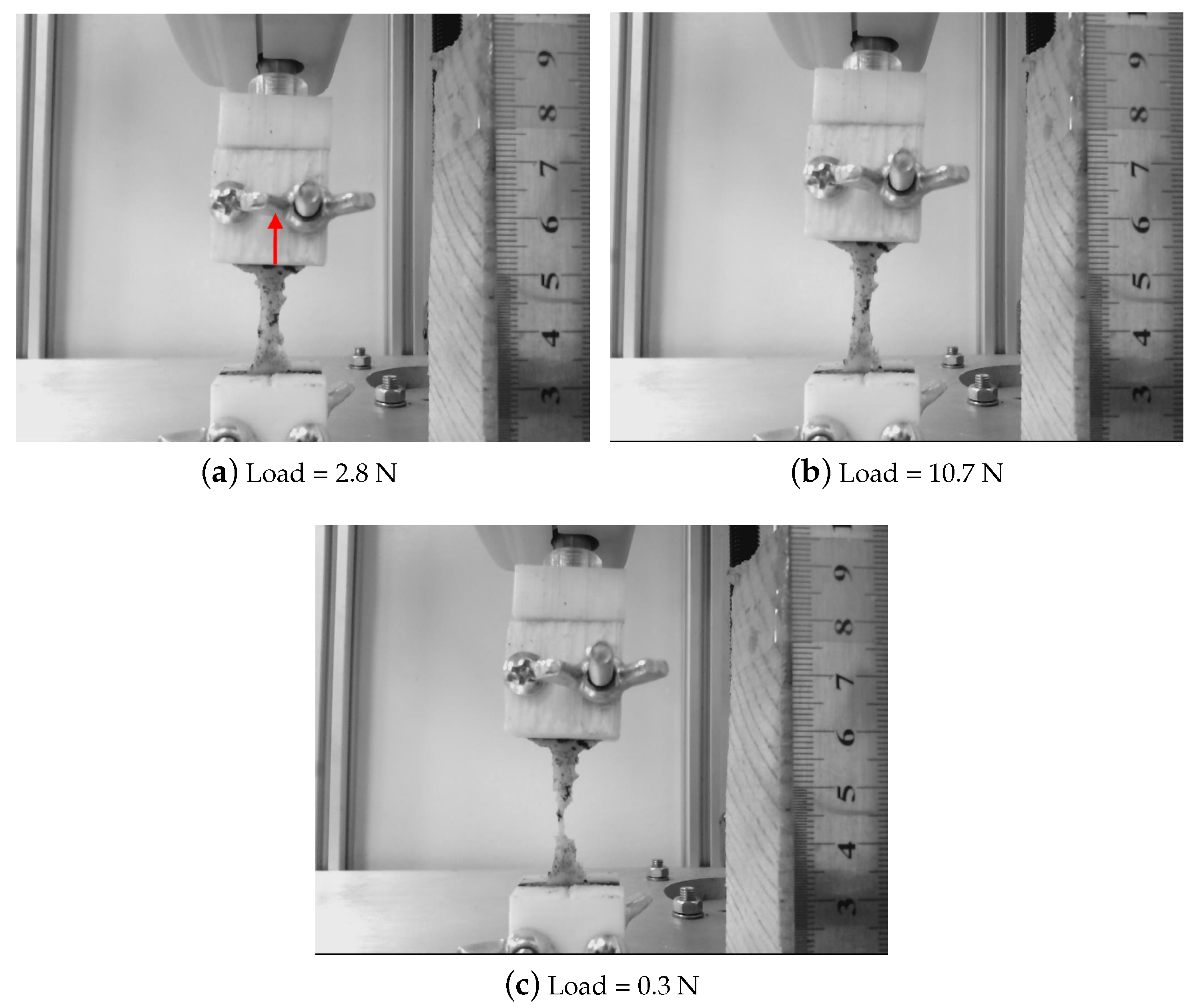
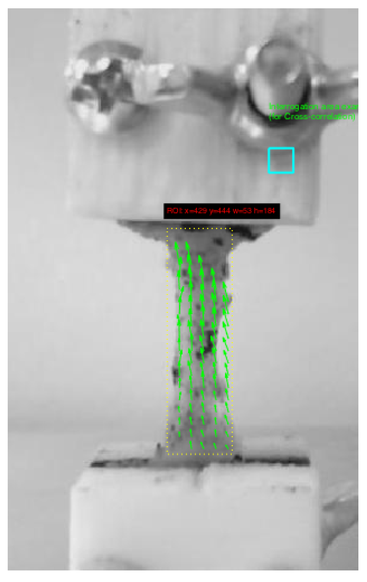
- A random dot pattern was used in the cervix to improve deformation monitoring carried out by a cross-correlation algorithm (PTVlab software), see Figure 5. For the speckle generation, acrylic black paint was used.
- An optimal contrast obtained by a good illumination and a uniform background help the tracking algorithm.
- It is worth underlining that the cervical tissue samples were kept continuously hydrated so as not to alter the mechanical properties during the experiment by spraying them with PBS.
- All the samples were preconditioned with 10 cycles at 1 N before the uniaxial tensile test.
3. Results
3.1. Comparison between Hyperelastic Models
3.2. Shear Modulus Estimation
4. Discussion
5. Conclusions
Author Contributions
Acknowledgments
Conflicts of Interest
Abbreviations
| FOEC | Fourth order elastic constants (FOEC) |
| ECM | Extracellular matrix |
| PBS | Phosphate buffered saline |
| ABS | Acrylonitrile butadiene styrene |
| GUI | Graphical user interface |
| LSPTV | Large scale particle tracking velocimetry |
| PTV | Particle tracking velocimetry |
| SSR | Sum of squares of the regression |
| SST | Total sum of squares |
References
- Atala, A.; Kasper, F.K.; Mikos, A.G. Engineering complex tissues. Sci. Transl. Med. 2012, 4, 160rv12. [Google Scholar] [CrossRef]
- Casey, J.; Crochet, M.J. Theoretical, Experimental, and Numerical Contributions to the Mechanics of Fluids and Solids: A Collection of Papers in Honor of Paul M. Naghdi; Birkhäuser: Basel, Switzerland, 2012. [Google Scholar]
- Langer, R.S.; Vacanti, J.P. Tissue engineering: The challenges ahead. Sci. Am. 1999, 280, 86–89. [Google Scholar] [CrossRef] [PubMed]
- Chatelin, S.; Deck, C.; Willinger, R. An anisotropic viscous hyperelastic constitutive law for brain material finite-element modeling. J. Biorheol. 2013, 27, 26–37. [Google Scholar] [CrossRef]
- Groves, R.B.; Coulman, S.A.; Birchall, J.C.; Evans, S.L. An anisotropic, hyperelastic model for skin: Experimental measurements, finite element modelling and identification of parameters for human and murine skin. J. Mech. Behav. Biomed. Mater. 2013, 18, 167–180. [Google Scholar] [CrossRef] [PubMed]
- Joldes, G.R.; Wittek, A.; Miller, K. Suite of finite element algorithms for accurate computation of soft tissue deformation for surgical simulation. Med. Image Anal. 2009, 13, 912–919. [Google Scholar] [CrossRef] [PubMed]
- Wu, T.; Alshareef, A.; Giudice, J.S.; Panzer, M.B. Explicit Modeling of White Matter Axonal Fiber Tracts in a Finite Element Brain Model. Ann. Biomed. Eng. 2019, 47, 1908–1922. [Google Scholar] [CrossRef]
- Bajka, M.; Haller, U. Virtual reality based surgery simulation for endoscopic gynaecology. In Medicine Meets Virtual Reality: The Convergence of Physical & Informational Technologies: Options for a New Era in Healthcare; IOS Press: Amsterdam, The Netherlands, 1999; pp. 351–357. [Google Scholar]
- Yoshida, E.A.; Castro, M.L.; Martins, V.F. Virtual reality and fetal medicine—A systematic review. In Proceedings of the 2017 XLIII Latin American Computer Conference (CLEI), Cordoba, Argentina, 4–8 September 2017; pp. 1–10. [Google Scholar]
- Misra, S.; Ramesh, K.; Okamura, A.M. Modeling of tool-tissue interactions for computer-based surgical simulation: A literature review. Presence Teleoperators Virtual Environ. 2008, 17, 463–491. [Google Scholar] [CrossRef]
- Fracczak, L.; Szaniewski, M.; Podsedkowski, L. Share control of surgery robot master manipulator guiding tool along the standard path. Int. J. Med. Robot. Comput. Assist. Surg. 2019, 15, e1984. [Google Scholar] [CrossRef]
- Ayache, N.; Cotin, S.; Delingette, H. Efficient Linear Elastic Models of Soft Tissues for Real-Time Surgery Simulation; Technical Report; Institut National de Recherche en Informoatique et en Automatique: Rocquencourt, France, 1998. [Google Scholar]
- Zhang, C.; Wang, M.; Song, Z. A brain-deformation framework based on a linear elastic model and evaluation using clinical data. IEEE Trans. Biomed. Eng. 2010, 58, 191–199. [Google Scholar] [CrossRef]
- Fung, Y.C. Biomechanics: Mechanical Properties of Living Tissues; Springer Science & Business Media: Berlin/Heidelberg, Germany, 2013. [Google Scholar]
- Veronda, D.; Westmann, R. Mechanical characterization of skin—finite deformations. J. Biomech. 1970, 3, 111–124. [Google Scholar] [CrossRef]
- Fung, Y. Elasticity of soft tissues in simple elongation. Am. J. Physiol.-Leg. Content 1967, 213, 1532–1544. [Google Scholar] [CrossRef]
- Wex, C.; Arndt, S.; Stoll, A.; Bruns, C.; Kupriyanova, Y. Isotropic incompressible hyperelastic models for modelling the mechanical behaviour of biological tissues: A review. Biomed. Eng./Biomed. Tech. 2015, 60, 577–592. [Google Scholar] [CrossRef]
- Kohandel, M.; Sivaloganathan, S.; Tenti, G.; Drake, J. The constitutive properties of the brain parenchyma: Part 1. Strain energy approach. Med. Eng. Phys. 2006, 28, 449–454. [Google Scholar] [CrossRef] [PubMed]
- Rashid, B.; Destrade, M.; Gilchrist, M.D. Mechanical characterization of brain tissue in simple shear at dynamic strain rates. J. Mech. Behav. Biomed. Mater. 2013, 28, 71–85. [Google Scholar] [CrossRef] [PubMed]
- Gao, Z.; Desai, J.P. Estimating zero-strain states of very soft tissue under gravity loading using digital image correlation. Med. Image Anal. 2010, 14, 126–137. [Google Scholar] [CrossRef] [PubMed]
- Lu, Y.C.; Kemper, A.R.; Untaroiu, C.D. Effect of storage on tensile material properties of bovine liver. J. Mech. Behav. Biomed. Mater. 2014, 29, 339–349. [Google Scholar] [CrossRef]
- Untaroiu, C.D.; Lu, Y.C. Material characterization of liver parenchyma using specimen-specific finite element models. J. Mech. Behav. Biomed. Mater. 2013, 26, 11–22. [Google Scholar] [CrossRef]
- Baah-Dwomoh, A.; McGuire, J.; Tan, T.; De Vita, R. Mechanical properties of female reproductive organs and supporting connective tissues: A review of the current state of knowledge. Appl. Mech. Rev. 2016, 68, 060801. [Google Scholar] [CrossRef]
- Barnum, C.E.; Fey, J.L.; Weiss, S.N.; Barila, G.; Brown, A.G.; Connizzo, B.K.; Shetye, S.S.; Elovitz, M.A.; Soslowsky, L.J. Tensile mechanical properties and dynamic collagen fiber re-alignment of the murine cervix are dramatically altered throughout pregnancy. J. Biomech. Eng. 2017, 139, 0610081–0610087. [Google Scholar] [CrossRef]
- Myers, K.M.; Paskaleva, A.; House, M.; Socrate, S. Mechanical and biochemical properties of human cervical tissue. Acta Biomater. 2008, 4, 104–116. [Google Scholar] [CrossRef]
- Poellmann, M.J.; Chien, E.K.; McFarlin, B.L.; Johnson, A.J.W. Mechanical and structural changes of the rat cervix in late-stage pregnancy. J. Mecha. Behav. Biomed. Mater. 2013, 17, 66–75. [Google Scholar] [CrossRef] [PubMed]
- Yoshida, K.; Mahendroo, M.; Vink, J.; Wapner, R.; Myers, K. Material properties of mouse cervical tissue in normal gestation. Acta Biomater. 2016, 36, 195–209. [Google Scholar] [CrossRef] [PubMed]
- Barone, W.R.; Feola, A.J.; Moalli, P.A.; Abramowitch, S.D. The effect of pregnancy and postpartum recovery on the viscoelastic behavior of the rat cervix. J. Mech. Med. Biol. 2012, 12, 1250009. [Google Scholar] [CrossRef] [PubMed]
- Myers, K.M.; Socrate, S.; Paskaleva, A.; House, M. A study of the anisotropy and tension/compression behavior of human cervical tissue. J. Biomech. Eng. 2010, 132, 021003. [Google Scholar] [CrossRef] [PubMed]
- Jayyosi, C.; Lee, N.; Willcockson, A.; Nallasamy, S.; Mahendroo, M.; Myers, K. The mechanical response of the mouse cervix to tensile cyclic loading in term and preterm pregnancy. Acta Biomater. 2018, 78, 308–319. [Google Scholar] [CrossRef] [PubMed]
- Jordan, J.; Singer, A.; Jones, H.; Shafi, M. The Cervix; John Wiley & Sons: Hoboken, NJ, USA, 2009. [Google Scholar]
- House, M.; Kaplan, D.L.; Socrate, S. Relationships between mechanical properties and extracellular matrix constituents of the cervical stroma during pregnancy. Semin. Perinatol. 2009, 33, 300–307. [Google Scholar] [CrossRef]
- Leppert, P.C. Anatomy and physiology of cervical ripening. Clin. Obstet. Gynecol. 1995, 38, 267–279. [Google Scholar] [CrossRef]
- Torres, J.; Faris, I.; Callejas, A. Histobiomechanical remodeling of the cervix during pregnancy: Proposed framework. Math. Probl. Eng. 2019, 2019, 5957432. [Google Scholar] [CrossRef]
- Myers, K.; Socrate, S.; Tzeranis, D.; House, M. Changes in the biochemical constituents and morphologic appearance of the human cervical stroma during pregnancy. Eur. J. Obstet. Gynecol. Reprod. Biol. 2009, 144, S82–S89. [Google Scholar] [CrossRef]
- Zork, N.M.; Myers, K.M.; Yoshida, K.; Cremers, S.; Jiang, H.; Ananth, C.V.; Wapner, R.J.; Kitajewski, J.; Vink, J. A systematic evaluation of collagen cross-links in the human cervix. Am. J. Obstet. Gynecol. 2015, 212, 321.e1–321.e8. [Google Scholar] [CrossRef]
- Marieb, E.N.; Hoehn, K. Human Anatomy & Physiology; Pearson Education: London, UK, 2007. [Google Scholar]
- Natali, A.N.; Carniel, E.L.; Gregersen, H. Biomechanical behaviour of oesophageal tissues: Material and structural configuration, experimental data and constitutive analysis. Med. Eng. Phys. 2009, 31, 1056–1062. [Google Scholar] [CrossRef] [PubMed]
- Massó, P.; Callejas, A.; Melchor, J.; Molina, F.S.; Rus, G. In Vivo Measurement of Cervical Elasticity on Pregnant Women by Torsional Wave Technique: A Preliminary Study. Sensors 2019, 19, 3249. [Google Scholar] [CrossRef] [PubMed]
- Rus, G.; Muñoz, R.; Melchor, J.; Molina, R.; Callejas, A.; Riveiro, M.; Massó, P.; Torres, J.; Moreu, G.; Molina, F.; et al. Torsion ultrasonic sensor for tissue mechanical characterization. In Proceedings of the 2016 IEEE International Ultrasonics Symposium (IUS), Tours, France, 18–21 September 2016; pp. 1–4. [Google Scholar]
- Callejas, A.; Gomez, A.; Melchor, J.; Riveiro, M.; Massó, P.; Torres, J.; López-López, M.; Rus, G. Performance study of a torsional wave sensor and cervical tissue characterization. Sensors 2017, 17, 2078. [Google Scholar] [CrossRef]
- Callejas, A.; Gomez, A.; Faris, I.H.; Melchor, J.; Rus, G. Kelvin–Voigt Parameters Reconstruction of Cervical Tissue-Mimicking Phantoms Using Torsional Wave Elastography. Sensors 2019, 19, 3281. [Google Scholar] [CrossRef]
- Landau, L.; Lifshitz, E. Elasticity Theory; Pergamon Press: Oxford, UK, 1975. [Google Scholar]
- Hamilton, M.F.; Ilinskii, Y.A.; Zabolotskaya, E.A. Separation of compressibility and shear deformation in the elastic energy density (L). J. Acoust. Soc. Am. 2004, 116, 41–44. [Google Scholar] [CrossRef]
- Destrade, M.; Ogden, R.W. On the third-and fourth-order constants of incompressible isotropic elasticity. J. Acoust. Soc. Am. 2010, 128, 3334–3343. [Google Scholar] [CrossRef] [PubMed]
- Eringen, A.C.; Suhubi, E. Nonlinear theory of simple micro-elastic solids. Int. J. Eng. Sci. 1964, 2, 189–203. [Google Scholar] [CrossRef]
- Muñoz, R.; Melchor, J. Nonlinear Classical Elasticity Model for Materials with Fluid and Matrix Phases. Math. Probl. Eng. 2018, 2018, 1–7. [Google Scholar] [CrossRef]
- Mooney, M. A theory of large elastic deformation. J. Appl. Phys. 1940, 11, 582–592. [Google Scholar] [CrossRef]
- Rivlin, R. Large elastic deformations of isotropic materials IV. Further developments of the general theory. Philos. Trans. R. Soc. A Math. Phys. Sci. 1948, 241, 379–397. [Google Scholar]
- Martins, P.; Natal Jorge, R.; Ferreira, A. A comparative study of several material models for prediction of hyperelastic properties: Application to silicone-rubber and soft tissues. Strain 2006, 42, 135–147. [Google Scholar] [CrossRef]
- Ogden, R.W. Large deformation isotropic elasticity–on the correlation of theory and experiment for incompressible rubberlike solids. Proc. R. Soc. A Math. Phys. Sci. 1972, 326, 565–584. [Google Scholar] [CrossRef]
- Mansouri, M.; Darijani, H. Constitutive modeling of isotropic hyperelastic materials in an exponential framework using a self-contained approach. Int. J. Solids Struct. 2014, 51, 4316–4326. [Google Scholar] [CrossRef]
- Aspden, R.M. Collagen organisation in the cervix and its relation to mechanical function. Collagen Relat. Res. 1988, 8, 103–112. [Google Scholar] [CrossRef]
- Patalano, A.; García, C.M.; Rodríguez, A. Rectification of image velocity results (RIVeR): A simple and user-friendly toolbox for large scale water surface particle image velocimetry (PIV) and particle tracking velocimetry (PTV). Comput. Geosci. 2017, 109, 323–330. [Google Scholar] [CrossRef]
- Patalano, A.; Garcia, C.; Brevis, W.; Bleninger, T.; Guillen, N.; Moreno, L.; Rodriguez, A. Recent advances in Eulerian and Lagragian large-scale particle image velocimetry. In Proceedings of the E-Proceedings of the 36th IAHR World Congress, The Hauge, The Netherlands, 28 June–3 July 2015. [Google Scholar]
- Landau, L.; Lifshitz, E. Theory of Elasticity; Pergamon Press: Oxford, UK, 1970. [Google Scholar]
- Rashid, B.; Destrade, M.; Gilchrist, M.D. Mechanical characterization of brain tissue in tension at dynamic strain rates. J. Mech. Behav. Biomed. Mater. 2014, 33, 43–54. [Google Scholar] [CrossRef]
- Zemánek, M.; Burša, J.; Děták, M. Biaxial tension tests with soft tissues of arterial wall. Eng. Mech. 2009, 16, 3–11. [Google Scholar]
- Gennisson, J.L.; Rénier, M.; Catheline, S.; Barrière, C.; Bercoff, J.; Tanter, M.; Fink, M. Acoustoelasticity in soft solids: Assessment of the nonlinear shear modulus with the acoustic radiation force. J. Acoust. Soc. Am. 2007, 122, 3211–3219. [Google Scholar] [CrossRef]
- Bernal, M.; Chamming’s, F.; Couade, M.; Bercoff, J.; Tanter, M.; Gennisson, J.L. In VivoQuantification of the Nonlinear Shear Modulus in Breast Lesions: Feasibility Study. IEEE Trans. Ultrason. Ferroelectr. Freq. Control 2015, 63, 101–109. [Google Scholar] [CrossRef]
- Shi, L.; Yao, W.; Gan, Y.; Zhao, L.; McKee, W.E.; Vink, J.; Wapner, R.; Hendon, C.; Myers, K. Anisotropic Material Characterization of Human Cervix Tissue based on Indentation. J. Biomech. Eng. 2019, 141, 091017. [Google Scholar] [CrossRef]
- Myers, K.M.; Feltovich, H.; Mazza, E.; Vink, J.; Bajka, M.; Wapner, R.J.; Hall, T.J.; House, M. The mechanical role of the cervix in pregnancy. J. Biomech. 2015, 48, 1511–1523. [Google Scholar] [CrossRef] [PubMed]


| Patient | Age | Hysterectomy Indication |
|---|---|---|
| 1 | 53 | Vaginal prolapse |
| 2 | 67 | Subserous myoma |
| 3 | 59 | Vaginal prolapse |
| 4 | 54 | Cervical prolapse |
| 5 | 50 | Cervical prolapse |
| 6 | 51 | Cervical prolapse |
| 7 | 71 | Cervical prolapse |
| Nonlinear Model | ||||
|---|---|---|---|---|
| Epithelial Layer | Connective Layer | |||
| Cervix | A | A | ||
| 1 | 1.13 | 22.6 | 3.58 | 3.49 |
| 2 | 1.22 | −6.08 | 4.72 | −7.63 |
| 3 | 1.35 | −3.06 | 2.64 | −5.92 |
| 4 | 1.57 | 28.3 | 3.30 | 27.6 |
| 5 | 1.35 | −2.35 | 3.51 | 73.6 |
| 6 | 1.13 | 2.32 | 3.49 | 70.1 |
| 7 | 1.27 | 30.72 | 3.96 | 25.7 |
| Median (IQR) | 1.27 (1.13 1.35) | 2.32 (−3.06 28.3) | 3.51 (3.30 3.96) | 25 (−5.92 70.1) |
| Ogden Model | ||||
|---|---|---|---|---|
| Epithelial Layer | Connective Layer | |||
| Cervix | ||||
| 1 | 0.41 | 7.94 | 0.941 | 6.01 |
| 2 | 1.01 | 1.62 | 1.16 | 5.63 |
| 3 | 0.42 | 4.54 | 0.97 | 4.13 |
| 4 | 0.35 | 9.94 | 0.85 | 11.1 |
| 5 | 0.47 | 4.31 | 0.82 | 10.25 |
| 6 | 0.39 | 5.27 | 0.57 | 11.54 |
| 7 | 0.40 | 9.05 | 1.29 | 6.40 |
| Median (IQR) | 0.41 (0.39 0.47) | 5.27 (4.31 9.05) | 0.94 (0.82 1.16) | 6.40 (5.63 11.1) |
| Mooney-Rivlin Model | ||||
|---|---|---|---|---|
| Epithelial Layer | Connective Layer | |||
| Cervix | ||||
| 1 | 6.93 | −6.73 | 5.87 | −4.77 |
| 2 | 0.33 | −0.08 | 4.7 | −3.15 |
| 3 | 1.22 | −0.78 | 2.51 | −1.68 |
| 4 | 8.25 | −7.84 | 59.9 | −59.3 |
| 5 | 1.47 | −1.05 | 20.56 | −19.67 |
| 6 | 2.35 | −2.06 | 15.7 | −15.9 |
| 7 | 8.69 | −8.44 | 12.1 | −11.1 |
| Median (IQR) | 2.35 (1.22 8.25) | −2.06 (−7.84 −0.78) | 12.10 (4.70 20.56) | −11.1 (−19.67 −3.15) |
| Shear Modulus | ||||||
|---|---|---|---|---|---|---|
| Epithelial Layer | Connective Layer | |||||
| Cervix | Nonlinear | Ogden | Curve | Nonlinear | Ogden | Curve |
| 1 | 1.13 | 1.65 | 0.82 | 3.58 | 2.83 | 4.17 |
| 2 | 1.22 | 0.82 | 0.69 | 4.72 | 3.28 | 3.78 |
| 3 | 1.35 | 0.95 | 1.43 | 2.64 | 2.01 | 3.62 |
| 4 | 1.57 | 1.77 | 1.82 | 3.30 | 4.71 | 3.26 |
| 5 | 1.35 | 1.02 | 0.44 | 3.51 | 4.22 | 5.25 |
| 6 | 1.13 | 1.03 | 0.90 | 3.49 | 3.30 | 4.42 |
| 7 | 1.27 | 1.84 | 1.08 | 3.96 | 4.15 | 3.17 |
| Mean ± Std | 1.29 ± 0.15 | 1.30 ± 0.43 | 1.02 ± 0.46 | 3.60 ± 0.63 | 3.50 ± 0.92 | 3.95 ± 0.72 |
© 2020 by the authors. Licensee MDPI, Basel, Switzerland. This article is an open access article distributed under the terms and conditions of the Creative Commons Attribution (CC BY) license (http://creativecommons.org/licenses/by/4.0/).
Share and Cite
Callejas, A.; Melchor, J.; Faris, I.H.; Rus, G. Hyperelastic Ex Vivo Cervical Tissue Mechanical Characterization. Sensors 2020, 20, 4362. https://doi.org/10.3390/s20164362
Callejas A, Melchor J, Faris IH, Rus G. Hyperelastic Ex Vivo Cervical Tissue Mechanical Characterization. Sensors. 2020; 20(16):4362. https://doi.org/10.3390/s20164362
Chicago/Turabian StyleCallejas, Antonio, Juan Melchor, Inas H. Faris, and Guillermo Rus. 2020. "Hyperelastic Ex Vivo Cervical Tissue Mechanical Characterization" Sensors 20, no. 16: 4362. https://doi.org/10.3390/s20164362
APA StyleCallejas, A., Melchor, J., Faris, I. H., & Rus, G. (2020). Hyperelastic Ex Vivo Cervical Tissue Mechanical Characterization. Sensors, 20(16), 4362. https://doi.org/10.3390/s20164362