Mo-Based Layered Nanostructures for the Electrochemical Sensing of Biomolecules


Abstract
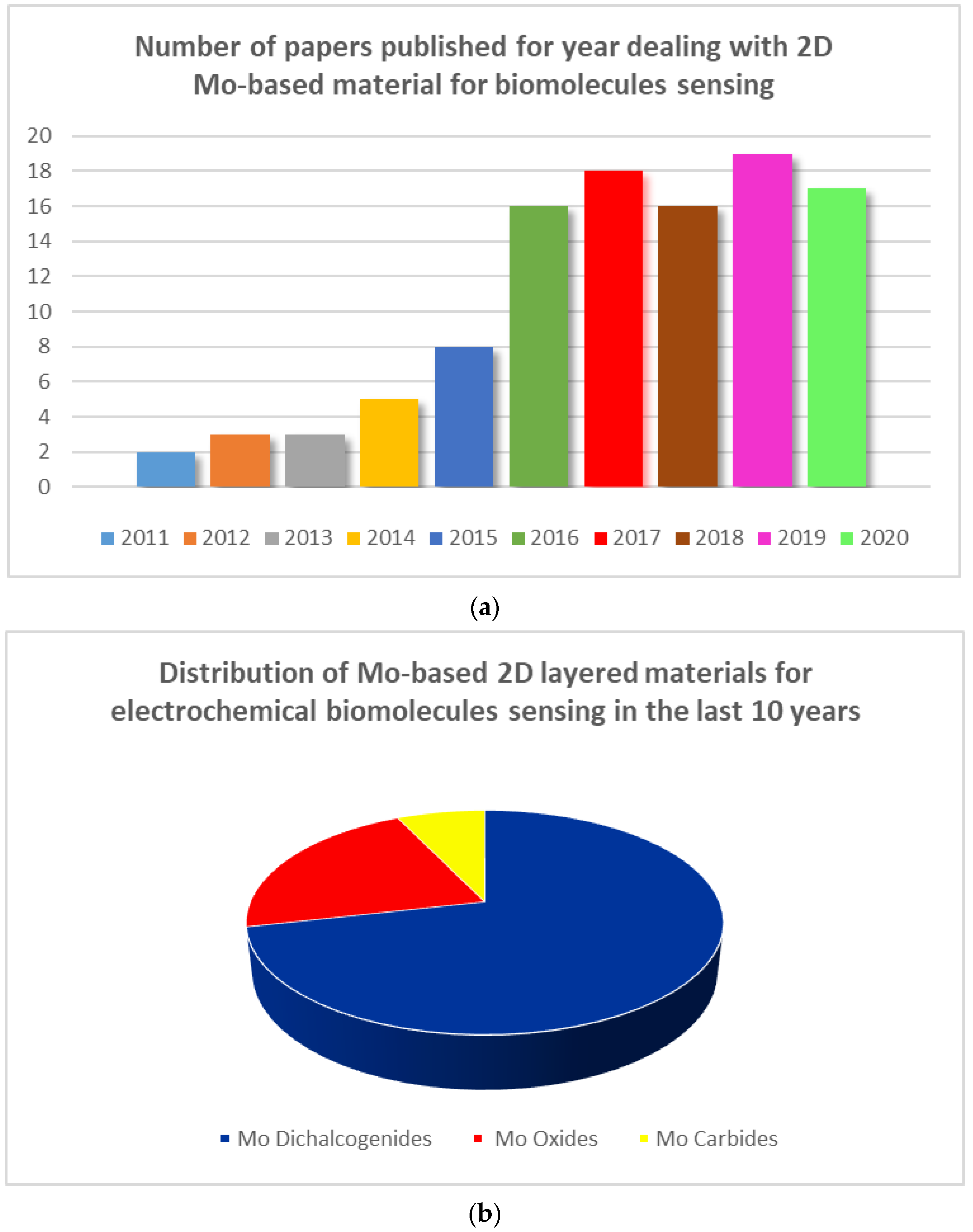
:1. Introduction
2. Mo-Based 2D Layered Materials
2.1. Mo Oxides (MoO3 and MoO2)
2.2. Mo Dichalcogenides (MoS2, MoSe2, MoTe2)
2.3. Molybdenum Carbides (MoC, Mo2C)
2.4. Hybrid Structures
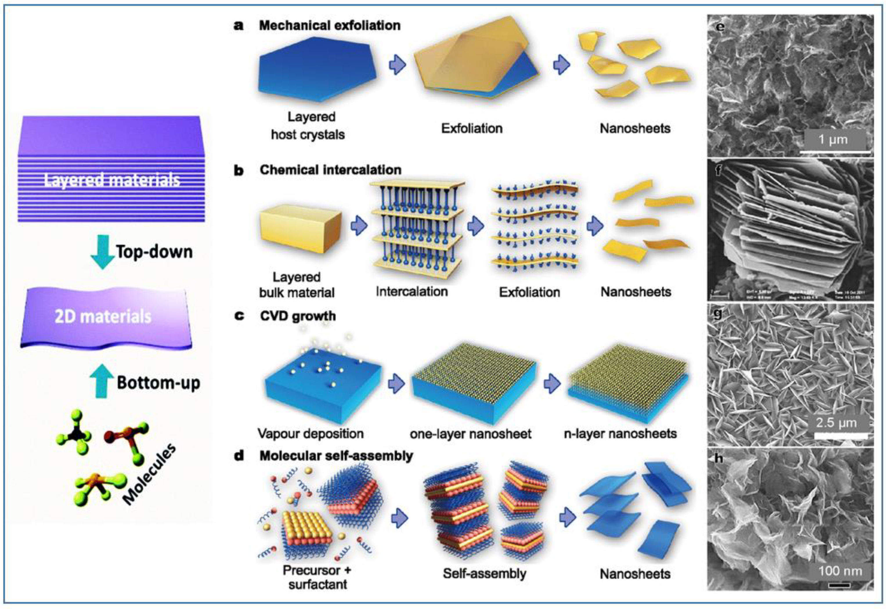
3. Synthesis Methods of Mo-Based 2D Layered Materials
3.1. Atomic Layer Deposition (ALD)
3.2. Pulsed Laser Deposition (PLD), Sputtering and Spray Pyrolysis
3.3. Chemical Etching
3.4. Chemical Vapor Deposition (CVD)
3.5. Hydrothermal Synthesis
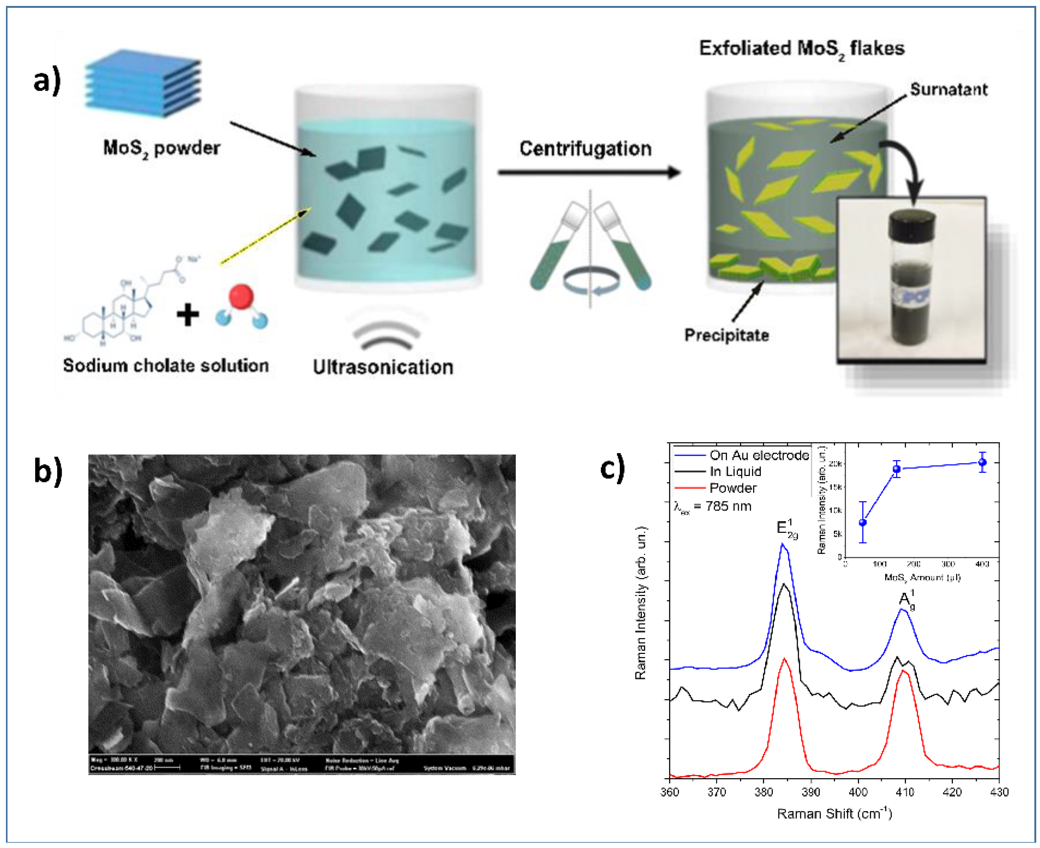
3.6. Exfoliation
3.7. Miscellaneous Synthesis
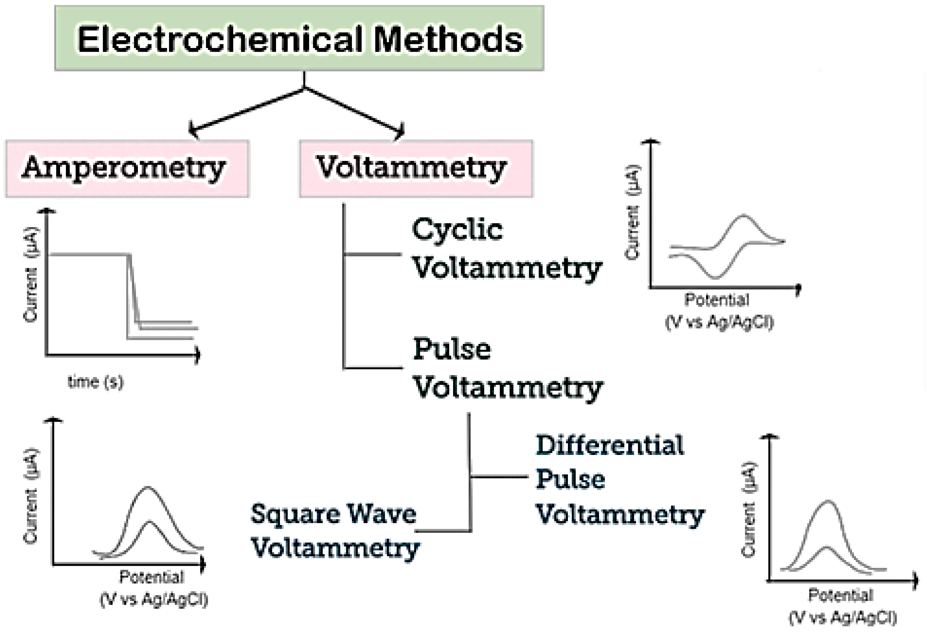
4. Electrochemical Sensors
5. Mo-Based 2D Layered Materials for Electrochemical Biomolecules Sensing
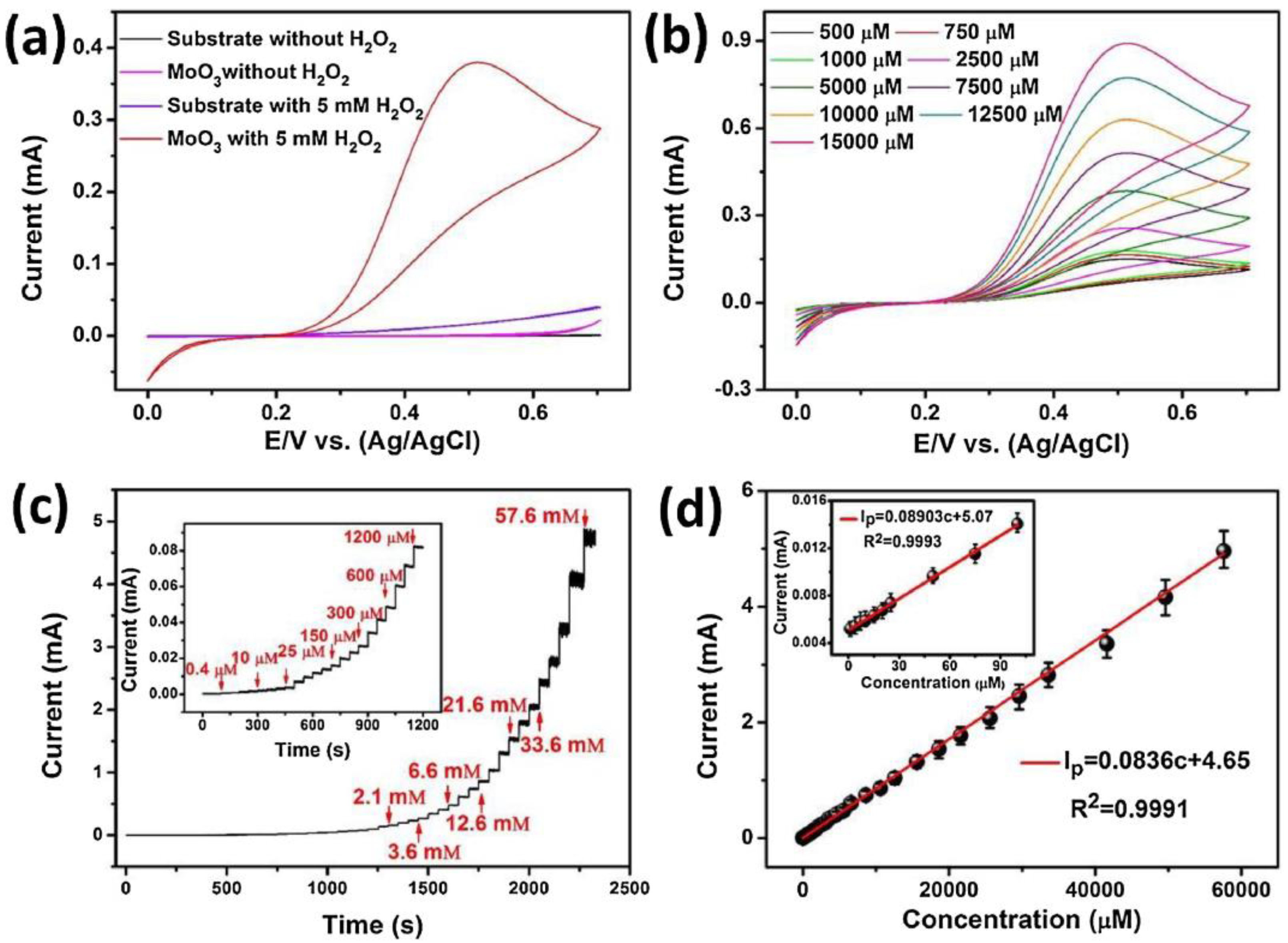
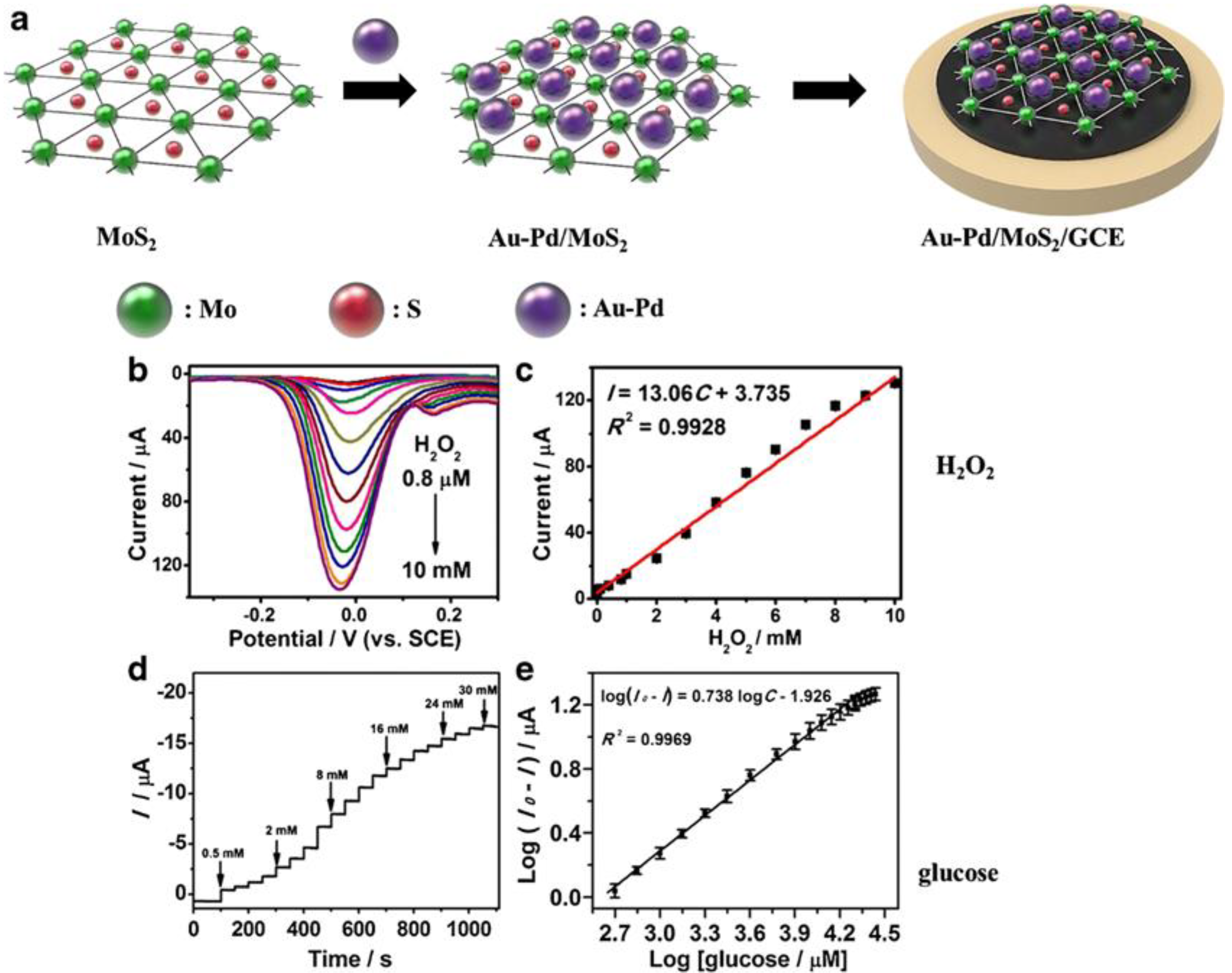
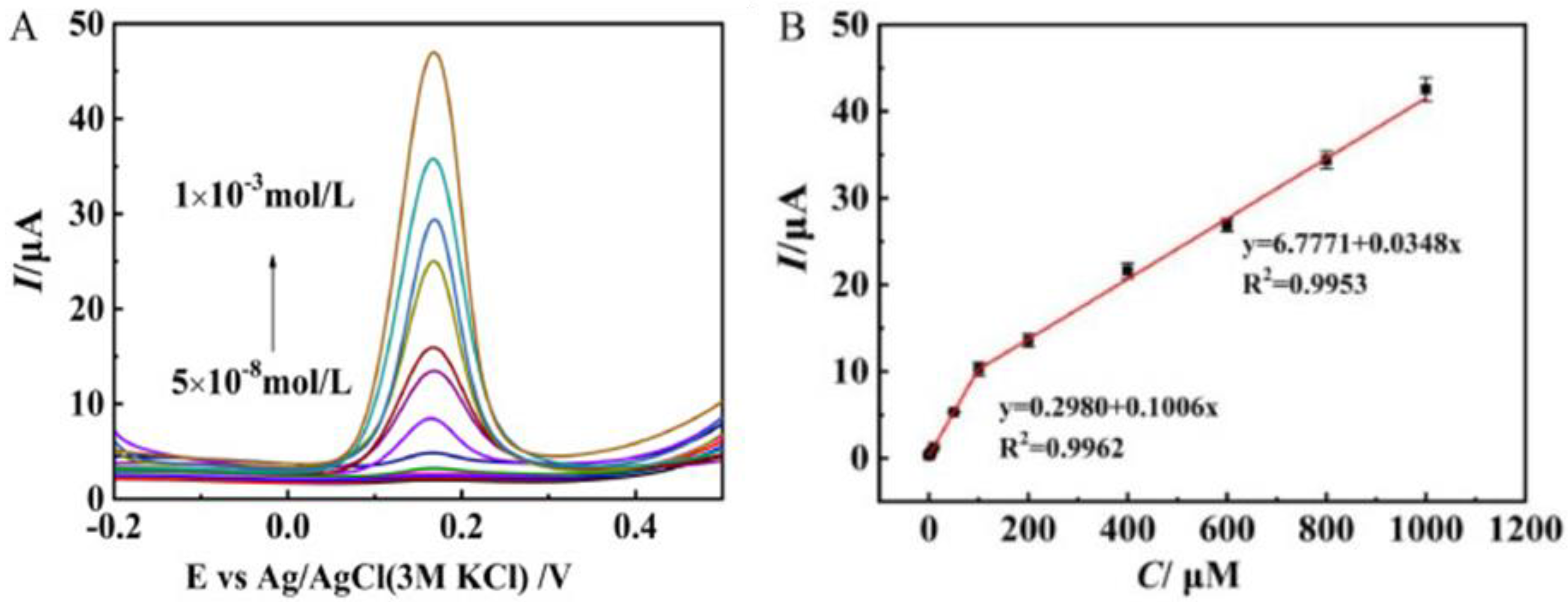
5.1. Hydrogen Peroxide
5.2. Glucose
5.3. Neurotransmitters
5.4. Vitamins
5.5. DNA
5.6. Other Biomolecules
6. Current and Future Perspectives

| Sensor | Target | Sensor Platform | Technique | Linear Range/Sensitivity | References |
|---|---|---|---|---|---|
| AuNPs@MoS2 | AA, DA, UA | GCE | DPV | 50 μM–100 mM, 0.05–30 μM, 50 μM–40 mM | [76] |
| AuNPs/MoS2/GN | NO- 2 | GCE | CV, Amp | 5.0 μM to 5.0 mM | [97] |
| 2D/MoS2 | Tyrosine | AuBSPE | CV | 1580 μA·mM−1·cm−2 | [49] |
| MoS2 NSs/N-GN | DA | GCE | CV | 3.2 μM–5.68 mM/75.49 μA·mM−1·cm−2 | [77] |
| ssDNA/MoS2-PANI | ssDNA | Pt | DPV | 10−15–10−6 M | [90] |
| ssRNA/AuNPs@MoS2-Ti3C2 | miRNA-182 | GCE | DPV | 1 fM–0.1 nM | [102] |
| Au-Pd/MoS2 | H2O2 | GCE | DPV, Amp | 0.8 µM–10 mM/−0.5–20 mM | [60] |
| AgCl/MoS2 | chloramphen | ITO | CV, Amp | 4–531 µM/3802 µA mM−1 cm−2 | [106] |
| MoS2 | UA | Flexible | DPV, Amp | 10–400 μM/98.3 ± 1 nA μM−1 | [93] |
| 2D-MoS2 | Cortisol | EIS | 1–500 ng/mL | [75] | |
| MoS2 | H2O2 | CC | CV, Amp | 5–3000 μM/5.3 μA mM−1 cm−2 | [61] |
| SrMoSe2 | MTZ | GCE | DPV | 0.05–914.92 μM/1.13 μA μM−1 cm−2 | [105] |
| MoSe2–GN | PDGF-BB | GCE | DPV | 0.0001–1 nM | [96] |
| HEG-MoSe2 | NADH | GCE | Amp | 1–280 μM/0.0814 µA⋅µM−1⋅cm−2 | [100] |
| MoSe2-GN/Ni | DA | - | CV | 0.01–10 μM | [78] |
| S-MoSe2/NSG/Au/MIPs | DA | GCE | CV, DPV | 0.05 μM–1000 μM | [83] |
| AuNPs/SiO2@MoSe2 | DNA | GCE | CV, DPV | 0.1 fM–100 pM | [91] |
| Mb@MnMoSe2 | H2O2 | GCE | CV, Amp | 0.09–60 μM/222.78 A cm−2 mM−1 | [63] |
| MoSe2-HGNs | SCCA | GCE | PhotoElectr | 1 pg∙mL−1 −50 ng∙mL−1 | [103] |
| Ag2Se-MoSe2-GSH | DA | GCE | Amp | 0.05−1110 μM/- | [79] |
| AuNPs-MoSe2-GN | CEA | GCE | CV, DPV | +0.001−100 ng∙mL−1/- | [84] |
| MoS2-Thi-AuNPs | MicroRNA-21 | GCE | SWV | 1.0 pM to 10.0 nM/- | [115] |
| MoS2–Au@Pt | Glucose | GCE | CV | 10 mM to 3 mM/- | [116] |
| Mn-MoS2 | DA | PGS | EIS, DPV | -/- | [117] |
| FMNs/MoS2 | PIK3CA Gene | GCE | CV, DPV | 10−16 mol l−1 to 10−8 mol l−1/- | [118] |
| Sensor | Target | Sensor Platform | Technique | Linear Range/Sensitivity | References |
|---|---|---|---|---|---|
| MoO3-GO | H2O2 | GCE | - | 0.92 μM–2.46 mM/391.32 μA mM−1 cm−2 | [66] |
| MoO3 | NO₂⁻ | GCE | CV | - | [98] |
| α-MoO3 | H2O2 | - | CV, EIS, Amp | 0.4 μM–57.6 mM/168.72 μA mM−1 cm−2 | [64] |
| MoO3·2H2O-GN | Thiourea | GCE | CV | 2.40 × 10⁻³–19.3 × 10⁻³ M | [120] |
| Sensor | Target | Sensor Platform | Technique | Linear Range/Sensitivity | References |
|---|---|---|---|---|---|
| MIP-Mo2C | FA | GCE | DPV | - | [86] |
| NCS/Mo2C | microRNA | GCE | CV, DPV, EIS, | 1 fM–1 nM | [92] |
| Luminol-AuNPs@Mo2C | α-fetoprotein | GCE | ECL | 0.1 pg·mL−1–30 ng·mL−1/- | [101] |
| MWCNTs-Mo2C | RIF | GCE | CV, DPV, CC | 0.5–74 μM/- | [99] |
7. Conclusions
Funding
Conflicts of Interest
References
- Neri, G. Thin 2D: The New Dimensionality in Gas Sensing. Chemosensors 2017, 5, 21. [Google Scholar] [CrossRef]
- Cao, X.; Halder, A.; Tang, Y.; Hou, C.; Wang, H.; Duus, J.O.; Chi, Q. Engineering two-dimensional layered nanomaterials for wearable biomedical sensors and power devices. Mater. Chem. Front. 2018, 2, 1944–1986. [Google Scholar] [CrossRef]
- Zeng, Y.; Lin, S.; Gu, D.; Li, X. Two-dimensional nanomaterials for gas sensing applications: The role of theoretical calculations. Nanomaterials 2018, 8, 851. [Google Scholar] [CrossRef] [PubMed] [Green Version]
- Duong, D.L.; Yun, S.J.; Lee, Y.H. Van der Waals Layered Materials: Opportunities and Challenges. ACS Nano 2017, 11, 11803–11830. [Google Scholar] [CrossRef] [PubMed]
- Dong, Y.; Wu, Z.S.; Ren, W.; Cheng, H.M.; Bao, X. Graphene: A promising 2D material for electrochemical energy storage. Sci. Bull. 2017, 62, 724–740. [Google Scholar] [CrossRef] [Green Version]
- Wang, Y.; Qiu, M.; Won, M.; Jung, E.; Fan, T.; Xie, N.; Chi, S.G.; Zhang, H.; Kim, J.S. Emerging 2D material-based nanocarrier for cancer therapy beyond graphene. Coord. Chem. Rev. 2019, 400, 213041. [Google Scholar] [CrossRef]
- Revuri, V.; Lee, Y.K. 2D material-based hybrid nanostructure for diagnosis and therapy. In Biomedical Applications of Graphene and 2D Nanomaterials; Nurunnabi, M., McCarthy, J.R., Eds.; Elsevier: Amsterdam, The Netherlands, 2018; Chapter 7; pp. 143–164. [Google Scholar]
- Ariga, K.; Ji, Q.; Hill, J.; Bando, P.; Aono, Y. Forming nanomaterials as layered functional structures toward materials nanoarchitectonics. NPG Asia Mater. 2012, 4, 17. [Google Scholar] [CrossRef] [Green Version]
- Zhang, X.; Ju, H.; Wang, J. Electrochemical Sensors, Biosensors and Their Biomedical Applications; Academic Press: Cambridge, MA, USA, 2007. [Google Scholar]
- Labib, M.; Sargent, E.H.; Kelley, S.O. Electrochemical methods for the analysis of clinically relevant biomolecules. Chem. Rev. 2016, 116, 9001–9090. [Google Scholar] [CrossRef]
- Cho, I.; Kim, D.H.; Park, S. Electrochemical biosensors: Perspective on functional nanomaterials for on-site analysis. Biomater. Res. 2020, 24, 6. [Google Scholar] [CrossRef] [Green Version]
- Maduraiveeran, G.; Sasidharan, M.; Ganesan, V. Electrochemical sensor and biosensor platforms based on advanced nanomaterials for biological and biomedical applications. Biosens. Bioelectron. 2018, 103, 113–129. [Google Scholar] [CrossRef]
- Bolotsky, A.; Butler, D.; Dong, C.; Gerace, K.; Glavin, N.R.; Muratore, C.; Robinson, J.A.; Ebrahimi, A. Two-dimensional materials in biosensing and healthcare: From in vitro diagnostics to optogenetics and beyond. ACS Nano 2019, 13, 9781–9810. [Google Scholar] [CrossRef] [PubMed] [Green Version]
- Yang, G.; Zhu, C.; Du, D.; Zhu, J.; Lin, Y. Graphene-like two-dimensional layered nanomaterials: Applications in biosensors and nanomedicine. Nanoscale 2015, 7, 14217–14231. [Google Scholar] [CrossRef] [PubMed]
- Kannan, P.K.; Late, D.J.; Morgan, H.; Rout, C.S. Recent developments in 2D layered inorganic nanomaterials for sensing. Nanoscale 2015, 7, 13293–13312. [Google Scholar] [CrossRef] [PubMed]
- Wang, Y.H.; Huang, K.J.; Wu, X. Recent advances in transition-metal dichalcogenides based electrochemical biosensors: A review. Biosens. Bioelectron. 2017, 97, 305–316. [Google Scholar] [CrossRef] [PubMed]
- Jin, W.; Maduraiveeran, G. Nanomaterial-based environmental sensing platforms using state-of-the-art electroanalytical strategies. J. Anal. Sci. Technol. 2018, 9, 18. [Google Scholar] [CrossRef]
- Bakir, M.Y.; Ozaydin, H.D.; Gorkan, T.; Aktürk, O.U.; Gokoglu, G.; Aktürk, E.; Ciraci, S. Free-standing and supported phosphorene nanoflakes: Shape- and size-dependent properties. Appl. Surf. Sci. 2020, 506, 144756. [Google Scholar] [CrossRef]
- Morgan, H.; Rout, C.S.; Late, D.J. Fundamentals and sensing applications of 2D materials. In Series in Electronic and Optical Materials; Woodhead Publishing: Sawston, UK, 2019; Chapter 4; p. 481. [Google Scholar]
- Gan, X.; Zhao, H.; Quan, X. Two-dimensional MoS2: A promising building block for biosensors. Biosens. Bioelectron. 2017, 89, 56–71. [Google Scholar] [CrossRef]
- Dalila, N.R.; Arshad, M.K.M.; Gopinath, S.C.B.; Norhaimi, W.M.W.; Fathil, M.F.M. Current and future envision on developing biosensors aided by 2D molybdenum disulfide (MoS2) productions. Biosens. Bioelectron. 2019, 132, 248–264. [Google Scholar] [CrossRef]
- Pauling, L. The Structural Chemistry of Molybdenum. In Molybdenum Compounds: Their Chemistry and Technology; Killeffer, D.H., Linz, A., Eds.; Interscience: New York, NY, USA, 1952; p. 407. [Google Scholar]
- Manthiram, K.; Alivisatos, A. Tunable localized surface plasmon resonances in tungsten oxide nanocrystals. J. Am. Chem. Soc. 2012, 134, 3995–3998. [Google Scholar] [CrossRef]
- Inzani, K.; Nematollahi, M.; Vullum-Bruer, F.; Grande, T.; Reenaas, T.W.; Selbach, S.M. Electronic properties of reduced molybdenum oxides. Phys. Chem. Chem. Phys. 2017, 19, 9232–9245. [Google Scholar] [CrossRef]
- De Castro, I.A.; Datta, R.S.; Ou, J.Z.; Castellanos-Gomez, A.; Sriram, S.; Daeneke, T.; Kalantar-zadeh, K. Molybdenum oxides—From fundamentals to functionality. Adv. Mater. 2017, 29, 1701619. [Google Scholar] [CrossRef] [PubMed]
- Halim, J.; Kota, S.; Lukatskaya, M.R.; Naguib, M.; Zhao, M.Q.; Moon, E.J.; Pitock, J.; Nanda, J.; May, S.J.; Gogotsi, Y.; et al. Synthesis and Characterization of 2D Molybdenum Carbide (MXene). Adv. Funct. Mater. 2016, 26, 3118–3127. [Google Scholar] [CrossRef]
- Dos Santos Politi, J.R.; Viñes, F.; Rodriguez, J.A.; Illa, F. Atomic and electronic structure of molybdenum carbide phases: Bulk and low Miller-index surfaces. Phys. Chem. Chem. Phys. 2013, 15, 12617–12625. [Google Scholar] [CrossRef] [PubMed]
- Wang, T.; Liu, X.W.; Wang, S.G.; Huo, C.F.; Li, Y.W.; Wang, J.G.; Jiao, H.J. Stability of β-Mo2C Facets from ab Initio Atomistic Thermodynamics. J. Phys. Chem. C 2011, 115, 22360–22368. [Google Scholar] [CrossRef]
- Chang, K.; Chen, W. l-Cysteine-Assisted Synthesis of Layered MoS2/Graphene Composites with Excellent Electrochemical Performances for Lithium Ion Batteries. ACS Nano 2011, 5, 4720–4728. [Google Scholar] [CrossRef]
- Jiang, Y.; Li, X.; Yu, S.; Jia, L.; Zhao, X.; Wang, C. Reduced Graphene oxide-modified carbon nanotube/polyimide film supported MoS2 nanoparticles for electrocatalytic hydrogen evolution. Adv. Funct. Mater. 2015, 25, 2693–2700. [Google Scholar] [CrossRef]
- Oakes, L.; Carter, R.; Hanken, T.; Cohn, A.P.; Share, K.; Schmidt, B.; Pint, C.L. Interface strain in vertically stacked two-dimensional heterostructured carbon-MOS2 nanosheets controls electrochemical reactivity. Nat. Commun. 2016, 7, 11796. [Google Scholar] [CrossRef]
- Zhang, W.; Wang, Q.; Chen, Y.; Wang, Z.; Wee, A.T.S. Van Der Waals stacked 2D layered materials for optoelectronics. 2D Mater. 2016, 3, 022001. [Google Scholar] [CrossRef]
- Jeon, J.; Park, Y.; Choi, S.; Lee, J.; Lim, S.S.; Lee, B.H.; Song, Y.J.; Cho, J.H.; Jang, Y.H.; Lee, S. Epitaxial synthesis of molybdenum carbide and formation of a Mo2C/MoS2 hybrid structure via chemical conversion of molybdenum disulfide. ACS Nano 2018, 12, 338–346. [Google Scholar] [CrossRef]
- Zhang, H.J.; Wang, K.X.; Wu, X.Y.; Jiang, Y.M.; Zhai, Y.B.; Wang, C.; Wei, X.; Chen, J.S. MoO2/Mo2C heteronanotubes function as high-performance Li-ion battery electrode. Adv. Funct. Mater. 2014, 24, 3399–3404. [Google Scholar] [CrossRef]
- Jauberteau, I.; Bessaudou, A.; Mayet, R.; Cornette, J.; Jauberteau, J.L.; Carles, P.; Merle-Méjean, T. Molybdenum nitride films: Crystal structures, synthesis, mechanical, electrical and some other properties. Coatings 2015, 5, 656–687. [Google Scholar] [CrossRef] [Green Version]
- Yang, L.; Chen, W.; Yang, R.; Chen, A.; Zhang, H.; Sun, Y.; Jia, Y.; Li, X.; Tang, Z.; Gui, X. Fabrication of MoOx/Mo2C-layered hybrid structures by direct thermal oxidation of Mo2C. ACS Appl. Mater. Interfaces 2020, 12, 10755–10762. [Google Scholar] [CrossRef] [PubMed]
- Kim, H.G.; Lee, H.B.R. Atomic Layer Deposition on 2D materials. Chem. Mater. 2017, 29, 3809–3826. [Google Scholar] [CrossRef]
- Venkata Subbaiah, Y.P.; Saji, K.J.; Tiwari, A. Atomically thin MoS2: A versatile nongraphene 2D material. Adv. Funct. Mater. 2016, 26, 2046–2069. [Google Scholar] [CrossRef]
- Rigi, V.J.C.; Jayaraj, M.K.; Saji, K.J. Envisaging radio frequency magnetron sputtering as an efficient method for large scale deposition of homogeneous two dimensional MoS2. Appl. Surf. Sci. 2020, 529, 147158. [Google Scholar] [CrossRef]
- Sun, Z.; Liao, T.; Kou, L. Strategies for designing metal oxide nanostructures. Sci. China Mater. 2017, 60, 1–24. [Google Scholar] [CrossRef] [Green Version]
- Bhagyaraj, S.M.; Oluwafemi, O.S.; Kalarikkal, N.; Thomas, S. Synthesis of inorganic nanomaterials: Advances and key technologies. In Micro and Nano Technologies; Woodhead Publishing: Sawston, UK, 2018; Chapter 3; pp. 59–88. [Google Scholar]
- Eftekhari, A.; Bagher Rahmani, M.; Masdarolomoor, F. Effect of Sn doping on structural and optical properties of 2D α-MoO3 nanostructures. J. Part. Sci. Technol. 2016, 2, 87–93. [Google Scholar]
- Meshkian, R.; Näslund, L.; Halim, J.; Lu, J.; Barsoum, M.W.; Rosen, J. Synthesis of two-dimensional molybdenum carbide, Mo2C, from the gallium based atomic laminate Mo2Ga2C. Scr. Mater. 2015, 108, 147–150. [Google Scholar] [CrossRef] [Green Version]
- Verger, L.; Natu, V.; Carey, M.; Barsoum, M.W. MXenes: An introduction of their synthesis, select properties, and applications. Trends. Chem. 2019, 1, 656–669. [Google Scholar] [CrossRef]
- Zhang, C.; Wu, W.; Lu, Y.; Hou, S. Strategies of fabricating graphene and graphene-analogous 2D nanosheets. Ceramics 2018, 62, 211–220. [Google Scholar] [CrossRef]
- Midya, A.; Ghorai, A.; Mukherjee, S.; Maiti, R.; Ray, S.K. Hydrothermal growth of few layer 2H-MoS2 for heterojunction photodetector and visible light induced photocatalytic applications. J. Mater. Chem. A 2016, 4, 534–4543. [Google Scholar] [CrossRef]
- Peng, Y.; Meng, Z.; Zhong, C.; Lu, J.; Yu, W.; Jia, Y.; Qian, Y. Hydrothermal Synthesis and Characterization of Single-Molecular-Layer MoS2and MoSe2. Chem. Lett. 2001, 8, 772–773. [Google Scholar] [CrossRef]
- Yao, J.D.; Zheng, Z.Q.; Yang, G.W. Production of large-area 2D materials for high-performance photodetectors by pulsed-laser deposition. Prog. Mater. Sci. 2019, 106, 100573. [Google Scholar] [CrossRef]
- Zribi, R.; Maalej, R.; Messina, E.; Gillibert, R.; Donato, M.G.; Maragò, O.M.; Gucciardi, P.G.; Leonardi, S.G.; Neri, G. Exfoliated 2D-MoS2 nanosheets on carbon and gold screen printed electrodes for enzyme-free electrochemical sensing of tyrosine. Sens. Actuators B 2020, 303, 127229. [Google Scholar] [CrossRef]
- Zribi, R.; Maalej, R.; Gillibert, R.; Donato, M.G.; Gucciardi, P.G.; Leonardi, S.G.; Neri, G. Simultaneous and selective determination of dopamine and tyrosine in the presence of uric acid with 2D-MoS2 nanosheets modified screen-printed carbon electrodes. Flat. Chem. 2020, in press. [Google Scholar] [CrossRef]
- Cheng, H.; Kamegawa, T.; Mori, K.; Yamashita, H. Surfactant-free nonaqueous synthesis of plasmonic molybdenum oxide nanosheets with enhanced catalytic activity for hydrogen generation from ammonia borane under visible light. Angew. Chem. Int. Ed. 2014, 126, 2954–2958. [Google Scholar] [CrossRef]
- Cheng, H.; Wen, M.; Ma, X.; Kuwahara, Y.; Mori, K.; Dai, Y.; Huang, B.; Yamashita, H. Hydrogen doped metal oxide semiconductors with exceptional and tunable localized surface plasmon resonances. J. Am. Chem. Soc. 2016, 138, 9316–9324. [Google Scholar] [CrossRef]
- Liu, W.; Xu, Q.; Cui, W.; Zhu, C.; Qi, Y. CO2-Assisted fabrication of two-dimensional amorphous molybdenum oxide nanosheets for enhanced plasmon resonances. Angew. Chem. Int. Ed. 2017, 56, 1600–1604. [Google Scholar] [CrossRef]
- Dong, W.; Ham, J.; Jung, G.; Son, J.; Lee, J. Ultrafast laser-assisted synthesis of hydrogenated molybdenum oxides for flexible organic solar cells. J. Mater. Chem. A 2016, 4, 4755–4762. [Google Scholar] [CrossRef] [Green Version]
- Li, R.; An, H.; Huang, W.; He, Y. Molybdenum oxide nanosheets meet ascorbic acid: Tunable surface plasmon resonance and visual colorimetric detection at room temperature. Sens. Actuators B 2018, 259, 59–63. [Google Scholar] [CrossRef]
- Vogiazi, V.; de la Cruz, A.; Mishra, S.; Shanov, V.; Heineman, W.R.; Dionysiou, D. A comprehensive review: Development of electrochemical biosensors for detection of cyanotoxins in freshwater. ACS Sens. 2019, 4, 1151–1173. [Google Scholar] [CrossRef] [PubMed]
- Stradiotto, N.R.; Yamanaka, H.; Zanoni, M.V.B. Electrochemical sensors: A powerful tool in analytical chemistry. J. Braz. Chem. Soc. 2003, 14, 2. [Google Scholar] [CrossRef] [Green Version]
- Chen, W.; Cai, S.; Ren, Q.; Wen, W.; Zhao, Y. Recent advances in electrochemical sensing for hydrogen peroxide: A review. Analyst 2012, 137, 49–58. [Google Scholar] [CrossRef]
- Grisham, M.B. Methods to detect hydrogen peroxide in living cells: Possibilities and pitfalls. Comp. Biochem. Physiol. Part A Mol. Integr. Physiol. 2013, 165, 429–438. [Google Scholar] [CrossRef]
- Li, X.; Du, X. Molybdenum disulfide nanosheets supported Au-Pd bimetallic nanoparticles for non-enzymatic electrochemical sensing of hydrogen peroxide and glucose. Sens. Actuators B Chem. 2017, 239, 536–543. [Google Scholar] [CrossRef]
- Du, H.T.; Zhang, X.Y.; Liu, Z.; Qu, F.L. A supersensitive biosensor based on MoS2 nanosheet arrays for the real-time detection of H2O2 secreted from living cells. Chem. Commun. 2019, 55, 9653–9656. [Google Scholar] [CrossRef] [PubMed]
- He, W.; Huang, Y.; Wu, J. Enzyme-free glucose biosensors based on MoS2 nanocomposites. Nanoscale Res. Lett. 2020, 15, 60. [Google Scholar] [CrossRef] [PubMed]
- Ramaraj, S.; Sakthivel, M.; Chen, S.M.; Lou, B.S.; Ho, K.C. Defect and additional active sites on the basal plane of manganese-doped molybdenum diselenide for effective enzyme immobilization: In vitro and in vivo real-time analyses of hydrogen peroxide sensing. ACS Appl. Mater. Interfaces 2019, 11, 7862–7871. [Google Scholar] [CrossRef]
- Wei, Z.; Hai, Z.; Akbari, M.K.; Qi, D.; Xing, K.; Zhao, Q.; Verpoort, F.; Hu, J.; Hyde, L.; Zhuiykov, S. Atomic layer deposition-developed two-dimensional α-MoO3 windows excellent hydrogen peroxide electrochemical sensing capabilities. Sens. Actuators B 2018, 262, 334–344. [Google Scholar] [CrossRef]
- Zhang, J.; Han, D.; Wang, Y.; Wang, L.; Chen, X.; Qiao, X.; Yu, X. Synergy between nanozymes and natural enzymes on the hybrid MoS2 nanosheets/graphite microfiber for enhanced voltammetric determination of hydrogen peroxide. Microchim. Acta 2020, 187, 321. [Google Scholar] [CrossRef]
- Li, B.; Song, H.-Y.; Deng, Z.-P.; Huo, L.-H.; Gao, S. Novel sensitive amperometric hydrogen peroxide sensor using layered hierarchical porous α-MoO3 and GO modified glass carbon electrode. Sens. Actuators B 2019, 288, 641–648. [Google Scholar] [CrossRef]
- Azharudeen, A.; Karthiga, R.; Rajarajan, M.; Suganthi, A. Selective enhancement of non-enzymatic glucose sensor by used PVP modified on α-MoO3 nanomaterials. Microchem. J. 2020, 157, 105006. [Google Scholar] [CrossRef]
- Zhai, Y.; Li, J.; Chu, X.; Xu, M.; Jin, F.; Li, X.; Fang, X.; Wei, Z.P.; Wang, X.H. MoS2 microflowers based electrochemical sensing platform for non-enzymatic glucose detection. J. Alloys Compd. 2016, 672, 600–608. [Google Scholar] [CrossRef]
- Parlak, O.; İncel, A.; Uzun, L.; Turner, A.P.F.; Tiwari, A. Structuring Au nanoparticles on two-dimensional MoS2 nanosheets for electrochemical glucose biosensors. Biosens. Bioelectron. 2017, 89, 545–550. [Google Scholar] [CrossRef] [PubMed]
- Arunbalaji, S.; Vasudevan, R.; Arivanandhan, M.; Alsalme, A.; Alghamdi, A.; Jayavel, R. CuO/MoS2 nanocomposites for rapid and high sensitive non-enzymatic glucose sensors. Ceram. Int. 2020, 46, 16879–16885. [Google Scholar] [CrossRef]
- Raza, M.H.; Movlaee, K.; Wu, Y.; El-Refaei, S.M.; Karg, M.; Leonardi, S.G.; Neri, G.; Pinna, N. Tuning the NiO thin film morphology on carbon nanotubes by atomic layer deposition for enzyme-free glucose sensing. ChemElectroChem 2019, 6, 383–392. [Google Scholar] [CrossRef]
- Huang, J.; He, Y.; Jin, J.; Li, Y.; Dong, Z.; Li, R. A novel glucose sensor based on MoS2 nanosheet functionalized with Ni nanoparticles. Electrochim. Acta 2014, 136, 41–46. [Google Scholar] [CrossRef]
- Ma, K.; Sinha, A.; Dang, X.; Zhao, H. Electrochemical preparation of gold nanoparticles-polypyrrole co-decorated 2D MoS2 nanocomposite sensor for sensitive detection of glucose. J. Electrochem. Soc. 2019, 166, B147. [Google Scholar] [CrossRef]
- Huang, J.; Dong, Z.; Li, Y.; Li, J.; Tang, W.; Yang, H.; Wang, J.; Bao, Y.; Jin, J.; Li, R. MoS2 nanosheet functionalized with Cu nanoparticles and its application for glucose detection. Mater. Res. Bull. 2013, 48, 4544–4547. [Google Scholar] [CrossRef]
- Kinnamon, D.; Ghanta, R.; Lin, K.; Muthukumar, S.; Prasad, S. Portable biosensor for monitoring cortisol in low-volume perspired human sweat. Sci. Rep. 2017, 7, 13312. [Google Scholar] [CrossRef] [Green Version]
- Sun, H.; Chao, J.; Zuo, X.; Su, S.; Liu, X.; Yuwen, L.; Fan, C.; Wang, L. Gold nanoparticle-decorated MoS2 nanosheets for simultaneous detection of ascorbic acid, dopamine and uric acid. RSC Adv. 2014, 4, 27625. [Google Scholar] [CrossRef]
- Bacha, L.G.; Nguyen, D.M.; Bui, Q.B.; Ai-Le, P.H.; Nhac-Vu, H.-T. Hierarchical molybdenum dichalcogenide nanosheets assembled nitrogen doped graphene layers for sensitive electrochemical dopamine detection. Mater. Chem. Phys. 2019, 236, 121814. [Google Scholar] [CrossRef]
- Huang, K.J.; Zhang, J.Z.; Cai, J.L. Preparation of porous layered molybdenum selenide-graphene composites on Ni foam for high-performance supercapacitor and electrochemical sensing. Electrochim. Acta. 2015, 180, 770–777. [Google Scholar] [CrossRef]
- Xia, X.; Shen, X.; Du, Y.; Ye, W.; Wang, C. Study on glutathione’s inhibition to dopamine polymerization and its application in dopamine determination in alkaline environment based on silver selenide/molybdenum selenide/glassy carbon electrode. Sens. Actuators B 2016, 237, 685–692. [Google Scholar] [CrossRef] [Green Version]
- Zou, H.L.; Li, B.L.; Luo, H.Q.; Li, N.B. 0D-2D heterostructures of Au nanoparticles and layered MoS2 for simultaneous detections of dopamine, ascorbic acid, uric acid, and nitrite. Sens. Actuators B Chem. 2017, 253, 352–360. [Google Scholar] [CrossRef]
- Mani, V.; Govindasamy, M.; Chen, S.-M.; Karthik, R.; Huang, S.-T. Determination of dopamine using a glassy carbon electrode modified with a graphene and carbon nanotube hybrid decorated with molybdenum disulfide flowers. Microchim. Acta 2016, 183, 2267–2275. [Google Scholar] [CrossRef]
- Murugesan, D.; Moulaee, K.; Neri, G.; Ponpandian, N.; Viswanathan, C. α-MoO3 nanostructure on carbon cloth substrate for dopamine detection. Nanotechnology 2019, 30, 265501. [Google Scholar] [CrossRef]
- Zang, Y.J.; Nie, J.; He, B.; Yin, W.; Zheng, J.; Hou, C.J.; Huo, D.Q.; Yang, M.; Liu, F.M.; Sun, Q.Q.; et al. Fabrication of S-MoSe2/NSG/Au/MIPs imprinted composites for electrochemical detection of dopamine based on synergistic effect. Microchem. J. 2020, 156, 104845. [Google Scholar] [CrossRef]
- Pramoda, K.; Moses, K.; Maitra, U.; Rao, C. Superior performance of a MoS2-RGO composite and a Borocarbonitride in the electrochemical detection of dopamine, uric acid and adenine. Electroanalysis 2015, 27, 1892–1898. [Google Scholar] [CrossRef]
- Horton, H.; Moran, L.; Ochs, R.; Rawn, J.; Scrimgeour, K. Principles of Biochemistry; Prentice-Hall: Saddle River, NJ, USA, 1996. [Google Scholar]
- Hussain, S.; Zaidi, S.A.; Vikraman, D.; Kim, H.-S.; Jung, J. Facile preparation of molybdenum carbide (Mo2C) nanoparticles and its effective utilization in electrochemical sensing of folic acid via imprinting. Biosens. Bioelectron. 2019, 140, 111330. [Google Scholar] [CrossRef]
- Mani, V.; Govindasamy, M.; Chen, S.-M.; Subramani, B.; Sathiyan, A.; Princy Merlin, J. Determination of Folic Acid Using Graphene/Molybdenum Disulfide Nanosheets/Gold Nanoparticles Ternary Composite. J. Electrochem. Sci. 2017, 12, 258–267. [Google Scholar] [CrossRef]
- Chekin, F.; Teodorescu, F.; Coffinier, Y.; Pan, G.-H.; Barras, A.; Boukherroub, R.; Szunerits, S. MoS2/reduced graphene oxide as active hybrid material for the electrochemical detection of folic acid in human serum. Biosens. Bioelectron. 2016, 85, 807–813. [Google Scholar] [CrossRef] [PubMed]
- Drummond, T.G.; Hill, M.G.; Barton, J.K. Electrochemical DNA sensors. Nat. Biotechnol. 2003, 21, 1192–1199. [Google Scholar] [CrossRef] [PubMed] [Green Version]
- Dutta, S.; Dutta Chowdhury, A.; Biswas, S.; Park, E.Y.; Agnihotri, N.; De, A.; De, S. Development of an effective electrochemical platform for highly sensitive DNA detection using MoS2-polyaniline nanocomposites. Biochem. Eng. J. 2018, 140, 130–139. [Google Scholar] [CrossRef]
- Shuai, H.L.; Wu, X.; Huang, K.J.; Zhai, Z.B. Ultrasensitive electrochemical biosensing platform based on spherical silicon dioxide/molybdenum selenide nanohybrids and triggered Hybridization Chain Reaction. Biosens. Bioelectron. 2017, 94, 616–625. [Google Scholar] [CrossRef]
- Tian, L.; Qi, J.; Ma, X.; Wang, X.; Yao, C.; Song, W.; Wang, Y. A facile DNA strand displacement reaction sensing strategy of electrochemical biosensor based on N-carboxymethyl chitosan/molybdenum carbide nanocomposite for microRNA-21 detection. Biosens. Bioelectron. 2018, 122, 43–50. [Google Scholar] [CrossRef]
- Sha, R.; Vishnu, N.; Badhulika, S. MoS2 based ultra-low-cost, flexible, non-enzymatic and non-invasive electrochemical sensor for highly selective detection of uric acid in human urine samples. Sens. Actuators B 2019, 279, 53–60. [Google Scholar] [CrossRef]
- Selvam, S.P.; Hansa, M.; Yun, K. Simultaneous differential pulse voltammetric detection of uric acid and melatonin based on a self-assembled Au nanoparticle–MoS2 nanoflake sensing platform. Sens. Actuators B 2020, 307, 127683. [Google Scholar] [CrossRef]
- Liu, J.; Xu, N.; Men, H.; Li, S.; Lu, Y.; Low, S.S.; Li, X.; Zhu, L.; Cheng, C.; Xu, G.; et al. Salivary cortisol determination on smartphone-based differential pulse voltammetry system. Sensors 2020, 20, 1422. [Google Scholar] [CrossRef] [Green Version]
- Huang, K.J.; Shuai, H.L.; Zhang, J.Z. Ultrasensitive sensing platform for platelet-derived growth factor BB detection based on layered molybdenum selenide–graphene composites and Exonuclease III assisted signal amplification. Biosens. Bioelectron. 2016, 77, 69–75. [Google Scholar] [CrossRef]
- Han, Y.; Zhang, R.; Dong, C.; Cheng, F.; Guo, Y. Sensitive electrochemical sensor for nitrite ions based on rose-like AuNPs/MoS2/graphene composite. Biosens. Bioelectron. 2019, 142, 111529. [Google Scholar] [CrossRef] [PubMed]
- Rocha, J.R.C.; Kosminsky, L.; Paixao, T.R.L.C.; Bertotti, M. Anodic oxidation of nitrite at a molybdenum oxide layer. Electroanalysis 2001, 13, 2. [Google Scholar] [CrossRef]
- Huang, Q.; Li, X.; Feng, S.; Zhuge, W.; Liu, F.; Peng, J.; Mo, S. An electrochemical sensor based on the composite of molybdenum carbides and a multiwalled carbon nanotube modified electrode for the ultrasensitive detection of rifampicin. Anal. Meth. 2018, 10, 3594–3601. [Google Scholar] [CrossRef]
- Karunagaran, S.; Prabhakaran, A.; Arumugam, P.; Berchmans, S.; Nayak, P. 2D MoSe2 sheets embedded over a high surface graphene hybrid for the amperometric detection of NADH. Microchim. Acta 2018, 185, 411. [Google Scholar]
- Zhu, X.; Zhai, Q.; Gu, W.; Li, J.; Wang, E. High-sensitivity electrochemiluminescence probe with molybdenum carbides as nanocarriers for α-fetoprotein sensing. Anal. Chem. 2017, 89, 12108–12114. [Google Scholar] [CrossRef]
- Liu, L.; Wei, Y.; Jiao, S.; Zhu, S.; Liu, X. A novel label-free strategy for the ultrasensitive miRNA-182 detection based on MoS2/Ti3C2 nanohybrids. Biosens. Bioelectron. 2019, 137, 45–51. [Google Scholar] [CrossRef]
- Ye, X.; Li, M.; Hu, Y.; Wang, Z.; Wang, Y.; Deng, H.; Xiong, X.; Li, C.; Sun, D. Sensitive photoelectrochemical immunosensor for squamous cell carcinoma antigen based on MoSe2, nanosheets and hollow gold nanospheres. Sens. Actuators B 2018, 275, 199–205. [Google Scholar] [CrossRef]
- He, B. Differential pulse voltammetric assay for the carcinoembryonic antigen using a glassy carbon electrode modified with layered molybdenum selenide, graphene, and gold nanoparticles. Microchim. Acta 2016, 184, 229–235. [Google Scholar] [CrossRef]
- Sakthivel, M.; Sukanya, R.; Chen, S.M.; Dinesh, B. Synthesis of two-dimensional Sr-doped MoSe2 nanosheets and its application for efficient electrochemical reduction of metronidazole. J. Phys. Chem. C 2018, 122, 12474–12484. [Google Scholar] [CrossRef]
- Li, Y.; Dai, H.; Feng, N.; Xie, X.; Zhang, J.; Li, W. Silver chloride nanoparticles-decorated molybdenum disulfide nanosheets for highly sensitive chloramphenicol detection. Mater. Express 2019, 9, 1. [Google Scholar] [CrossRef]
- Zhang, H.; Wang, T.; Qiu, Y.; Fu, F.; Yu, Y. Electrochemical behavior and determination of baicalin on a glassy carbon electrode modified with molybdenum disulfide nano-sheets. J. Electroanal. Chem. 2016, 775, 286–291. [Google Scholar] [CrossRef]
- Yang, R.; Zhao, J.; Chen, M.; Yang, T.; Luo, S.; Jiao, K. Electrocatalytic determination of chloramphenicol based on molybdenum disulfide nanosheets and self-doped polyaniline. Talanta 2015, 131, 619–623. [Google Scholar] [CrossRef] [PubMed]
- Zhang, C.; Ping, J.; Ye, Z.; Ying, Y. Two-dimensional nanocomposite-based electrochemical sensor for rapid determination of trans-resveratrol. Sci. Total Environ. 2020, 742, 140351. [Google Scholar] [CrossRef] [PubMed]
- Veeralingam, S.; Badhulika, S. X (metal: Al, Cu, Sn, Ti)-functionalized tunable 2D-MoS2 nanostructure assembled biosensor arrays for qualitative and quantitative analysis of vital neurological drugs. Nanoscale 2020, 12, 15336–15347. [Google Scholar] [CrossRef] [PubMed]
- Rawat, B.; Mishra, K.K.; Barman, U.; Arors, L.; Pal, D.; Paily, R.P. Two-dimensional MoS2-based electrochemical biosensor for highly selective detection of glutathione. IEEE Sens. J. 2020, 20, 6937–6944. [Google Scholar] [CrossRef]
- Sagar, P.; Srivastava, M.; Prakash, R.; Srivastava, S.K. The fabrication of an MoS2 QD-AuNP modified screen-printed electrode for the improved electrochemical detection of cefixime. Anal. Methods 2020, 12, 3014–3024. [Google Scholar] [CrossRef]
- Balasubramanian, P.; Annalakshmi, M.; Chen, S.-M.; Chen, T.-W. Sonochemical synthesis of molybdenum oxide (MoO3) microspheres anchored graphitic carbon nitride (g-C3N4) ultrathin sheets for enhanced electrochemical sensing of Furazolidone. Ultrason. Sonochem. 2019, 50, 96–104. [Google Scholar] [CrossRef]
- Sun, Y.; Wang, Y.; Chen, J.Y.C.; Fujisawa, K.; Holder, C.F.; Miller, J.T.; Crespi, V.H.; Terrones, M.; Schaak, R.E. Interface-mediated noble metal deposition on transition metal dichalcogenide nanostructures. Nature Chem. 2020, 12, 284–293. [Google Scholar] [CrossRef]
- Zhu, D.; Liu, W.; Zhao, D.; Hao, Q.; Li, J.; Huang, J.; Shi, J.; Chao, J.; Su, S.; Wang, L. Label-free electrochemical sensing platform for microRNA-21 detection using thionine and gold nanoparticles co-functionalized MoS2 nanosheet. ACS Appl. Mater. Interfaces 2017, 9, 35597–35603. [Google Scholar] [CrossRef]
- Su, S.; Lu, Z.; Li, J.; Hao, Q.; Liu, W.; Zhu, C.; Shen, X.; Shi, J.; Wang, L. MoS2–Au@Pt nanohybrids as a sensing platform for electrochemical non-enzymatic glucose detection. New J. Chem. 2018, 42, 6750–6755. [Google Scholar] [CrossRef]
- Lei, Y.; Derrick Butler, D.; Lucking, M.C.; Zhang, F.; Xia, T.; Fujisawa, K.; Granzier-Nakajima, T.; Cruz-Silva, R.; Endo, M.; Terrones, H.; et al. Single-atom doping of MoS2 with manganese enables ultrasensitive detection of dopamine: Experimental and computational approach. Sci. Adv. 2020, 6. [Google Scholar] [CrossRef] [PubMed]
- Zhang, W.; Yang, J.; Wu, D. Surface-functionalized MoS2 nanosheets sensor for direct electrochemical detection of PIK3CA gene related to lung cancer. J. Electrochem. Soc. 2020, 167, 027501. [Google Scholar] [CrossRef]
- Kukkar, M.; Mohanta, G.C.; Tuteja, S.K.; Kumar, P.; Bhadwal, A.S.; Samaddar, P.; Kim, K.H.; Deep, A. A comprehensive review on nano-molybdenum disulfide/DNA interfaces as emerging biosensing platforms. Biosens. Bioelectron. 2018, 107, 244–258. [Google Scholar] [CrossRef] [PubMed]
- Zhang, X.; Li, K.; Li, H.; Lu, J.; Zhang, L. Molybdenum trioxide dihydrate-graphene composite for electrochemical detection of thiourea molecule. Nano 2016, 11, 1650036. [Google Scholar] [CrossRef]
- Chen, X.; Berner, N.C.; Backes, C.; Duesberg, G.S.; McDonald, A.R. Functionalization of two-dimensional MoS2: On the reaction between MoS2 and organic thiols. Angew. Chem. Int. Ed. 2016, 55, 5803–5808. [Google Scholar] [CrossRef] [PubMed]

© 2020 by the authors. Licensee MDPI, Basel, Switzerland. This article is an open access article distributed under the terms and conditions of the Creative Commons Attribution (CC BY) license (http://creativecommons.org/licenses/by/4.0/).
Share and Cite
Zribi, R.; Neri, G. Mo-Based Layered Nanostructures for the Electrochemical Sensing of Biomolecules. Sensors 2020, 20, 5404. https://doi.org/10.3390/s20185404
Zribi R, Neri G. Mo-Based Layered Nanostructures for the Electrochemical Sensing of Biomolecules. Sensors. 2020; 20(18):5404. https://doi.org/10.3390/s20185404
Chicago/Turabian StyleZribi, Rayhane, and Giovanni Neri. 2020. "Mo-Based Layered Nanostructures for the Electrochemical Sensing of Biomolecules" Sensors 20, no. 18: 5404. https://doi.org/10.3390/s20185404