Ultrasensitive and Highly Selective Graphene-Based Field-Effect Transistor Biosensor for Anti-Diuretic Hormone Detection

 ,
,
Abstract
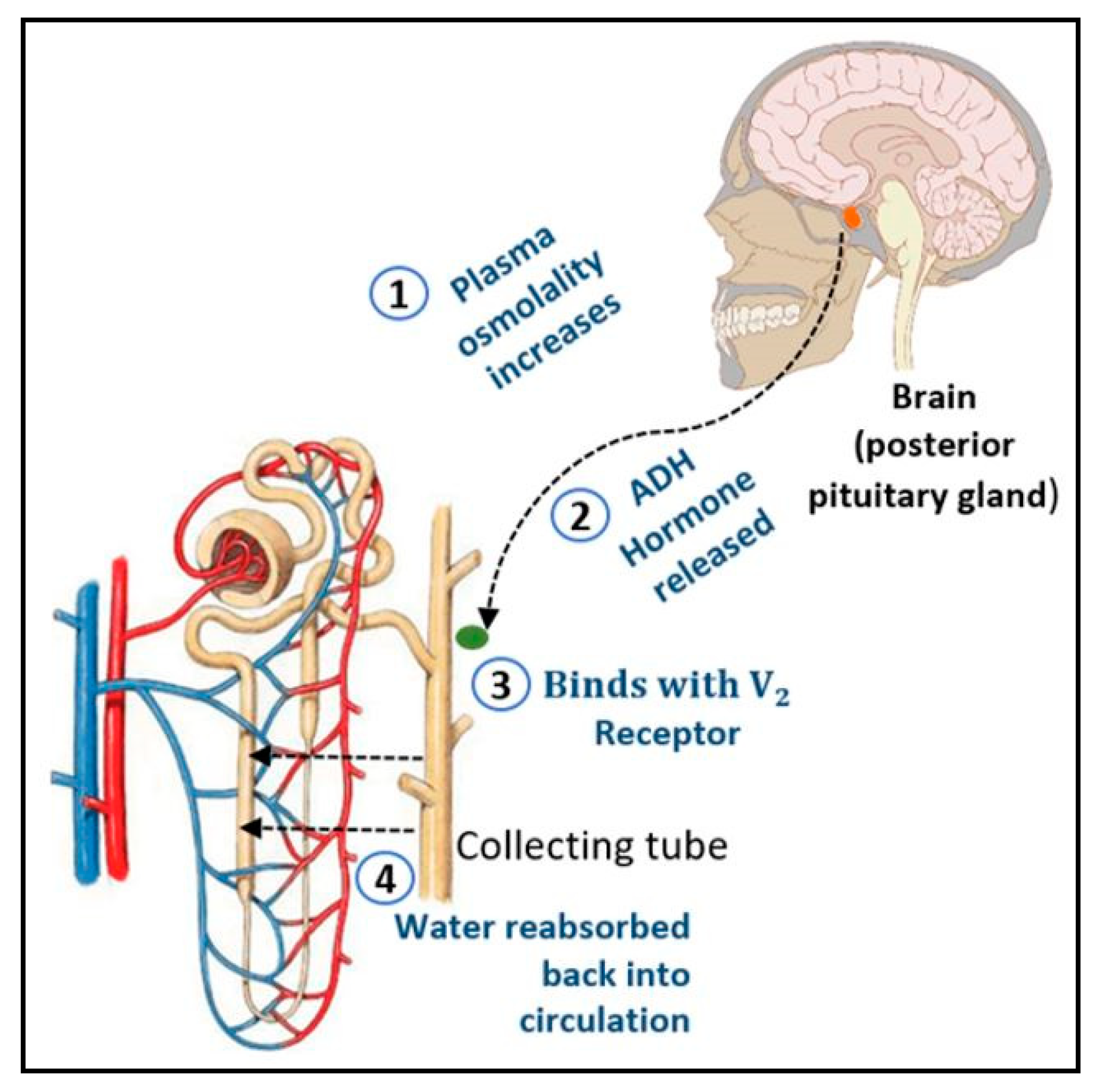
:1. Introduction
2. Materials and Methods
2.1. Field-Effect Transistor (FET) Fabrication and Graphene Transfer
2.2. Functionalization of Graphene Surface in Graphene-Based Field-Effect Transistor (GFET)
2.3. Anti-Diuretic Hormone (ADH) Detection
2.4. Specificity Analysis
3. Results and Discussion
3.1. Surface and Morphological Characterization of GFET
3.2. Raman Characterization for before and after Graphene Transfer
3.2.1. Raman Characterization for Graphene on Copper
3.2.2. Raman Characterization for Graphene Transferred on Field-Effect Transistor (FET)
3.3. Attenuated Total Reflectance Fourier Transform Infrared (ATR-FTIR) Characterization for Functionalization of Graphene
3.4. Analytical Performance of GFET
3.4.1. I–V Curve for Surface Functionalization
3.4.2. I–V Curve for Various ADH Concentrations
3.4.3. Sensitivity
3.4.4. Limit of Detection (LOD)
3.5. Specificity Analysis
3.5.1. Specificity Analysis for ADH Spiked in Phosphate-Buffered Saline (PBS) Buffer
3.5.2. Specificity Analysis for ADH-Spiked in Human Serum
4. Conclusions
Author Contributions
Funding
Conflicts of Interest
References
- Verbalis, J.G. Vasopressin receptors. In Hormones, Brain and Behavior Online, 1599—2010; Elsevier: Washington, DC, USA, 2010. [Google Scholar] [CrossRef]
- Davison, J.M.; Gilmore, E.A.; Durr, J.; Robertson, G.L.; Lindheimer, M.D. Altered osmotic thresholds for vasopressin secretion and thirst in human pregnancy. Am. J. Physiol. Ren. Physiol. 2017, 246, F105–F109. [Google Scholar] [CrossRef] [PubMed]
- Ma, T.; Song, Y.; Yang, B.; Gillespie, A.; Carlson, E.J.; Epstein, C.J.; Verkman, A.S. Nephrogenic diabetes insipidus in mice lacking aquaporin-3 water channels. Proc. Natl. Acad. Sci. USA 2000, 97, 4386–4391. [Google Scholar] [CrossRef] [PubMed] [Green Version]
- Menzel, W.; Zahn, V.; Maiss, E. Multiplex RT-PCR-ELISA compared with bioassay for the detection of four apple viruses. J. Virol. Methods 2003, 110, 153–157. [Google Scholar] [CrossRef]
- Koshimizu, T.; Nakamura, K.; Egashira, N.; Hiroyama, M.; Nonoguchi, H.; Tanoue, A. Vasopressin V1a and V1b receptors: From molecules to physiological systems. Physiol. Rev. 2012, 92, 1813–1864. [Google Scholar] [CrossRef] [Green Version]
- He, Q.; Chen, Y.; Shen, D.; Cui, X.; Zhang, C.; Yang, H.; Zhong, W.; Eremin, S.A.; Fang, Y.; Zhao, S. Development of a surface plasmon resonance immunosensor and ELISA for 3-nitrotyrosine in human urine. Talanta 2019, 195, 655–661. [Google Scholar] [CrossRef]
- Shang, L.; Xue, G.; Gong, L.; Zhang, Y.; Peng, S.; Yuan, C.; Huang, M. A novel ELISA for the detection of active form of plasminogen activator inhibitor-1 based on a highly specific trapping agent. Anal. Chim. Acta 2019, 1053, 98–104. [Google Scholar] [CrossRef]
- Wiederschain, G.Y. The ELISA guidebook. In Biochemistry (Moscow); Springer Nature: Cham, Switzerland, 2009; Volume 74. [Google Scholar] [CrossRef]
- Sanaka, T.; Funaki, T.; Tanaka, T.; Hoshi, S.; Niwayama, J.; Taitoh, T.; Nishimura, H.; Higuchi, C. Plasma pentosidine levels measured by a newly developed method using ELISA in patients with chronic renal failure. Nephron 2002, 91, 64–73. [Google Scholar] [CrossRef]
- Hara, M.; Yamagata, K.; Tomino, Y.; Saito, Y.; Hirayama, Y.; Ogasawara, S.; Kurusawa, H.; Sekine, S.; Yan, K. Urinary podocalyxin is an early marker for podocyte injury in patients with diabetes: Establishment of a highly sensitive ELISA to detect urinary podocalyxin. Diabetologia 2012, 55, 2913–2919. [Google Scholar] [CrossRef] [Green Version]
- Biocompare, N.D. ADH ELISA Kits. Available online: http://www.biocompare.com/pfu/110627/soids/3982/ELISA_Kit/ADH (accessed on 4 March 2020).
- Bankir, L.; Bichet, D.G.; Morgenthaler, N.G. Vasopressin: Physiology, assessment and osmosensation. J. Intern. Med. 2017, 282, 284–297. [Google Scholar] [CrossRef] [Green Version]
- Baumann, G.; Dingman, G.F. Distribution, blood transport, and degradation of antidiuretic hormone in man. J. Clin. Investig. 1976, 57, 1109–1116. [Google Scholar] [CrossRef]
- Caiazza, R.; Bigliardi, B. Business models for biosensors in the food industry. In Handbook of Cell Biosensors; Springer: Cham, Switzerland, 2019; pp. 1–20. [Google Scholar] [CrossRef]
- Ligler, F.S.; Gooding, J.J. Lighting up Biosensors: Now and the decade to come. Anal. Chem. 2019, 91, 8732–8738, Review-article. [Google Scholar] [CrossRef]
- Cheng, S.; Hideshima, S.; Kuroiwa, S.; Nakanishi, T.; Osaka, T. Label-free detection of tumor markers using field effect transistor (FET)-based biosensors for lung cancer diagnosis. Sens. Actuators B Chem. 2015, 212, 329–334. [Google Scholar] [CrossRef]
- Chaudhary, R.; Sharma, A.; Sinha, S.; Yadav, J.; Sharma, R.; Mukhiya, R.; Khanna, V.K. Fabrication and characterisation of Al gate n-metal-oxide-semiconductor field-effect transistor, on-chip fabricated with silicon nitride ion-sensitive field-effect transistor. IET Comput. Digit. Tech. 2016, 10, 268–272. [Google Scholar] [CrossRef]
- Zhang, B.; Cui, T. An ultrasensitive and low-cost graphene sensor based on layer-by-layer nano self-assembly. Appl. Phys. Lett. 2011, 98, 2011–2014. [Google Scholar] [CrossRef]
- Bolotin, K.I.; Sikes, K.J.; Jiang, Z.; Klima, M.; Fudenberg, G.; Hone, J.; Kim, P.; Stormer, H.L. Ultrahigh electron mobility in suspended graphene. Solid State Commun. 2008, 146, 351–355. [Google Scholar] [CrossRef] [Green Version]
- Wang, H.; Zhao, Y.; Xie, Y.; Ma, X.; Zhang, X. Recent progress in synthesis of two-dimensional hexagonal boron nitride. J. Semicond. 2017, 38. [Google Scholar] [CrossRef]
- Cheng, Z.; Li, Q.; Li, Z.; Zhou, Q.; Fang, Y. Suspended graphene sensors with improved signal and reduced noise. Nano Lett. 2010, 10, 1864–1868. [Google Scholar] [CrossRef]
- Li, P.; Zhang, B.; Cui, T. Towards instrinsic graphene biosensor: A label-free, suspended single crystalline graphene sensor for multiplex lung cancer tumor markers detection. Biosens. Bioelectron. 2015, 72, 168–174. [Google Scholar] [CrossRef]
- Selvarajan, R.S.; Majlis, B.Y.; Mohamed, M.A.; Hamzah, A.A. Optimization of lift off process in electrode patterning for graphene based field effect transistors. ASM Sci. J. 2019, 12, 76–82. [Google Scholar]
- He, P.; Oncescu, V.; Lee, S.; Choi, I.; Erickson, D. Label-free electrochemical monitoring of vasopressin in aptamer-based microfluidic biosensors. Anal. Chim. Acta 2013, 759, 74–80. [Google Scholar] [CrossRef]
- Hamzah, A.A.; Selvarajan, R.S.; Majlis, B.Y. Graphene for biomedical applications: A review. Sains Malays. 2017, 46, 1125–1139. [Google Scholar] [CrossRef]
- Tang, B.; Guoxin, H.; Gao, H. Raman spectroscopic characterization of graphene. Appl. Spectrosc. Rev. 2010, 45, 369–407. [Google Scholar] [CrossRef]
- Sagar, R.R.; Zhang, X.; Xiong, C. Growth of graphene on copper and nickel foils via chemical vapour deposition using ethylene. Mater. Res. Innov. 2014, 18, 706–710. [Google Scholar] [CrossRef]
- Anindya, D.; Biswanath, C.; Sood, A.K. Raman spectroscopy of graphene on different substrates and influence of defects. Bull. Mater. Sci. 2008, 31, 579–584. [Google Scholar]
- Stojanović, D.; Matković, A.; Aškrabić, S.; Beltaos, A.; Ralević, U.; Jovanović, D.; Gajić, R. Raman spectroscopy of graphene: Doping and mapping. Physica Scripta 2013, T157. [Google Scholar] [CrossRef]
- Malard, L.M.; Pimenta, M.A.; Dresselhaus, G.; Dresselhaus, M.S. Raman spectroscopy in graphene. Phys. Rep. 2009, 473, 51–87. [Google Scholar] [CrossRef]
- Shimada, T.; Sugai, T.; Souza, M.; Cancado, L.G.; Jorio, A.; Pimenta, M.A.; Saito, R.; Gruneis, A.; Dresselhaus, G.; Dresselhaus, M.S.; et al. Origin of the 2450 cm-1 Raman bands in HOPG, single-wall and double-wall carbon nanotubes. Carbon 2015. [Google Scholar] [CrossRef]
- Cinzia, C. Probing disorder and charged impurities in graphene by Raman spectroscopy. Phys. Status Solidi Rapid Res. Lett. 2009, 3, 175–177. [Google Scholar] [CrossRef]
- Dietzek, B.; Cialla, D.; Schmitt, M.; Popp, J. Confocal Raman microscopy. In Confocal Raman Microscopy; Springer: Cham, Switzerland, 2010. [Google Scholar]
- Ferrari, A.C.; Basko, D.M. Raman spectroscopy as a versatile tool for studying the properties of graphene. Nat. Nanotechnol. 2013, 8, 235–246. [Google Scholar] [CrossRef] [Green Version]
- Nautiyal, P.; Loganathan, A.; Agrawal, R.; Boesl, B.; Wang, C.; Agarwal, A. Oxidative unzipping and transformation of high aspect ratio boron nitride nanotubes into “white Graphene oxide” platelets. Sci. Rep. 2016. [Google Scholar] [CrossRef]
- Ma, J.; Sun, Y.; Yu, F. Efficient removal of tetracycline with KOH-activated grapheme from aqueous solution. R. Soc. Open Sci. 2017, 4. [Google Scholar] [CrossRef] [Green Version]
- Fan, M.; Dai, D.; Huang, B. Fourier transform infrared spectroscopy for natural fibres. Fourier Transform Mater. Anal. 2012. [Google Scholar] [CrossRef] [Green Version]
- Li, K.; Luo, X.; Lin, X.; Qi, F.; Wu, P. Novel NiCoMnO4 thermocatalyst for low-temperature catalytic degradation of methylene blue. J. Mol. Catal. A Chem. 2014, 383–384, 1–9. [Google Scholar] [CrossRef]
- Li, Y.; Van Zijll, M.; Chiang, S.; Pan, N. KOH modified graphene nanosheets for supercapacitor electrodes. J. Power Sources 2011, 196, 6003–6006. [Google Scholar] [CrossRef]
- Gunda, N.S.K.; Singh, M.; Norman, L.; Kaur, K.; Mitra, S.K. Optimization and characterization of biomolecule immobilization on silicon substrates using (3-aminopropyl)triethoxysilane (APTES) and glutaraldehyde linker. Appl. Surf. Sci. 2014, 305, 522–530. [Google Scholar] [CrossRef]
- Fathil, M.F.M.; Md Arshad, M.K.; Ruslinda, A.R.; Gopinath, S.C.B.; Nuzaihan, M.M.N.; Adzhri, R.; Hashim, U.; Lam, H.Y. Substrate-gate coupling in ZnO-FET biosensor for cardiac troponin I detection. Sens. Actuators B Chem. 2017, 242, 1142–1154. [Google Scholar] [CrossRef]
- Pumera, M. Graphene in biosensing. Mater. Today 2011, 14, 308–315. [Google Scholar] [CrossRef]
- Letchumanan, I.; Md Arshad, M.K.; Balakrishnan, S.R.; Gopinath, S.C.B. Gold-nanorod enhances dielectric voltammetry detection of c-reactive protein: A predictive strategy for cardiac failure. Biosens. Bioelectron. 2019, 130, 40–47. [Google Scholar] [CrossRef]
- Huh, Y.S.; Erickson, D. Aptamer based surface enhanced Raman scattering detection of vasopressin using multilayer nanotube arrays. Biosens. Bioelectron. 2010, 25, 1240–1243. [Google Scholar] [CrossRef] [Green Version]
- Yang, J.; Wang, C.; Zhu, Y.; Liu, G.; Lin, Q. A microfluidic aptasensor integrating specific enrichment with a graphene nanosensor for label-free detection of small biomolecules. In Proceedings of the IEEE International Conference on Micro Electro Mechanical Systems (MEMS) 2015, Estoril, Portugal, 18–22 January 2015; pp. 569–572. [Google Scholar] [CrossRef]
- Yang, J.; Zhu, J.; Pei, R.; Oliver, J.A.; Landry, D.W.; Stojanovic, M.N.; Lin, Q. Integrated microfluidic aptasensor for mass spectrometric detection of vasopressin in human plasma ultrafiltrate. Anal. Methods 2016, 8, 5190–5196. [Google Scholar] [CrossRef]
- Konieczna, I.; Zarnowiec, P.; Kwinkowski, M.; Kolesinska, B.; Fraczyk, J.; Kaminski, Z.; Kaca, W. Bacterial urease and its role in long-lasting human diseases. Curr. Protein Pept. Sci. 2013, 13, 789–806. [Google Scholar] [CrossRef] [PubMed]


| Point 1 | 1580.00 | 2670.00 | 1.92 |
| Point 2 | 1580.00 | 2670.00 | 2.53 |
| Point 3 | 1590.69 | 2674.49 | 2.22 |
| Average | 1582.98 | 2666.78 | 2.21 |
| Peak D | Peak G | Peak 2D | |||
|---|---|---|---|---|---|
| Point 1 | 1332.28 | 1581.05 | 2670.63 | 2.84 | 0.19 |
| Point 2 | 1336.13 | 1581.05 | 2674.49 | 2.22 | 0.46 |
| Point 3 | 1338.06 | 1579.12 | 2670.63 | 2.70 | 0.38 |
| Average | 1338.06 | 1581.05 | 2668.70 | 2.58 | 0.34 |
© 2020 by the authors. Licensee MDPI, Basel, Switzerland. This article is an open access article distributed under the terms and conditions of the Creative Commons Attribution (CC BY) license (http://creativecommons.org/licenses/by/4.0/).
Share and Cite
Selvarajan, R.S.; Rahim, R.A.; Majlis, B.Y.; Gopinath, S.C.B.; Hamzah, A.A. Ultrasensitive and Highly Selective Graphene-Based Field-Effect Transistor Biosensor for Anti-Diuretic Hormone Detection. Sensors 2020, 20, 2642. https://doi.org/10.3390/s20092642
Selvarajan RS, Rahim RA, Majlis BY, Gopinath SCB, Hamzah AA. Ultrasensitive and Highly Selective Graphene-Based Field-Effect Transistor Biosensor for Anti-Diuretic Hormone Detection. Sensors. 2020; 20(9):2642. https://doi.org/10.3390/s20092642
Chicago/Turabian StyleSelvarajan, Reena Sri, Ruslinda A. Rahim, Burhanuddin Yeop Majlis, Subash C. B. Gopinath, and Azrul Azlan Hamzah. 2020. "Ultrasensitive and Highly Selective Graphene-Based Field-Effect Transistor Biosensor for Anti-Diuretic Hormone Detection" Sensors 20, no. 9: 2642. https://doi.org/10.3390/s20092642