
Time Evolution of the Skin–Electrode Interface Impedance under Different Skin Treatments

 , ,
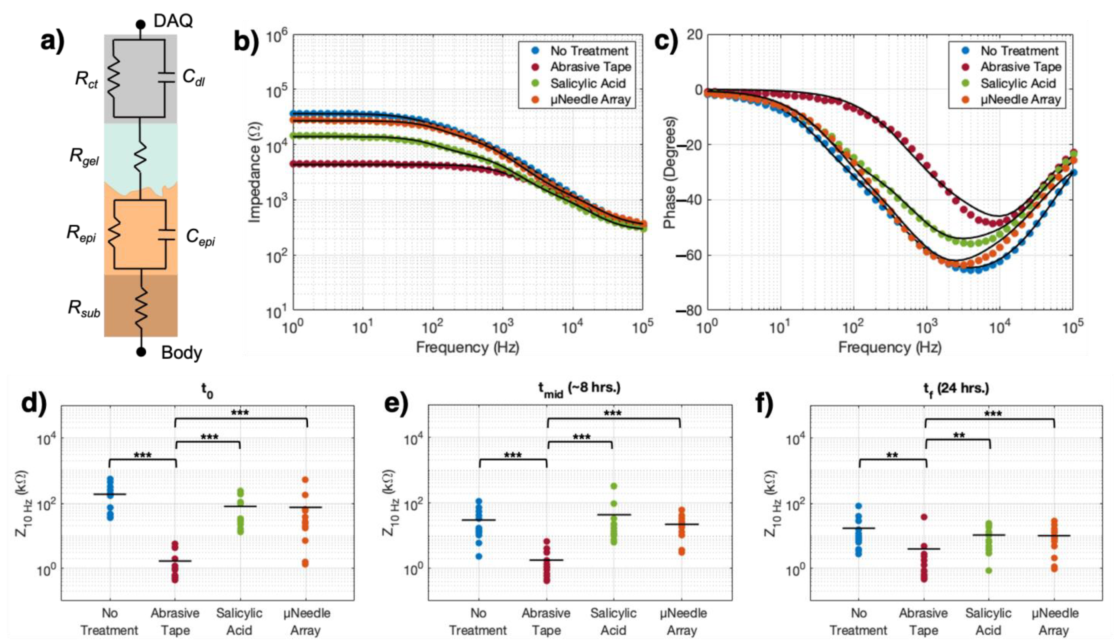
, ,
Abstract
:1. Introduction
2. Materials and Methods
2.1. Experimental Procedures
2.2. Data Analysis and Statistics
3. Results
4. Discussion
5. Conclusions
Author Contributions
Funding
Institutional Review Board Statement
Informed Consent Statement
Data Availability Statement
Acknowledgments
Conflicts of Interest
References
- Nag, A.; Mukhopadhyay, S.C.; Kosel, J. Wearable Flexible Sensors: A Review. IEEE Sens. J. 2017, 17, 3949–3960. [Google Scholar] [CrossRef] [Green Version]
- Bloss, R. Wearable sensors bring new benefits to continuous medical monitoring, real time physical activity assessment, baby monitoring and industrial applications. Sens. Rev. 2015, 35, 141–145. [Google Scholar] [CrossRef]
- Koydemir, H.C.; Ozcan, A. Wearable, epidermal and implantable sensors for medical applications. Annu. Rev. Anal. Chem. 2018, 11, 127–146. [Google Scholar] [CrossRef]
- Heikenfeld, J.; Jajack, A.; Rogers, J.; Gutruf, P.; Tian, L.; Pan, T.; Li, R. Wearable sensors: Modalities, challenges, and prospects. Lab Chip 2017, 18, 217–248. [Google Scholar] [CrossRef] [Green Version]
- Scalia, M.; Sperini, M.; Guidi, F. The Johnson noise in biological matter. Math. Probl. Eng. 2012, 2012. [Google Scholar] [CrossRef]
- Lempka, S.F.; Johnson, M.D.; Moffitt, M.A.; Otto, K.J.; Kipke, D.R.; McIntyre, C.C. Theoretical analysis of intracortical microelectrode recordings. J. Neural Eng. 2011, 8, 045006. [Google Scholar] [CrossRef]
- Huigen, E.; Peper, A.; Grimbergen, C.A. Investigation into the origin of the noise of surface electrodes. Med. Biol. Eng. Comput. 2002, 40, 332–338. [Google Scholar] [CrossRef]
- Saelim, W.; Phukpattaranont, P.; Thongpull, K. Effect of electrode skin impedance on electromyography signal quality. In Proceedings of the 2018 15th International Conference on Electrical Engineering/Electronics, Computer, Telecommunications and Information Technology (ECTI-CON), Chiang Rai, Thailand, 18–21 July 2018; pp. 748–751. [Google Scholar] [CrossRef]
- Taji, B.; Shirmohammadi, S.; Groza, V.; Batkin, I. Impact of skin-electrode interface on electrocardiogram measurements using conductive textile electrodes. IEEE Trans. Instrum. Meas. 2014, 63, 1412–1422. [Google Scholar] [CrossRef]
- Buxi, D.; Kim, S.; van Helleputte, N.; Altini, M.; Wijsman, J.; Yazicioglu, R.F.; Penders, J.; van Hoof, C. Correlation Between Electrode-Tissue Impedance and Motion Artifact in Biopotential Recordings. IEEE Sens. J. 2012, 12, 3373–3383. [Google Scholar] [CrossRef]
- Wiese, S.R.; Anheier, P.; Connemara, R.D.; Mollner, A.T.; Neils, T.F.; Kahn, J.A.; Webster, J.G. Electrocardiographic motion artifact versus electrode impedance. IEEE Trans. Biomed. Eng. 2005, 52, 136–139. [Google Scholar] [CrossRef] [PubMed]
- Matsukawa, R.; Miyamoto, A.; Yokota, T.; Someya, T. Skin Impedance Measurements with Nanomesh Electrodes for Monitoring Skin Hydration. Adv. Healthc. Mater. 2020, 9, 1–6. [Google Scholar] [CrossRef]
- Lozano, A.; Rosell, J.; Pallas-Areny, R. Errors in prolonged electrical impedance measurements due to electrode repositioning and postural changes. Physiol. Meas. 1995, 16, 121–130. [Google Scholar] [CrossRef]
- Newbold, C.; Risi, F.; Hollow, R.; Yusof, Y.; Dowell, R. Long-term electrode impedance changes and failure prevalence in cochlear implants. Int. J. Audiol. 2015, 54, 1–8. [Google Scholar] [CrossRef]
- Puurtinen, M.M.; Komulainen, S.M.; Kauppinen, P.K.; Malmivuo, J.A.V.; Hyttinen, J.A.K. Measurement of noise and impedance of dry and wet textile electrodes, and textile electrodes with hydrogel. In Proceedings of the Annual International Conference of the IEEE Engineering in Medicine and Biology, New York, NY, USA, 30 August–3 September 2006; pp. 6012–6015. [Google Scholar]
- Radüntz, T. Signal quality evaluation of emerging EEG devices. Front. Physiol. 2018, 9, 1–12. [Google Scholar] [CrossRef] [Green Version]
- Cattarello, P.; Merletti, R. Characterization of dry and wet Electrode-Skin interfaces on different skin treatments for HDsEMG. In Proceedings of the 2016 IEEE International Symposium on Medical Measurements and Applications (MeMeA), Benevento, Italy, 15–18 May 2016. [Google Scholar] [CrossRef]
- Piervirgili, G.; Petracca, F.; Merletti, R. A new method to assess skin treatments for lowering the impedance and noise of individual gelled Ag-AgCl electrodes. Physiol. Meas. 2014, 35, 2101–2118. [Google Scholar] [CrossRef]
- Merletti, R.; Botter, A.; Troiano, A.; Merlo, E.; Minetto, M.A. Technology and instrumentation for detection and conditioning of the surface electromyographic signal: State of the art. Clin. Biomech. 2009, 24, 122–134. [Google Scholar] [CrossRef]
- Li, G.; Wang, S.; Duan, Y.Y. Towards gel-free electrodes: A systematic study of electrode-skin impedance. Sens. Actuators B Chem. 2017, 241, 1244–1255. [Google Scholar] [CrossRef]
- Yamagami, M.; Peters, K.M.; Milovanovic, I.; Kuang, I.; Yang, Z.; Lu, N.; Steele, K.M. Assessment of dry epidermal electrodes for long-term electromyography measurements. Sensors 2018, 18, 1269. [Google Scholar] [CrossRef] [Green Version]
- Peng, H.L.; Liu, J.Q.; Dong, Y.Z.; Yang, B.; Chen, X.; Yang, C.S. Parylene-based flexible dry electrode for bioptential recording. Sens. Actuators B Chem. 2016, 231, 1–11. [Google Scholar] [CrossRef]
- Mihajlovic, V.; Grundlehner, B. The effect of force and electrode material on electrode-to-skin impedance. In Proceedings of the 2012 IEEE Biomedical Circuits and Systems Conference (BioCAS), Hsinchu, Taiwan, 28–30 November 2012; pp. 57–60. [Google Scholar] [CrossRef]
- White, E.A.; Orazem, M.E.; Bunge, A.L. Characterization of damaged skin by impedance spectroscopy: Mechanical damage. Pharm. Res. 2013, 30, 2036–2049. [Google Scholar] [CrossRef]
- Hewson, D.J.; Hogrel, J.Y.; Langeron, Y.; Duchêne, J. Evolution in impedance at the electrode-skin interface of two types of surface EMG electrodes during long-term recordings. J. Electromyogr. Kinesiol. 2003, 13, 273–279. [Google Scholar] [CrossRef]
- Ferree, T.C.; Luu, P.; Russell, G.S.; Tucker, D.M. Scalp electrode impedance, infection risk, and EEG data quality. Clin. Neurophysiol. 2001, 112, 1–9. [Google Scholar] [CrossRef]
- Baek, J.Y.; An, J.H.; Choi, J.M.; Park, K.S.; Lee, S.H. Flexible polymeric dry electrodes for the long-term monitoring of ECG. Sens. Actuators A Phys. 2008, 143, 423–429. [Google Scholar] [CrossRef]
- Pylatiuk, C.; Müller-Riederer, M.; Kargov, A.; Schulz, S.; Schill, O.; Reischl, M.; Bretthauer, G. Comparison of surface EMG monitoring electrodes for long-term use in rehabilitation device control. In Proceedings of the 2009 IEEE International Conference on Rehabilitation Robotics, Kyoto, Japan, 23–26 June 2009; pp. 300–304. [Google Scholar] [CrossRef]
- Oliveira, C.C.; da Machado Silva, J.; Trindade, I.G.; Martins, F. Characterization of the electrode-skin impedance of textile electrodes. In Proceedings of the Proceedings of the 2014 29th Conference on Design of Circuits and Integrated Systems, Madrid, Spain, 26–28 November 2014; pp. 1–6. [Google Scholar]
- Lin, C.T.; de Liao, L.; Liu, Y.H.; Wang, I.J.; Lin, B.S.; Chang, J.Y. Novel dry polymer foam electrodes for long-term EEG measurement. IEEE Trans. Biomed. Eng. 2011, 58, 1200–1207. [Google Scholar] [CrossRef]
- Beckmann, L.; Neuhaus, C.; Medrano, G.; Jungbecker, N.; Walter, M.; Gries, T.; Leonhardt, S. Characterization of textile electrodes and conductors using standardized measurement setups. Physiol. Meas. 2010, 31, 233. [Google Scholar] [CrossRef]
- Decker, A.; Graber, E.M. Over-the-counter acne treatments: A review. J. Clin. Aesthet. Dermatol. 2012, 5, 32–40. [Google Scholar]
- Arif, T. Salicylic acid as a peeling agent: A comprehensive review. Clin. Cosmet. Investig. Dermatol. 2015, 8, 455–461. [Google Scholar] [CrossRef] [Green Version]
- Cormier, M.; Johnson, B.; Ameri, M.; Nyam, K.; Libiran, L.; Zhang, D.D.; Daddona, P. Transdermal delivery of desmopressin using a coated microneedle array patch system. J. Control. Release 2004, 97, 503–511. [Google Scholar] [CrossRef]
- Nguyen, T.T.; Oh, Y.; Kim, Y.; Shin, Y.; Baek, S.K.; Park, J.H. Progress in microneedle array patch (MAP) for vaccine delivery. Hum. Vaccines Immunother. 2020, 00, 1–12. [Google Scholar] [CrossRef]
- Ilić, T.; Savić, S.; Batinić, B.; Marković, B.; Schmidberger, M.; Lunter, D.; Savić, M.; Savić, S. Combined use of biocompatible nanoemulsions and solid microneedles to improve transport of a model NSAID across the skin: In Vitro and in Vivo studies. Eur. J. Pharm. Sci. 2018, 125, 110–119. [Google Scholar] [CrossRef]
- Zhang, D.; Das, D.B.; Rielly, C.D. Microneedle Assisted Micro-Particle Delivery from Gene Guns: Experiments using skin-mimicking agarose gel. J. Pharm. Sci. 2014, 103, 613–627. [Google Scholar] [CrossRef] [Green Version]
- Roberts, T.; De Graaf, J.B.; Nicol, C.; Hervé, T.; Fiocchi, M.; Sanaur, S. Flexible Inkjet-Printed Multielectrode Arrays for Neuromuscular Cartography. Adv. Healthc. Mater. 2016, 5, 1462–1470. [Google Scholar] [CrossRef]
- Leleux, P.; Johnson, C.; Strakosas, X.; Rivnay, J.; Hervé, T.; Owens, R.M.; Malliaras, G.G. Ionic liquid gel-assisted electrodes for long-term cutaneous recordings. Adv. Healthc. Mater. 2014, 3, 1377–1380. [Google Scholar] [CrossRef]
- Murphy, B.B.; Mulcahey, P.J.; Driscoll, N.; Richardson, A.G.; Robbins, G.T.; Apollo, N.V.; Maleski, K.; Lucas, T.H.; Gogotsi, Y.; Dillingham, T.; et al. A Gel-Free Ti3C2Tx-Based Electrode Array for High-Density, High-Resolution Surface Electromyography. Adv. Mater. Technol. 2020, 5, 2000325–2000335. [Google Scholar] [CrossRef]
- Franks, W.; Schenker, I.; Schmutz, P.; Hierlemann, A. Impedance Characterization and Modeling of Electrodes for Biomedical Applications. IEEE Trans. Biomed. Eng. 2005, 52, 1295–1302. [Google Scholar] [CrossRef]
- Bates, D.; Mächler, M.; Bolker, B.M.; Walker, S.C. Fitting linear mixed-effects models using lme4. J. Stat. Softw. 2015, 67. [Google Scholar] [CrossRef]
- Boehler, C.; Carli, S.; Fadiga, L.; Stieglitz, T.; Asplund, M. Tutorial: Guidelines for standardized performance tests for electrodes intended for neural interfaces and bioelectronics. Nat. Protoc. 2020, 15, 3557–3578. [Google Scholar] [CrossRef]
- Cutaneous Electrodes for Recording Purposes—Performance Criteria for Safety and Performance Based Pathway. United States of America: Food & Drug Administration. Available online: https://www.fda.gov/regulatory-information/search-fda-guidance-documents/cutaneous-electrodes-recording-purposes-performance-criteria-safety-and-performance-based-pathway (accessed on 30 June 2021).
- ANSI/AAMI EC12:2000/(R)2015–Disposable ECG Electrodes. Available online: file:///C:/Users/MDPI/AppData/Local/Temp/previews_1642226_pre.pdf (accessed on 30 June 2021).
- Yazicioglu, R.F.; van Hoof, C.; Puers, R. Common Biopotentials and Electrodes. In Biopotential Readout Circuits for Portable Acquisition Systems; Springer Science: New York, NY, USA, 2009; ISBN 9781402090929. [Google Scholar]
- Andrews, S.; Lee, J.W.; Prausnitz, M. Recovery of skin barrier after stratum corneum removal by microdermabrasion. AAPS Pharm. Sci. Tech. 2011, 12, 1393–1400. [Google Scholar] [CrossRef] [Green Version]
- Del Rosso, J.; Zeichner, J.; Alexis, A.; Cohen, D.; Berson, D. Understanding the Epidermal Barrier in Healthy and Compromised Skin: Clinically Relevant Information for the Dermatology Practitioner. J. Clin. Aesthet. Dermatol. 2011, 9, 7–9. [Google Scholar]
- Garmirian, L.P.; Chin, A.B.; Rutkove, S.B. Discriminating Neurogenic From Myopathic Disease Via Measurement of Muscle Anisotropy. Muscle Nerve 2009, 39, 1–7. [Google Scholar] [CrossRef] [Green Version]
- Li, L.; Li, X.; Hu, H.; Shin, H.; Zhou, P. The effect of subcutaneous fat on electrical impedance myography: Electrode configuration and multi-frequency analyses. PLoS ONE 2016, 11. [Google Scholar] [CrossRef]
- Yamamoto, T.; Yamamoto, Y. Electrical properties of the epidermal stratum corneum. Med. Biol. Eng. 1976, 14, 151–158. [Google Scholar] [CrossRef]
- Yapici, M.K.; Alkhidir, T.; Samad, Y.A.; Liao, K. Graphene-clad textile electrodes for electrocardiogram monitoring. Sens. Actuators B Chem. 2015, 221, 1469–1474. [Google Scholar] [CrossRef]
- Giannos, S.A. Skin microporation: Strategies to enhance and expand transdermal drug delivery. J. Drug Deliv. Sci. Technol. 2014, 24, 293–299. [Google Scholar] [CrossRef]
- Lu, F.; Wang, C.; Zhao, R.; Du, L.; Fang, Z.; Guo, X.; Zhao, Z. Review of stratum corneum impedance measurement in non-invasive penetration application. Biosensors 2018, 8, 31. [Google Scholar] [CrossRef] [PubMed] [Green Version]
- Hoffmann, K.P.; Ruff, R. Flexible dry surface-electrodes for ECG long-term monitoring. Annu. Int. Conf. IEEE Eng. Med. Biol. Proc. 2007, 4, 5739–5742. [Google Scholar] [CrossRef]
- Sawangjai, P.; Hompoonsup, S.; Leelaarporn, P.; Kongwudhikunakorn, S.; Wilaiprasitporn, T. Consumer Grade EEG Measuring Sensors as Research Tools: A Review. IEEE Sens. J. 2020, 20, 3996–4024. [Google Scholar] [CrossRef]
- LaMonte, M.P. Ceribell EEG shortens seizure diagnosis and workforce time and is useful for COVID isolation. Epilepsia Open 2021, 1–8. [Google Scholar] [CrossRef]


| Rsub [kΩ cm2] | Repi [kΩ cm2] | Cepi [nF cm−2] | Rgel [kΩ cm2] | Rct [kΩ cm2] | Cdl [nF cm−2] | Model Fit (×102) | ||
|---|---|---|---|---|---|---|---|---|
| Lower-Upper Parameter bounds | 0.05–5.00 | 0.10–500 | 0.10–500 | 0.05–5.00 | 0.5–5.0 × 10³ | 1.0–1.0 × 10⁵ | — | |
| No Treatment | t0 | 0.71 ± 0.16 | 86.38 ± 29.43 | 242.62 ± 148.41 | 0.87 ± 0.46 | (1.31 ± 1.27) × 103 | 5.38 ± 2.19 | 3.75 ± 0.71 |
| tmid | 0.74 ± 0.19 | 47.07 ± 33.09 | 27.98 ± 26.86 | 1.07 ± 0.90 | 147.93 ± 101.27 | 63.42 ± 25.62 | 2.18 ± 1.85 | |
| tf | 0.71 ± 0.20 | 40.90 ± 30.70 | 34.70 ± 19.11 | 0.72 ± 0.42 | 66.54 ± 31.96 | 36.29 ± 15.24 | 3.09 ± 0.84 | |
| Abrasive Tape | t0 | 0.70 ± 0.20 | 6.61 ± 5.02 | 4.95 ± 3.73 | 0.59 ± 0.13 | 2.90 ± 2.39 | 7.00 ± 2.90 | 2.97 ± 0.75 |
| tmid | 0.72 ± 0.18 | 1.80 ± 1.45 | 8.49 ± 2.46 | 0.59 ± 0.11 | 3.30 ± 2.66 | 10.50 ± 6.56 | 0.27 ± 0.08 | |
| tf | 0.67 ± 0.22 | 2.27 ± 1.39 | 28.28 ± 20.65 | 0.76 ± 0.48 | 1.26 ± 0.49 | 12.23 ± 4.27 | 0.36 ± 0.18 | |
| Salicylic Acid | t0 | 0.79 ± 0.14 | 69.43 ± 30.09 | 97.63 ± 76.73 | 0.65 ± 0.14 | 675.80 ± 501.10 | 9.97 ± 5.40 | 4.72 ± 0.97 |
| tmid | 0.77 ± 0.18 | 48.39 ± 32.86 | 47.81 ± 27.09 | 1.17 ± 1.02 | 17.67 ± 14.97 | 9.88 ± 9.54 | 0.97 ± 0.05 | |
| tf | 0.77 ± 0.16 | 35.97 ± 33.36 | 44.74 ± 31.84 | 1.36 ± 1.18 | 12.76 ± 9.71 | 7.70 ± 3.40 | 1.15 ± 0.49 | |
| µNeedle Array | t0 | 0.81 ± 0.14 | 81.56 ± 31.34 | 29.46 ± 22.32 | 0.67 ± 0.17 | 807.42 ± 394.97 | 5.05 ± 2.17 | 4.15 ± 1.40 |
| tmid | 0.74 ± 0.18 | 46.50 ± 35.41 | 24.59 ± 21.80 | 1.19 ± 1.09 | 92.16 ± 60.74 | 9.50 ± 5.52 | 2.62 ± 1.34 | |
| tf | 0.71 ± 0.16 | 35.36 ± 32.01 | 33.65 ± 28.34 | 0.91 ± 0.67 | 72.29 ± 30.89 | 5.38 ± 2.19 | 1.22 ± 0.40 | |
| Element | Interaction Effect (df = 6) (p-Value) | Main Effect F(df = 2) (p-Value) | Simple Effect F(df = 2) (p-Value) | |||
|---|---|---|---|---|---|---|
| NT | AT | SA | µNA | |||
| Rsub | 3.0 (0.81) | 0.9 (0.4) | -- | -- | -- | -- |
| Repi | 8.8 (0.19) | 16.8 (2.7 × 10⁻⁷) | -- | -- | -- | -- |
| Cepi | 34.2 (6.13 × 10⁻⁶) | -- | 9.5 (4.6 × 10⁻⁴) | 7.0 (0.003) | 0.9 (0.4) | 2.1 (0.1) |
| Rgel | 6.0 (0.43) | 0.8 (0.4) | -- | -- | -- | -- |
| Rct | 58.0 (1.16 × 10⁻¹⁰) | -- | 30.2 (1.4 × 10⁻⁸) | 3.7 (0.03) | 21.2 (5.9 × 10⁻⁷) | 3.1 (0.06) |
| Cdl | 58.0 (1.15 × 10⁻⁸) | -- | 1.0 (0.4) | 10.0 (3.9 × 10⁻⁴) | 1.2 (0.32) | 1.9 (0.16) |
Publisher’s Note: MDPI stays neutral with regard to jurisdictional claims in published maps and institutional affiliations. |
© 2021 by the authors. Licensee MDPI, Basel, Switzerland. This article is an open access article distributed under the terms and conditions of the Creative Commons Attribution (CC BY) license (https://creativecommons.org/licenses/by/4.0/).
Share and Cite
Murphy, B.B.; Scheid, B.H.; Hendricks, Q.; Apollo, N.V.; Litt, B.; Vitale, F. Time Evolution of the Skin–Electrode Interface Impedance under Different Skin Treatments. Sensors 2021, 21, 5210. https://doi.org/10.3390/s21155210
Murphy BB, Scheid BH, Hendricks Q, Apollo NV, Litt B, Vitale F. Time Evolution of the Skin–Electrode Interface Impedance under Different Skin Treatments. Sensors. 2021; 21(15):5210. https://doi.org/10.3390/s21155210
Chicago/Turabian StyleMurphy, Brendan B., Brittany H. Scheid, Quincy Hendricks, Nicholas V. Apollo, Brian Litt, and Flavia Vitale. 2021. "Time Evolution of the Skin–Electrode Interface Impedance under Different Skin Treatments" Sensors 21, no. 15: 5210. https://doi.org/10.3390/s21155210