Monitoring Bone Density Using Microwave Tomography of Human Legs: A Numerical Feasibility Study
Abstract
:1. Introduction
2. Materials and Methods
2.1. Generation of Anatomically-Realistic Models and the Forward Problem
2.1.1. Numerical Models of Human Leg
2.1.2. Solving the Forward Problem in MWT
- Numerical model: 2D triangular mesh (discussed in Section 2.1.1).
- Operating Frequency: 0.8 GHz.
- Transmitters’ (TX) and receivers’ (RX) configuration: 24 point-sources (TX) and 24 point-sinks (RX) collocated and equally distributed around the model outer most layer.
| Region | Relative Complex Permittivity () |
|---|---|
| Skin | |
| Fat | |
| Muscles | |
| Healthy Bone |
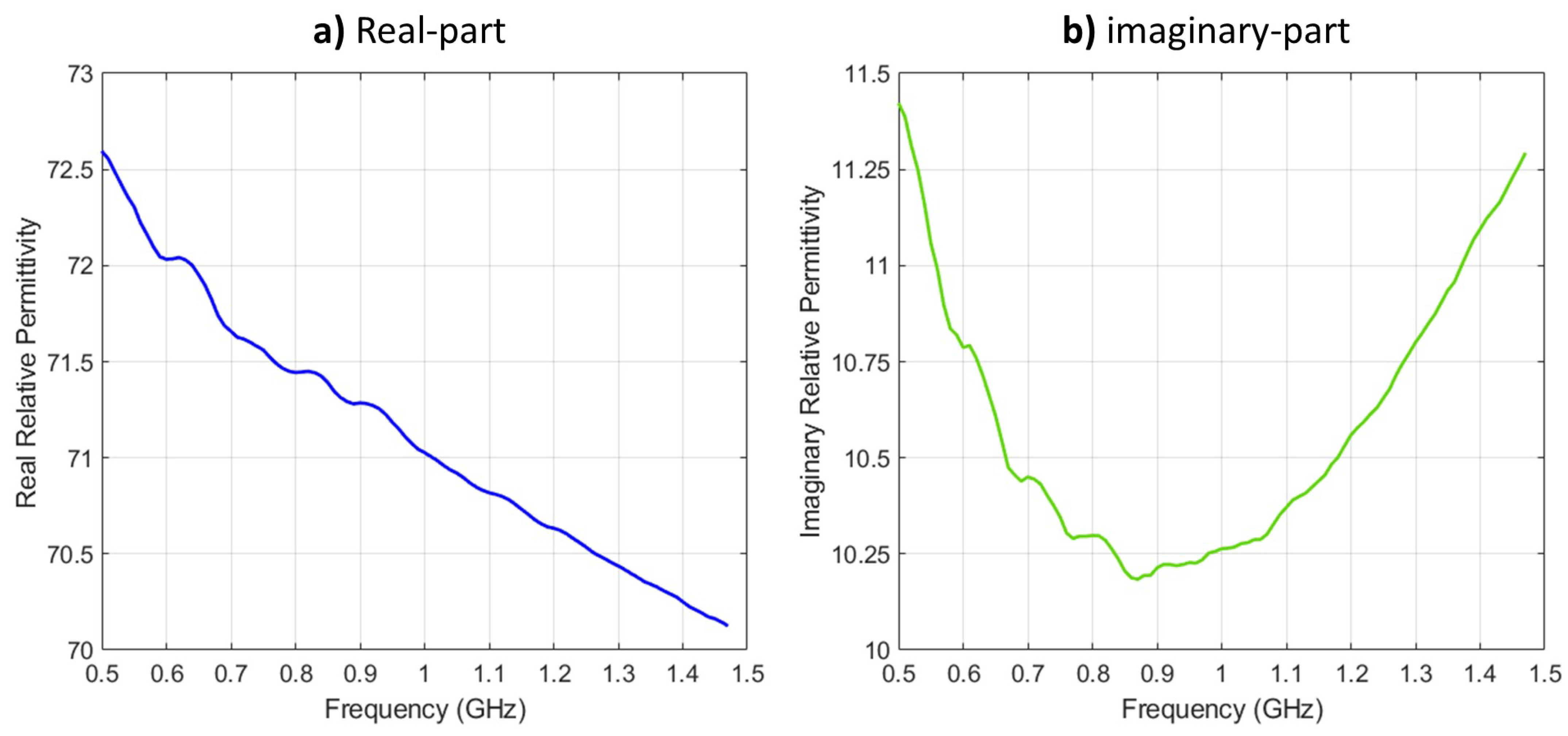
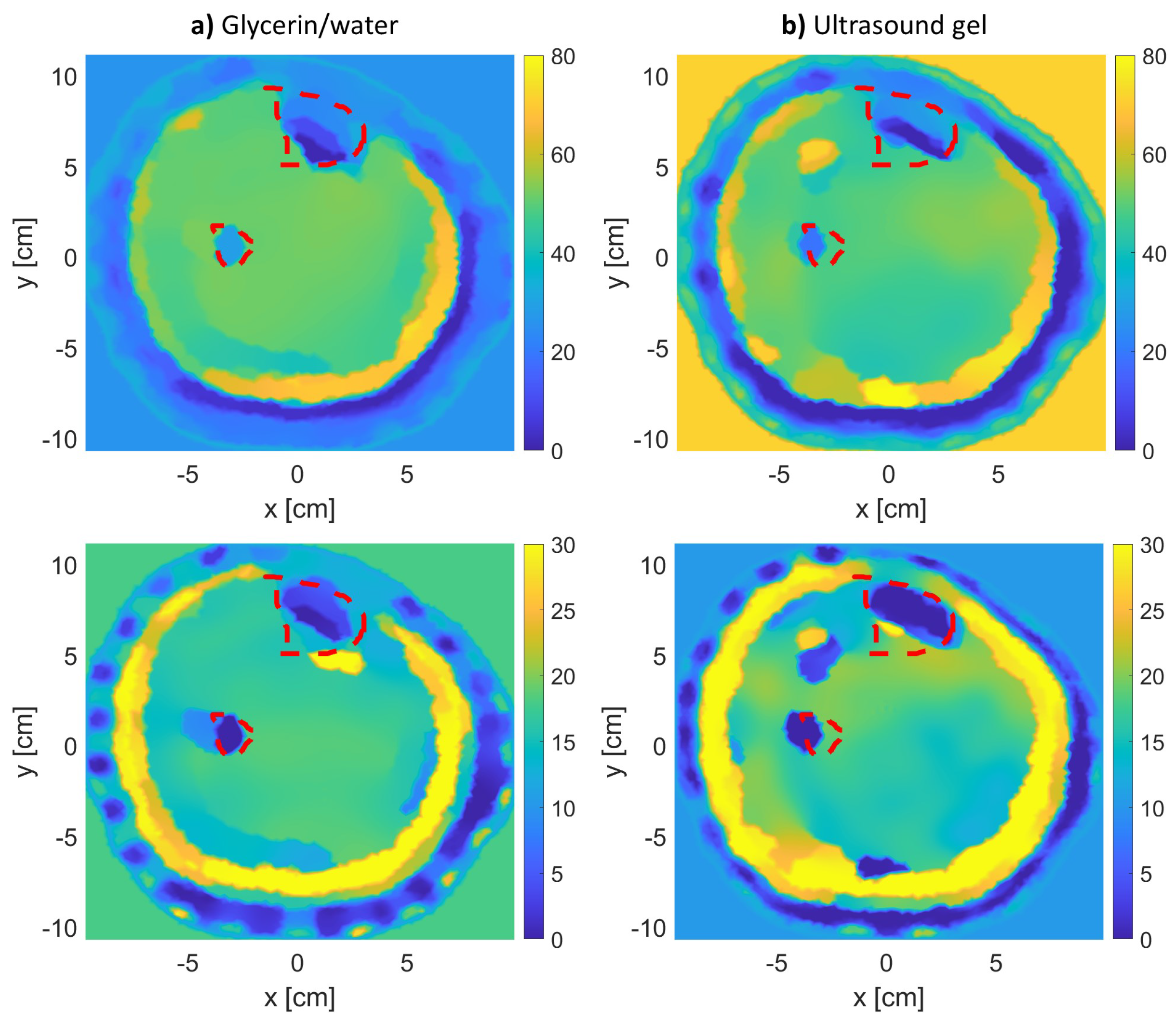
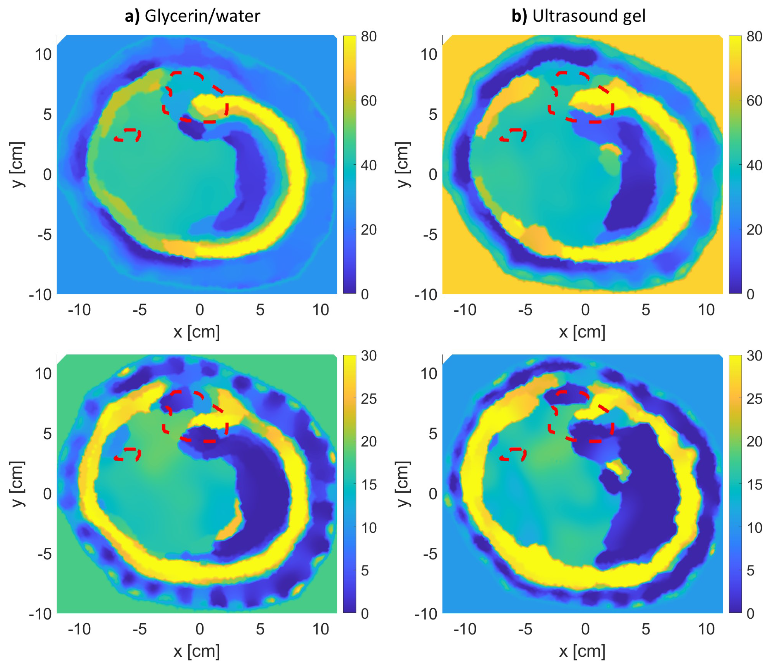
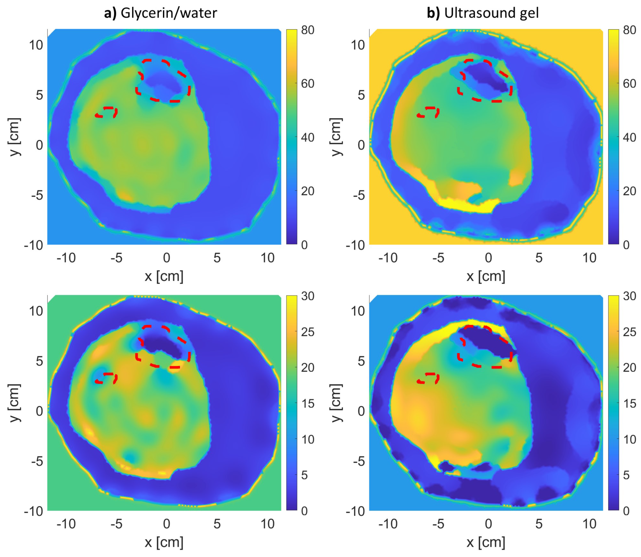
2.1.3. Utilization of an Ultrasound Gel as a Matching Medium
2.2. Image Reconstruction and the Inverse Problem
- is a vector of size R of the scattered electric field values measured at the receivers for a transmitter t. The receivers are located on measurement surface S.
- is a vector of size N for the contrast source values inside the imaging domain D for a transmitter t.
- is a vector of size I for the incident field values inside the imaging domain D for transmitter t.
- is a vector of size I for the contrast values inside the imaging domain D.
- is an FEM matrix operator. This operator contains information about the problem’s boundary and background (whether it is homogeneous or inhomogeneous).
- is a matrix operator that calculates the scattered field values at the receiver locations on measurement surface S.
- is a matrix operator that calculates the scattered field values inside the imaging domain D.
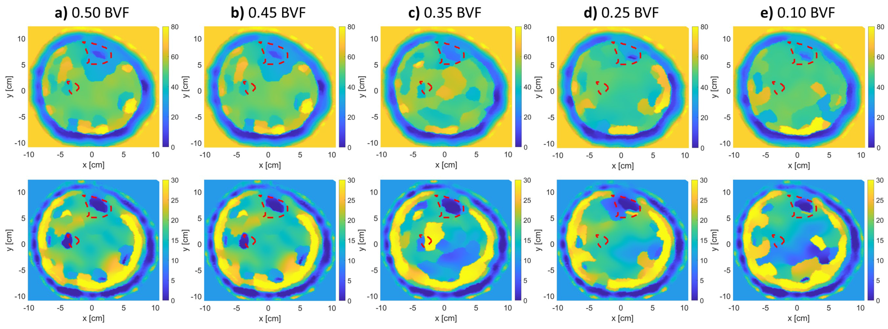
2.3. Detection of Bone Density Variations
- Bones with 0.50 BVF: ,
- Bones with 0.45 BVF: ,
- Bones with 0.35 BVF: ,
- Bones with 0.25 BVF: ,
- Bones with 0.10 BVF: .
Expert-Eye Localization of Bones
3. Results
3.1. Additional Numerical Simulations
3.1.1. Additional BVF Scenarios
- Bones with 0.50 BVF: .
- Bones with 0.45 BVF: .
- Bones with 0.40 BVF: .
- Bones with 0.35 BVF: .
- Bones with 0.30 BVF: .
- Bones with 0.25 BVF: .
- Bones with 0.20 BVF: .
- Bones with 0.15 BVF: .
- Bones with 0.10 BVF: .
3.1.2. Varying Number of Antennas
4. Discussions
4.1. MWT as a Promising Imaging Modality
4.2. Fat Thickness
4.3. Number of Antennas
4.4. Study Limitations
5. Conclusions
Author Contributions
Funding
Institutional Review Board Statement
Informed Consent Statement
Conflicts of Interest
Abbreviations
| BVF | Bone volume fraction |
| CSI | Contrast source inversion |
| DXA | dual-energy X-ray absorptiometry |
| FEM | Finite-element method |
| MWT | Microwave tomography |
| MRI | Magnetic resonance imaging |
| NOF | National osteoporosis foundation |
| QCT | Quantitative computed tomography |
| TX | Transmitters |
| RX | Receivers |
References
- Watts, N.; Lewiecki, E.; Miller, P.; Baim, S. National psteoporosis foundation 2008 clinician’s guide to prevention and treatment of osteoporosis and the world health organization fracture risk assessment tool (FRAX): What they mean to the bone densitometrist and bone technologist. J. Clin. Densitom. 2008, 11, 473–477. [Google Scholar] [CrossRef] [PubMed]
- Holick, M.F. Vitamin D: Importance in the prevention of cancers, type 1 diabetes, heart disease, and osteoporosis. Am. J. Clin. Nutr. 2004, 79, 362–371. [Google Scholar] [CrossRef] [Green Version]
- Lips, P.; Van Schoor, N.M. The effect of vitamin D on bone and osteoporosis. Best Pract. Res. Clin. Endocrinol. Metab. 2011, 25, 585–591. [Google Scholar] [CrossRef] [PubMed]
- Haq, A.; Wimalawansa, S.J.; Pludowski, P.; Al Anouti, F. Clinical practice guidelines for vitamin D in the United Arab Emirates. J. Steroid Biochem. Mol. Biol. 2018, 175, 4–11. [Google Scholar] [CrossRef] [PubMed]
- Meaney, P.; Goodwin, D.; Golnabi, A.; Zhou, T.; Pallone, M.; Geimer, S.; Burke, G.; Paulsen, K. Clinical Microwave Tomographic Imaging of the Calcaneus: A First-in-Human Case Study of Two Subjects. IEEE Trans. Biomed. Eng. 2012, 59, 3304–3313. [Google Scholar] [CrossRef] [PubMed] [Green Version]
- Tian, Z.; Meaney, P.; Pallone, M.; Geimer, S.; Paulsen, K. Microwave Tomographic Imaging for Osteoporosis Screening: A Pilot Clinical Study. In Proceedings of the 2010 Annual International Conference of the IEEE Engineering in Medicine and Biology, Buenos Aires, Argentina, 31 August–4 September 2010; pp. 1218–1221. [Google Scholar]
- Majumdar, S.; Genant, H.; Grampp, S.; Newitt, D.; Truong, V.H.; Lin, J.; Mathur, A. Correlation of trabecular bone structure with age, bone mineral density, and osteoporotic status: In vivo studies in the distal radius using high resolution magnetic resonance imaging. J. Bone Miner. Res. 1997, 12, 111–118. [Google Scholar] [CrossRef] [PubMed]
- Strzelecki, M. Texture boundary detection using network of synchronised oscillators. Electron. Lett. 2004, 40, 466–467. [Google Scholar] [CrossRef]
- Chang, G.; Boone, S.; Martel, D.; Rajapakse, C.S.; Hallyburton, R.S.; Valko, M.; Honig, S.; Regatte, R.R. MRI assessment of bone structure and microarchitecture. J. Magn. Reson. Imaging 2017, 46, 323–337. [Google Scholar] [CrossRef]
- Fathi Kazerooni, A.; Pozo, J.M.; McCloskey, E.V.; Saligheh Rad, H.; Frangi, A.F. Diffusion MRI for assessment of bone quality: A review of findings in healthy aging and osteoporosis. J. Magn. Reson. Imaging 2020, 51, 975–992. [Google Scholar] [CrossRef] [Green Version]
- Njeh, C.; Boivin, C.; Langton, C. The role of ultrasound in the assessment of osteoporosis: A review. Osteoporos. Int. 1997, 7, 7–22. [Google Scholar] [CrossRef]
- Ron, A.; Abboud, S.; Arad, M. Home monitoring of bone density in the wrist—A parametric EIT computer modeling study. Biomed. Phys. Eng. Express 2016, 2, 035002. [Google Scholar] [CrossRef]
- Hamidipour, A.; Henriksson, T.; Hopfer, M.; Planas, R.; Semenov, S. Electromagnetic tomography for brain imaging and stroke diagnostics: Progress towards clinical application. In Emerging Electromagnetic Technologies for Brain Diseases Diagnostics, Monitoring and Therapy; Springer: Berlin/Heidelberg, Germany, 2018; pp. 59–86. [Google Scholar]
- Zamani, A.; Rezaeieh, S.; Bialkowski, K.; Abbosh, A. Boundary estimation of imaged object in microwave medical imaging using antenna resonant frequency shift. IEEE Trans. Antennas Propag. 2018, 66, 927–936. [Google Scholar] [CrossRef]
- Semenov, S. Microwave tomography: Review of the progress towards clinical applications. Philos. Trans. R. Soc. A 2009, 367, 3021–3042. [Google Scholar] [CrossRef] [Green Version]
- Pastorino, M. Microwave Imaging; John Wiley & Sons: Hoboken, NJ, USA, 2010; Volume 208. [Google Scholar]
- Meaney, P.; Zhou, T.; Goodwin, D.; Golnabi, A.; Attardo, E.; Paulsen, K. Bone dielectric property variation as a function of mineralization at microwave frequencies. Int. J. Biomed. Imaging 2012, 2012, 9. [Google Scholar] [CrossRef] [Green Version]
- Amin, B.; Elahi, M.A.; Shahzad, A.; Porter, E.; McDermott, B.; O’Halloran, M. Dielectric properties of bones for the monitoring of osteoporosis. Med. Biol. Eng. Comput. 2019, 57, 1–13. [Google Scholar] [CrossRef]
- Nazarian, A.; Stechow, D. Bone volume fraction explains the variation in strength and stiffness of cancellous bone affected by metastatic cancer and osteoporosis. Calcif. Tissue Int. 2008, 83, 368–379. [Google Scholar] [CrossRef]
- Fajardo, J.E.; Vericat, F.; Irastorza, G.; Carlevaro, C.M.; Irastorza, R.M. Sensitivity analysis on imaging the calcaneus using microwaves. arXiv 2017, arXiv:1709.04934. [Google Scholar] [CrossRef] [Green Version]
- Fajardo, J.E.; Lotto, F.P.; Vericat, F.; Carlevaro, C.M.; Irastorza, R.M. Microwave tomography with phaseless data on the calcaneus by means of artificial neural networks. arXiv 2019, arXiv:1902.07777. [Google Scholar] [CrossRef] [Green Version]
- Zakaria, A.; LoVetri, J. Application of multiplicative regularization to the finite-element contrast source inversion method. IEEE Trans. Antennas Propag. 2011, 59, 3495–3498. [Google Scholar] [CrossRef]
- Zakaria, A.; Gilmore, C.; Pistorius, S.; LoVetri, J. Balanced multiplicative regularization for the contrast source inversion method. In Proceedings of the 28th International Review of Progress in Applied Computational Electromagnetics Conference (ACES’12), Columbus, OH, USA, 10–14 April 2012. [Google Scholar]
- Alkhodari, M.; Zakaria, A.; Qaddoumi, N. Preliminary numerical analysis of monitoring bone density using microwave tomography. In Proceedings of the 2018 Asia-Pacific Microwave Conference (APMC), Kyoto, Japan, 6–9 November 2018; pp. 563–565. [Google Scholar]
- Mohanad, A.; Amer, Z.; Nasser, Q. Guidelines towards a wearable microwave tomography system. In Proceedings of the 2019 Asia-Pacific Microwave Conference (APMC), Singapore, 10–13 December 2019. [Google Scholar]
- Alkhodari, M.; Zakaria, A.; Qaddoumi, N. Image Classification in Microwave Tomography using a Parametric Intensity Model. In Proceedings of the 2020 International Conference on Communications, Signal Processing, and Their Applications (ICCSPA), Sharjah, United Arab Emirates, 16–18 March 2021; pp. 1–4. [Google Scholar]
- Micheau, A.; Hoa, D. MRI of The Lower Extremity Anatomy-Atlas of the Human Body Using Cross-Sectional Imaging. 2017. Available online: https://doi.org/10.37019/e-anatomy/185 (accessed on 13 December 2017). [CrossRef]
- Tatco, V. Normal MRI of the Leg. 2016. Available online: https://radiopaedia.org/cases/43617 (accessed on 19 March 2016).
- George, R.; Cruz, J.; Singh, R.; Ilangovan, R. Lower Leg Images. 2017. Available online: https://mrimaster.com (accessed on 22 December 2017).
- Geuzaine, C.; Remacle, J. GMSH: A three-dimensional finite element mesh generator with built-in pre- and post-processing facilities. Int. J. Numer. Method Eng. 2009, 79, 1–24. [Google Scholar] [CrossRef]
- Zakaria, A. The Finite-Element Contrast Source Inversion Method for Microwave Imaging Applications. Ph.D. Thesis, University of Manitoba, Winnipeg, MB, Canada, 2012. [Google Scholar]
- Fang, Q. Computational Methods for Microwave Medical Imaging. Ph.D. Thesis, Dartmouth College, Hanover, NH, USA, 2004. [Google Scholar]
- Meaney, P.; Pendergrass, S.; Fanning, M.; Li, D.; Paulsen, K. Importance of using a reduced contrast coupling medium in 2D microwave breast imaging. J. Electromagn. Waves Appl. 2003, 17, 333–355. [Google Scholar] [CrossRef]
- Gabriel, C.; Gabriel, S.; Corthout, E. The dielectric properties of biological tissues: I. literature survey. Phys. Med. Biol. 1996, 41, 2231–2249. [Google Scholar] [CrossRef] [PubMed] [Green Version]
- Labs, P. AquaSonic 100: Ultrasound Transmission Gel. Available online: https://www.parkerlabs.com/aquasonic-100.asp (accessed on 13 November 2018).
- Keysight. N1501A Dielectric Probe Kit. Available online: https://www.keysight.com/en/pd-2492144-pn-N1501A/dielectric-probe-kit?cc=AE&lc=eng (accessed on 20 December 2018).
- Zakaria, A.; Gilmore, C.; LoVetri, J. Finite-element contrast source inversion method for microwave imaging. Inverse Probl. 2010, 26, 115010. [Google Scholar] [CrossRef] [Green Version]
- Gilmore, C.; Zakaria, A.; Pistorius, S.; LoVetri, J. Microwave Imaging of Human Forearms: Pilot Study and Image Enhancement. Int. J. Biomed. Imaging 2013, 11, 1–17. [Google Scholar] [CrossRef] [PubMed]
- Zakaria, A.; Baran, A.; LoVetri, J. Estimation and use of prior information in FEM-CSI for biomedical microwave tomography. IEEE Antennas Wirel. Propag. Lett. 2012, 11, 1606–1609. [Google Scholar] [CrossRef]
- Baran, A.; Kurrant, D.J.; Zakaria, A.; Fear, E.C.; LoVetri, J. Breast imaging using microwave tomography with radar-based tissue-regions estimation. Prog. Electromagn. Res. 2014, 149, 161–171. [Google Scholar] [CrossRef] [Green Version]
- Abdollahi, N.; Kurrant, D.; Mojabi, P.; Omer, M.; Fear, E.; LoVetri, J. Incorporation of Ultrasonic Prior Information for Improving Quantitative Microwave Imaging of Breast. IEEE J. Multiscale Multiphys. Comput. Tech. 2019, 4, 98–110. [Google Scholar] [CrossRef]
- Gilmore, C.; Mojabi, P.; Zakaria, A.; Ostadrahimi, M.; Kaye, C.; Noghanian, S.; Shafai, L.; Pistorius, S.; LoVetri, J. A wideband microwave tomography system with a novel frequency selection procedure. IEEE Trans. Biomed. Eng. 2010, 57, 894–904. [Google Scholar] [CrossRef]
- Gilmore, C.; Abubakar, A.; Hu, W.; Habashy, T.M.; van den Berg, P.M. Microwave biomedical data inversion using the finite-difference contrast source inversion method. IEEE Trans. Antennas Propag. 2009, 57, 1528–1538. [Google Scholar] [CrossRef]
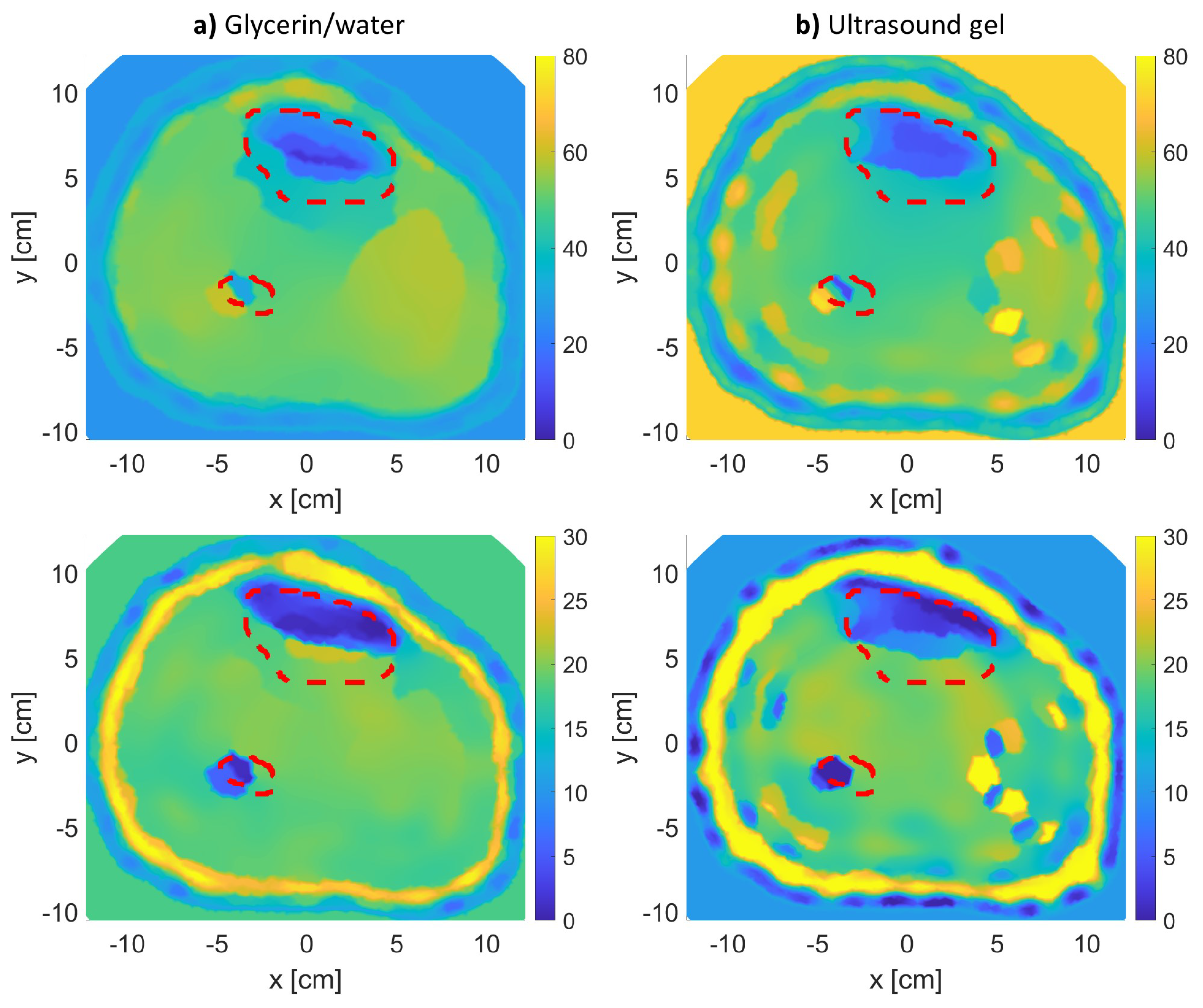


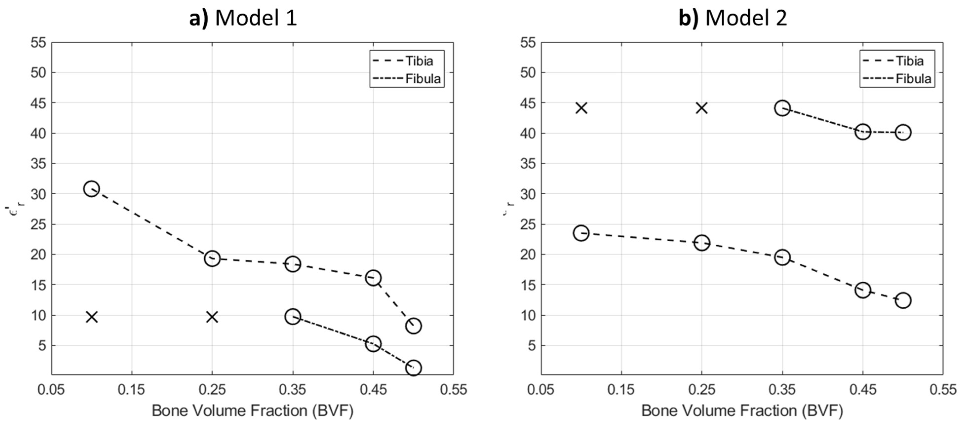

| Model 1 | Model 2 | |
|---|---|---|
| Tibia Bone | 4.52 | 2.54 |
| Fibula Bone | 8.56 | 27.36 |
Publisher’s Note: MDPI stays neutral with regard to jurisdictional claims in published maps and institutional affiliations. |
© 2021 by the authors. Licensee MDPI, Basel, Switzerland. This article is an open access article distributed under the terms and conditions of the Creative Commons Attribution (CC BY) license (https://creativecommons.org/licenses/by/4.0/).
Share and Cite
Alkhodari, M.; Zakaria, A.; Qaddoumi, N. Monitoring Bone Density Using Microwave Tomography of Human Legs: A Numerical Feasibility Study. Sensors 2021, 21, 7078. https://doi.org/10.3390/s21217078
Alkhodari M, Zakaria A, Qaddoumi N. Monitoring Bone Density Using Microwave Tomography of Human Legs: A Numerical Feasibility Study. Sensors. 2021; 21(21):7078. https://doi.org/10.3390/s21217078
Chicago/Turabian StyleAlkhodari, Mohanad, Amer Zakaria, and Nasser Qaddoumi. 2021. "Monitoring Bone Density Using Microwave Tomography of Human Legs: A Numerical Feasibility Study" Sensors 21, no. 21: 7078. https://doi.org/10.3390/s21217078
APA StyleAlkhodari, M., Zakaria, A., & Qaddoumi, N. (2021). Monitoring Bone Density Using Microwave Tomography of Human Legs: A Numerical Feasibility Study. Sensors, 21(21), 7078. https://doi.org/10.3390/s21217078


