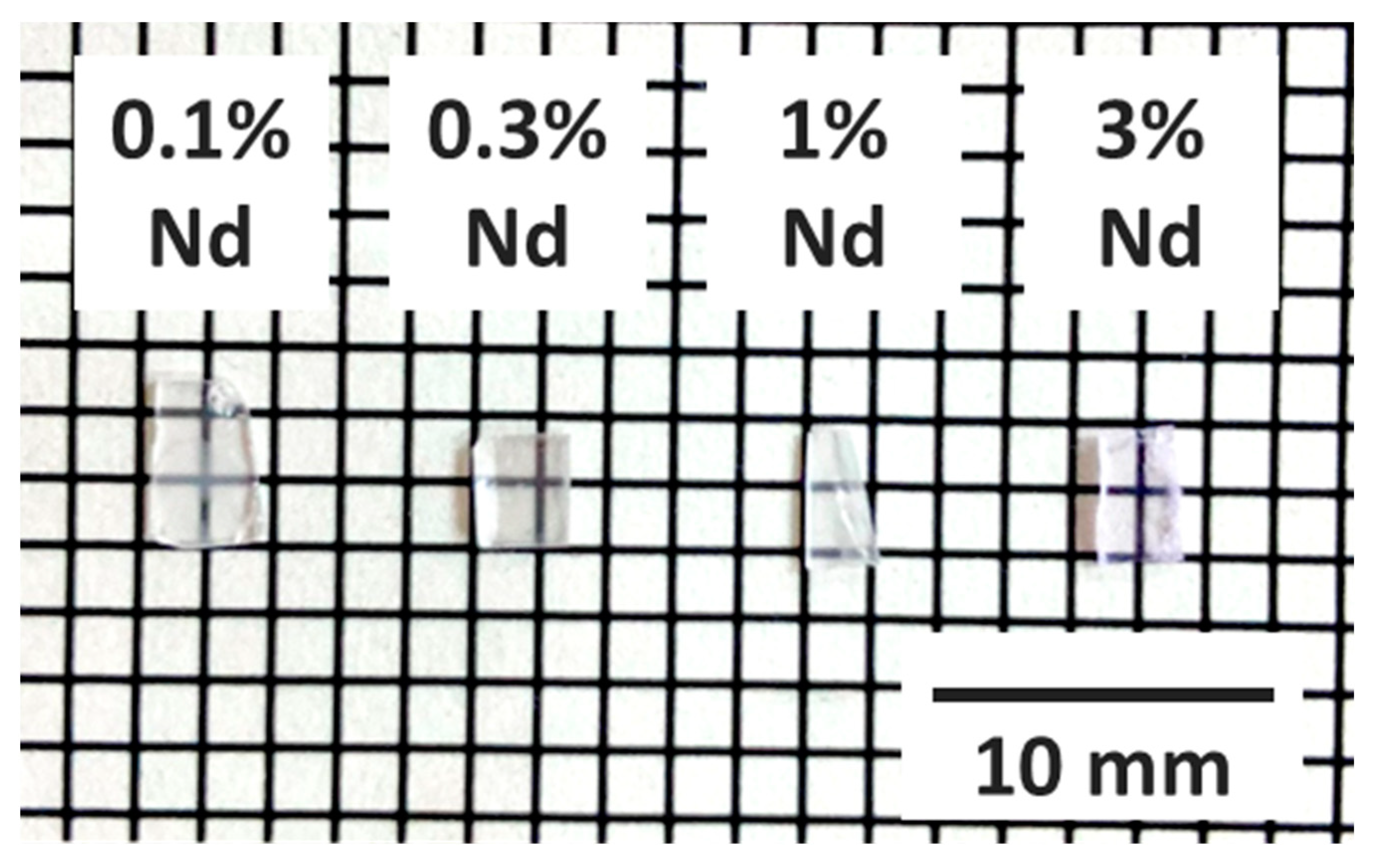
Scintillation Response of Nd-Doped LaMgAl11O19 Single Crystals Emitting NIR Photons for High-Dose Monitoring
Abstract
1. Introduction
2. Materials and Methods
3. Results
4. Conclusions
Author Contributions
Funding
Institutional Review Board Statement
Informed Consent Statement
Data Availability Statement
Conflicts of Interest
References
- van Eijk, C.W.E. Inorganic scintillators in medical imaging. Phys. Med. Biol. 2002, 47, R85–R106. [Google Scholar] [CrossRef]
- van Eijk, C.W.E. Inorganic scintillators in medical imaging detectors. Nucl. Instrum. Methods Phys. Res. A 2003, 509, 17–25. [Google Scholar] [CrossRef]
- William, W.; Moses, W.W. Scintillator Requirements for Medical Imaging. In Proceedings of the International Conference on Inorganic Scintillators and Their Applications: SCINT99, Moscow, Russia, 16–20 August 1999; pp. 11–21. [Google Scholar]
- Glodo, J.; Wang, Y.; Shawgo, R.; Brecher, C.; Hawrami, R.H.; Tower, J.; Shah, K.S. New Developments in Scintillators for Security Applications. Phys. Procedia 2017, 90, 285–290. [Google Scholar] [CrossRef]
- Wells, K.; Bradley, D.A. A review of X-ray explosives detection techniques for checked baggage. Appl. Radiat. Isot. 2012, 70, 1729–1746. [Google Scholar] [CrossRef] [PubMed]
- Kim, K.H.; Jun, I.S.; Eun, Y.S. New sandwich type detector module and its characteristics for dual x-ray baggage inspection system. In Proceedings of the 2007 IEEE Nuclear Science Symposium Conference Record, Honolulu, HI, USA, 27 October–3 November 2007; pp. 1191–1194. [Google Scholar]
- Mazoochi, A.; Rahmani, F.; Abbasi Davani, F.; Ghaderi, R. A novel numerical method to eliminate thickness effect in dual energy X-ray imaging used in baggage inspection. Nucl. Instrum. Methods Phys. Res. A 2014, 763, 538–542. [Google Scholar] [CrossRef]
- Nikitin, A.; Bliven, S. Needs of well logging industry in new nuclear detectors. In Proceedings of the IEEE Nuclear Science Symposuim & Medical Imaging Conference, Knoxville, TN, USA, 30 October–6 November 2010; pp. 1214–1219. [Google Scholar]
- Melcher, C.L. Scintillators for well logging applications. Nucl. Instrum. Methods Phys. Res. B 1989, 40–41, 1214–1218. [Google Scholar] [CrossRef]
- Melcher, C.L.; Schweitzer, J.S.; Manente, R.S.; Peterson, C.A. Applicability of GSO scintillators for well logging. IEEE Trans. Nucl. Sci. 1991, 38, 506–509. [Google Scholar] [CrossRef]
- Zaffalon, M.L.; Cova, F.; Liu, M.; Cemmi, A.; Di Sarcina, I.; Rossi, F.; Carulli, F.; Erroi, A.; Rodà, C.; Perego, J.; et al. Extreme γ-ray radiation hardness and high scintillation yield in perovskite nanocrystals. Nat. Photonics 2022, 16, 860–868. [Google Scholar] [CrossRef]
- Chen, Q.; Wu, J.; Ou, X.; Huang, B.; Almutlaq, J.; Zhumekenov, A.A.; Guan, X.; Han, S.; Liang, L.; Yi, Z.; et al. All-inorganic perovskite nanocrystal scintillators. Nature 2018, 561, 88–93. [Google Scholar] [CrossRef]
- Dorenbos, P. The quest for high resolution γ-ray scintillators. Opt. Mater. X 2019, 1, 100021. [Google Scholar] [CrossRef]
- Weber, M.J. Scintillation: Mechanisms and new crystals. Nucl. Instrum. Methods Phys. Res. A 2004, 527, 9–14. [Google Scholar] [CrossRef]
- Holl, I.; Lorenz, E.; Mageras, G. A measurement of the light yield of common inorganic scintillators. IEEE Trans. Nucl. Sci. 1988, 35, 105–109. [Google Scholar] [CrossRef]
- van Eijk, C.W.E. Inorganic-scintillator development. Nucl. Instrum. Methods Phys. Res. A 2001, 460, 1–14. [Google Scholar] [CrossRef]
- Awual, M.R.; Miyazaki, Y.; Taguchi, T.; Shiwaku, H.; Yaita, T. Encapsulation of cesium from contaminated water with highly selective facial organic–inorganic mesoporous hybrid adsorbent. Chem. Eng. J. 2016, 291, 128–137. [Google Scholar] [CrossRef]
- Khandaker, S.; Chowdhury, M.F.; Awual, M.R.; Islam, A.; Kuba, T. Efficient cesium encapsulation from contaminated water by cellulosic biomass based activated wood charcoal. Chemosphere 2021, 262, 127801. [Google Scholar] [CrossRef] [PubMed]
- Awual, M.R.; Yaita, T.; Miyazaki, Y.; Matsumura, D.; Shiwaku, H.; Taguchi, T. A Reliable Hybrid Adsorbent for Efficient Radioactive Cesium Accumulation from Contaminated Wastewater. Sci. Rep. 2016, 6, 19937. [Google Scholar] [CrossRef]
- Awual, M.R.; Suzuki, S.; Taguchi, T.; Shiwaku, H.; Okamoto, Y.; Yaita, T. Radioactive cesium removal from nuclear wastewater by novel inorganic and conjugate adsorbents. Chem. Eng. J. 2014, 242, 127–135. [Google Scholar] [CrossRef]
- Takada, E.; Kimura, A.; Hosono, Y.; Takahashi, H.; Nakazawa, M. Radiation Distribution Sensor with Optical Fibers for High Radiation Fields. J. Nucl. Sci. Technol. 1999, 36, 641–645. [Google Scholar] [CrossRef]
- Takada, E. Application of red and near infrared emission from rare earth ions for radiation measurements based on optical fibers. IEEE Trans. Nucl. Sci. 1998, 45, 556–560. [Google Scholar] [CrossRef]
- Kimura, A.; Takada, E.; Hosono, Y.; Nakazawa, M.; Takahashi, H.; Hayami, H. New techniques to apple optical fiber image guide to nucileas facilities. J. Nucl. Sci. Technol. 2002, 39, 603–607. [Google Scholar] [CrossRef]
- Huston, A.; Justus, B.; Falkenstein, P.; Miller, R.; Ning, H.; Altemus, R. Remote optical fiber dosimetry. Nucl. Instrum. Methods Phys. Res. B 2001, 184, 55–67. [Google Scholar] [CrossRef]
- O’Keeffe, S.; McCarthy, D.; Woulfe, P.; Grattan, M.W.D.; Hounsell, A.R.; Sporea, D.; Mihai, L.; Vata, I.; Leen, G.; Lewis, E. A review of recent advances in optical fibre sensors for in vivo dosimetry during radiotherapy. Br. J. Radiol. 2015, 88, 20140702. [Google Scholar] [CrossRef] [PubMed]
- Bartesaghi, G.; Conti, V.; Prest, M.; Mascagna, V.; Scazzi, S.; Cappelletti, P.; Frigerio, M.; Gelosa, S.; Monti, A.; Ostinelli, A.; et al. A real time scintillating fiber dosimeter for gamma and neutron monitoring on radiotherapy accelerators. Nucl. Instrum. Methods Phys. Res. A 2007, 572, 228–230. [Google Scholar] [CrossRef]
- Vedda, A.; Chiodini, N.; Di Martino, D.; Fasoli, M.; Keffer, S.; Lauria, A.; Martini, M.; Moretti, F.; Spinolo, G.; Nikl, M.; et al. Ce3+-doped fibers for remote radiation dosimetry. Appl. Phys. Lett. 2004, 85, 6356–6358. [Google Scholar] [CrossRef]
- Ishikawa, M.; Ono, K.; Sakurai, Y.; Unesaki, H.; Uritani, A.; Bengua, G.; Kobayashi, T.; Tanaka, K.; Kosako, T. Development of real-time thermal neutron monitor using boron-loaded plastic scintillator with optical fiber for boron neutron capture therapy. Appl. Radiat. Isot. 2004, 61, 775–779. [Google Scholar] [CrossRef]
- Ding, M. Handbook of Optical Fibers; Springer: Berlin/Heidelberg, Germany, 2018. [Google Scholar]
- Christie, S.; Scorsone, E.; Persaud, K.; Kvasnik, F. Remote detection of gaseous ammonia using the near infrared transmission properties of polyaniline. Sensors Actuators B Chem. 2003, 90, 163–169. [Google Scholar] [CrossRef]
- Chan, K.; Ito, H.; Inaba, H.; Furuya, T. 10 km-long fibre-optic remote sensing of CH4 gas by near infrared absorption. Appl. Phys. B Photophysics Laser Chem. 1985, 38, 11–15. [Google Scholar] [CrossRef]
- Chan, K.; Ito, H.; Inaba, H. Remote sensing system for near-infrared differential absorption of CH4 gas using low-loss optical fiber link. Appl. Opt. 1984, 23, 3415. [Google Scholar] [CrossRef]
- Stewart, G.; Jin, W.; Culshaw, B. Prospects for fibre-optic evanescent-field gas sensors using absorption in the near-infrared. Sensors Actuators B Chem. 1997, 38, 42–47. [Google Scholar] [CrossRef]
- Fukushima, H.; Akatsuka, M.; Kimura, H.; Onoda, D.; Shiratori, D.; Nakauchi, D.; Kato, T.; Kawaguchi, N.; Yanagida, T. Optical and Scintillation Properties of Nd-doped Strontium Yttrate Single Crystals. Sens. Mater. 2021, 33, 2235. [Google Scholar] [CrossRef]
- Akatsuka, M.; Kimura, H.; Onoda, D.; Shiratori, D.; Nakauchi, D.; Kato, T.; Kawaguchi, N.; Yanagida, T. X-ray-induced Luminescence Properties of Nd-doped GdVO4. Sens. Mater. 2021, 33, 2243. [Google Scholar] [CrossRef]
- Okazaki, K.; Onoda, D.; Fukushima, H.; Nakauchi, D.; Kato, T.; Kawaguchi, N.; Yanagida, T. Characterization of scintillation properties of Nd-doped Bi4Ge3O12 single crystals with near-infrared luminescence. J. Mater. Sci. Mater. Electron. 2021, 32, 21677–21684. [Google Scholar] [CrossRef]
- Akatsuka, M.; Usui, Y.; Nakauchi, D.; Kato, T.; Kawano, N.; Okada, G.; Kawaguchi, N.; Yanagida, T. Scintillation properties of YAlO3 doped with Lu and Nd perovskite single crystals. Opt. Mater. 2018, 79, 428–434. [Google Scholar] [CrossRef]
- Nakauchi, D.; Okada, G.; Koshimizu, M.; Yanagida, T. Optical and scintillation properties of Nd-doped SrAl2O4 crystals. J. Rare Earths 2016, 34, 757–762. [Google Scholar] [CrossRef]
- Gao, Z.Y.; Xia, G.Q.; Yan, X.; Zhu, J.F.; Xu, X.D.; Wu, Z.M. A high-repetition-rate diode-pumped Q-switched Nd:LaMgAl11O19 laser operating at single wavelength or dual wavelength. Opt. Rev. 2021, 28, 404–410. [Google Scholar] [CrossRef]
- Guyot, Y.; Moncorge, R. Excited-state absorption in the infrared emission domain of Nd3+-doped Y3Al5O12, YLiF4, and LaMgAl11O19. J. Appl. Phys. 1993, 73, 8526–8530. [Google Scholar] [CrossRef]
- Viana, B.; Garapon, C.; Lejus, A.M.; Vivien, D. Cr3+ → Nd3+ energy transfer in the LaMgAl11O19:Cr, Nd laser material. J. Lumin. 1990, 47, 73–83. [Google Scholar] [CrossRef]
- Wang, J.; Zhang, Y.; Guan, X.; Xu, B.; Xu, H.; Cai, Z.; Pan, Y.; Liu, J.; Xu, X.; Xu, J. High-efficiency diode-pumped continuous-wave and passively Q-switched c-cut Nd:LaMgAl11O19 (Nd:LMA) lasers. Opt. Mater. 2018, 86, 512–516. [Google Scholar] [CrossRef]
- Min, X.; Fang, M.; Huang, Z.; Liu, Y.; Tang, C.; Wu, X. Luminescent properties of white-light-emitting phosphor LaMgAl11O19:Dy3+. Mater. Lett. 2014, 125, 140–142. [Google Scholar] [CrossRef]
- Min, X.; Fang, M.; Huang, Z.; Liu, Y.; Tang, C.; Wu, X. Synthesis and optical properties of Pr3+-doped LaMgAl11O19-A novel blue converting yellow phosphor for white light emitting diodes. Ceram. Int. 2015, 41, 4238–4242. [Google Scholar] [CrossRef]
- Min, X.; Fang, M.; Huang, Z.; Liu, Y.; Tang, C.; Wu, X. Luminescence properties and energy-transfer behavior of a novel and color-tunable LaMgAl11O19:Tm3+, Dy3+ phosphor for white light-emitting diodes. J. Am. Ceram. Soc. 2015, 98, 788–794. [Google Scholar] [CrossRef]
- Min, X.; Fang, M.; Huang, Z.; Liu, Y.; Tang, C.; Zhu, H.; Wu, X. Preparation and luminescent properties of orange reddish emitting phosphor LaMgAl11O19:Sm3+. Opt. Mater. 2014, 37, 110–114. [Google Scholar] [CrossRef]
- Li, W.; Fang, G.; Wang, Y.; You, Z.; Li, J.; Zhu, Z.; Tu, C.; Xu, Y.; Jie, W. Luminescent properties of Dy3+ activated LaMgAl11O19 yellow emitting phosphors for application in white-LEDs. Vacuum 2021, 188, 110215. [Google Scholar] [CrossRef]
- Min, X.; Huang, Z.; Fang, M.; Liu, Y.; Tang, C.; Wu, X. Energy Transfer from Sm3+ to Eu3+ in Red-Emitting Phosphor LaMgAl11O19:Sm3+, Eu3+ for Solar Cells and Near-Ultraviolet White Light-Emitting Diodes. Inorg. Chem. 2014, 53, 6060–6065. [Google Scholar] [CrossRef] [PubMed]
- Rodnyi, P.A.; Mikhrin, S.B.; Dorenbos, P.; van Der Kolk, E.; van Eijk, C.W.E.; Vink, A.P.; Avanesov, A.G. The observation of photon cascade emission in Pr3+-doped compounds under X-ray excitation. Opt. Commun. 2002, 204, 237–245. [Google Scholar] [CrossRef]
- Verstegen, J.M.P.J.; Sommerdijk, J.L.; Verriet, J.G. Cerium and terbium luminescence in LaMgAl11O19. J. Lumin. 1973, 6, 425–431. [Google Scholar] [CrossRef]
- Singh, V.; Chakradhar, R.P.S.; Rao, J.L.; Kwak, H.Y. Enhanced blue emission and EPR study of LaMgAl11O19:Eu phosphors. J. Lumin. 2011, 131, 247–252. [Google Scholar] [CrossRef]
- Zhu, J.; Xia, Z.; Liu, Q. Synthesis and energy transfer studies of LaMgAl11O19:Cr3+, Nd3+ phosphors. Mater. Res. Bull. 2016, 74, 9–14. [Google Scholar] [CrossRef]
- Sommerdijk, J.L.; van der Does de Bye, J.A.W.; Verberne, P.H.J.M. Decay of the Ce3+ luminescence of LaMgAl11O19: Ce3+ and of CeMgAl11O19 activated with Tb3+ or Eu3+. J. Lumin. 1976, 14, 91–99. [Google Scholar] [CrossRef]
- Brandle, C.D.; Berkstresser, G.W.; Shmulovich, J.; Valentino, A.J. Czochralski growth and evaluation of LaMgAl11O19 based phosphors. J. Cryst. Growth 1987, 85, 234–239. [Google Scholar] [CrossRef]
- Luo, X.; Yang, X.; Xiao, S. Conversion of broadband UV-visible to near infrared emission by LaMgAl11O19: Cr3+, Yb3+ phosphors. Mater. Res. Bull. 2018, 101, 73–82. [Google Scholar] [CrossRef]
- Pidol, L.; Kahn-Harari, A.; Viana, B.; Virey, E.; Ferrand, B.; Dorenbos, P.; De Haas, J.T.M.; van Eijk, C.W.E. High efficiency of lutetium silicate scintillators, Ce-doped LPS, and LYSO crystals. IEEE Trans. Nucl. Sci. 2004, 51, 1084–1087. [Google Scholar] [CrossRef]
- Pepin, C.M.; Forgues, J.; Kurata, Y.; Shimura, N.; Usui, T.; Takeyama, T.; Ishibashi, H.; Lecomte, R. Physical Characterization of the LabPET TM LGSO and LYSO Scintillators. In Proceedings of the IEEE Nuclear Science Symposium conference record. Nuclear Science Symposium, Honolulu, HI, USA, 27 October–3 November 2007. [Google Scholar]
- Gerasymov, I.; Sidletskiy, O.; Neicheva, S.; Grinyov, B.; Baumer, V.; Galenin, E.; Katrunov, K.; Tkachenko, S.; Voloshina, O.; Zhukov, A. Growth of bulk gadolinium pyrosilicate single crystals for scintillators. J. Cryst. Growth 2011, 318, 805–808. [Google Scholar] [CrossRef]
- Tanaka, M.; Hara, K.; Kim, S.; Kondo, K.; Takano, H.; Kobayashi, M.; Ishibashi, H.; Kurashige, K.; Susa, K.; Ishii, M. Applications of cerium-doped gadolinium silicate Gd2SiO5:Ce scintillator to calorimeters in high-radiation environment. Nucl. Instrum. Methods Phys. Res. A 1998, 404, 283–294. [Google Scholar] [CrossRef]
- Moses, W.W.; Derenzo, S.E.; Fyodorov, A.; Korzhik, M.; Gektin, A.; Minkov, B.; Aslanov, V. LuAlO3:Ce-a high density, high speed scintillator for gamma detection. IEEE Trans. Nucl. Sci. 1995, 42, 275–279. [Google Scholar] [CrossRef]
- Mares, J.A.; Beitlerova, A.; Nikl, M.; Solovieva, N.; D’Ambrosio, C.; Blazek, K.; Maly, P.; Nejezchleb, K.; De Notaristefani, F. Scintillation response of Ce-doped or intrinsic scintillating crystals in the range up to 1 MeV. Radiat. Meas. 2004, 38, 353–357. [Google Scholar] [CrossRef]
- Iwanowska, J.; Swiderski, L.; Szczesniak, T.; Sibczynski, P.; Moszynski, M.; Grodzicka, M.; Kamada, K.; Tsutsumi, K.; Usuki, Y.; Yanagida, T.; et al. Performance of cerium-doped Gd3Al2Ga3O12 (GAGG:Ce) scintillator in gamma-ray spectrometry. Nucl. Instrum. Methods Phys. Res. A 2013, 712, 34–40. [Google Scholar] [CrossRef]
- Moszyński, M.; Wolski, D.; Ludziejewski, T.; Kapusta, M.; Lempicki, A.; Brecher, C.; Wiśniewski, D.; Wojtowicz, A. Properties of the new LuAP:Ce scintillator. Nucl. Instrum. Methods Phys. Res. A 1997, 385, 123–131. [Google Scholar] [CrossRef]
- van Eijk, C.W.E.; Andriessen, J.; Dorenbos, P.; Visser, R. Ce3+ doped inorganic scintillators. Nucl. Instrum. Methods Phys. Res. A 1994, 348, 546–550. [Google Scholar] [CrossRef]
- Yanagida, T.; Kamada, K.; Fujimoto, Y.; Yagi, H.; Yanagitani, T. Comparative study of ceramic and single crystal Ce:GAGG scintillator. Opt. Mater. 2013, 35, 2480–2485. [Google Scholar] [CrossRef]
- Yanagida, T.; Fujimoto, Y.; Ito, T.; Uchiyama, K.; Mori, K. Development of X-ray-induced afterglow characterization system. Appl. Phys. Express 2014, 7, 062401. [Google Scholar] [CrossRef]
- Xu, P.; Xia, C.; Di, J.; Xu, X.; Sai, Q.; Wang, L. Growth and optical properties of Co,Nd:LaMgAl11O19. J. Cryst. Growth 2012, 361, 11–15. [Google Scholar] [CrossRef]
- Ryadun, A.A.; Rakhmanova, M.I.; Trifonov, V.A.; Pavluk, A.A. Optical and near-infrared luminescence studies of Nd3+- doped CsGd(MoO4)2 crystals. Mater. Lett. 2021, 285, 129062. [Google Scholar] [CrossRef]
- Ceci-Ginistrelli, E.; Smith, C.; Pugliese, D.; Lousteau, J.; Boetti, N.G.; Clarkson, W.A.; Poletti, F.; Milanese, D. Nd-doped phosphate glass cane laser: From materials fabrication to power scaling tests. J. Alloys Compd. 2017, 722, 599–605. [Google Scholar] [CrossRef]
- Pugliese, D.; Gobber, F.S.; Forno, I.; Milanese, D.; Actis Grande, M. Design and Manufacturing of a Nd-Doped Phosphate Glass-Based Jewel. Materials 2020, 13, 2321. [Google Scholar] [CrossRef]
- Cavalli, E.; Zannoni, E.; Belletti, A.; Carozzo, V.; Toncelli, A.; Tonelli, M.; Bettinelli, M. Spectroscopic analysis and laser parameters of Nd3+ in Ca3Sc2Ge3O12 garnet crystals. Appl. Phys. B Lasers Opt. 1999, 68, 677–681. [Google Scholar] [CrossRef]
- Mhlongo, M.R.; Koao, L.F.; Motaung, T.; Kroon, R.E.; Motloung, S.V. Analysis of Nd3+ concentration on the structure, morphology and photoluminescence of sol-gel Sr3ZnAl2O7 nanophosphor. Results Phys. 2019, 12, 1786–1796. [Google Scholar] [CrossRef]
- Ning, L.; Tanner, P.A.; Harutunyan, V.V.; Aleksanyan, E.; Makhov, V.N.; Kirm, M. Luminescence and excitation spectra of YAG:Nd3+ excited by synchrotron radiation. J. Lumin. 2007, 127, 397–403. [Google Scholar] [CrossRef][Green Version]
- Guyot, Y.; Garapon, C.; Moncorgé, R. Optical properties and laser performance of nearstoichiometric and non-stoichiometric Nd3+-doped LaMgAl11O19 single crystals. Opt. Mater. 1994, 3, 275–286. [Google Scholar] [CrossRef]
- Nakauchi, D.; Kato, T.; Kawaguchi, N.; Yanagida, T. Characterization of Eu-doped Ba2SiO4, a high light yield scintillator. Appl. Phys. Express 2020, 13, 122001. [Google Scholar] [CrossRef]


| PL QY [%] | τ [s] | kf [s−1] | knr [s−1] | |
|---|---|---|---|---|
| 0.1% Nd | 91.0 | 3.32 × 10⁻⁴ | 2.74 × 10³ | 2.71 × 10² |
| 0.3% Nd | 73.8 | 3.13 × 10⁻⁴ | 2.36 × 10³ | 8.37 × 10² |
| 1% Nd | 72.1 | 3.08 × 10⁻⁴ | 2.34 × 10³ | 9.06 × 10² |
| 3% Nd | 70.9 | 2.90 × 10⁻⁴ | 2.45 × 10³ | 1.00 × 10³ |
| τ1 [μs] | τ2 [μs] | |
|---|---|---|
| 0.1% Nd | 114 | 762 |
| 0.3% Nd | 94 | 739 |
| 1% Nd | 84 | 631 |
| 3% Nd | 50 | 564 |
Publisher’s Note: MDPI stays neutral with regard to jurisdictional claims in published maps and institutional affiliations. |
© 2022 by the authors. Licensee MDPI, Basel, Switzerland. This article is an open access article distributed under the terms and conditions of the Creative Commons Attribution (CC BY) license (https://creativecommons.org/licenses/by/4.0/).
Share and Cite
Nakauchi, D.; Kato, T.; Kawaguchi, N.; Yanagida, T. Scintillation Response of Nd-Doped LaMgAl11O19 Single Crystals Emitting NIR Photons for High-Dose Monitoring. Sensors 2022, 22, 9818. https://doi.org/10.3390/s22249818
Nakauchi D, Kato T, Kawaguchi N, Yanagida T. Scintillation Response of Nd-Doped LaMgAl11O19 Single Crystals Emitting NIR Photons for High-Dose Monitoring. Sensors. 2022; 22(24):9818. https://doi.org/10.3390/s22249818
Chicago/Turabian StyleNakauchi, Daisuke, Takumi Kato, Noriaki Kawaguchi, and Takayuki Yanagida. 2022. "Scintillation Response of Nd-Doped LaMgAl11O19 Single Crystals Emitting NIR Photons for High-Dose Monitoring" Sensors 22, no. 24: 9818. https://doi.org/10.3390/s22249818
APA StyleNakauchi, D., Kato, T., Kawaguchi, N., & Yanagida, T. (2022). Scintillation Response of Nd-Doped LaMgAl11O19 Single Crystals Emitting NIR Photons for High-Dose Monitoring. Sensors, 22(24), 9818. https://doi.org/10.3390/s22249818


