
Monitoring Respiratory Motion during VMAT Treatment Delivery Using Ultra-Wideband Radar

Abstract
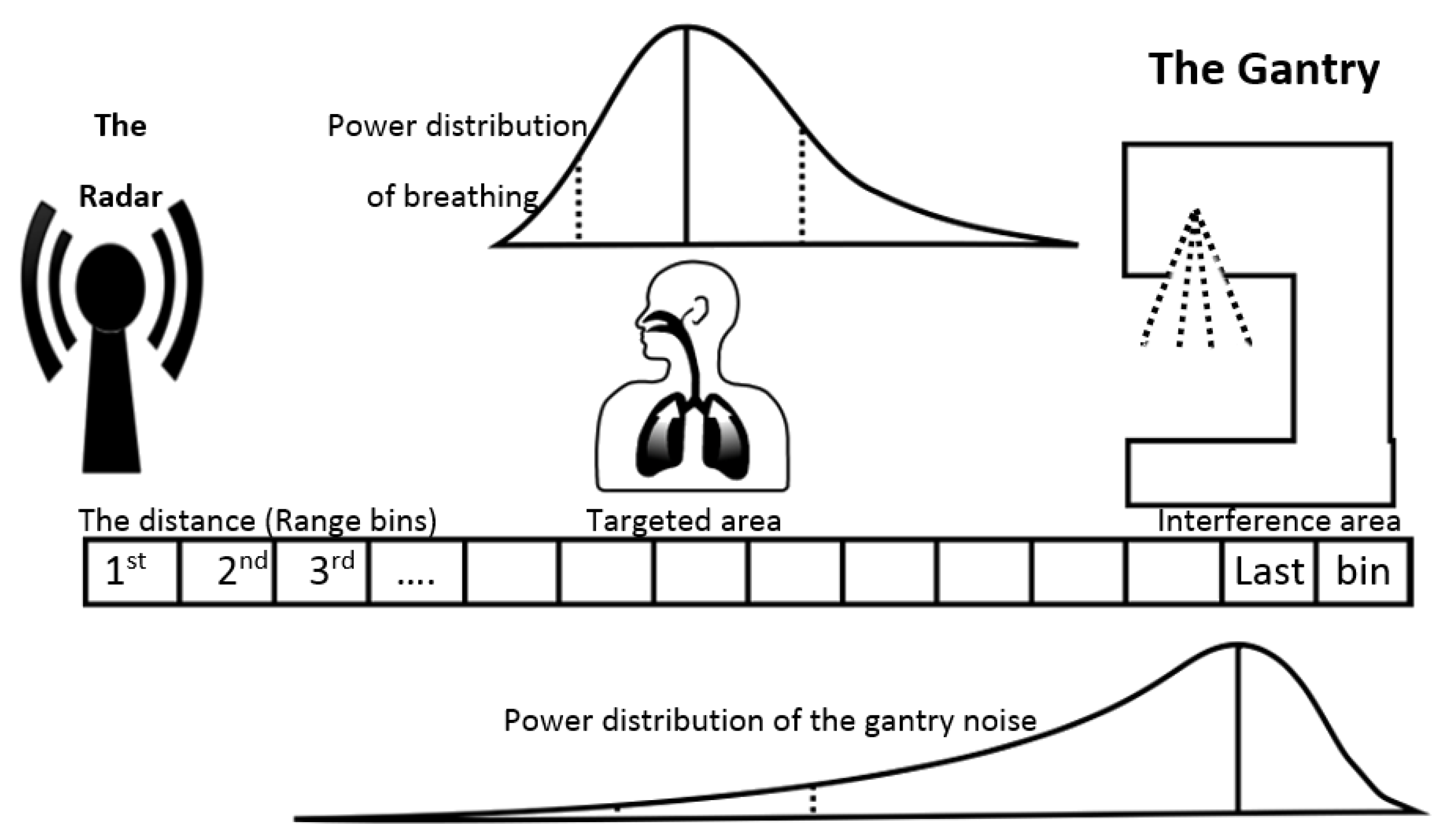
:1. Introduction
2. Materials and Methods
2.1. Experimental Design
2.2. Breathing Signal Recovery
2.2.1. Single-Bin Artificial Neural Network Noise Estimation
2.2.2. Range-Bin Artificial Neural Network Signal Noise Estimation
2.2.3. Signal Enhancement with Range Bin Weighted Averaging
2.3. Combining ANN Noise Estimation, Range Bin Weighted Averaging and Filtering
2.4. Classification
3. Results
3.1. Uwb Radar Signals from Target Range Bins
3.2. Breathing Signal Recovery from High Noise Background
3.2.1. Performance of the Single-Bin Artificial Neural Network Noise Estimation
3.2.2. Performance of the Range-Bin Artificial Neural Network Noise Estimation
3.2.3. Signal Enhancement Using Range Bin Weighted Averaging
3.3. Combining ANN Noise Estimation, Range Bin Weighted Averaging and Filtering
3.4. Classification Performance
4. Discussion
5. Conclusions
Author Contributions
Funding
Institutional Review Board Statement
Informed Consent Statement
Data Availability Statement
Acknowledgments
Conflicts of Interest
Abbreviations
| VMAT | Volumetric modulated arc therapy |
| DIBH | deep-inspiration breath hold |
| UWB | ultra-wideband |
| RMP | respiratory motion phantom |
| ANN | artificial neural network |
| SB-ANN | single-bin ANN |
| RB-ANN | range-bin ANN |
| RBWA | range-bin weighted averaging |
| SNR | Signal to Noise Ratio |
| MSE | Mean Square Error |
| MA | moving-average |
References
- Keall, P.J.; Mageras, G.S.; Balter, J.M.; Emery, R.S.; Forster, K.M.; Jiang, S.B.; Kapatoes, J.M.; Low, D.A.; Murphy, M.J.; Murray, B.R.; et al. The management of respiratory motion in radiation oncology report of AAPM Task Group 76. Med. Phys. 2006, 33, 3874–3900. [Google Scholar] [CrossRef] [PubMed]
- Benedict, S.H.; Yenice, K.M.; Followill, D.; Galvin, J.M.; Hinson, W.; Kavanagh, B.; Keall, P.; Lovelock, M.; Meeks, S.; Papiez, L.; et al. Stereotactic body radiation therapy: The report of AAPM Task Group 101. Med. Phys. 2010, 37, 4078–4101. [Google Scholar] [CrossRef] [PubMed] [Green Version]
- Giraud, P.; Morvan, E.; Claude, L.; Mornex, F.; Le Pechoux, C.; Bachaud, J.M.; Boisselier, P.; Beckendorf, V.; Morelle, M.; Carrère, M.O.; et al. Respiratory gating techniques for optimization of lung cancer radiotherapy. J. Thorac. Oncol. 2011, 6, 2058–2068. [Google Scholar] [CrossRef] [PubMed] [Green Version]
- Giraud, P.; Houle, A. Respiratory Gating for Radiotherapy: Main Technical Aspects and Clinical Benefits. Int. Sch. Res. Not. 2013, 2013, 519602. [Google Scholar] [CrossRef]
- Darby, S.C.; Ewertz, M.; McGale, P.; Bennet, A.M.; Blom-Goldman, U.; Brønnum, D.; Correa, C.; Cutter, D.; Gagliardi, G.; Gigante, B.; et al. Risk of Ischemic Heart Disease in Women after Radiotherapy for Breast Cancer. N. Engl. J. Med. 2013, 368, 987–998. [Google Scholar] [CrossRef] [PubMed] [Green Version]
- Farzaneh, K.M.; Momennezhad, M.; Naseri, S. Gated Radiotherapy Development and its Expansion. J. Biomed. Phys. Eng. 2021, 2, 93–100. [Google Scholar] [CrossRef] [Green Version]
- Wong, J.W.; Sharpe, M.B.; Jaffray, D.A.; Kini, V.R.; Robertson, J.M.; Stromberg, J.S.; Martinez, A.A. The use of active breathing control (ABC) to reduce margin for breathing motion. Int. J. Radiat. Oncol. Biol. Phys. 1999, 44, 911–919. [Google Scholar] [CrossRef]
- Fayad, H.; Pan, T.; François Clement, J.; Visvikis, D. Technical Note: Correlation of respiratory motion between external patient surface and internal anatomical landmarks. Med. Phys. 2011, 2015, 3157–3164. [Google Scholar] [CrossRef] [Green Version]
- Girbau, D.; Lázaro, A.; Ramos, Á.; Villarino, R. Remote sensing of vital signs using a doppler radar and diversity to overcome null detection. IEEE Sens. J. 2012, 12, 512–518. [Google Scholar] [CrossRef] [Green Version]
- Massagram, W.; Lubecke, V.M.; Høst-Madsen, A.; Boric-Lubecke, O. Assessment of heart rate variability and respiratory sinus arrhythmia via doppler radar. IEEE Trans. Microw. Theory Tech. 2009, 57, 2542–2549. [Google Scholar] [CrossRef]
- Kim, Y.; Ling, H. Human activity classification based on micro-doppler signatures using a support vector machine. IEEE Trans. Geosci. Remote Sens. 2009, 47, 1328–1337. [Google Scholar] [CrossRef]
- He, S.; Mehta, V.; Bolic, M. A Joint Localization Assisted Respiratory Rate Estimation using IR-UWB Radars. In Proceedings of the 2020 42nd Annual International Conference of the IEEE Engineering in Medicine & Biology Society (EMBC), Montreal, QC, Canada, 20–24 July 2020; pp. 489–493. [Google Scholar]
- Yarovoy, A.G.; Ligthart, L.P.; Matuzas, J.; Levitas, B. UWB Radar for human being detection. IEEE Aerosp. Electron. Syst. Mag. 2008, 23, 36–40. [Google Scholar] [CrossRef]
- Kakouche, I.; Maali, A.; El Korso, M.N.; Mesloub, A.; Azzaz, M.S. Fast and cost-effective method for non-contact respiration rate tracking using UWB impulse radar. Sens. Actuators Phys. 2021, 329, 112814. [Google Scholar] [CrossRef]
- Soldovieri, F.; Catapano, I.; Crocco, L.; Anishchenko, L.N.; Ivashov, S.I. A feasibility study for life signs monitoring via a continuous-wave radar. Hindawi 2012, 2021, 420178. [Google Scholar] [CrossRef]
- Li, L.; Cjangzhan, G.; Changzhi, L. Doppler Radar Noncontact Vital Signs Monitoring; Springer: New York, NY, USA, 2014. [Google Scholar] [CrossRef]
- Gu, C.; Li, R.; Li, C.; Jiang, S. Doppler radar respiration measurement for gated lung cancer radiotherapy. In Proceedings of the 2011 IEEE Topical Conference on Biomedical Wireless Technologies, Networks, and Sensing Systems, Phoenix, AZ, USA, 16–19 January 2011; pp. 91–94. [Google Scholar] [CrossRef]
- Li, C.; Gu, C.; Li, R.; Jiang, S. Radar motion sensing for accurate tumor tracking in radiation therapy. In Proceedings of the IEEE 12th Annu Wirel Microw Technol Conference WAMICON 2011i, Clearwater Beach, FL, USA, 18–19 April 2011; pp. 1–6. [Google Scholar] [CrossRef]
- Gu, C.; Salmani, Z.; Zhang, H.; Li, C. Antenna array technology for radar respiration measurement in motion-adaptive lung cancer radiotherapy. In Proceedings of the 2012 IEEE Topical Conference on Biomedical Wireless Technologies, Networks, and Sensing Systems (BioWireleSS), Santa Clara, CA, USA, 15–18 January 2012; pp. 1221–1224. [Google Scholar] [CrossRef]
- Zeeshan, Z.; Gu, C.; Li, C.; Zhang, H. High gain Fermi antenna array for radar-aided radiotherapy system. Microw. Opt. Technol. Lett. 2012, 54, 1649–1654. [Google Scholar] [CrossRef]
- Ren, H.; Shao, J.; Arigong, B.; Zhang, H.; Gu, C.; Li, C. Application of phased array antenna for radar respiration measurement. In Proceedings of the 2012 IEEE International Symposium on Antennas and Propagation, Chicago, IL, USA, 8–14 July 2012; pp. 4–5. [Google Scholar] [CrossRef]
- Gu, C.; Li, R.; Zhang, H.; Fung, A.Y.; Torres, C.; Jiang, S.B.; Li, C. Accurate respiration measurement using DC-coupled continuous-wave radar sensor for motion-adaptive cancer radiotherapy. IEEE Trans. Biomed. Eng. 2012, 59, 3117–3123. [Google Scholar] [CrossRef]
- Gu, C.; Li, C. Distortion analysis of continuous-wave radar sensor for complete respiration pattern monitoring. In Proceedings of the 2013 IEEE Topical Conference on Biomedical Wireless Technologies, Networks, and Sensing Systems, Austin, TX, USA, 20–23 January 2013; pp. 20137–20139. [Google Scholar] [CrossRef]
- Muragaki, M.; Okumura, S.; Sakamoto, T.; Sato, T. Non-Contact Respiration Measurement Using Ultra-wideband Array Radar with Adaptive Beamforming Technique for Cancer Radiotherapy. In Proceedings of the 2016 International Symposium on Antennas and Propagation (ISAP), Okinawa, Japan, 24–28 October 2016; pp. 440–441. [Google Scholar]
- Kakouche, I.; Abadlia, H.; El Korso, M.; Mesloub, A.; Maali, A.; Azzaz, M. Joint Vital Signs and Position Estimation of Multiple Persons Using SIMO Radar. Electronics 2021, 10, 2805. [Google Scholar] [CrossRef]
- He, S.; Han, Z.; Iglesias, C.; Mehta, V.; Bolic, M. A Real-Time Respiration Monitoring and Classification System using a Depth Camera and Radars. Front. Physiol. 2022, 13. [Google Scholar] [CrossRef]
- Forouzanfar, M.; Mabrouk, M.; Rajan, S.; Bolic, M.; Dajani, H.R.; Groza, V. Event Recognition for Contactless Activity Monitoring Using Phase-Modulated Continuous Wave Radar. IEEE Trans. Biomed. Eng. 2017, 64, 479–491. [Google Scholar] [CrossRef]
- Cardillo, E.; Li, C.; Caddemi, A. Millimeter-Wave Radar Cane: A Blind People Aid with Moving Human Recognition Capabilities. IEEE J. Electromagn. Microw. Med. Biol. 2021, 1–8. [Google Scholar] [CrossRef]
- Feng, C.; Zhao, H.; Liu, Q.; Hong, H.; Gu, C.; Zhu, X. Implementation of Radar-based Breathing Disorder Recognition Using FPGA. In Proceedings of the 2019 IEEE MTT-S International Microwave Biomedical Conference (IMBioC), Nanjing, China, 6–8 May 2019; pp. 1–3. [Google Scholar] [CrossRef]
- Zhao, H.; Hong, H.; Miao, D.; Li, Y.; Zhang, H.; Zhang, Y.; Li, C.; Zhu, X. A Noncontact Breathing Disorder Recognition System Using 2.4-GHz Digital-IF Doppler Radar. IEEE J. Biomed. Health Inform. 2019, 23, 208–217. [Google Scholar] [CrossRef] [PubMed]
- Berbeco, R.I.; Nishioka, S.; Shirato, H.; Gty, C.; Jiang, S. Residual motion of lung tumours in gated radiotherapy with external respiratory surrogates. Phys. Med. Biol. 2005, 50, 3655–3667. [Google Scholar] [CrossRef] [PubMed]
- Xia, J.; Siochi, R. A real-time respiratory motion monitoring system using KINECT: Proof of concept. Med. Phys. 2012, 39, 2682–2685. [Google Scholar] [CrossRef] [PubMed]
- Edmunds, D.M.; Bashforth, S.E.; Tahavori, F.; Wells, K.; Donovan, E. The feasibility of using Microsoft Kinect v2 sensors during radiotherapy delivery. J. Appl. Clin. Med. Phys. 2016, 17, 446–453. [Google Scholar] [CrossRef] [PubMed] [Green Version]
- Edmunds, D.M.; Gothard, L.; Khabra, K.; Kirby, A.; Madhale, P.; McNair, H.; Roberts, D.; Tang, K.K.; Symonds-Tayler, R.; Tahavori, F.; et al. Low-cost Kinect Version 2 imaging system for breath hold monitoring and gating: Proof of concept study for breast cancer VMAT radiotherapy. J. Appl. Clin. Med. Phys. 2018, 19, 71–78. [Google Scholar] [CrossRef] [PubMed]
- Nazir, S.; Rihana, S.; Visvikis, D.; Fayad, H. Technical Note: Kinect V2 surface filtering during gantry motion for radiotherapy applications. Med. Phys. 2018, 45, 1400–1407. [Google Scholar] [CrossRef]
- Dunn, L.; Kron, T.; Johnston, P.N.; McDermott, L.N.; Taylor, M.L.; Callahan, J.; Franich, R.D. A programmable motion phantom for quality assurance of motion management in radiotherapy. Australas Phys. Eng. Sci. Med. 2012, 35, 93–100. [Google Scholar] [CrossRef]
- Zywec, A.; Bogner, R. Coherent averaging of range profiles. In Proceedings of the International Radar Conference, Alexandria, VA, USA, 8–11 May 1995; pp. 456–461. [Google Scholar]
- Freislederer, P.; Kügele, M.; Öllers, M.; Swinnen, A.; Sauer, T.O.; Bert, C.; Giantsoudi, D.; Corradini, S.; Batista, V. Recent advances in Surface Guided Radiation Therapy. Radiat. Oncol. 2020, 15, 187. [Google Scholar] [CrossRef]
- Yeh, O.; Tavallaei, M.A.; Adve, R.S.; Wrigh, G.A. Fine Position Estimation of a Myocardium Phantom Using a UWB Radar. In Proceedings of the 2019 IEEE Radar Conference, Boston, MA, USA, 22–26 April 2019; pp. 1–6. [Google Scholar] [CrossRef]


| No-Gantry Noise | Gantry Noise | ||||
|---|---|---|---|---|---|
| Sensitivity | Specificity | Sensitivity | Specificity | ||
| SB-ANN | No breathing | 0.86 | 0.97 | 0.86 | 0.86 |
| Hold breathing | 0.73 | 0.89 | 0.47 | 0.87 | |
| Regular breathing | 0.90 | 0.96 | 0.66 | 0.87 | |
| Deep inspiration | 1 | 0.99 | 0.83 | 0.98 | |
| Overall accuracy | 0.85 | 0.72 | |||
| RB-ANN | No breathing | 0.90 | 0.97 | 0.98 | 0.98 |
| Hold breathing | 0.72 | 0.92 | 0.78 | 0.78 | |
| Regular breathing | 0.79 | 0.96 | 0.87 | 0.87 | |
| Deep inspiration | 1 | 0.99 | 0.97 | 0.97 | |
| Overall accuracy | 0.88 | 0.70 | |||
Publisher’s Note: MDPI stays neutral with regard to jurisdictional claims in published maps and institutional affiliations. |
© 2022 by the authors. Licensee MDPI, Basel, Switzerland. This article is an open access article distributed under the terms and conditions of the Creative Commons Attribution (CC BY) license (https://creativecommons.org/licenses/by/4.0/).
Share and Cite
Fallatah, A.; Bolic, M.; MacPherson, M.; La Russa, D.J. Monitoring Respiratory Motion during VMAT Treatment Delivery Using Ultra-Wideband Radar. Sensors 2022, 22, 2287. https://doi.org/10.3390/s22062287
Fallatah A, Bolic M, MacPherson M, La Russa DJ. Monitoring Respiratory Motion during VMAT Treatment Delivery Using Ultra-Wideband Radar. Sensors. 2022; 22(6):2287. https://doi.org/10.3390/s22062287
Chicago/Turabian StyleFallatah, Anwar, Miodrag Bolic, Miller MacPherson, and Daniel J. La Russa. 2022. "Monitoring Respiratory Motion during VMAT Treatment Delivery Using Ultra-Wideband Radar" Sensors 22, no. 6: 2287. https://doi.org/10.3390/s22062287