Article

S-Scheme BiOCl/MoSe2 Heterostructure with Enhanced Photocatalytic Activity for Dyes and Antibiotics Degradation under Sunlight Irradiation

1
Key Laboratory for Advanced Materials and Feringa Nobel Prize Scientist Joint Research Center, Institute of Fine Chemicals, School of Chemistry and Molecular Engineering, East China University of Science and Technology, 130 Meilong Road, Shanghai 200237, China
2
Research Institute of Physical and Chemical Engineering of Nuclear Industry, 168 Jintang Road, Tianjin 300180, China
3
State Environmental Protection Key Lab of Environmental Risk Assessment and Control on Chemical Processes, School of Resources & Environmental Engineering, East China University of Science and Technology, 130 Meilong Road, Shanghai 200237, China
4
Key Laboratory of Specially Functional Polymeric Materials and Related Technology (Ministry of Education), East China University of Science and Technology, 130 Meilong Road, Shanghai 200237, China
*
Authors to whom correspondence should be addressed.
Sensors 2022, 22(9), 3344; https://doi.org/10.3390/s22093344
Submission received: 31 March 2022 / Revised: 24 April 2022 / Accepted: 25 April 2022 / Published: 27 April 2022
(This article belongs to the Section Chemical Sensors)

Abstract

:
Semiconductor photocatalysis is considered to be a promising technique to completely eliminate the organic pollutants in wastewater. Recently, S-scheme heterojunction photocatalysts have received much attention due to their high solar efficiency, superior transfer efficiency of charge carriers, and strong redox ability. Herein, we fabricated an S-scheme heterostructure BiOCl/MoSe2 by loading MoSe2 nanosheets on the surface of BiOCl microcrystals, using a solvothermal method. The microstructures, light absorption, and photoelectrochemical performances of the samples were characterized by the means of SEM, TEM, XRD, transient photocurrents, electrochemical impedance, and photoluminescence (PL) spectra. The photocatalytic activities of BiOCl, MoSe2, and the BiOCl/MoSe2 samples with different MoSe2 contents were evaluated by the degradation of methyl orange (MO) and antibiotic sulfadiazine (SD) under simulated sunlight irradiation. It was found that BiOCl/MoSe2 displayed an evidently enhanced photocatalytic activity compared to single BiOCl and MoSe2, and 30 wt.% was an optimal loading amount for obtaining the highest photocatalytic activity. On the basis of radical trapping experiments and energy level analyses, it was deduced that BiOCl/MoSe2 follows an S-scheme charge transfer pathway and •O2, •OH, and h+ all take part in the degradation of organic pollutants.

1. Introduction

With the rapid development of urbanization and industrialization, water pollution has become more and more serious and imparted huge adverse effects on aquatic ecosystems, human health, and the development of economy and society [1,2,3]. In recent decades, the removal of noxious organic pollutants in wastewater, such as drugs [4,5], dyes [6,7], and antibiotics [8], has become a big challenge that must be managed. For instance, the antibiotics always bring about side effects on ecosystems and human health by inducing the proliferation of bacterial drug resistance [6]. The carcinogenic and teratogenic dyes can enter the human body along with the polluted water, leading to the appearance of cancers and other serious illnesses. To eliminate these organic pollutants, a series of techniques, such as physical adsorption, micro-biological degradation, and chemical oxidation, have been applied in the remediation of organic pollutants [1,9]. However, these strategies are still insufficient to completely remove the water-borne organic pollutants because of their low efficiency, as well as the formation of secondary waste products [10,11,12]. Alternatively, semiconductor photocatalysis has received much attention as a promising solution to completely eliminate the organic contaminants in wastewater [1,2,3,4,5,6,7,13,14,15,16,17,18,19]. As a semiconductor with wide bandgap, BiOCl is considered to be an ideal photocatalyst for the decomposition of organic pollutants in wastewater under UV light [20,21]. The main weakness of BiOCl is that it cannot respond to visible light, severely blocking its application in the whole solar spectrum. To solve this problem, the researchers have developed many strategies, such as fabricating oxygen vacancies, depositing with metals, constructing heterojunctions, and so on [22,23,24,25,26,27,28,29,30]. Although these approaches can extend the light response of BiOCl to the visible region, they inevitably decrease the redox ability of the photogenerated electrons and holes. In this regard, the researchers further exploited a series of all-solid-state and direct Z-scheme composite semiconductors to avoid decreasing the redox ability of photogenerated charge carriers [13,14,31,32,33,34,35,36,37,38]. Recently, Yu et al. proposed a novel S-scheme heterojunction theory and reasonably explained the transfer pathway of photogenerated charge carriers in the two semiconductors [39]. From then on, a series of S-scheme photocatalytic materials have been reported and successfully applied in the fields of environment and energy [40,41,42,43,44,45,46,47].
Layer-structured molybdenum selenide (MoSe2) has a narrow band gap (about 1.3–1.9 eV) [48,49], which means it can respond to the whole UV-visible-near-infrared (UV-Vis-NIR) light. However, its multilayer structure and narrow bandgap usually lead to the high recombination rate of photogenerated charge carriers [50]. In this regard, coupling MoSe2 with other semiconductors with wide bandgaps is an ideal strategy to take its advantages and simultaneously avoid its flaws. So far, several composite MoSe2-based photocatalysts have been exploited [51,52,53,54]. However, to the best our knowledge, the S-scheme heterojunction photocatalyst based on MoSe2 and BiOCl has never been studied.
Herein, we first constructed the S-scheme heterojunction BiOCl/MoSe2 photocatalyst by loading MoSe2 nanosheets on the surface of BiOCl microcrystals, using a solvothermal method. The morphology and crystalline structures of the as-prepared samples were characterized by the means of scanning electron microscopy (SEM), transmission electron microscopy (TEM), and high-resolution transmission electron microscopy (HR-TEM). The light absorption properties of the samples were analyzed by UV-Vis diffuse reflectance spectroscope (DRS). The photoelectric properties and the separation rate of charge carriers were investigated using transient photocurrents, electrochemical impedance, and photoluminescent (PL) spectra. The photocatalytic activities of BiOCl and the different BiOCl/MoSe2 samples were evaluated by the degradation of azo dye methyl orange (MO) and antibiotic sulfadiazine (SD) under simulated sunlight irradiation. On the basis of the radical trapping experiments and potential analyses of BiOCl and MoSe2 conduction bands (CB) and valence bands (VB), the possible photocatalytic mechanism of S-scheme BiOCl/MoSe2 was proposed.

2. Materials and Methods

2.1. Materials

Bismuth nitrate pentahydrate (Bi(NO3)3·5H2O) and absolute ethanol (C2H5OH) were provided by Sinopharm Chemical Reagent Co., Ltd., Shanghai, China. Selenium powder, sodium molybdate dihydrate (Na2MoO4·2H2O), and sodium borohydride (NaBH4) were purchased from Shanghai Adamas Reagent Co., Ltd., Shanghai, China. Potassium chloride (KCl) was obtained from Shanghai Lingfeng Chemical Reagent Co., Ltd., Shanghai, China. All the reagents were analytically pure grade and used as received without further purification. Milli-Q water was homemade and the resistivity was 18.2 MΩ cm.

2.2. Synthesis of BiOCl/MoSe2

BiOCl nanosheets were prepared using a hydrothermal method, similar to the previous report [55]. The detailed procedures were as follows: Firstly, 1 mmol Bi(NO3)3·5H2O and 1 mmol KCl were successively dispersed in 15 mL deionized water and stirred at room temperature for 1 h. Then, the mixture was transferred into a 50 mL Teflon-lined stainless-steel autoclave and placed in an oven to react at 160 °C for 24 h. Subsequently, the suspension was cooled to room temperature and the precipitation was washed, respectively, with deionized water and ethanol three times. Finally, the product was dried in a vacuum drying oven at 70 °C for 8 h, denoted as BiOCl.
BiOCl/MoSe2 was synthesized via a modified solvothermal method [56]: Firstly, 200.5 mg BiOCl, 0.079 mmol Na2MoO4·2H2O, 0.158 mmol selenium powder, and 0.079 mmol NaBH4 were added into a 25 mL mixture solution of ethanol and water with a volume ratio of 1:1. After the mixture was stirred at room temperature for 1 h, the obtained homogeneous mixture was transferred into a 50 mL Teflon-lined stainless-steel autoclave and kept at 180 °C for 12 h. Then, the autoclave was cooled to room temperature and the obtained precipitate was washed with deionized water and ethanol three times, respectively. Finally, the obtained product was dried in a vacuum drying oven at 70 °C for 8 h. The theoretical loading amount of the MoSe2 sample was 10 wt.%, denoted as BiOCl/MoSe2-10. By changing the dosages of Na2MoO4·2H2O, selenium powder, and NaBH4, the BiOCl/MoSe2 samples with 30 wt.% and 50 wt.% MoSe2 contents were also synthesized, denoted as BiOCl/MoSe2-30 and BiOCl/MoSe2-50, respectively. Pure MoSe2 was further prepared by the same method, except that BiOCl was not added.

2.3. Characterization

The morphologies of the obtained samples were observed via scanning electron microscope (SEM, TESCAN VEGA 3 SBH), transmission electron microscope (TEM, JEM2000EX), and high-resolution transmission electron microscope (HR-TEM, JEOJ JEM2100). The crystalline structures of the samples were analyzed using a Riguku D/Max 2550 VB/PC X-ray diffractometer with Cu Kα (λ = 1.5406 A) radiation, operated at a voltage of 40 kV and a current of 40 mA. The UV-Vis diffuse reflectance spectra of the samples were recorded on a SHIMADZU UV-2450 spectrophotometer and a Lambda 950 spectrophotometer, equipped with an integrating sphere assembly, using BaSO4 as the reference material. The photoluminescence (PL) spectra were tested on a Shimadzu RF5301PC fluorescence spectrophotometer and the 320 nm line of Xe lamp was used as the excitation source. The transient photocurrents, electrochemical impedance, and Mott–Schottky spectra were measured by a Zahner electrochemical workstation equipped with a three-electrode system, in which the platinum electrode and saturated calomel electrode were used as the counter electrode and reference electrode, respectively, and 0.2 mg photocatalyst sample was coated on 1.5 cm2 FTO glass as the working electrode. The transient photocurrent and Mott–Schottky tests were performed in a 0.5 M Na2SO4 aqueous solution and a 300 W Xe lamp with AM 1.5 filter as the light source. A mixed aqueous solution of 2.0 mM K3[Fe(CN)6], 2.0 mM K4[Fe(CN)6], and 0.5 M KCl was used as the electrolyte for the electrochemical impedance tests.

2.4. Photocatalytic Activity Measurement

The photocatalytic activities of the prepared samples were evaluated by the degradation of methyl orange (MO) and sulfadiazine (SD) under simulated sunlight irradiation, using a 300 W Xe lamp with AM1.5 as the light source. For each measurement, a 50 mg photocatalyst was dispersed in a 50 mL MO/or SD (20 mg/L) solution in a quartz tube and stirred in the dark for 30 min to achieve the adsorption–desorption of MO/or SD on the surface of the photocatalyst. At a given time interval, 5 mL of the mixture solution was withdrawn, centrifuged, and filtered to remove the remaining particles. The residual concentrations of MO and SD were determined using a UV-Vis spectrophotometer and a high-performance liquid chromatograph, respectively.

3. Results and Discussion

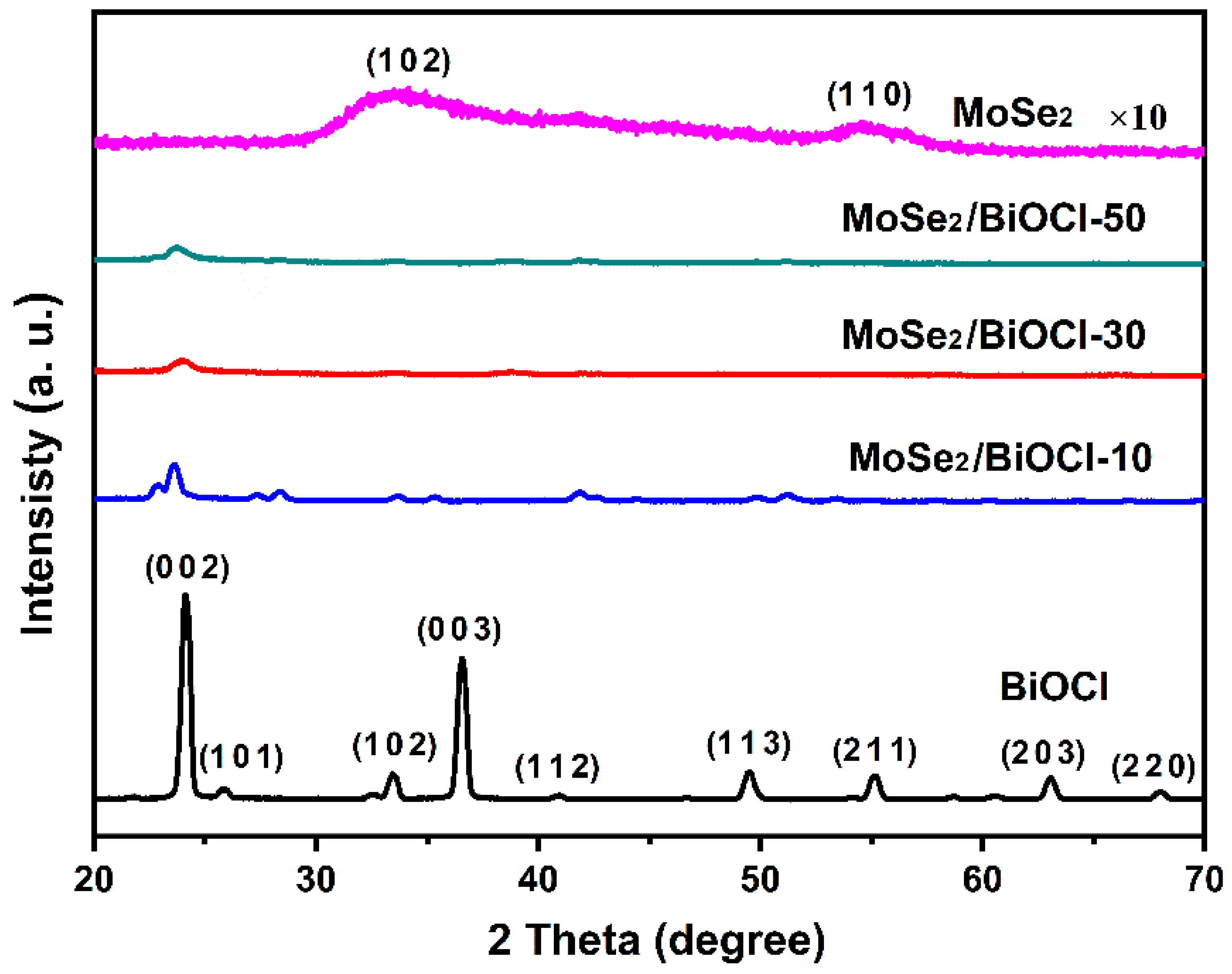
3.1. Morphological and Crystalline Structures

The morphological structures of the samples were observed by SEM, TEM, and HR-TEM images. As shown in Figure 1A,B, the surface of BiOCl sheets seems to be smooth and the width and thickness of BiOCl sheets are in the range of 1−4.5 μm and 300−400 nm, respectively. From the TEM images of BiOCl and BiOCl/MoSe2-30, it can be seen that the block-structured MoSe2 consists of many thin nanosheets (Figure 1C), which are uniformly wrapped on the surface of BiOCl sheets to form a shell structure (Figure 1D). The lattice structure of BiOCl/MoSe2-30 was further analyzed using HR-TEM images. In Figure 1E, the lattice spacing was measured to be 0.65 nm, attributed to the (0 0 2) crystal planes of 2H phase MoSe2 [57]. In Figure 1F, the lattice spacing of 0.275 nm corresponds to BiOCl (1 1 0) crystal planes, while that of 0.28 nm is attributed to MoSe2 (1 0 0) crystal planes. These results demonstrate the formation of a BiOCl/MoSe2 heterojunction structure [58].
The crystalline structures of the synthesized samples were analyzed by X-ray diffraction patterns (XRD). As shown in Figure 2, BiOCl presents the diffraction peaks at 2θ = 24.1°, 25.9°, 33.4°, 36.5°, 40.9°, 49.7°, 54.1°, 63.1°, and 68.1°, attributed to BiOCl (0 0 2), (1 0 1), (1 0 2), (0 0 3), (1 1 2), (1 1 3), (2 1 1), (2 0 3), and (2 2 0) crystal planes, respectively (JCPDS No. 06-0249) [54]. In contrast, BiOCl/MoSe2-10, BiOCl/MoSe2-30, and BiOCl/MoSe2-50 exhibit an obvious diffraction peak at 24.1°, while the other characteristic peaks become very weak, due to the resistance of the thick MoSe2 shell layer to X-ray. Even enlarged 10 times in intensity, the diffraction peaks of MoSe2 (1 0 2) and (1 1 0) are still very weak and broad, which is probably ascribable to both its low crystallinity as well as the very thin sheet structure.

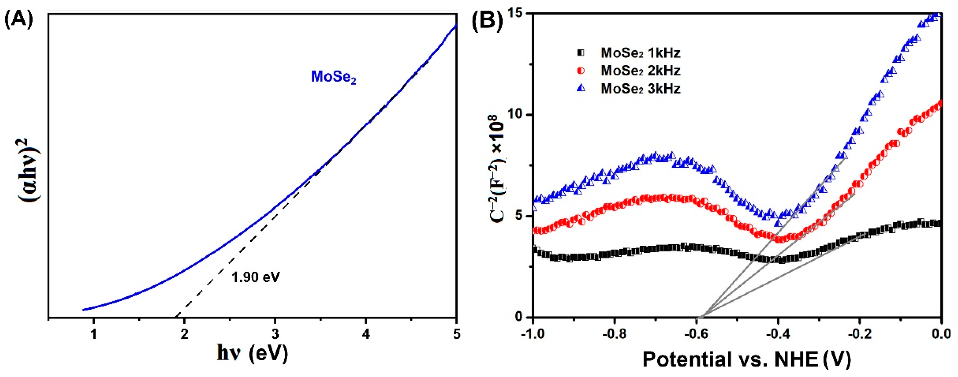
3.2. Light Absorption and PL Properties

The optical properties of MoSe2, BiOCl, and BiOCl/MoSe2 were investigated by UV-Vis DRS and PL spectra. As shown in Figure 3A,B, pure BiOCl only can absorb UV light, while MoSe2 displays strong light absorption in the whole UV-Vis-NIR region. After coupling with MoSe2, all the BiOCl/MoSe2 samples exhibit a significantly enhanced absorption in the visible and NIR region, and the absorption intensity gradually increases with the increase of MoSe2 content. PL spectrum is a useful technique to investigate the trapping, migration, and transfer efficiency of the photogenerated charge carriers in semiconductor photocatalysts [31,59,60]. Herein, we tested the PL spectra of BiOCl and the different BiOCl/MoSe2 samples at room temperature with an excitation wavelength of 320 nm. As displayed in Figure 3C, BiOCl exhibits a strong PL emission band in the range of 350–550 nm, while all the BiOCl/MoSe2 samples only have a very weak PL emission peak at 470 nm. After increasing the luminous flux of excitation light, the three BiOCl/MoSe2 samples also exhibit the PL emission bands in the range of 350–550 nm, similar to that of BiOCl (Figure 3D). The PL intensity of BiOCl/MoSe2-30 is near to that of BiOCl/MoSe2-50 and obviously weaker than that of BiOCl/MoSe2-10. These results indicate that the coupling of BiOCl and MoSe2 can effectively restrain the recombination of photogenerated charge carriers and that 30 wt.% is the optimal MoSe2 loading amount for effectively separating the photogenerated electrons and holes.

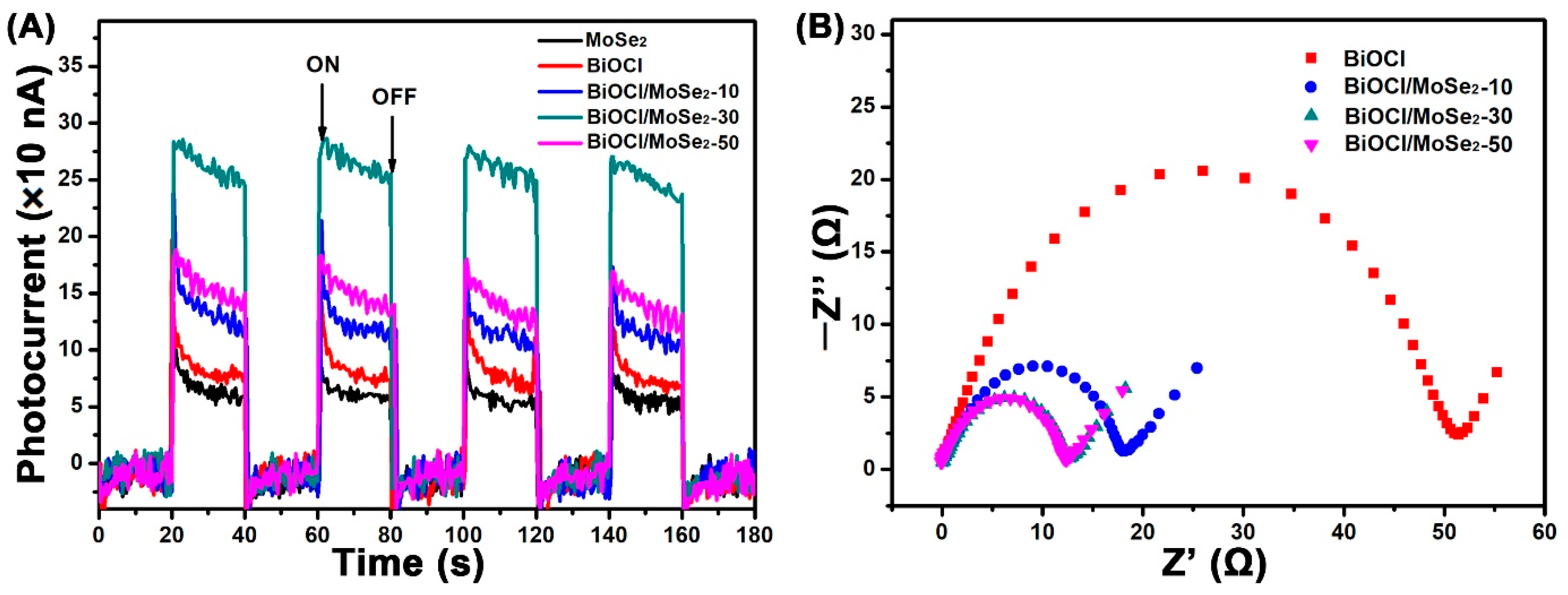
3.3. Photoelectric Characteristics

The photoelectric characteristics of BiOCl and the different BiOCl/MoSe2 samples were investigated by transient photocurrent measurements, which can further disclose the production, separation, and transfer efficiency of photogenerated charge carriers in these samples. As shown in Figure 4A, both BiOCl and MoSe2 exhibit very weak photocurrent intensity due to the low sunlight response ability and the high recombination rate of photo-generated electrons and holes, respectively. In contrast, all the BiOCl/MoSe2 composite photocatalysts display obviously enhanced current photocurrent intensity, indicating that the formation of a heterojunction structure can effectively promote the separation and transfer of photogenerated charge carriers. Amongst these samples, BiOCl/MoSe2-30 shows the highest photocurrent intensity, which is about four times that of pure BiOCl. For BiOCl/MoSe2-50, its photocurrent intensity is evidently weaker than that of BiOCl/MoSe2-30, resulting from the shielding of excess MoSe2 to light [61]. The electrochemical impedance spectra (EIS) can be used to disclose the dynamics of the mobile and bound charges in the interfacial or bulk regions of semiconductors, and the smaller curvature radius usually implies the weaker resistance to charge transfer [14,62,63]. In the EIS Nyquist spectra of Figure 4B, all the BiOCl/MoSe2 samples exhibit much smaller semicircle diameters than BiOCl, implying that coupling MoSe2 can effectively decrease the transfer resistance of the carriers in BiOCl. As the loading amount of MoSe2 increases from 10 wt.% to 30 wt.%, the semicircle diameter of the EIS curve obviously becomes smaller and it almost has no change when the loading amount of MoSe2 is further increased to 30 wt.%. Combining the results of the transient photocurrents and EIS spectra, it can be concluded that 30 wt.% is the optimal MoSe2 loading amount for effectively facilitating the production, separation, and transfer of photogenerated change carriers.

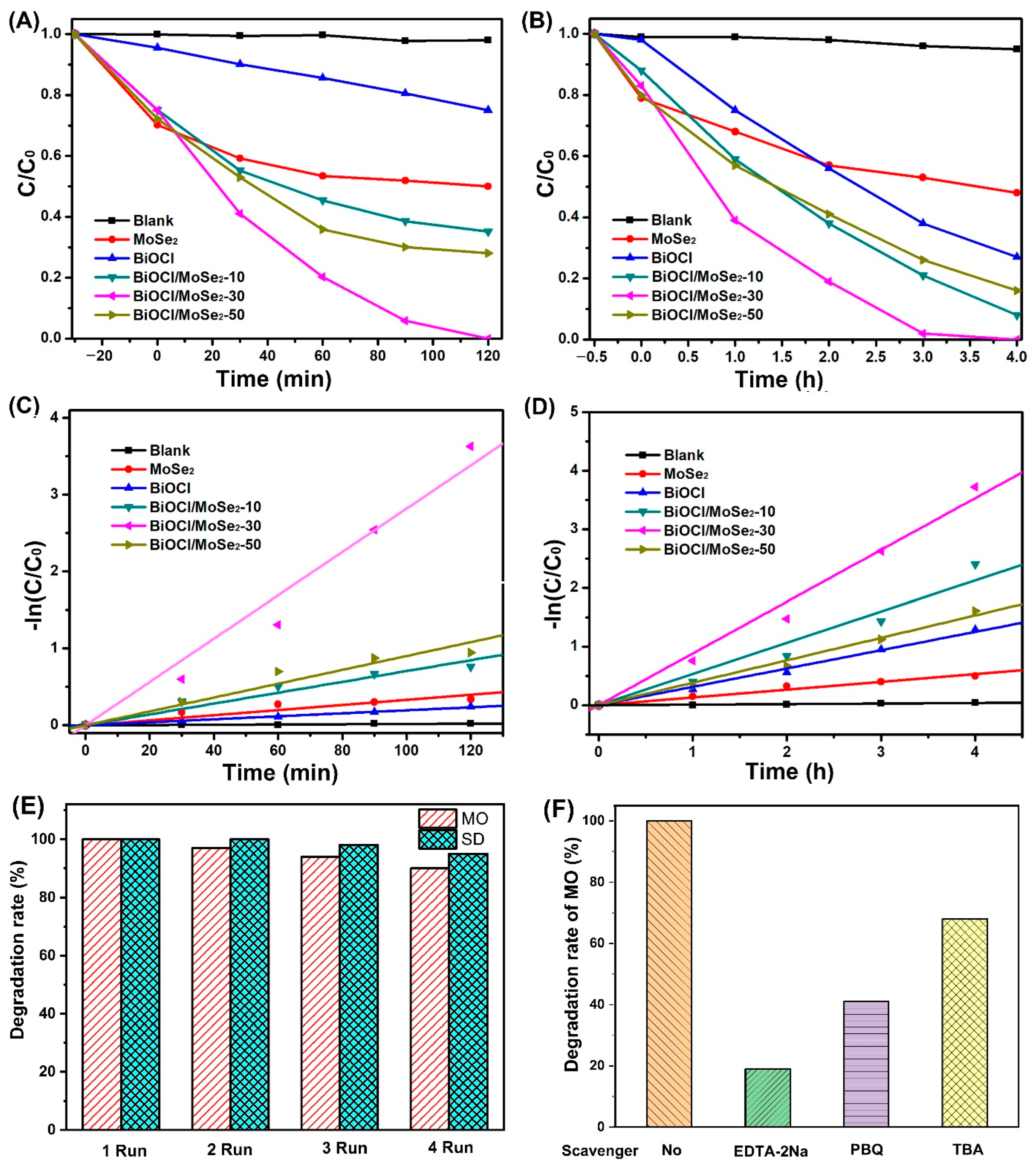
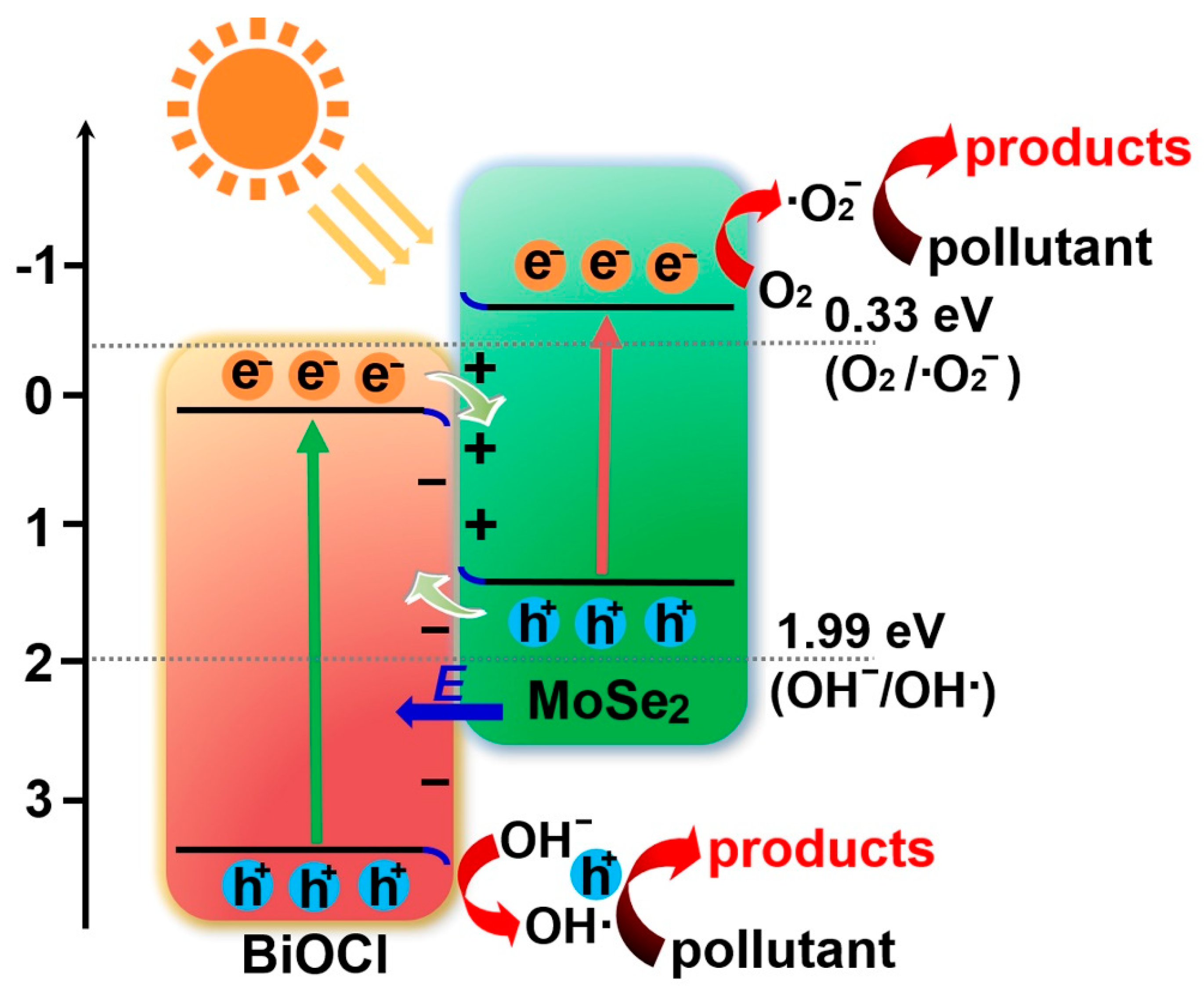
3.4. Photocatalytic Activity and Mechanism

Figure 5A,B presents the degradation curves of MO and SD over the different photocatalysts under simulated sunlight irradiation, respectively. In the absence of photocatalyst, the concentrations of MO and SD almost have no change under simulated sunlight irradiation, indicating that they have high photostability. Both pure BiOCl and MoSe2 exhibit low photocatalytic activity for MO and SD degradation, which is because BiOCl cannot respond to visible light while MoSe2 has the high recombination rate of photogenerated electrons and holes. Compared to pure MoSe2 and BiOCl, all the BiOCl/MoSe2 samples show evidently enhanced photocatalytic activity for MO and SD degradation, because the heterojunction structure between MoSe2 and BiOCl can effectively restrain the recombination of photogenerated electrons and holes. To more accurately compare the photocatalytic activities of BiOCl and the different BiOCl/MoSe2 samples, we further fitted the kinetic curves of MO and SD degradations over these samples. From Figure 5C,D, it can be seen that the degradations of MO and SD over these photocatalysts follow the pseudo first-order kinetic reaction. By comparing the reaction kinetic constants in Table 1, we know that BiOCl/MoSe2-30 possesses the highest photocatalytic activity among all the samples.
Given that photostability is very important to a photocatalyst for its practical applications, we further tested the photostability of BiOCl/MoSe2-30 using the cyclic degradation experiments of MO and SD under simulated sunlight irradiation. As shown in Figure 5E, the degradation rates of MO and SD only display a slight decrease after four cycles, probably due to the inevitable loss of photocatalysts during the recycle runs. This result indicates that BiOCl/MoSe2-30 is a stable photocatalyst under simulated sunlight irradiation. In the photocatalytic degradation process, the reactive species that take part in the organic pollutant decomposition mainly include hydroxyl radical (•OH), superoxide radical (•O2), and hole (h+). Herein, we identified the produced reactive species over BiOCl/MoSe2-30 in the organic decomposition process by addition of radical trapping agents. It is known that •OH, h+, and •O2 can be quenched by tert-butanol (TBA), EDTA-2Na, and p-benzoquinone (PBQ), respectively. As shown in Figure 5F, the degradation rate of MO was evidently inhibited after addition of EDTA-2Na, PBQ, and TBA, implying that all h+, •O2, and •OH take part in the degradation of MO. The effect of these species for MO degradation deceases in the order of h+ > •O2 > •OH.
To clarify the migration pathways of photogenerated charge carriers in BiOCl/MoSe2, it is necessary to identify the conduction band (CB) and valence band (VB) potentials of MoSe2 and BiOCl. In our previous studies [14,62], we have calculated the potentials of BiOCl CB and VB, which are +0.14 eV and +3.51 eV, respectively. Herein, we estimated the potentials of MoSe2 CB and VB by analyzing its UV-Vis absorption spectrum and Mott–Schottky curve.
Firstly, the bandgap energy of MoSe2 nanosheets was calculated using Tauc plot via the following Kubelka–Munk equation [64]:
(αhν)2 = A(hν − Eg)
where h, α, ν, A, and Eg are the Planck constant, absorption coefficient, light frequency, constant value, and bandgap energy, respectively. As shown in Figure 6A, the bandgap energy of MoSe2 was estimated to be 1.9 eV, similar to the value of the previous reports [52,65,66]. Then, the potential of MoSe2 CB edge was determined by Mott–Schottky analysis [67]. As shown in Figure 6B, the potential of MoSe2 CB (ECB) was estimated using the extrapolation of the Mott–Schottky plots at different frequencies (1 kHz, 2 kHz, and 3 kHz) to be −0.59 V (vs. NHE). According to the equation of EVB = ECB + Eg (EVB is the potential of VB), the potential of MoSe2 VB was further calculated to be 1.31 eV.
On the basis of the CB and VB potentials of BiOCl and MoSe2, BiOCl/MoSe2 should be ascribed to one of the three types of heterojunction, i.e., Type-II, direct Z-scheme, and S-scheme. Firstly, assuming that BiOCl/MoSe2 is a Type-II semiconductor, the electrons on MoSe2 CB would migrate to BiOCl CB. Given that the potential of BiOCl CB (0.14 eV vs. NHE) is more positive than E0(O2/•O2) (−0.33 eV vs. MHE) [68,69,70], the adsorbed O2 cannot be reduced by the electrons on BiOCl CB to form •O2. Similarly, since the potential of MoSe2 VB (1.31 eV vs. NHE) is more negative than E0(•OH/OH) (1.99 eV vs. NHE) [68,69,70], the holes on MoSe2 VB cannot oxidize OH into •OH. However, the presence of •O2 and •OH has been proved by the radical trapping experiments (Figure 5F), implying that BiOCl/MoSe2 is not a traditional Type-II semiconductor and the electrons for •O2 production and the holes for •OH production come from the MoSe2 CB and BiOCl VB, respectively. Moreover, Z-scheme heterojunction also has a theoretical problem in explaining the transfer pathway of photogenerated electrons and holes in BiOCl/MoSe2: from the perspective of charge transfer, the electrons on MoSe2 CB will preferentially recombine with the holes on BiOCl VB, rather than the electrons on BiOCl CB recombine with the holes on MoSe2 VB.
The S-scheme heterojunction is more reasonable to illustrate the transfer pathway of photogenerated electrons and holes in BiOCl/MoSe2 [39,41,71,72]—in this composite photocatalytic system, BiOCl is the oxidation photocatalyst (OP) and MoSe2 is the reduction photocatalyst (RP), both of which form an S-scheme heterojunction [39,41,71,72]. After the two components are in close contact, the electrons in MoSe2 spontaneously transfer to BiOCl, producing an electron depletion layer and electron accumulation layer near the interface of MoSe2 and BiOCl, respectively. Thus, MoSe2 would be positively charged and BiOCl would be positively charged, forming an internal electric field directing from MoSe2 to BiOCl. Meanwhile, after BiOCl and MoSe2 contact together, their Fermi energy should be aligned to the same level. Thus, the Fermi levels of BiOCl and should upward shift and upward shift, respectively, together with the band bending at their interfaces. Both the coulomb force of electric field and the band bending urge the photogenerated electrons from BiOCl to recombine with the holes from MoSe2 VB. Due to the band bending, the electrons on MoSe2 CB and holes on BiOCl will be reserved.
Based on the above experimental results and analyses, the degradation mechanism of organic pollutants over S-scheme BiOCl/MoSe2 was proposed: As illustrated in Figure 7, under simulated sunlight irradiation, both BiOCl and MoSe2 can produce holes on their VB and electrons on their CB. Using the acceleration of internal electric field, the photogenerated electrons on BiOCl CB and the holes on MoSe2 would be recombined. As a result, the powerful electrons on MoSe2 CB and the powerful holes on BiOCl VB would be reserved. Subsequently, the electrons on MoSe2 CB would react with adsorbed O2 to form •O2. Meanwhile, some holes on the BiOCl VB would oxidize OH to produce •OH. All of •O2, •OH, and h+ take part in the degradation of organic pollutants.

4. Conclusions

In summary, S-scheme BiOCl/MoSe2 heterojunction was fabricated via a modified solvothermal method. It was found that the thin MoSe2 nanosheets are uniformly wrapped on the surface of BiOCl microcrystals to form a shell structure. The MoSe2 diffraction peaks of MoSe2 and the different BiOCl/MoSe2 samples are very weak due to its low crystallinity and thin layer structure. The UV-Vis diffuse reflectance spectra show that all the BiOCl/MoSe2 samples exhibit a significantly enhanced absorption in the visible and near-infrared light region when compared with BiOCl, and the absorption intensity gradually increases with the increase of MoSe2 content. From the photoluminescence spectra, transient photocurrents, and electrochemical impedance spectra, it can be concluded that the BiOCl/MoSe2 heterojunction can effectively promote the transfer of photogenerated charge carriers. The results of MO and SD degradations indicate that all the BiOCl/MoSe2 samples display an evidently enhanced photocatalytic activity compared to single BiOCl and MoSe2, and the optimal MoSe2 loading amount for obtaining the highest photocatalytic activity is 30 wt.%. The radical trapping experiments disclosed that all h+, •O2, and •OH take part in the degradation of organic pollutants and h+ plays a more important role than •O2 and •OH. By further analyzing the potentials of BiOCl and MoSe2 CB and VB, it can be deduced that the BiOCl/MoSe2 follows an S-scheme photocatalytic mechanism. We think that this study provides a reference for fabricating the S-scheme photocatalytic materials to eliminate the organic pollutants in wastewater under sunlight irradiation.

Author Contributions

Conceptualization, B.T., J.Z. and Z.G.; methodology, Y.H., F.C. and Z.G.; formal analysis, Y.H. and Z.G.; Investigation, Y.L. (Yusheng Luo); data curation, F.C.; writing—original draft preparation, Y.H.; writing—review and editing, B.T.; visualization, L.Z. and Y.L. (Yufeng Lu); supervision, B.T. and J.Z. All authors have read and agreed to the published version of the manuscript.

Funding

This research was funded by the National Natural Science Foundation of China (U1862112 and 21573069), Shanghai Municipal Science and Technology Major Project (2018SHZDZX03), Program of Introducing Talents of Discipline to Universities (B16017), Fundamental Research Funds for the Central Universities (50321041917001 and 50321042017001), and Fundamental Research Funds for the Central Universities (JKD01211701).

Institutional Review Board Statement

Not applicable.

Informed Consent Statement

Not applicable.

Acknowledgments

All authors gratefully acknowledge the support of Science and Technology on Particle Transport and Separation Laboratory.

Conflicts of Interest

The authors declare no conflict of interest.

References

  1. Som, I.; Roy, M.; Saha, R. Advances in nanomaterial-based water treatment approaches for photocatalytic degradation of water pollutants. ChemCatChem 2020, 12, 3409–3433. [Google Scholar] [CrossRef]
  2. Ren, G.M.; Han, H.T.; Wang, Y.X.; Liu, S.T.; Zhao, J.Y.; Meng, X.C.; Li, Z.Z. Recent advances of photocatalytic application in watertreatment: A review. Nanomaterials 2021, 11, 1804. [Google Scholar] [CrossRef] [PubMed]
  3. Natarajan, S.; Bajaj, H.C.; Tayade, R.J. Recent advances based on the synergetic effect of adsorption for removal of dyes from waste water using photocatalytic process. J. Environ. Sci. 2018, 65, 201–222. [Google Scholar] [CrossRef] [PubMed]
  4. Molinari, R.; Pirillo, F.; Loddo, V.; Palmisano, L. Heterogeneous photocatalytic degradation of pharmaceuticals in water by using polycrystalline TiO2 and a nanofiltration membrane reactor. Catal. Today 2006, 118, 205–213. [Google Scholar] [CrossRef] [Green Version]
  5. Mendez-Arriaga, F.; Esplugas, S.; Giménez, J. Photocatalytic degradation of non−steroidal anti−inflammatory drugs with TiO2 and simulated solar irradiation. Water Res. 2008, 42, 585–594. [Google Scholar] [CrossRef]
  6. Hu, J.L.; Yang, Q.H.; Lin, H.; Ye, Y.P.; He, Q.; Zhang, J.N.; Qian, H.S. Mesoporous silica nanospheres decorated with CdS nanocrystals for enhanced photocatalytic and excellent antibacterial activities. Nanoscale 2013, 5, 6327–6332. [Google Scholar] [CrossRef]
  7. Dong, R.F.; Tian, B.Z.; Zhang, J.L.; Wang, T.T.; Tao, Q.S.; Bao, S.Y.; Yang, F.; Zeng, C.Y. AgBr@Ag/TiO2 core−shell composite with excellent visible light photocatalytic activity and hydrothermal stability. Catal. Commun. 2013, 38, 16–20. [Google Scholar] [CrossRef]
  8. Elmolla, E.S.; Chaudhuri, M. Photocatalytic degradation of amoxicillin, ampicillin and cloxacillin antibiotics in aqueous solution using UV/TiO2 and UV/H2O2/TiO2 photocatalysis. Desalination 2010, 252, 46–52. [Google Scholar] [CrossRef]
  9. Sarayu, K.; Sandhya, S. Current technologies for biological treatment of textile wastewater—A review. Appl. Biochem. Biotechnol. 2012, 167, 645–661. [Google Scholar] [CrossRef]
  10. Palominos, R.A.; Mondaca, M.A.; Giraldo, A.; Peñuela, G.; Perez-Moya, M.; Mansilla, H.D. Photocatalytic oxidation of the antibiotic tetracycline on TiO2 and ZnO suspensions. Catal. Today 2009, 144, 100–105. [Google Scholar] [CrossRef]
  11. Wang, P.H.; Yap, P.S.; Lim, T.T. C−N−S tridoped TiO2 for photocatalytic degradation of tetracycline under visible−light irradiation. Appl. Catal. A 2011, 399, 252–261. [Google Scholar] [CrossRef]
  12. Crini, G.; Lichtfouse, E. Advantages and disadvantages of techniques used for wastewater treatment. Environ. Chem. Lett. 2019, 17, 145–155. [Google Scholar] [CrossRef]
  13. Bao, S.Y.; Wu, Q.F.; Chang, S.Z.; Tian, B.Z.; Zhang, J.L. Z-scheme CdS−Au−BiVO4 with enhanced photocatalytic activity for organic contaminant decomposition. Catal. Sci. Technol. 2017, 7, 124–132. [Google Scholar] [CrossRef]
  14. Li, Q.Y.; Guan, Z.P.; Wu, D.; Zhao, X.G.; Bao, S.Y.; Tian, B.Z.; Zhang, J.L. Z-scheme BiOCl-Au-CdS heterostructure with enhanced sunlight-driven photocatalytic activity in degrading water dyes and antibiotics. ACS Sustain. Chem. Eng. 2017, 5, 6958–6968. [Google Scholar] [CrossRef]
  15. Kong, W.H.; Wang, S.L.; Wu, D.; Chen, C.R.; Luo, Y.S.; Pei, Y.T.; Tian, B.Z.; Zhang, J.L. Fabrication of 3D sponge@AgBr-AgCl/Ag and tubular photoreactor for continuous wastewater purification under sunlight irradiation. ACS Sustain. Chem. Eng. 2019, 7, 14051–14063. [Google Scholar] [CrossRef]
  16. Cheng, T.T.; Gao, H.J.; Liu, G.R.; Pu, Z.S.; Wang, S.F.; Yi, Z.; Wu, X.W.; Yang, H. Preparation of core-shell heterojunction photocatalysts by coating CdS nanoparticles onto Bi4Ti3O12 hierarchical microspheres and their photocatalytic removal of organic pollutants and Cr(VI) ions. Colloid Surf. A 2022, 633, 127918. [Google Scholar] [CrossRef]
  17. Li, L.X.; Gao, H.J.; Yi, Z.; Wang, S.F.; Wu, X.W.; Li, R.S.; Yang, H. Comparative investigation on synthesis, morphological tailoring and photocatalytic activities of Bi2O2CO3 nanostructures. Colloid Surf. A 2022, 644, 128758. [Google Scholar] [CrossRef]
  18. Chen, H.; Chen, Z.H.; Yang, H.; Wen, L.H.; Yi, Z.; Zhou, Z.G.; Dai, B.; Zhang, J.G.; Wu, X.W.; Wu, P.H. Multi-mode surface plasmon resonance absorber based on dart-type single-layer graphene. RSC Adv. 2022, 12, 7821–7829. [Google Scholar] [CrossRef]
  19. Li, L.X.; Sun, X.F.; Xian, T.; Gao, H.J.; Wang, S.F.; Yi, Z.; Wu, X.W.; Yang, H. Template-free synthesis of Bi2O2CO3 hierarchical nanotubes self-assembled from ordered nanoplates for promising photocatalytic application. Phys. Chem. Chem. Phys. 2022, 24, 8279–8295. [Google Scholar] [CrossRef]
  20. Lei, Y.Q.; Wang, G.H.; Song, S.Y.; Fan, W.Q.; Zhang, H.J. Synthesis, characterization and assembly of BiOCl nanostructure and their photocatalytic properties. CrystEngComm 2009, 11, 1857–1862. [Google Scholar] [CrossRef]
  21. Zhang, X.; Ai, Z.H.; Jia, F.L.; Zhang, L.Z. Generalized one-pot synthesis, characterization, and photocatalytic activity of hierarchical BiOX (X = Cl, Br, I) nanoplate microspheres. J. Phys. Chem. C 2008, 112, 747–753. [Google Scholar] [CrossRef]
  22. Guan, M.L.; Xiao, C.; Zhang, J.; Fan, S.J.; An, R.; Cheng, Q.M.; Xie, J.F.; Zhou, M.; Ye, B.J.; Xie, Y. Vacancy associates promoting solar–driven photocatalytic activity of ultrathin bismuth oxychloride nanosheets. J. Am. Chem. Soc. 2013, 135, 10411–10417. [Google Scholar] [CrossRef] [PubMed]
  23. Zhao, K.; Zhang, L.Z.; Wang, J.J.; Li, Q.X.; He, W.W.; Yin, J.J. Surface structure–dependent molecular oxygen activation of BiOCl single–crystalline nanosheets. J. Am. Chem. Soc. 2013, 135, 15750–15753. [Google Scholar] [CrossRef] [PubMed]
  24. Ye, L.Q.; Deng, K.J.; Xu, F.; Tian, L.H.; Peng, T.Y.; Zan, L. Increasing visible–light absorption for photocatalysis with black BiOCl. Phys. Chem. Chem. Phys. 2012, 14, 82–85. [Google Scholar] [CrossRef] [PubMed]
  25. Weng, S.X.; Chen, B.B.; Xie, L.Y.; Zheng, Z.Y.; Liu, P. Facile in situ synthesis of a Bi/BiOCl nanocomposite with high photocatalytic activity. J. Mater. Chem. A 2013, 1, 3068–3075. [Google Scholar] [CrossRef]
  26. Dong, F.; Xiong, T.; Yan, S.; Wang, H.Q.; Sun, Y.J.; Zhang, Y.X.; Huang, H.W.; Wu, Z.B. Facets and defects cooperatively promote visible light plasmonic photocatalysis with Bi nanowires@BiOCl nanosheets. J. Catal. 2016, 344, 401–410. [Google Scholar] [CrossRef]
  27. Jiang, J.; Zhang, L.Z.; Li, H.; He, W.W.; Yin, J.J. Self–doping and surface plasmon modification induced visible light photocatalysis of BiOCl. Nanoscale 2013, 5, 10573–10581. [Google Scholar] [CrossRef]
  28. Tan, C.W.; Zhu, G.Q.; Hojamberdiev, M.; Okada, K.; Liang, J.C.; Luo, X.; Liu, P.; Liu, Y. Co3O4 nanoparticles–loaded BiOCl nanoplates with the dominant {001} facets: Efficient photodegradation of organic dyes under visible light. Appl. Catal. B 2014, 152–153, 425–436. [Google Scholar] [CrossRef]
  29. Li, T.B.; Chen, G.; Zhou, C.; Shen, Z.Y.; Jin, R.C.; Sun, J.X. New photocatalyst BiOCl/BiOI composites with highly enhanced visible light photocatalytic performances. Dalton Trans. 2011, 40, 6751–6758. [Google Scholar] [CrossRef]
  30. Yu, L.H.; Zhang, X.Y.; Li, G.W.; Cao, Y.T.; Shao, Y.; Li, D.Z. Highly efficient Bi2O2CO3/BiOCl photocatalyst based on heterojunction with enhanced dye–sensitization. Appl. Catal. B 2016, 187, 301–309. [Google Scholar] [CrossRef]
  31. Bao, S.Y.; Wang, Z.; Zhang, J.L.; Tian, B.Z. Facet-heterojunction-based Z-Scheme BiVO4{010} microplates decorated with AgBr-Ag nanoparticles for the photocatalytic inactivation of bacteria and the decomposition of organic contaminants. ACS Appl. Nano Mater. 2020, 3, 8604–8617. [Google Scholar] [CrossRef]
  32. Yuan, Z.; Huang, H.; Li, N.; Chen, D.; Xu, Q.; Li, H.; He, J.; Lu, J. All-solid-state WO3/TQDs/In2S3 Z-scheme heterojunctions bridged by Ti3C2 quantum dots for efficient removal of hexavalent chromium and bisphenol A. J. Hazard. Mater. 2021, 409, 125027. [Google Scholar] [CrossRef] [PubMed]
  33. Wen, X.J.; Niu, C.G.; Zhang, L.; Liang, C.; Guo, H.; Zeng, G.M. Photocatalytic degradation of ciprofloxacin by a novel Z-scheme CeO2–Ag/AgBr photocatalyst: Influencing factors, possible degradation pathways, and mechanism insight. J. Catal. 2018, 358, 141–154. [Google Scholar] [CrossRef]
  34. Jo, W.K.; Natarajan, T.S. Influence of TiO2 morphology on the photocatalytic efficiency of direct Z-scheme g-C3N4/TiO2 photocatalysts for isoniazid degradation. Chem. Eng. J. 2015, 281, 549–565. [Google Scholar] [CrossRef]
  35. Jin, Z.; Hu, R.; Wang, H.; Hu, J.; Ren, T. One-step impregnation method to prepare direct Z-scheme LaCoO3/g-C3N4 heterojunction photocatalysts for phenol degradation under visible light. Appl. Surf. Sci. 2019, 491, 432–442. [Google Scholar] [CrossRef]
  36. Huang, S.; Zhang, J.; Qin, Y.; Song, F.; Du, C.; Su, Y. Direct Z-scheme SnO2/Bi2Sn2O7 photocatalyst for antibiotics removal: Insight on the enhanced photocatalytic performance and promoted charge separation mechanism. J. Photochem. Photobiol. A 2021, 404, 112947. [Google Scholar] [CrossRef]
  37. Shangguan, X.Y.; Fang, B.L.; Xu, C.X.; Tan, Y.; Chen, Y.G.; Xia, Z.J.; Chen, W. Fabrication of direct Z-scheme FeIn2S4/Bi2WO6 hierarchical heterostructures with enhanced photocatalytic activity for tetracycline hydrochloride photodagradation. Ceram. Int. 2021, 47, 6318–6328. [Google Scholar] [CrossRef]
  38. Li, G.; Wang, B.; Zhang, J.; Wang, R.; Liu, H. Rational construction of a direct Z-scheme g-C3N4/CdS photocatalyst with enhanced visible light photocatalytic activity and degradation of erythromycin and tetracycline. Appl. Surf. Sci. 2019, 478, 1056–1064. [Google Scholar] [CrossRef]
  39. Xu, Q.L.; Zhang, L.Y.; Cheng, B.; Fan, J.J.; Yu, J.G. S-Scheme heterojunction photocatalyst. Chem 2020, 6, 1543–1559. [Google Scholar] [CrossRef]
  40. Bao, Y.J.; Song, S.Q.; Yao, G.J.; Jiang, S.J. S-Scheme photocatalytic systems. Sol. RRL 2021, 5, 2100118. [Google Scholar] [CrossRef]
  41. Luo, Y.S.; Chi, Z.L.; Zhang, J.L.; Tian, B.Z. Photothermocatalytic system designed by facet-heterojunction to enhance the synergistic effect of toluene oxidation. ChemCatChem 2022, 14, e202101958. [Google Scholar] [CrossRef]
  42. Wang, J.; Zhang, Q.; Deng, F.; Luo, X.B.; Dionysiou, D.D. Rapid toxicity elimination of organic pollutants by the photocatalysis of environment-friendly and magnetically recoverable step-scheme SnFe2O4/ZnFe2O4 nano-heterojunctions. Chem. Eng. J. 2020, 379, 122264. [Google Scholar] [CrossRef]
  43. He, R.A.; Liu, H.J.; Liu, H.M.; Xu, D.F.; Zhang, L.Y. S-scheme photocatalyst Bi2O3/TiO2 nanofiber with improved photocatalytic performance. J. Mater. Sci. Technol. 2020, 52, 145–151. [Google Scholar]
  44. Gogoi, D.; Makkar, P.; Ghosh, N.N. Solar light-irradiated photocatalytic degradation of model dyes and industrial dyes by a magnetic CoFe2O4–gC3N4 S-scheme heterojunction photocatalyst. ACS Omega 2021, 6, 4831–4841. [Google Scholar] [CrossRef]
  45. Jia, X.M.; Han, Q.F.; Liu, H.Z.; Li, S.Z.; Bi, H.P. A dual strategy to construct flowerlike S-scheme BiOBr/BiOAc1−xBrx heterojunction with enhanced visible-light photocatalytic activity. Chem. Eng. J. 2020, 399, 125701. [Google Scholar] [CrossRef]
  46. Dou, L.; Jin, X.Y.; Chen, J.F.; Zhong, J.B.; Li, J.Z.; Zeng, Y.; Duan, R. One-pot solvothermal fabrication of S-scheme OVs-Bi2O3/Bi2SiO5 microsphere heterojunctions with enhanced photocatalytic performance toward decontamination of organic pollutants. Appl. Surf. Sci. 2020, 527, 146775. [Google Scholar] [CrossRef]
  47. Lian, X.; Xue, W.H.; Dong, S.; Liu, E.Z.; Li, H.; Xu, K.Z. Construction of S-scheme Bi2WO6/g-C3N4 heterostructure nanosheets with enhanced visible-light photocatalytic degradation for ammonium dinitramide. J. Hazard. Mater. 2021, 412, 125217. [Google Scholar] [CrossRef]
  48. Huang, K.J.; Zhang, J.Z.; Fan, Y. Preparation of layered MoSe2 nanosheets on Ni-foam substrate with enhanced supercapacitor performance. Mater. Lett. 2015, 152, 244–247. [Google Scholar] [CrossRef]
  49. Eftekhari, A. Molybdenum diselenide (MoSe2) for energy storage, catalysis, and optoelectronics. Appl. Mater. Today 2017, 8, 1–7. [Google Scholar] [CrossRef]
  50. Liu, Z.; Li, N.; Zhao, H.; Du, Y. Colloidally synthesized MoSe2/graphene hybrid nanostructures as efficient electrocatalysts for hydrogen evolution. J. Mater. Chem. A 2015, 3, 19706–19710. [Google Scholar] [CrossRef]
  51. Zhang, H.; Tang, G.G.; Wan, X.; Xu, J.; Tang, H. High-efficiency all-solid-state Z-scheme Ag3PO4/g-C3N4/MoSe2 photocatalyst with boosted visible-light photocatalytic performance for antibiotic elimination. Appl. Surf. Sci. 2020, 530, 147234. [Google Scholar] [CrossRef]
  52. Zheng, X.T.; Yang, L.M.; Li, Y.B.; Yang, L.X.; Luo, S.L. Direct Z-scheme MoSe2 decorating TiO2 nanotube arrays photocatalyst for water decontamination. Electrochim. Acta 2019, 298, 663–669. [Google Scholar] [CrossRef]
  53. Tahir, M.B.; Asiri, A.M.; Nabi, G.; Rafique, M.; Sagir, M. Fabrication of heterogeneous photocatalysts for insight role of carbon nanofibre in hierarchical WO3/MoSe2 composite for enhanced photocatalytic hydrogen generation. Ceram. Int. 2019, 45, 5547–5552. [Google Scholar] [CrossRef]
  54. Zhang, S.M.; Chen, L.; Shen, J.; Li, Z.F.; Wu, Z.H.; Feng, W.H.; Xu, K.Q.; Xu, D.F.; Chen, X.H.; Zhang, S.Y. TiO2@MoSe2 line-to-face heterostructure: An advanced photocatalyst for highly efficient reduction of Cr (VI). Ceram. Int. 2019, 45, 18065–18072. [Google Scholar] [CrossRef]
  55. Jiang, J.; Zhao, K.; Xiao, X.Y.; Zhang, L.Z. Synthesis and facet-dependent phoyoreactivity of BiOCl single-crystalline nanosheets. J. Am. Chem. Soc. 2012, 134, 4473–4476. [Google Scholar] [CrossRef]
  56. Yang, S.; Shao, C.L.; Zhou, X.J.; Li, X.H.; Tao, R.; Li, X.W.; Liu, S.; Liu, Y.C. MoSe2/TiO2 nanofibers for cycling photocatalytic removing water pollutants under UV-Vis-NIR light. ACS Appl. Nano Mater. 2020, 3, 2278–2287. [Google Scholar] [CrossRef]
  57. Fan, C.; Wei, Z.; Yang, S.; Li, J. Synthesis of MoSe2 flower-like nanostructures and their photo-responsive properties. RSC Adv. 2014, 4, 775–778. [Google Scholar] [CrossRef]
  58. Xiao, P.; Lou, J.; Zhang, H.; Song, W.; Wu, X.L.; Lin, H.; Chen, J.; Liu, S.; Wang, X. Enhanced visible-light-driven photocatalysis from WS2 quantum dots coupled to BiOCl nanosheets: Synergistic effect and mechanism insight. Catal. Sci. Technol. 2018, 8, 201–209. [Google Scholar] [CrossRef]
  59. Liu, Y.; Zhang, P.; Tian, B.Z.; Zhang, J.L. Core-shell structural CdS@SnO2 nanorods with excellent visible-light photocatalytic activity for the selective oxidation of benzyl alcohol to benzaldehyde. ACS Appl. Mater. Interfaces 2015, 7, 13849–13858. [Google Scholar] [CrossRef]
  60. Wang, S.; Han, Y.; Luo, Y.; Ma, Y.; Zhang, J.; Tian, B. Au thorn-decorated TiO2 hierarchical microspheres with superior photocatalytic bactericidal activity under red and NIR light irradiation. J. Alloys Compd. 2022, 910, 164485. [Google Scholar] [CrossRef]
  61. Li, H.; Yang, C.; Wang, X.; Zhang, J.; Xi, J.; Du, G.; Ji, Z. Mixed 3D/2D dimensional TiO2 nanoflowers/MoSe2 nanosheets for enhanced photoelectrochemical hydrogen generation. J. Am. Chem. Soc. 2019, 103, 1187–1196. [Google Scholar]
  62. Han, Y.Q.; Li, Q.Y.; Bao, S.Y.; Lu, Y.F.; Guan, Z.P.; Zhang, J.L.; Tian, B.Z. Z-scheme heterostructure BiOCl-Ag-AgBr with enhanced sunlight-driven photocatalytic activity in simultaneous removal of Cr6+ and phenol contaminants. Catal. Today 2021, 376, 151–161. [Google Scholar]
  63. Guan, Z.P.; Li, Q.Y.; Shen, B.; Bao, S.Y.; Zhang, J.L.; Tian, B.Z. Fabrication of Co3O4 and Au co-modified BiOBr flower-like microspheres with high photocatalytic efficiency for sulfadiazine degradation. Sep. Purif. Technol. 2020, 234, 116100. [Google Scholar] [CrossRef]
  64. Dion, M.C.L.; Fauzia, V.; Imawan, C. The Effect of deposition of MoSe2 nanosheets on the performance of a ZnO-based UV detector. J. Phys. Conf. Ser. 2021, 1951, 012006. [Google Scholar] [CrossRef]
  65. Kwon, I.S.; Kwak, I.H.; Debela, T.T.; Abbas, H.G.; Park, Y.C.; Ahn, J.; Park, J.; Kang, H.S. Se-rich MoSe2 nanosheets and their superior electrocatalytic performance for hydrogen evolution reaction. ACS Nano 2020, 14, 6295–6304. [Google Scholar] [CrossRef]
  66. Wang, G.Y.; Zhang, Y.Z.; You, C.Y.; Liu, B.Y.; Yang, Y.H.; Li, H.J.W.; Cui, A.J.; Liu, D.M.; Yan, H. Two dimensional materials based photodetectors. Infrared Phys. Technol. 2018, 88, 149–173. [Google Scholar] [CrossRef]
  67. Bao, S.Y.; Wang, Z.Q.; Gong, X.Q.; Zeng, C.Y.; Wu, Q.F.; Tian, B.Z.; Zhang, J.L. AgBr tetradecahedrons with co-exposed {100} and {111} facets: Simple fabrication and enhancing spatial charge separation using facet heterojunctions. J. Mater. Chem. A 2016, 4, 18570–18577. [Google Scholar] [CrossRef]
  68. Hong, X.D.; Li, Y.; Wang, X.; Long, J.P.; Liang, B. Carbon nanosheet/MnO2/BiOCl ternary composite for degradation of organic pollutants. J. Alloys Compd. 2022, 891, 162090. [Google Scholar] [CrossRef]
  69. Acharya, L.; Nayak, S.; Pattnaik, S.P.; Acharya, R.; Parida, K. Resurrection of boron nitride in p-n type-II boron nitride/B-doped-g-C3N4 nanocomposite during solid-state Z-scheme charge transfer path for the degradation of tetracycline hydrochloride. J. Colloid Interface Sci. 2020, 566, 211–223. [Google Scholar] [CrossRef]
  70. Tang, M.L.; Ao, Y.H.; Wang, P.F.; Wang, C. All-solid-state Z-scheme WO3 nanorod/ZnIn2S4 composite photocatalysts for the effective degradation of nitenpyram under visible light irradiation. J. Hazard. Mater. 2020, 387, 121713. [Google Scholar] [CrossRef]
  71. Cheng, C.; He, B.W.; Fan, J.J.; Cheng, B.; Cao, S.W.; Yu, J.G. An Inorganic/Organic S-scheme heterojunction H2-production photocatalyst and its charge transfer mechanism. Adv. Mater. 2021, 33, 2100317. [Google Scholar] [CrossRef] [PubMed]
  72. Deng, H.Z.; Fei, X.G.; Yang, Y.; Fan, J.J.; Yu, J.G.; Cheng, B.; Zhang, L.Y. S-scheme heterojunction based on p-type ZnMn2O4 and n-type ZnO with improved photocatalytic CO2 reduction activity. Chem. Eng. J. 2021, 409, 127377. [Google Scholar] [CrossRef]
Figure 1. (A) SEM image of BiOCl. (BD) TEM images of (B) BiOCl, (C) MoSe2, and (D) BiOCl/MoSe2-30. (E,F) HR-TEM images of BiOCl/MoSe2-30.
Sensors 22 03344 g001
Figure 2. XRD patterns of MoSe2, BiOCl, BiOCl/MoSe2-10, BiOCl/MoSe2-30, and BiOCl/MoSe2-50.
Sensors 22 03344 g002
Figure 3. (A) UV−Vis DRS spectra of MoSe2, BiOCl, BiOCl/MoSe2-10, BiOCl/MoSe2-30, and BiOCl/MoSe2-50 in the UV and visible light region. (B) DRS spectra of MoSe2 in the UV, visible light, and NIR region. (C) PL spectra of BiOCl, BiOCl/MoSe2-10, BiOCl/MoSe2-30, and BiOCl/MoSe2-50. (D) Enhanced PL spectra of BiOCl/MoSe2-10, BiOCl/MoSe2-30, and BiOCl/MoSe2-50.
Sensors 22 03344 g003
Figure 4. (A) Transient photocurrents of MoSe2, BiOCl, BiOCl/MoSe2-10, BiOCl/MoSe2-30, and BiOCl/MoSe2-50 under simulated sunlight irradiation; (B) Nyquist plots of the electrochemical impedance spectra of BiOCl, BiOCl/MoSe2-10, BiOCl/MoSe2-30, and BiOCl/MoSe2-50.
Sensors 22 03344 g004
Figure 5. (A,B) Photocatalytic degradation curves of (A) MO and (B) SD over the different photocatalysts under simulated sunlight irradiation. Corresponding fitted degradation kinetic curves of (C) MO and (D) SD. (E) Cyclic photocatalytic degradations of MO and SD over BiOCl/MoSe2-30. The reaction time of each cycle experiment for MO is 120 min and that for SD is 4 h. (F) Photocatalytic degradation rates of MO over BiOCl/MoSe2-30 in the presence of different radical scavengers.
Sensors 22 03344 g005
Figure 6. (A) Plots of (αhν)1/2 (MoSe2) versus photon energy (hν); (B) Mott–Schottky plots of MoSe2.
Sensors 22 03344 g006
Figure 7. Proposed photocatalytic mechanism of S-scheme BiOCl/MoSe2.
Sensors 22 03344 g007
Table 1. The kinetic constants of photocatalytic degradation of MO and SD over the different samples.
SampleMoSe2BiOClBiOCl/MoSe2-10BiOCl/MoSe2-30BiOCl/MoSe2-50
MO (min−1)0.00200.00270.00630.03070.0082
SD (h−1)0.12460.32580.58290.93230.4004
Publisher’s Note: MDPI stays neutral with regard to jurisdictional claims in published maps and institutional affiliations.

