Article

One-Pot Synthesis of Enzyme and Antibody/CaHPO4 Nanoflowers for Magnetic Chemiluminescence Immunoassay of Salmonella enteritidis

College of Science, Huazhong Agricultural University, Wuhan 430070, China
*
Author to whom correspondence should be addressed.
Sensors 2023, 23(5), 2779; https://doi.org/10.3390/s23052779
Submission received: 2 February 2023 / Revised: 24 February 2023 / Accepted: 27 February 2023 / Published: 3 March 2023
(This article belongs to the Special Issue Advanced Biosensors for Foodborne Pathogens)

Abstract

:
In this study, through a bioinspired strategy, the horseradish peroxidase (HRP) and antibody (Ab) were co-embedded into CaHPO4 to prepare HRP-Ab-CaHPO4 (HAC) bifunctional hybrid nanoflowers by one-pot mild coprecipitation. The as-prepared HAC hybrid nanoflowers then were utilized as the signal tag in a magnetic chemiluminescence immunoassay for application in the detection of Salmonella enteritidis (S. enteritidis). The proposed method exhibited excellent detection performance in the linear range of 10–105 CFU/mL, with the limit of detection (LOD) of 10 CFU/mL. This study indicates great potential in the sensitive detection of foodborne pathogenic bacteria in milk with this new magnetic chemiluminescence biosensing platform.

1. Introduction

With the continuous improvement of people’s living standards, food safety issues have been widely discussed and emphasized by the public. A significant percentage of foodborne diseases and deaths are ascribed to bacterial pathogens, according to the World Health Organization’s report [1,2]. Salmonella is well known to be the one of the most prevalent foodborne pathogenic bacteria, which can cause food poisoning and acute gastroenteritis or other health problems [3]. In order to opportunely and effectively monitor the possible outbreak of an infection in the future, it is very important to develop a reliable technology for the rapid and sensitive detection of Salmonella.
Currently, there are two main types of methods for the detection of Salmonella. One is traditional cultivation, and the other is the rapid detection methods. The traditional cultivation method has the merits of uncomplicated equipment and good reliability, but suffers from the shortcoming of time-consuming preparation procedures, including enrichment and colony isolation [4]. For the rapid detection methods, such as matrix-assisted laser desorption ionization time-of-flight mass spectrometry [5] and colony-scattering-based optical methods [6,7], these techniques are more suitable for the enumeration or identification of Salmonella and still have complexity in sample preparation. Despite the relatively rapid quantification methods based on immunological assays and nucleic acid probes, such as enzyme-linked immunosorbent assay (ELISA) and polymerase chain reaction (PCR), they still face the challenge of improving their detection sensitivity without enrichment [8,9,10].
A biosensor is an analytical device that is commonly used to determine the concentrations of substances or specific parameters of biological interest. With simple platforms such as lateral flow [11] and smartphones [12], etc., the biosensor has great potential for the rapid detection of Salmonella, allowing its use by nonscientific/nontechnical personnel and detection in different food samples (meat, milk, eggs, vegetables, etc.). For non-antibody- or non-enzyme-based biosensors, such as aptamer-based biosensors [13] and surface positively charged nitrogen-rich carbon nanoparticle-based biosensors [14], they offer acceptable affinity and sensitivity to the targets. Nanotechnology plays a very important role in the development of biosensors. The specificity and sensitivity of a biosensor can be significantly improved by employing antibody-(Ab) or enzyme-functionalized nanomaterials for its construction [15,16,17]. The functionalization of nanomaterials by conjugating Ab or enzyme proteins is usually achieved by covalent attachment or physical adsorption on presynthesized nanomaterials, which is the key to realizing efficient recognition and signal amplification [18,19]. However, these two routes for conjugation have the disadvantages of easy detachment, a loss in enzyme activity or stability or cumbersome operation and purification processes [20].
Recently, a newly developed bioinspired strategy for the synthesis of protein–inorganic hybrid nanoflowers has become an efficient solution for the conjugation of nanomaterials with proteins. Working through this methodology, various enzymes, such as amylase [21], laccase [22], sucrase [23], glucose oxidase [24], horseradish peroxidase (HRP) [25] and protease [26], etc., can be embedded with metal phosphates (e.g., Cu3(PO4)2) or metal hydrogen phosphates (e.g., CaHPO4) by one-pot mild coprecipitation to form diversified hybrid nanoflowers with simplified operation and improved enzyme activity or stability. In addition, these protein–inorganic hybrid nanoflowers can simultaneously conjugate two different proteins, such as Ab and enzymes, and have been successfully applied in the sensing of various analytes.
At present, there are many techniques combined with immunoassays, including the surface-enhanced Raman scattering [27,28,29], surface plasmon resonance [30,31], fluorescence [32,33], chemiluminescence [34], electrochemistry [35], colorimetry [36], magnetic beads [37] and phage-based technologies [38], which have been developed for the biosensing of pathogens owing to their advantages of simplicity, sensitivity, specificity or affording readout signals. In particular, chemiluminescence immunoassays have higher sensitivity, a wider dynamic range and a faster reaction rate than other traditional analytical methods [39]. However, to the best of our knowledge, the combination of such hybrid nanoflowers and magnetic chemiluminescence immunoassays for the detection of S. enteritidis has not yet been developed. Therefore, in this study, the HRP and Ab were co-embedded into CaHPO4 to prepare HRP-Ab-CaHPO4 (HAC) bifunctional hybrid nanoflowers by one-pot mild coprecipitation. The morphology, size, composition and structure of the prepared HAC hybrid nanoflowers were carefully characterized. Under optimized conditions, the as-prepared HAC hybrid nanoflowers then were utilized as the signal tag in a magnetic chemiluminescence immunoassay for application in the detection of S. enteritidis. The performance, including the sensitivity, specificity, accuracy and applicability, of the proposed method was also studied and discussed in detail.

2. Materials and Methods

2.1. Reagents and Materials

Calcium chloride (CaCl2), dimethylformamide (DMF), sodium dihydrogen phosphate (NaH2PO4), fluorescein isothiocyanate isomer I (FITC), sodium monohydrogen phosphate (Na2HPO4), dimethylsulfoxide (DMSO), HRP, luminol, 4-iodophenol and streptavidin-coated magnetic beads (SMB, diameter 2.8 μm) were purchased from Sigma-Aldrich. Polycolonal antibody (pAb) and biotinylated monoclonal antibody (mAb) to S. enteritidis were obtained from the KPL company in the United States. S. enteritidis 50041, Listeria monocytogenes 54004 and E. coli O157:H7 44939 were from the Chinese National Center for Medical Culture Collections (CMCC). Sulfo-cyanine5 (Cy5) N-hydroxysuccinimide ester was purchased from the Lumiprobe company in the United States.

2.2. Synthesis and Characterization of HAC

The synthesis of HAC was conducted according to a previous study, with modification [40]. In a typical synthesis procedure, 980 μL phosphate-buffered saline (PBS) (3 mM, pH 6.8) containing 181.8 μg HRP and 18.18 μg pAb was mixed with 20 μL 200 mM CaCl2 solution to react at room temperature for 12 h. The obtained product was centrifuged at 10,000× g rpm for 5 min, and then washed with deionized water 3 times. The purified product was dispersed in PBS. A drop of the obtained product was dried naturally on a filter membrane and then treated by gold spraying for the scanning electron microscope (SEM, JSM-6390LV, NTC Ltd., Tokyo, Japan) data collection. A drop of the obtained product was dried naturally on a carbon grid for the transmission electron microscope (TEM, Hitachi H-7650, Hitachi High-Technologies Co., Tokyo, Japan) data collection. The obtained product was treated by vacuum freeze-drying for the X-ray diffraction (XRD, Rigaku MiniFlex 600 Rigaku Co., Tokyo, Japan) analysis. A drop of the obtained product was dried naturally on a glass slide for fluorescence confocal microscopy (Leica TCS SP5, Leica Microsystems GmbH, Wetzlar, Germany) data collection.

2.3. Synthesis of HRP-Cy5 and pAb-FITC Conjugate

The synthesis was conducted according to a previous study, with modification [41]. The HRP-Cy5 conjugate was synthesized as follows: 100 μL DMF containing Sulfo-Cyanine5 NHS ester (0.30 mg) was mixed with 900 μL 0.1 M sodium bicarbonate solution containing 10 mg HRP and allowed to react in the fridge at 4 °C overnight. The obtained HRP-Cy5 conjugate was purified by centrifugal devices (Nanosep) with ultrafiltration membranes (3000 molecular weight cut-off). The pAb-FITC conjugate was synthesized as follows: 5 μL DMSO containing 1 mg/mL FITC was mixed with 100 μL 2 mg/mL pAb to react in the fridge at 4 °C for 12 h. The obtained pAb-FITC conjugate was purified by centrifugal devices (Nanosep) with ultrafiltration membranes (3000 molecular weight cut-off).

2.4. Immobilization Efficiency of HRP and pAb in HAC

The procedure was followed according to a previous study, with modification [41]. In order to calculate the immobilization efficiency of HRP and pAb in HAC hybrid nanoflowers, a series of 3 mM PBS containing pAb-FITC with concentrations of 0, 1, 10, 15 and 25 μg/mL were prepared to establish a standard curve of correlation between the concentration of pAb-FITC and the fluorescence intensity. The fluorescence intensity of pAb-FITC was detected at 525 nm. A series of 3 mM PBS containing HRP-Cy5 with concentrations of 0, 50, 100, 150 and 250 μg/mL were prepared to establish a standard curve of correlation between the concentration of HRP-Cy5 and the fluorescence intensity. The fluorescence intensity of HRP-Cy5 was detected at 670 nm. The amount of HRP immobilized on HAC was determined in the following way: the fluorescence intensity of free HRP-Cy5 in the supernatant (10 μL) of the as-prepared HAC solution before washing with deionized water was measured to determine the amount of free HRP-Cy5 based on the standard curve. The amount of immobilized HRP was the difference value between the total amount of HRP-Cy5 added and the amount of free HRP. The immobilization efficiency of pAb-FITC was determined in the same way.

2.5. Preparation of SMB-Conjugated mAb (SMB-mAb)

The SMB-mAb was used as a capture probe and prepared as follows: 400 μL 5 mg/mL SMB was mixed with 100 μL 1 mg/mL biotinylated mAb prepared with PBS and incubated for 30 min in a rotary mixer at room temperature. The resulting SMB-mAb was washed with PBS containing 0.1% BSA four times and reconstituted with 500 μL PBS and stored in the fridge at 4 °C.

2.6. Procedure for S. enteritidis Detection

For the biorecognition of the target of S. enteritidis, 5 μL SMB-mAb was mixed with 10 μL of different concentrations of S. enteritidis. The mixture was incubated for 60 min at 35 °C and magnetically separated (by magnetic separation rack with 96 wells, Beyotime Biotech. Inc., Shanghai, China) by washing with PBS three times. The supernatant was totally removed and 50 μL synthesized HAC with an appropriate dilution (dilution buffer: PBS containing 1% bovine serum protein (BSA) and 0.5% Tween-20) was added before incubation for 30 min at 35 °C. The unbound HAC was removed by magnetic separation and washed with PBS three times. Then, 100 μL of a chemiluminescent substrate solution (2 mM luminol, 4 mM H2O2 and 0.5 mM p-iodophenol dissolved in 0.1 M pH 8.5 Tris–HCl) was added and mixed in the microplate to catalyze the chemiluminescent reaction. The chemiluminescent signal was measured using the SpectraMax i3 microplate reader (Molecular Devices, Sunnyvale, CA, USA). The relative chemiluminescent intensity (RCI) was represented as C/C0, where C and C0 are the RCI in the presence and absence of S. enteritidis, respectively.

2.7. Selectivity and Recovery Experiment

Two non-target bacteria of E. coli O157:H7 and Listeria monocytogenes and the mixture of S. enteritidis and two non-target bacteria were determined to investigate the specificity of this method (all bacteria at the same concentration of 1 × 10⁴ CFU/mL). For recovery experiments, milk purchased from a local supermarket was spiked with S. enteritidis at the concentrations of 1 × 10², 1 × 10³ and 1 × 10⁴ CFU/mL, which were detected based on the standard curve.

3. Results and Discussion

3.1. Preparation and Sensing Principle of HAC Nanoflowers

Scheme 1 depicts the mechanism and process of the synthesis of HAC nanoflowers and their application in the magnetic chemiluminescence immunoassay of S. enteritidis. The formation of HAC organic–inorganic hybrid nanoflowers was similar to the biomineralization. The Ca2+ could react with HPO42− to form CaHPO4 in PBS. Due to the introduction of organic components (HRP and pAb) in the reaction system, the Ca2+ could also form a complex with protein (HRP and pAb) molecules via their carboxylic groups [41], and then served as the nucleation site for the formation of primary nanoparticles of CaHPO4 that were employed as the inorganic component and provided a biocompatible interface for the efficient immobilization of HRP and pAb by one-pot coprecipitation. The synthesized HAC nanoflowers were then applied as the signal tags in the magnetic chemiluminescence immunoassay for the detection of S. enteritidis. A large amount of immobilized HRP on HAC executed the function of signal amplification and the pAb on HAC ensured the recognition of the target S. enteritidis. After the formation of a sandwich immunocomplex and magnetic separation, the indirect chemiluminescence signal was produced based on the oxidation of luminol by hydrogen peroxide catalyzed by HRP, with p-iodophenol as an enhancer, which exhibited maximal emission around 425 nm [42]. The intensity was proportional to the concentration of S. enteritidis, which allowed the establishment of a standard curve for the quantitative detection of S. enteritidis.

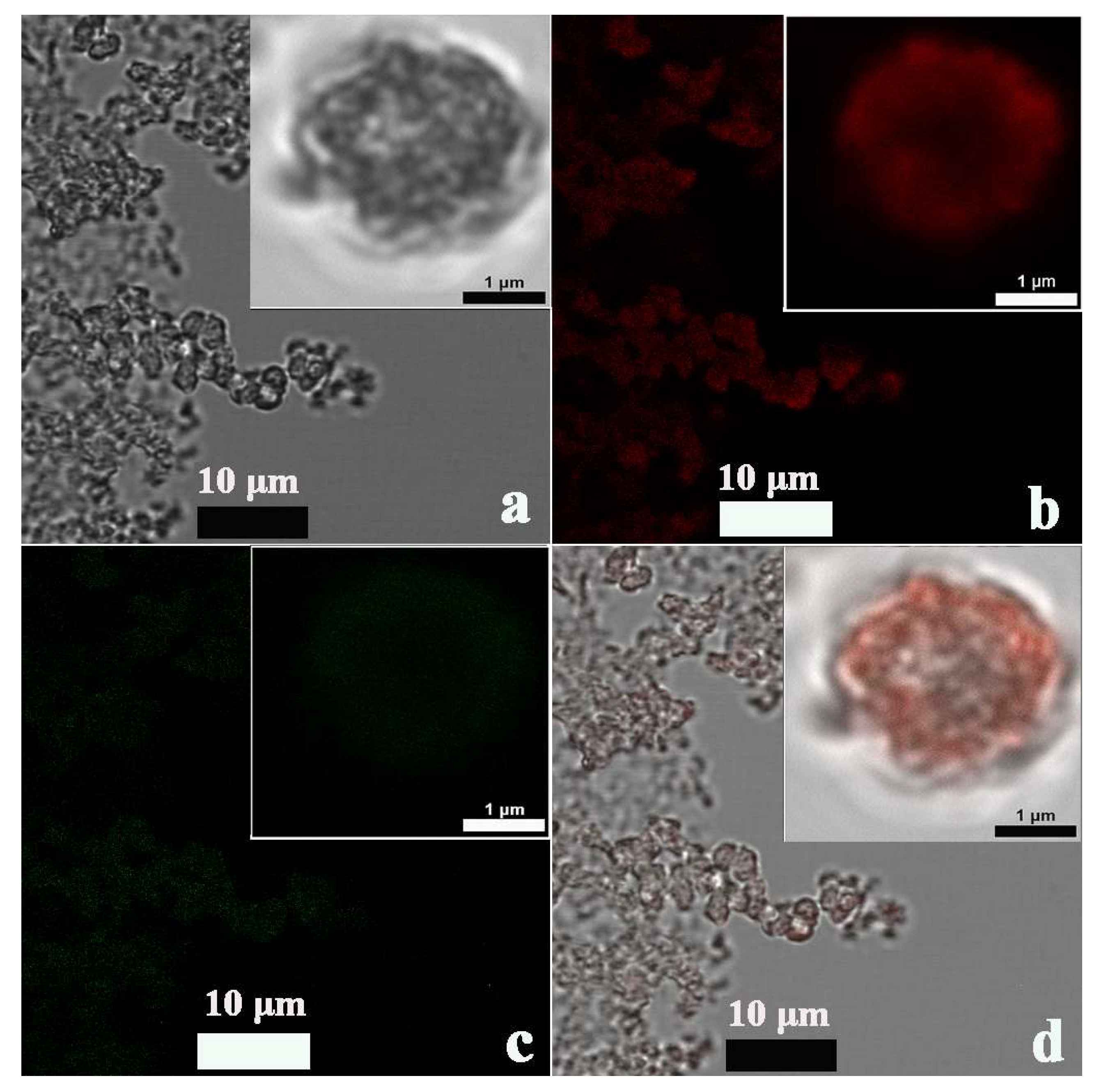
3.2. Characterization of HAC Nanoflowers

To investigate their size and morphology, the HAC were firstly characterized by SEM and TEM (Figure 1). The SEM image showed that the shape of the HAC nanohybrids was flower-like, with a size of ~1.5 um (Figure 1a). The same observation was also verified by the TEM images, and the petal-like structure in the HAC nanoflowers seemed to be composed of nanosheets (Figure 1b,c). XRD analysis was performed to study the crystalline structure and composition of the HAC nanoflowers. The XRD patterns had good agreement with the characteristic peaks of CaHPO4·2H2O (Figure 1d), which confirmed the inorganic composition of the HAC nanoflowers. In addition to the elements of calcium, oxygen and phosphorus detected by the dispersive X-ray (EDX) analysis (Figure S1, Supporting Materials), the element of carbon was also found, indicating that the organic component of HRP and pAb could be hybridized with the CaHPO4. To corroborate this, HRP and pAb were both immobilized in the HAC nanoflowers, and the HRP and pAb were fluorescently labeled with Cy5 and FITC, respectively, to monitor the synthesis of HAC (Figure 2). The characteristic fluorescence of Cy5 (red, Figure 2b) and FITC (green, Figure 2c) was displayed in the fluorescence confocal microscopy images, suggesting the successful coimmobilization of HRP and pAb in the HAC nanoflowers. The immobilization efficiency of HRP and pAb in HAC was determined to be 25.1% and 65.7%, respectively, based on an established standard curve of correlation between the concentration of HRP-Cy5 (or pAb-FITC) and the fluorescence intensity.

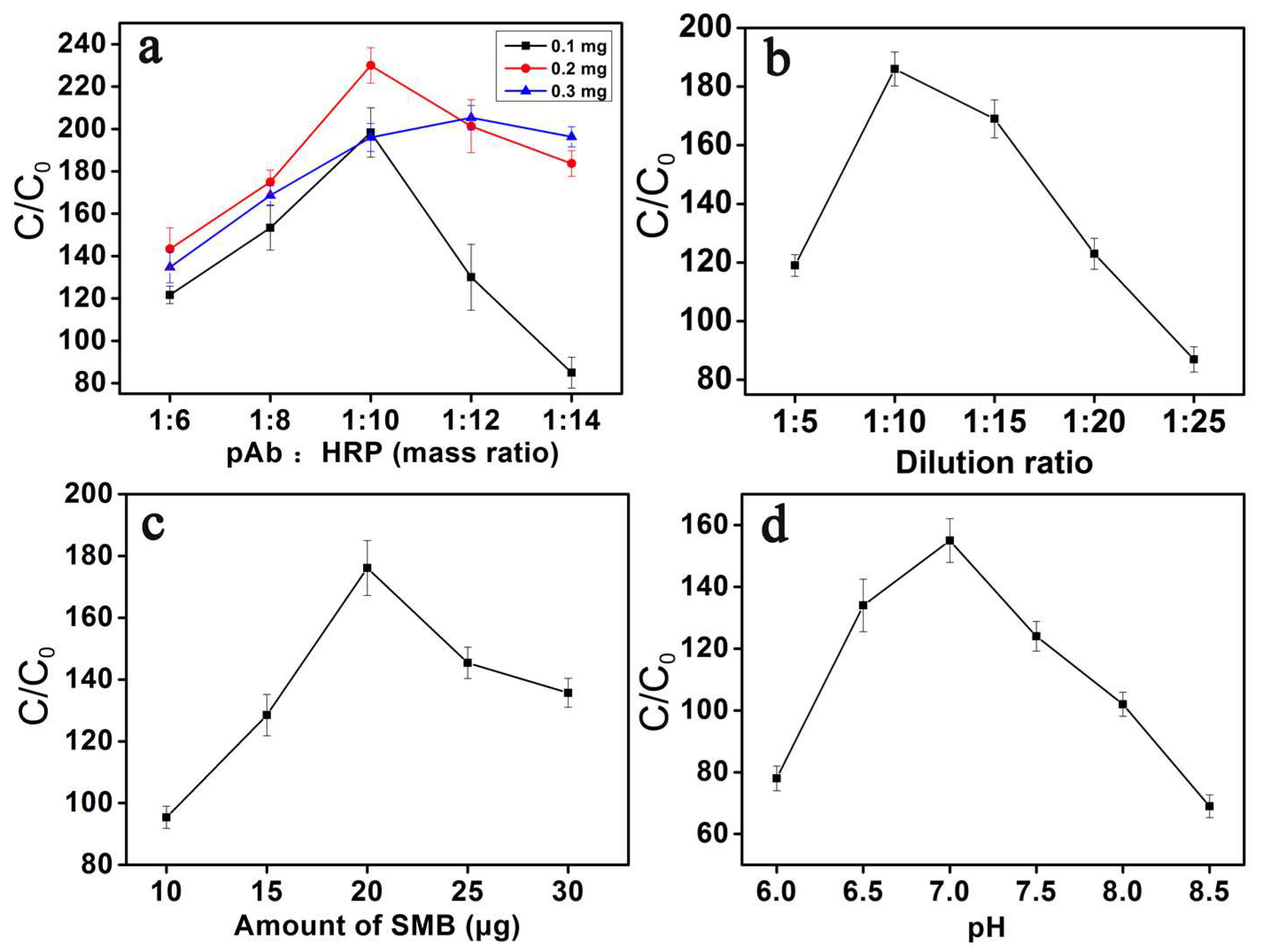
3.3. Optimization of Detection Conditions

To acquire improved analytical performance, several parameters of the experiment were optimized (Figure 3). Firstly, the amount of HRP immobilized on the HAC nanoflowers was directly related to the catalytic efficiency and RCI. The amount of pAb affected the recognition efficiency for the target S. enteritidis and was indirectly related to the RCI. Thus, the total amount of HRP and pAb and their mass ratio played an important role in the sensitivity of this method. As shown in Figure 3a, a significant increase in RCI occurred within the mass ratio of pAb to HRP from 1:6 to 1:10, and it then reached the strongest RCI at 1:10 with the total amount of 0.2 mg. Other proportions and total amounts of pAb and HRP could not achieve a higher RCI, which may due to the lack of sufficient pAb or HRP to ensure the strongest RCI. Secondly, the optimum application amount of synthesized HAC nanoflowers was verified (Figure 3b). The strongest RCI occurred when the 10-fold dilution of synthesized HAC nanoflowers was applied to the detection, and the higher dilutions could have reduced the amount of HRP involved in the reaction. Thirdly, the amount of SMB determining the amount of mAb required for the capture of target S. enteritidis was also investigated (Figure 3c). Different amounts of SMB were tested, ranging from 10 μg to 30 μg, and 20 μg of SMB could obtain the strongest RCI. A higher amount of SMB may disrupt signal measurement. Fourthly, the effect of the pH value on HAC binding to the target was also studied (Figure 3d). The strongest RCI was acquired when the pH value was 7.0. The optimum detection conditions were concluded as follows: (1) pAb and HRP (total amount of 0.2 mg) with the mass ratio of 1:10; (2) 10-fold dilution of synthesized HAC nanoflowers; (3) 20 μg of SMB; (4) pH 7.0.

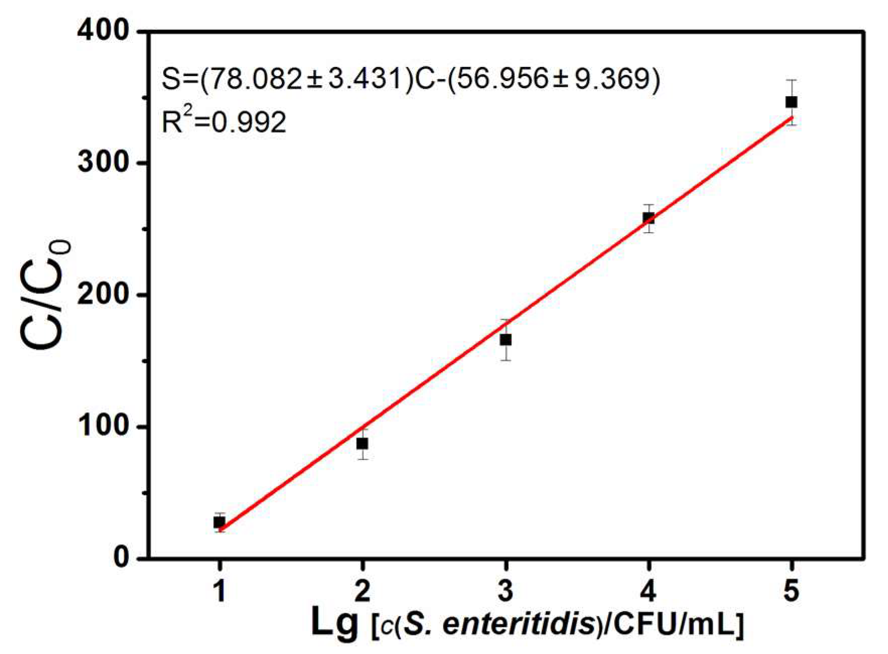
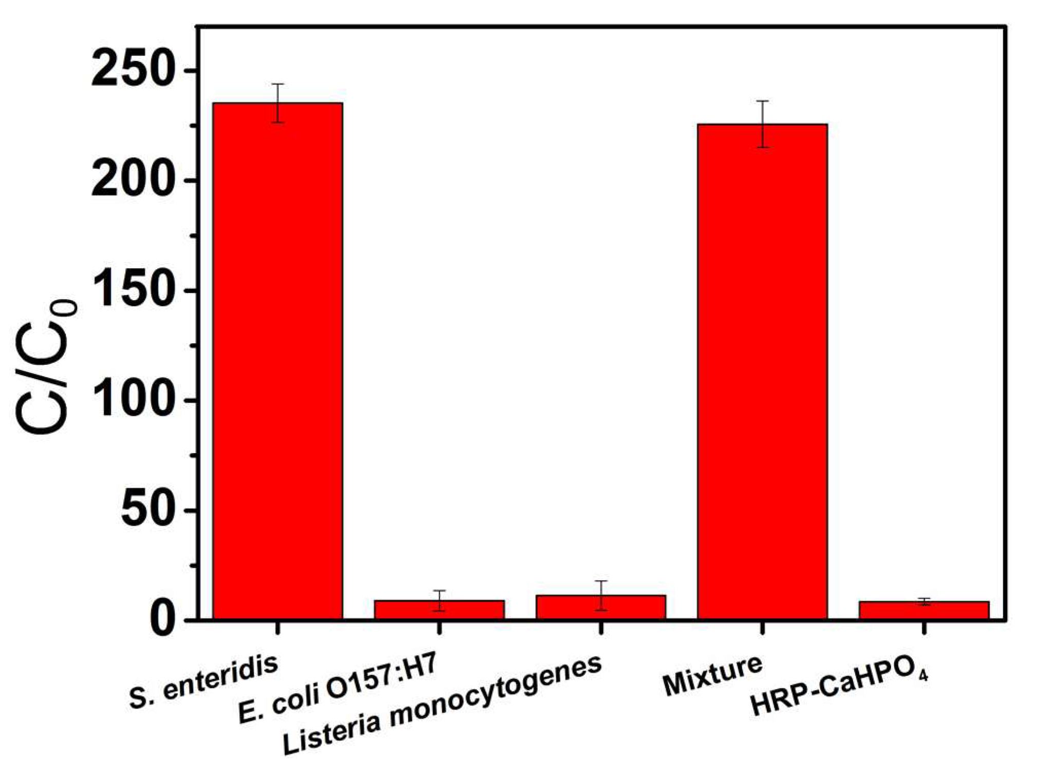
3.4. Detection of S. enteritidis Using HAC Nanoflowers

The detection of S. enteritidis was based on the combination of immunomagnetic separation and a sandwich-type immunoreaction. According to the process shown in Scheme 1, S. enteritidis was firstly captured by SMB-mAb and then the sandwich immunocomplex was formed by the introduction of HAC. After the magnetic separation, the HRP enriched on the HAC nanoflowers catalyzed the substrate, luminol, in the presence of H2O2 to produce the chemiluminescent signal. As shown in Figure 4, the RCI significantly increased as the concentration of S. enteritidis increased. The RCI was linearly dependent on the logarithm of the concentration of S. enteritidis ranging from 10 to 105 CFU/mL with the LOD of 10 CFU/mL (3σ criterion) [43]. The linear equation was represented as S = (78.082 ± 3.431) C—(56.956 ± 9.369) (R2 = 0.992), where S is the RCI, C denotes the common logarithm of the concentration of S. enteritidis, and R2 is the correlation coefficient. In the previous study, only the morphology and size of such nanoflowers were characterized [40]. We have provided a more comprehensive analysis of their crystalline structure and composition, as well as the immobilization efficiency, content and proportion of HRP and Ab in the proposed HAC nanoflowers, which can contribute to the better understanding and application of this material. Moreover, the previous nanoflowers with a size of around ~5 μm were relatively large and could easily lead to non-specific adsorption. We modified the synthesis method to control the size of the proposed HAC nanoflowers to be around ~1.5 μm, and 1% BSA and 0.5% Tween-20 were added to the dilution buffer of the HAC nanoflowers to reduce the non-specific binding to the target, so as to ensure the precision of the proposed method. To assess the precision of the proposed method, a series of 10 consecutive measurements of S. enteritidis (103 CFU/mL) were collected and a low relative standard deviation (RSD) of 4.2% was obtained, indicating the good precision of the proposed method. In addition, to obtain reproducible results, every step of the operation, from synthesis to detection, including the time, temperature, volume, concentration, feeding ratio, etc., should be exactly followed. The good reproducibility of the proposed method was also proven by the low RSD of 5.8% for six measurements of S. enteritidis (103 CFU/mL) using the HAC synthesized at six different times. In terms of the procedure for the synthesis, compared with most of the nanomaterial-based biosensors in Table 1, such as graphene oxide, gold, magnetic, carbon, copper nanoparticles and polyvalent directed peptide polymers, this proposed synthesis method involves no organic or harmful reagents, has a less complicated conjugation and purification process and is only performed in one step, with green and mild coprecipitation. Regarding the aspect of the detection platform and time, the lateral flow strip provides a low cost and more rapid processing, but the sensitivity is not optimal. Although the smartphone-based biosensor achieves more flexibility and portability, as well as higher sensitivity and a wider linear range, it takes approximately two more hours to complete the detection, and there is no specific information about the precision and reproducibility of the method. On the basis of the sensitivity and linear range, the proposed method is more sensitive or has a wider linear range than most of the reported methods in Table 1. To evaluate the specificity of the proposed method, two non-target bacteria of E. coli O157:H7 and Listeria monocytogenes and a mixture of S. enteritidis and two non-target bacteria were detected (Figure 5). The RCI for the detection of E. coli O157:H7 and Listeria monocytogenes in the absence of S. enteritidis was similar to that of the blank. In contrast, in the presence of S. enteritidis, a relatively strong RCI was observed, suggesting the good specificity of the proposed method. Moreover, recovery experiments were conducted to further investigated the applicability of this method. Milk was spiked with S. enteritidis at the concentrations of 1 × 10², 1 × 10³ and 1 × 10⁴ CFU/mL. The average recovery of spiked S. enteritidis was between 92.6% and 102.5% (Table 2), indicating the satisfactory application potential of the proposed method in milk.

4. Conclusions

In summary, this study reported a green, mild, simple and efficient method for the conjugation of HRP and Ab with CaHPO4 to prepare HAC bifunctional hybrid nanoflowers. The combination of a magnetic chemiluminescence immunoassay and HAC nanoflowers showed excellent sensitivity, specificity, accuracy and applicability for the detection of S. enteritidis. The proposed method provides a universal approach for the sensitive detection of foodborne pathogenic bacteria in milk.

Supplementary Materials

The following supporting information can be downloaded at: https://www.mdpi.com/article/10.3390/s23052779/s1, Figure S1: EDX spectrum of the HAC hybrid nanoflowers.

Author Contributions

Conceptualization and methodology, R.Y.; experiments, R.Y. and X.M.; investigation, R.Y. and X.M.; writing—original draft preparation, R.Y.; writing—review and editing, R.Y. All authors have read and agreed to the published version of the manuscript.

Funding

Funder: National Natural Science Foundation of China (Funding number: 21804047).

Institutional Review Board Statement

Not applicable.

Informed Consent Statement

Not applicable.

Data Availability Statement

The data presented in this study are available on request from the corresponding author.

Acknowledgments

The authors thank the reviewers for their valuable comments.

Conflicts of Interest

The authors declare no conflict of interest.

References

  1. DeFlorio, W.; Liu, S.; White, A.R.; Taylor, T.M.; Cisneros-Zevallos, L.Y.; Min, E.M. Scholar, Recent developments in antimicrobial and antifouling coatings to reduce or prevent contamination and cross-contamination of food contact surfaces by bacteria. Compr. Rev. Food Sci. Food Saf. 2021, 20, 3093–3134. [Google Scholar] [CrossRef] [PubMed]
  2. Liu, J.-M.; Wang, Z.-H.; Ma, H.; Wang, S. Probing and Quantifying the Food-Borne Pathogens and Toxins: From In Vitro to In Vivo. J. Agric. Food Chem. 2018, 66, 1061–1066. [Google Scholar] [CrossRef] [PubMed]
  3. Nair, D.V.T.; Venkitanarayanan, K.; Kollanoor Johny, A. Antibiotic-Resistant Salmonella in the Food Supply and the Potential Role of Antibiotic Alternatives for Control. Foods 2018, 7, 24. [Google Scholar]
  4. Priyanka, B.; Patil, R.K.; Dwarakanath, S. A review on detection methods used for foodborne pathogens. Indian J. Med. Res. 2016, 144, 327–338. [Google Scholar] [CrossRef]
  5. Yang, S.M.; Kim, E.; Kim, D.; Baek, J.; Yoon, H.; Kim, H.Y. Rapid detection of Salmonella Enteritidis, Typhimurium, and Thompson by specific peak analysis using matrix-assisted laser desorption ionization time-of-flight mass spectrometry. Foods 2021, 10, 933. [Google Scholar] [CrossRef]
  6. McGoverin, C.; Steed, C.; Esan, A.; Robertson, J.; Swift, S.; Vanholsbeeck, F. Optical methods for bacterial detection and characterization. APL Photonics 2021, 6, 080903. [Google Scholar] [CrossRef]
  7. Buzalewicz, I.; Karwańska, M.; Wieliczko, A.; Podbielska, H. On the application of multi-parametric optical phenotyping of bacterial colonies for multipurpose microbiological diagnostics. Biosens. Bioelectron. 2021, 172, 112761. [Google Scholar] [CrossRef]
  8. Velusamy, V.; Arshak, K.; Korostynska, O.; Oliwa, K.; Adley, C. An overview of foodborne pathogen detection: In the perspective of biosensors. Biotechnol. Adv. 2010, 28, 232–254. [Google Scholar] [CrossRef]
  9. Zhang, X.; Tsuji, S.; Kitaoka, H.; Kobayashi, H.; Tamai, M.; Honjoh, K.I.; Miyamoto, T. Simultaneous Detection of Escherichia coli O157:H7, Salmonella enteritidis and Listeria monocytogenes at a Very Low Level Using Simultaneous Enrichment Broth and Multichannel SPR Biosensor. J. Food Sci. 2017, 82, 2357–2363. [Google Scholar] [CrossRef]
  10. Farahani, R.K.; Meskini, M.; Langeroudi, A.G.; Gharibzadeh, S.; Ghosh, S.; Farahani, A.H.K. Evaluation of the different methods to detect Salmonella in poultry feces samples. Arch. Microbiol. 2022, 204, 269. [Google Scholar] [CrossRef]
  11. Gao, P.; Wang, L.; He, Y.; Wang, Y.; Yang, X.; Fu, S.; Qin, X.; Chen, Q.; Man, C.; Jiang, Y. An enhanced lateral flow assay based on aptamer–magnetic separation and multifold AuNPs for ultrasensitive detection of Salmonella typhimurium in milk. Foods 2021, 10, 1605. [Google Scholar] [CrossRef]
  12. Zeinhom, M.M.A.; Wang, Y.; Song, Y.; Zhu, M.J.; Lin, Y.; Du, D. A portable smart-phone device for rapid and sensitive detection of E. coli O157: H7 in Yoghurt and Egg. Biosens. Bioelectron. 2018, 99, 479–485. [Google Scholar] [CrossRef] [PubMed]
  13. Zhang, P.; Song, M.; Dou, L.; Xiao, Y.; Li, K.; Shen, G.; Ying, B.; Geng, J.; Yang, D.; Wu, Z. Development of a fluorescent DNA nanomachine for ultrasensitive detection of Salmonella enteritidis without labeling and enzymes. Microchim. Acta 2020, 187, 376. [Google Scholar] [CrossRef] [PubMed]
  14. Wang, Z.; Yao, X.; Wang, R.; Ji, Y.; Yue, T.; Sun, J.; Zhang, D. Label-free strip sensor based on surface positively charged nitrogen-rich carbon nanoparticles for rapid detection of Salmonella enteritidis. Biosens. Bioelectron. 2019, 132, 360–367. [Google Scholar] [CrossRef] [PubMed]
  15. Chen, J.; Miao, Y.; He, N.; Wu, X.; Li, S. Nanotechnology and biosensors. Biotechnol. Adv. 2004, 22, 505–518. [Google Scholar]
  16. Fenzl, C.; Hirsch, T.; Baeumner, A.J. Nanomaterials as versatile tools for signal amplification in (bio)analytical applications. TrAC Trends Anal. Chem. 2016, 79, 306–316. [Google Scholar] [CrossRef]
  17. Lei, J.P.; Ju, H.X. Signal amplification using functional nanomaterials for biosensing. Chem. Soc. Rev. 2012, 41, 2122–2134. [Google Scholar] [CrossRef] [PubMed]
  18. Wang, L.; Zhi, W.J.; Lian, D.S.; Wang, Y.; Han, J.; Wang, Y. HRP@ZIF-8/DNA hybrids: Functionality integration of ZIF-8 via biomineralization and surface absorption. ACS Sustain. Chem. Eng. 2019, 7, 14611–14620. [Google Scholar] [CrossRef]
  19. Sheldon, R.A.; Pelt, S.V. Enzyme immobilisation in biocatalysis: Why, what and how. Chem. Soc. Rev. 2013, 42, 6223–6235. [Google Scholar] [CrossRef] [Green Version]
  20. Ye, R.; Xu, H.; Gu, J.; Chen, H. Bioinspired synthesis of protein-posnjakite organic-inorganic nanobiohybrid for biosensing applications. Anal. Chim. Acta 2021, 1143, 31–36. [Google Scholar] [CrossRef]
  21. Wang, L.B.; Wang, Y.C.; He, R.; Zhuang, A.; Wang, X.P.; Zeng, J.; Hou, J.G. A new nanobiocatalytic system based on allosteric effect with dramatically enhanced enzymatic performance. J. Am. Chem. Soc. 2013, 135, 1272–1275. [Google Scholar] [CrossRef] [PubMed]
  22. Ge, J.; Lei, J.D.; Zare, R.N. Protein–inorganic hybrid nanoflowers. Nat. Nanotechnol. 2012, 7, 428–432. [Google Scholar] [CrossRef] [PubMed]
  23. Ye, R.; Zhu, C.; Song, Y.; Song, J.; Fu, S.; Lu, Q.; Yang, X.; Zhu, M.; Du, D.; Li, H.; et al. One-pot bioinspired synthesis of all-inclusive protein-protein nanoflowers for point-of-care bioassay: Detection of E. coli O157:H7 from milk. Nanoscale 2016, 8, 18980–18986. [Google Scholar] [CrossRef] [PubMed]
  24. Li, Z.; Ding, Y.; Li, S.; Jiang, Y.; Liu, Z.; Ge, J. Highly active, stable and self-antimicrobial enzyme catalysts prepared by biomimetic mineralization of copper hydroxysulfate. Nanoscale 2016, 8, 17440–17445. [Google Scholar] [CrossRef]
  25. Lin, Z.; Xiao, Y.; Yin, Y.; Hu, W.; Liu, W.; Yang, H. Facile synthesis of enzyme-inorganic hybrid nanoflowers and its application as a colorimetric platform for visual detection of hydrogen peroxide and phenol. ACS Appl. Mater. Interfaces 2014, 6, 10775–10782. [Google Scholar] [CrossRef]
  26. Zhang, Z.; Zhang, Y.; Song, R.; Wang, M.; Yan, F.; He, L.; Feng, X.; Fang, S.; Zhao, J.; Zhang, H. Manganese (II) phosphate nanoflowers as electrochemical biosensors for the high-sensitivity detection of ractopamine. Sens. Actuators B Chem. 2015, 211, 310–317. [Google Scholar] [CrossRef]
  27. Neng, J.; Li, Y.; Driscoll, A.J.; Wilson, W.C.; Johnson, P.A. Detection of multiple pathogens in serum using silica-encapsulated nanotags in a surface-enhanced Raman scattering-based immunoassay. J. Agric. Food Chem. 2018, 66, 5707–5712. [Google Scholar] [CrossRef]
  28. Yin, B.; Ho, W.K.H.; Zhang, Q.; Li, C.; Huang, Y.; Yan, J.; Yang, H.; Hao, J.; Wong, S.H.D.; Yang, M. Magnetic-responsive surface-enhanced Raman scattering platform with tunable hot spot for ultrasensitive virus nucleic acid detection. ACS Appl. Mater. Interfaces 2022, 14, 4714–4724. [Google Scholar] [CrossRef]
  29. Yin, B.; Zhang, Q.; Xia, X.; Li, C.; Ho, W.K.H.; Yan, J.; Huang, Y.; Wu, H.; Wang, P.; Yi, C.; et al. A CRISPR-Cas12a integrated SERS nanoplatform with chimeric DNA/RNA hairpin guide for ultrasensitive nucleic acid detection. Theranostics 2022, 12, 5914. [Google Scholar] [CrossRef]
  30. Zhang, Y.; Wu, H.; Wang, H.; Yin, B.; Wong, S.H.D.; Zhang, A.P.; Tam, H.Y. Ultraminiature optical fiber-tip directly-printed plasmonic biosensors for label-free biodetection. Biosens. Bioelectron. 2022, 218, 114761. [Google Scholar] [CrossRef]
  31. Bhandari, D.; Chen, F.C.; Bridgman, R.C. Detection of Salmonella typhimurium in romaine lettuce using a surface plasmon resonance biosensor. Biosensors 2019, 9, 94. [Google Scholar] [CrossRef] [Green Version]
  32. Liu, Y.; Wang, B.; Ji, X.; He, Z. Self-assembled protein-enzyme nanoflower-based fluorescent sensing for protein biomarker. Anal. Bioanal. Chem. 2018, 410, 7591–7598. [Google Scholar] [CrossRef] [PubMed]
  33. Zhang, Q.; Yin, B.; Hao, J.; Ma, L.; Huang, Y.; Shao, X.; Li, C.; Chu, Z.; Yi, C.; Wong, S.H.D.; et al. An AIEgen/graphene oxide nanocomposite (AIEgen@GO)-based two-stage “turn-on” nucleic acid biosensor for rapid detection of SARS-CoV-2 viral sequence. Aggregate 2022, 4, e195. [Google Scholar] [CrossRef]
  34. Xiao, Q.; Xu, C. Research progress on chemiluminescence immunoassay combined with novel technologies. TrAC Trends Anal. Chem. 2020, 124, 115780. [Google Scholar] [CrossRef]
  35. Mahari, S.; Gandhi, S. Recent advances in electrochemical biosensors for the detection of salmonellosis: Current prospective and challenges. Biosensors 2022, 12, 365. [Google Scholar] [CrossRef]
  36. Liang, X.; Liu, Y.; Wen, K.; Jiang, W.; Li, Q. Immobilized enzymes in inorganic hybrid nanoflowers for biocatalytic and biosensing applications. J. Mater. Chem. B 2021, 9, 7597–7607. [Google Scholar] [CrossRef]
  37. Kubo, I.; Kajiya, M.; Aramaki, N.; Furutani, S. Detection of Salmonella enterica in egg yolk by PCR on a microfluidic disc device using immunomagnetic beads. Sensors 2020, 20, 1060. [Google Scholar] [CrossRef] [Green Version]
  38. Ye, J.; Guo, J.; Li, T.; Tian, J.; Yu, M.; Wang, X.; Majeed, U.; Song, W.; Xiao, J.; Luo., Y.; et al. Phage-based technologies for highly sensitive luminescent detection of foodborne pathogens and microbial toxins: A review. Compr. Rev. Food Sci. Food Saf. 2022, 21, 1843–1867. [Google Scholar] [CrossRef]
  39. Al Yahyai, I.; Al-Lawati, H.A. A review of recent developments based on chemiluminescence detection systems for pesticides analysis. Luminescence 2021, 36, 266–277. [Google Scholar] [CrossRef]
  40. Zeinhom, M.M.A.; Wang, Y.; Sheng, L.; Du, D.; Li, L.; Zhu, M.J.; Lin, Y. Smart phone based immunosensor coupled with nanoflower signal amplification for rapid detection of Salmonella Enteritidis in milk, cheese and water. Sens. Actuators B Chem. 2018, 261, 75–82. [Google Scholar] [CrossRef]
  41. Ye, R.; Zhu, C.; Song, Y.; Lu, Q.; Ge, X.; Yang, X.; Zhu, M.; Du, D.; Li, H.; Lin, Y. Bioinspired synthesis of all-in-one organic-inorganic hybrid nanoflowers combined with a handheld pH meter for on-site detection of food pathogen. Small 2016, 12, 3094–3100. [Google Scholar] [CrossRef] [PubMed]
  42. Zhang, Z.; Lai, J.; Wu, K.; Huang, X.; Guo, S.; Zhang, L.; Liu, J. Peroxidase-catalyzed chemiluminescence system and its application in immunoassay. Talanta 2018, 180, 260–270. [Google Scholar] [CrossRef] [PubMed]
  43. Ye, R.; Chen, H.; Li, H. One-Pot Synthesis of HRP&SA/ZIF-8 Nanocomposite and Its Application in the Detection of Insecticidal Crystalline Protein Cry1Ab. Nanomaterials 2022, 12, 2679. [Google Scholar] [PubMed]
  44. Ma, X.; Jiang, Y.; Jia, F.; Yu, Y.; Chen, J.; Wang, Z. An aptamer-based electrochemical biosensor for the detection of Salmonella. J. Microbiol. Methods 2014, 98, 94–98. [Google Scholar] [CrossRef]
  45. Waswa, J.W.; Debroy, C.; Irudayaraj, J. Rapid detection of Salmonella enteritidis and Escherichia coli using surface plasmon resonance biosensor. J. Food Process. Eng. 2006, 29, 373–385. [Google Scholar] [CrossRef]
  46. Mahari, S.; Roberts, A.; Gandhi, S. Probe-free nanosensor for the detection of Salmonella using gold nanorods as an electroactive modulator. Food Chem. 2022, 390, 133219. [Google Scholar] [CrossRef]
  47. Bu, T.; Yao, X.; Huang, L.; Dou, L.; Zhao, B.; Yang, B.; Li, T.; Wang, J.; Zhang, D. Dual recognition strategy and magnetic enrichment based lateral flow assay toward Salmonella enteritidis detection. Talanta 2020, 206, 120204. [Google Scholar] [CrossRef]
  48. Lee, S.C.; Kim, M.S.; Yoo, K.C.; Ha, N.R.; Moon, J.Y.; Lee, S.J.; Yoon, M.Y. Sensitive fluorescent imaging of Salmonella enteritidis and Salmonella typhimurium using a polyvalent directed peptide polymer. Microchim. Acta 2017, 184, 2611–2620. [Google Scholar] [CrossRef]
Scheme 1. Illustration of the preparation process for the HAC nanoflowers and their application in the detection of S. enteritidis. (a) The synthsis of HAC; (b)The application in the detection of S. enteritidis.
Sensors 23 02779 sch001
Figure 1. (a) SEM image of HAC nanoflowers; (b) TEM image of HAC nanoflowers; (c) TEM image of HAC nanoflowers (partial magnification from (b)); (d) XRD pattern of HAC nanoflowers (JCPDS No. 72–0713).
Sensors 23 02779 g001
Figure 2. Confocal fluorescence microscopy images of HAC. (a) Bright-field image of HAC; (b) Cy5-labeled HRP in HAC; (c) FITC-labeled pAb in HAC; (d) overlap image of bright-field and fluorescence images. Insert is the magnification of a single HAC.
Sensors 23 02779 g002
Figure 3. Optimization of detection conditions. (a) Different total amounts of HRP and pAb and their mass ratios for HAC preparation. (b) Different dilution ratios of HAC for detection. (c) Different amounts of SMB for detection. (d) Different pH values for HAC binding.
Sensors 23 02779 g003
Figure 4. Linear calibration curve for the detection of S. enteritidis.
Sensors 23 02779 g004
Figure 5. Specificity of the proposed method.
Sensors 23 02779 g005
Table 1. Comparison of different methods for S. enteritidis detection.
MaterialsTechniqueLinear RangeLODDetection Time
(h)
Reference
CFU/mLCFU/mL
Copper nanoparticlesFluorescent biosensor50–10425~2.6[13]
Carbon nanoparticlesLateral flow102–108102~0.25[14]
Magnetic nanocompositeSmartphone-based colorimetric immunosensor1–1061~3.4[40]
AptamerElectrochemical biosensor2.4–2.4 × 1033~1.5[44]
GoldSurface plasmon resonance0–10725~0.61[45]
Gold nanorodsElectrochemical immunosensor1–105105Not given[46]
Magnetic nanoparticlesLateral flow103–107102–103~0.5[47]
Polyvalent directed peptide polymerFluorescence microscope102–108102~2.0[48]
HACMagnetic chemiluminescence immunoassay10–10510~1.5This work
Table 2. Recovery of S. enteritidis-spiked milk samples.
S. enteritidis Added
(CFU/mL)
S. enteritidis Found
(CFU/mL)
Recovery (%)
1 × 10²(0.926 ± 0.036) × 10292.6
1 × 10³(0.945 ± 0.057) × 10394.5
1 × 10⁴(1.025 ± 0.063) × 104102.5
0Not found-
Disclaimer/Publisher’s Note: The statements, opinions and data contained in all publications are solely those of the individual author(s) and contributor(s) and not of MDPI and/or the editor(s). MDPI and/or the editor(s) disclaim responsibility for any injury to people or property resulting from any ideas, methods, instructions or products referred to in the content.

