Review

Sensing with Molecularly Imprinted Membranes on Two-Dimensional Solid-Supported Substrates

by
Lishuang Wang
1,
Nan Li
1,
Xiaoyan Zhang
1,*,
Ivan Bobrinetskiy
2,
Ivana Gadjanski
2 and
Wangyang Fu
3,*
1
School of Pharmaceutical Sciences, Capital Medical University, Beijing 100069, China
2
BioSense Institute, University of Novi Sad, Dr Zorana Đinđića 1a, 21000 Novi Sad, Serbia
3
School of Materials Science and Engineering, Tsinghua University, No. 1 Tsinghua Yuan, Haidian District, Beijing 100084, China
*
Authors to whom correspondence should be addressed.
Sensors 2024, 24(16), 5119; https://doi.org/10.3390/s24165119
Submission received: 9 June 2024 / Revised: 25 July 2024 / Accepted: 27 July 2024 / Published: 7 August 2024
(This article belongs to the Special Issue Biosensors for Point-of-Care Diagnostics)

Abstract

:
Molecularly imprinted membranes (MIMs) have been a focal research interest since 1990, representing a breakthrough in the integration of target molecules into membrane structures for cutting-edge sensing applications. This paper traces the developmental history of MIMs, elucidating the diverse methodologies employed in their preparation and characterization on two-dimensional solid-supported substrates. We then explore the principles and diverse applications of MIMs, particularly in the context of emerging technologies encompassing electrochemistry, surface-enhanced Raman scattering (SERS), surface plasmon resonance (SPR), and the quartz crystal microbalance (QCM). Furthermore, we shed light on the unique features of ion-sensitive field-effect transistor (ISFET) biosensors that rely on MIMs, with the notable advancements and challenges of point-of-care biochemical sensors highlighted. By providing a comprehensive overview of the latest innovations and future trajectories, this paper aims to inspire further exploration and progress in the field of MIM-driven sensing technologies.

1. Introduction

Molecular-specific recognition plays a crucial role in regulating biological systems, involving organic functional groups characterized by distinct chemical or biological properties. These groups interact via highly specialized and efficient bonding methods, resulting in unique biological effects manifested as physiological activities or physicochemical interactions with the external environment [1,2,3,4,5]. Over time, natural systems have evolved an array of specific affinities to meet functional demands, exemplified by interactions such as enzyme–inhibitor complexes [6,7], sugar–lectins [8], RNA–ribosomes [9,10,11], and antibody–antigen pairings [12,13]. Precise molecular recognition is essential for biological processes and represents a sophisticated form of sensing. Given its importance in catalysis, separation, and detection, this area has become a central focus of chemical research. Although a biosystem is capable of producing antibodies for a wide range of foreign entities, employing receptors in chemical processes leads to difficulties such as high expenses and vulnerability to environmental factors [14]. Replicating the specific ligand recognition found in biological systems presents a significant challenge for researchers. Inspired by natural receptor–ligand interactions, scientists have developed molecular imprinting technology (MIT). This technique involves the formation of synthetic polymers with highly specific cavities designed for target molecules, thereby mimicking natural recognition systems [15,16,17].
Wulff and Klotz pioneered the concept of imprinting with organic polymers, coining the term “molecularly imprinted polymers” (MIPs) in 1972. The initial MIPs were conceptualized using a silica-based imprinting approach [18]. Over the decades, spherical MIPs have evolved and gained popularity due to their ability to selectively recognize target molecules within complex mixtures. Researchers have utilized spherical silica [19], quantum dots [20], and magnetic particle nanoparticles [21] as substrates to prepare molecularly imprinted particles with core-shell structures. However, spherical MIPs have limitations, including relatively low binding properties and restricted binding signaling mechanisms. Consequently, a specialized membrane that recognizes specific molecules, termed a molecularly imprinted membrane (MIM), was developed [22,23]. MIMs exemplify a unique variety of imprinted polymers that merge particular recognition attributes with conventional aspects of membrane technology.
Furthermore, MIMs can be combined with various two-dimensional (2D) solid-supported substrates to enhance their performance and broaden the application field of MIT [24,25,26]. The huge specific surface area and high porosity of 2D solid-supported substrates provide more recognition sites, enabling the better separation and detection of target molecules. Additionally, 2D solid-supported substrates serve as excellent support structures, simplifying the preparation process of MIMs and allowing precise control over the membrane thickness, pore structure, and surface chemistry, thus modulating membrane properties. Numerous reports have detailed the combination of MIMs with 2D solid-supported substrates to enable coupling with electrical, optical, and mechanical detection techniques [27,28,29].
Compared to naturally derived ligands like antibodies, MITs have become prominent in materials science focused on molecular affinity. MITs offer cost-effectiveness, adaptability, and superior stability. This review progresses from discussing MIT particles in solution—including their state-of-the-art advancements, benefits, and limitations—to exploring the development of MIT membranes on solid surfaces. The aim is to introduce the applications of solid-supported MIMs in various fields, with a particular focus on sensing. We will primarily discuss and summarize the latest technological advancements and applications of MIMs in combination with electrochemistry sensing, surface-enhanced Raman scattering (SERS), surface plasmon resonance (SPR), and quartz crystal microbalance (QCM) technologies. Additionally, we will explore future prospects for innovative applications of MIMs in field-effect transistor biochemical sensors. Figure 1 shows the basic principle and classification of MIMs based on 2D solid-supported substrates.

1.1. Molecular Imprinting Technology

MIT involves the polymerization and assembly of specifically designed monomers around a target molecule, creating a customized “artificial lock” that can specifically recognize the “key” (template). This technology has its origins in 1931, when Polyakov first reported the binding of silica particles derived from sodium silicate to target molecules in the presence of organic solvents like benzene, toluene, or xylene. Theoretically, MIPs can be customized to target a diverse array of target analytes, such as ions, proteins, and peptides [5,30]. For instance, a recent study introduced a novel method that is enzyme-free, cost-effective, and user-friendly for detecting α-fetoprotein (AFP) and carcinoembryonic antigen (CEA) cancer biomarkers built on fluorescent molecularly imprinted coupled polythiophene (FMICP) [31]. The sensors exhibited a high sensitivity to CEA concentrations within the range of 0.001 to 200 ng/mL, with detection limits of 40 fg/mL and 3.5 fg/mL, respectively. Additionally, a distinctive double-imprinted electrochemical paper-based analytical instrument (Di-ePAD) was developed, capable of concurrently detecting 8-hydroxy-20-deoxyguanosine (8-OHdG) and 3-nitrotyrosine (3-NT) in a complex matrix (urine and plasma), along with oxidation and nitrification biomarkers [32].

1.2. Development of Molecular Imprinting Membranes

Piletsky et al. initially employed MIMs to selectively separate adenosine monophosphate (AMP) and guanosine monophosphate (GMP) in 1990 [33]. Subsequently, Mathew-Klotz et al. independently prepared MIMs using 9-ethyladenine, which served as a template, and AIBN, which acted as an initiator, by polymerizing glycol dimethacrylate and methyl methacrylate. They further selected adenosine for the infiltration of adenosine from an adenosine–guanosine mixture [34]. Following this, a novel type of imprinted membrane for theophylline (THO) using the phase-inversion method was developed [35]. Polyacrylonitrile–acrylic acid copolymers were prepared using THO molecules as template molecules and dimethylsulfoxide (DMSO) copolymers as functional monomers by the phase-inversion precipitation method. The selectivity was confirmed through the structural analogue of caffeine (CAF), highlighting the specific hydrogen bonding between the THO molecule and the polyacrylonitrile–co-acrylic acid group.
The synthesis process of MIMs closely mirrors that of MIPs. Covalent or non-covalent template molecules are introduced during the synthesis process of a membrane, followed by the leaching out of the template, leaving specific pores in the membrane with an affinity for template-like molecules [23]. Initially, MIMs could only selectively identify target molecules in solution through simple static adsorption or osmosis via membrane devices [23]. However, with advancements in science and technology, MIMs can now imprint a variety of template analytes, such as inorganic ions, drugs, nucleic acids, proteins, viruses, and even cells [36,37,38,39,40]. Numerous reports have detailed the combination of MIMs with 2D solid-supported substrates to enable coupling with electrical, optical, and mechanical detection techniques. For instance, the combination of MIMs with semiconducting substrates facilitates the development of ultrasensitive FET biochemical sensors, the promising application of which will be discussed in the prospect part of this review. Although electrochemical sensors may not match FET sensors’ sensitivity, they have become popular in recent years due to their simplicity, ease of production, and low equipment costs. Furthermore, electrochemical sensors necessitate less complex electrode preparation than FET sensors [41,42]. Electrodes can also be coated with a single layer of MIMs. There are numerous techniques for covering an electrode’s surface with MIMs, including electropolymerization. The sensing signal depends on the rate of mass transfer of the electrochemically active analyte to the MIM-covered electrodes. In addition to electrical coupling, MIMs can also be combined with optics, mechanics, and other disciplines. Optical MIPs sensors, often designated as molecularly imprinted photosensitive materials (MIOMs), can be applied to the sensing of both gas and liquid phases [43,44,45]. These optical sensors typically detect the specific fluorescence signal of the object under measurement. Furthermore, QCM technology, as a piezoelectric mass sensing method, is simple to operate and enables the accurate detection of resonant frequency changes associated with mass loading [46,47]. By merging molecular imprinting with a slender piezoelectric quartz crystal, which is layered between two metallic contacts, the attachment of the desired molecule to the surface of the MIMs leads to an increase in the crystal’s mass, consequently altering its resonant frequency.

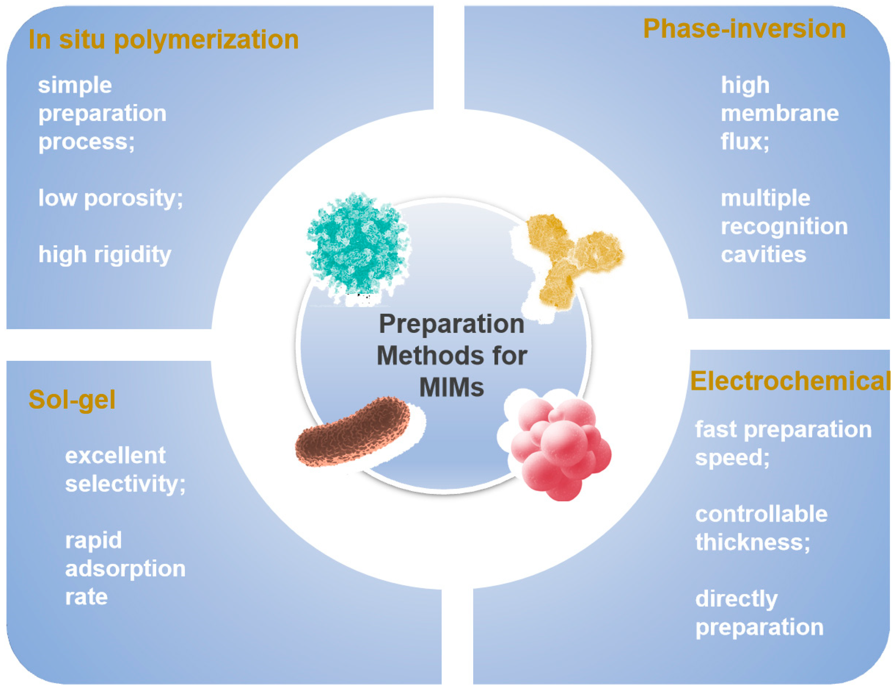
2. Preparation Methods for MIMs

There are two primary types of chemical interactions that play a role during the development process of polymers with molecular imprinting techniques: covalent bonding and noncovalent associations [48,49]. Covalent interactions, which were introduced by Wulff, rely on covalent bonds that can be reversed between template and functional monomers within pre-polymerized blends [18]. Following polymerization, these bonds are chemically broken to release and remove the template. In the recombination process, covalent bonds are formed through interactions involving functional monomers and the template. Covalent imprinting involves the formation of reversible covalent bonds between functional monomers and target analytes. Examples include carboxylate bonds, borate bonds [50], and Schiff bases [51]. In contrast, non-covalent molecular imprinting, as introduced by Mosbach and Sellergren, utilizes non-covalent interactions of templates and monomers. These engagements encompass hydrogen bonding, hydrophobic interactions, van der Waals forces, and metal chelation [52,53,54,55]. During polymerization, a solvent–template–monomer–cross-linker–initiator system is used to ensure the precise spatial arrangement of these components. Afterward, tailored solvents are employed to remove the target molecules, resulting in the creation of the target three-dimensional MIPs.
Each of these synthesis types has its own advantages and disadvantages. The covalent method allows for control over the stoichiometry of imprinted materials, resulting in more uniform identification sites compared to the non-covalent method. Covalently imprinted polymers are known for their stability and selectivity. However, they have limitations such as a restricted number of functional groups interacting with the target molecule and slow binding dynamics. Additionally, their reuse in cleaving and rebinding templates can be challenging due to these limited interactions. Furthermore, covalent interactions differ from the natural recognition mechanisms observed in biological systems at the molecular level. They replicate the size, shape, and chemical groups of template molecules leading to a high specificity in molecular detection. On the other hand, non-covalent methods rely on weaker template–functional monomer interactions, which are not as robust as covalent bonds. This allows for a broader range of imprinting targets. This method benefits from the ease with which monomer/template complexes can form through self-assembly, with the template being released under gentle conditions, potentially yielding a high density of recognition cavities [56]. Currently, non-covalent MIT is the most commonly employed due to its simple synthesis process, applicability to various templates, and compatibility with many functional monomers. The synthesis methods of MIMs are continually expanding based on non-covalent binding methods. Each synthesis method possesses distinct characteristics. Figure 2 contains the current most common synthesis methods and summarizes their advantages. For instance, the in situ polymerization method offers a straightforward preparation process, the phase-inversion method enhances membrane flux, the sol-gel method demonstrates excellent selectivity, and the electrochemical method boasts a rapid preparation speed. The comprehensive examination of the advantages and disadvantages associated with each method of preparation, along with its corresponding procedure, can be found in Table 1.

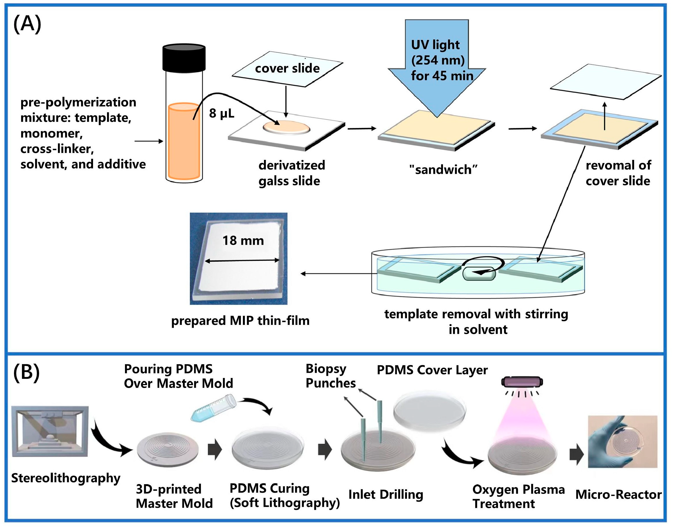
2.1. In Situ Polymerization Method

Piletsky et al. pioneered the successful development of MIMs, selecting adenylate as a template molecule through in situ polymerization in 1990 [33]. This method involves dissolving and mixing a precise ratio of template molecules, functional group monomers, and cross-linkers between two plates. The cross-linking reaction is initiated by UV irradiation or heat, which promotes direct polymerization on the carrier surface, resulting in the formation of an MIM [57,71,72]. As illustrated in Figure 3A, a custom porous MIM was created using in situ photo-radical polymerization conducted on a slide. This technique offers a viable method for selectively detecting trace polycyclic aromatic sulfur heterocycles (PASHs) in seawater [73]. Subsequently, researchers prepared additional MIMs using in situ photo-radical polymerization on a glass plate [58]. These membranes were utilized as microextraction adsorbents to detect trace levels of polycyclic aromatic sulfur heterocycles of seawater, achieving a detection limit between 0.029 and 0.166 μg/L. As illustrated in Figure 3B, MIPs were synthesized through in situ polymerization within a small reaction vessel. This approach accelerates reaction rates and allows for controlled in situ growth, with the continuous synthesis of trillions of MIP nanoparticles taking approximately 5 to 30 min [58]. Additionally, a straightforward, rapid, and highly sensitive method was devised for determining artemisinin (ART) using electrochemical sensors [74]. The optimization of the MIM on the graphene sensor was achieved by using acrylamide (AM) as the functional monomers. The wrinkled structure of the graphene enhanced the sensor’s construction by providing a large surface area for the MIM to serve as a recognition area. Concurrently, the research group expanded their investigation into the structure of a Pd electrochemical sensor [75]. The polymerization process was completed by applying the solution to the electrode surface, covering it with glass, and using an infrared lamp for heating. Although this preparation method is simple and manageable, the extensive degree of cross-linking results in high membrane rigidity and minimal porosity. This, in turn, leads to reduced membrane flux and increased mass transfer resistance, thereby limiting the practical application of membranes prepared via in situ polymerization.

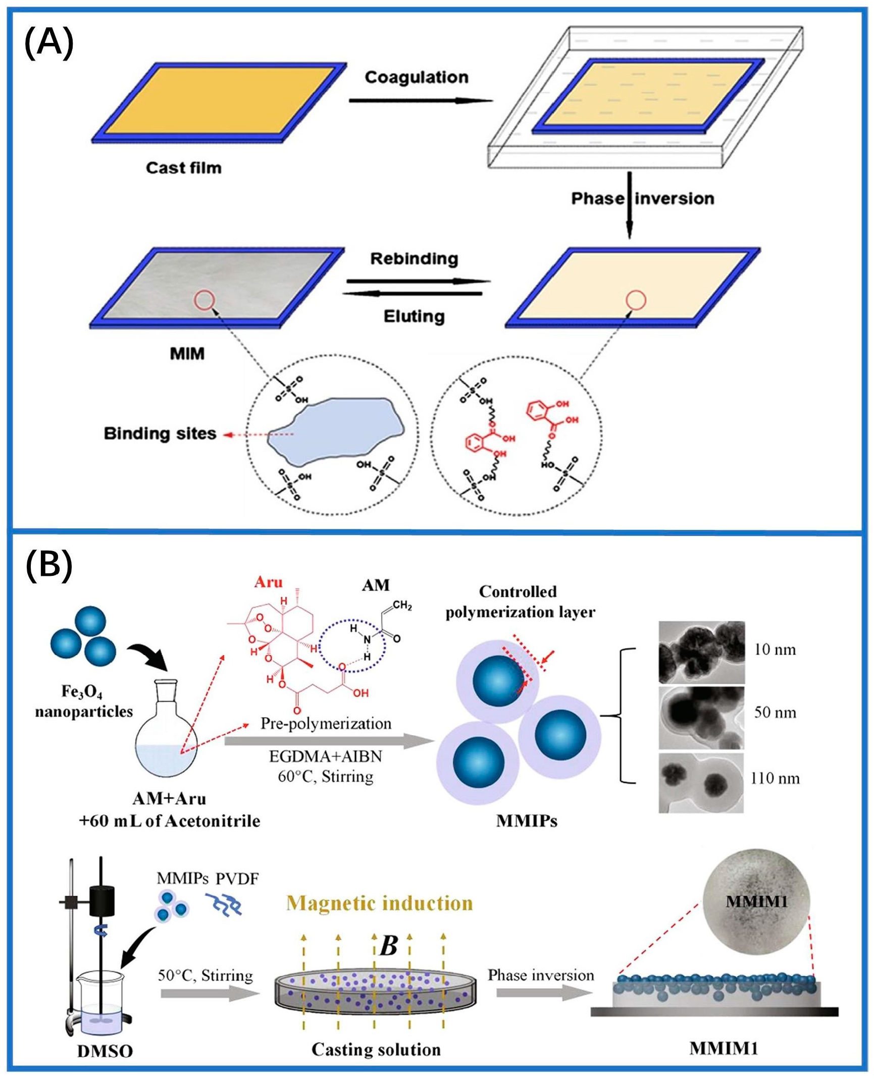
2.2. Phase-Inversion Method

Phase-inversion technology entails dissolving templates, membrane materials, cross-linkers, and functional monomers mixed in an appropriate solvent. The resulting solution is then applied onto a carrier, such as clean glasses. Bidirectional diffusion occurs between the casting and precipitating solution, leading to the phase separation of the casting solution from the uniform mixture and complete detachment. This process results in MIMs with specific molecular recognition capabilities. Currently, phase-inversion techniques include dry phase, thermogelation, wet phase, and reverse phase using water vapor inhalation [60,61]. Recently, researchers prepared MIMs by combining polystyrene with polypeptide recognition groups containing matrix polymers via the “dry reverse transfer” method. The resulting membranes demonstrated a higher permeability compared to that of non-imprinted membranes (NIMs) [76]. Early studies utilizing phase-inversion technology for MIMs employed cellulose diacetate as a matrix polymer. Optimization efforts concentrated on the solvent, reagent ratio, and total polymer concentration to improve membrane effectiveness and achieve molecular imprinting effects [77]. Figure 4A shows the MIMs for salicylic acid (SA) created by the reverse transfer technique. After first preparing the casting solution containing the template molecules, the solution was cast on a polished glass plate at room temperature. The glass plate was swiftly submerged in a gelling bath after 30 s. The phase transition between the polymer and deionized water then commences at 18 °C [78]. As depicted in Figure 4B, researchers developed an innovative MIM by the phase-inversion technique [62]. In subsequent research, a highly efficient ion-imprinted membrane was created employing the non-solvent-induced phase separation (NIPS) technology with ionized platinum (IV) serving as the template. This approach achieved the selective separation of platinum (IV) [79]. Additionally, poly(vinylidene fluoride) (PVDF) composite membranes were produced using the inverse transference method, which demonstrated a strong rebinding capability (31.25 mg/g) in adsorption tests with environmental effluents [80]. The primary distinction between phase transition and in situ polymerized methods for membrane fabrication lies in the incorporation of recognition sites into the polymer. In phase transition, the recognition sites are directly introduced into the polymer. MIMs are created from the polymer solution with target molecules present. Throughout the imprinting process, both recognition binding sites and the penetration channels are formed. The phase-inversion process has been demonstrated to be partially influenced by kinetic and thermodynamic factors and typically is completed within a few milliseconds. Therefore, understanding the mechanism of this process poses challenges due to its complexity.

2.3. Sol-Gel Method

The sol-gel method for preparing MIMs involves constructing films by incorporating templates into network structures composed of inorganic and organic inorganic. This technique has seen significant advancement in recent years and is increasingly utilized in mixture separation and sensor development. Generally, functional group monomers and cross-linking agents are initially dissolved together, followed by the addition of template compounds. Subsequently, a polymer network forms through polymerization or cross-linking reactions. Once the template molecules are removed, MIMs are formed. This technology involves a gentle reaction environment and is straightforward to execute. Additionally, during the reaction process, the properties of the synthesized film can be finely tuned through precisely controlling the reagent composition, density, and pH value belonging to the reaction solution [63,64,65]. Figure 5A depicts the MIP-modified electrode prepared using the sol-gel technique for the electrochemical detection of the drug naloxone (NLX). In this setup, pyrrole imparts conductivity to the sensor, while triethoxyphenylsilane (TEPS) functions as the functional group monomer and tetraethoxysilane (TES) acts as the cross-linker. This sensor is excellent for the quantitative testing of NLX in artificial urine samples [81]. Figure 5B illustrates the process of preparing borate affinity sol-gel molecular imprinting. This approach enhances selectivity and binding affinity through the synergistic interaction of imprinted cavities and boric acid, achieving an imprinting factor greater than 10 [82]. Additionally, another study focused on developing sensing membranes by adding microcystin-LR templates into the sol-gel matrix [64]. During the hydrolysis of the mixture, the substrate undergoes polymerization, forming a network. Ultimately, the templates were extracted from the imprinted polymer, creating selectively imprinted cavities behind it. In another investigation, molecularly imprinted sol-gel hollow-fiber membranes (MSHMs) was used to detect horse uric acid from plasma and urine samples by the sol-gel method [66]. The experimental results indicated the detection limit was 0.3 nmol/L and 1.0 nmol/L in the human plasma and urine samples, respectively.

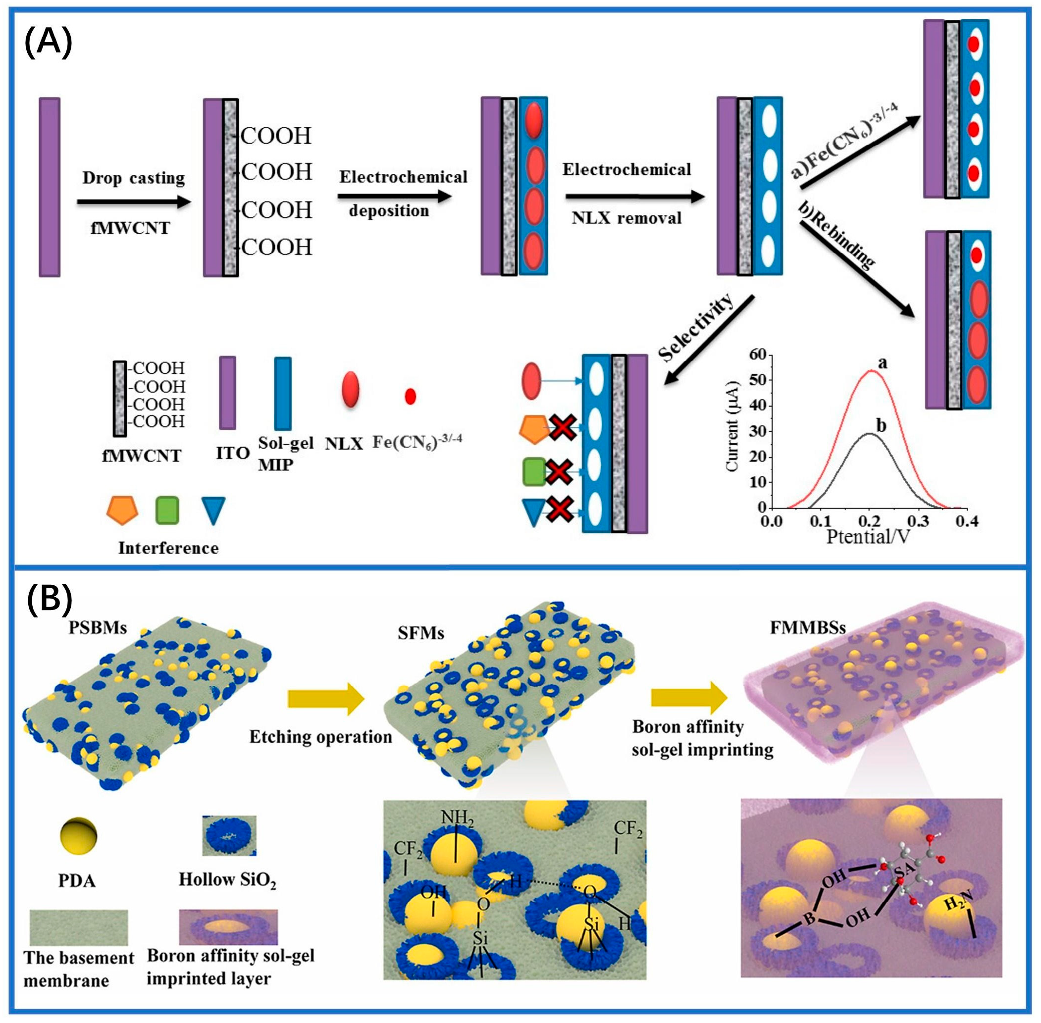
2.4. Electrochemical Method

Electrochemical polymerization is a widely used technique for fabricating electrochemical membrane sensors. This method generally involves the creation of MIMs on the electrode surface through electrochemical polymerization or deposition techniques [26,67,68]. The process includes selecting and cleaning the electrode, preparing a casting solution, performing electrochemical polymerization, and subsequently removing the template molecules to yield the MIMs. Electrochemical polymerization offers several advantages over alternative polymerization techniques, such as a faster preparation rate and indirect control over film thickness by adjusting the polymerization solution concentration and scanning cycles. In 2003, Blanco-Lopez and colleagues developed a voltametric sensor to detect vanillylmandelic acid (VMA) with a monomer mixture on a glassy carbon electrode (GCE). This involved spin-coating a layer of MIPs with a blend of monomers (template, cross-linker, solvent, and methacrylic acid), followed by in situ photopolymerization [83]. The membranes obtained exhibited a proper pore size and suitable density, facilitating the diffusion of the analyte toward the electrode surface for effective molecule detection. However, challenges persisted regarding membrane stability and the control of membrane thickness. Furthermore, the selectivity of imprinted membranes in water systems is relatively limited, thereby constraining their potential applications to some extent. Building upon these findings, an electrochemical surface molecular self-assembly strategy was proposed. Figure 6A demonstrates the fabrication process of a glassy carbon electrode coated with a magnetically attracted MIP (Fe3O4@MIP) to electrochemically detect luteolin. The sensor exhibits a stable performance for a duration of 6 days, with a linear detection range spanning from 2.5 pM to 0.1 μM [84]. As depicted in Figure 6B, a composite chiral sensor was developed by immobilizing MIPs onto the glass carbon electrode surface. The chiral sensor possessed a linear identification range at 5–6000 × 10−11 mol/L. In addition, this particular sensor exhibited detection recoveries ranging from 93.8% to 109.0% when used for identifying levamisole in chicken and various authentic samples [85]. Furthermore, enzymatic biosensors with a single-layered membrane structure were developed for the detection of pesticides, utilizing an asymmetric base film made of bromomethylated poly(2,6-dimethyl-1,4-phenylene oxide), BPPO [86]. The biosensor membranes were fabricated utilizing a graphite carbon electrode, which identifies pesticides through the reduction current generated by the enzymatic reaction and subsequent formation of quinone as its end product. Catechol diffused across the solution and permeated into the enzyme domain, where it underwent conversion into quinone and a subsequent reduction atop the electrode’s outer layer. However, an inhibitor-like pesticide resulted in a delayed enzymatic reaction, causing a reduction in the observed current. Meanwhile, another study employed an in situ electrochemical reduction and polymerization process to gradually form MIMs directly on a GCE surface [70]. The prepared electrochemical sensor showed outstanding achievement in determining dihydromyricetin with an extremely sensitive detection threshold of 1.2 × 10⁻⁸ M.

3. Important Applications of MIMs in Various Fields

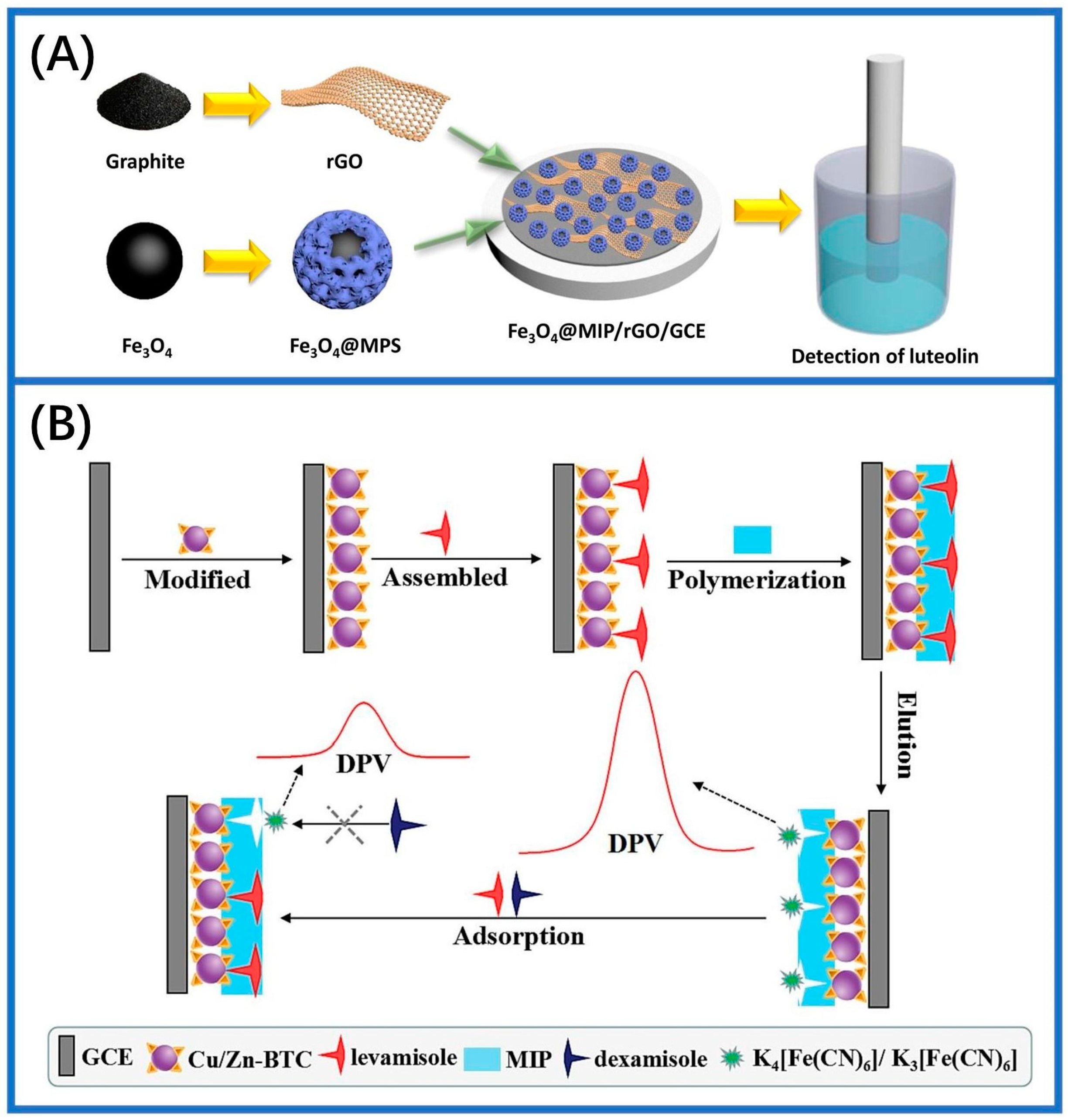
3.1. Electrochemical Biochemical Sensors

The integration of MIT into electrochemical methods facilitates the conversion of reactions occurring on imprinted membranes into monitorable electrical signals, thereby building an efficient and reliable analysis platform [26,87]. MIM functionalized electrochemical sensors are assessed by monitoring changes in current and voltage before and after the analyte enters the imprinted cavity [88,89]. Typically, electrochemical sensors consist of electrodes and electrolytes. The electrodes, a three-electrode system, comprise a reference electrode (RE), a working electrode (WE), and a counter electrode (CE) [90]. Electrolytes are substances that conduct electricity by dissociating into cations and anions when dissolved in water or in their molten state, examples of which include NaCl and PBS. A key feature of electrochemical reactions is the oxidation or reduction of analytes through electron transfer at the electrode. Analytes can be detected either qualitatively or quantitatively by measuring electrical signals such as current, voltage, and resistance. Based on these signals, electrochemical sensors are classified into various types, including amperometric, potentiometric, and impedimetric sensors [91,92,93].
Different detection techniques can be utilized during the electrochemical measurement process, including chronoamperometry (CA), differential pulse voltammetry (DPV), cyclic voltammetry (CV), and square wave voltammetry (SWV) [94,95]. As an exemplification of a prototypical point-of-care testing (PoCT) scenario, an MIM-electrochemical method was constructed for the immediate detection of 3-NT by modifying zeolite imidazolate backbone-67 (ZIF-67) on a laser-induced graphene (LIG) substrate (Figure 7A). The sensor is controlled using a tablet or smartphone and requires only 50 μL of a sample solution to complete the test. Figure 7A depicts the DPV results, which showed that the five sensors prepared under identical conditions exhibit excellent repeatability. With a turnaround time of 10 min and minimum detectable level of 6.71 nM, this platform demonstrated promising potential as a point-of-care (PoC) diagnostic tool. By optimizing performance, cost, and biocompatibility, this sensor effectively meets the requirements for routine disease detection [96]. In addition to graphene, GCE is also commonly employed as an electrochemical substrate. An MIM immunosensor was developed for the analysis of AFP by modifying the GCE surface with polythionine (PTh) and gold nanoparticles (AuNPs). The DPV analysis demonstrated that the maximum sensor current remained unaltered in the presence of interferences, indicating its exceptional specificity. Even after storage at low temperatures for seven days, the sensor exhibited a negligible decline in its peak current, showcasing remarkable stability [97]. The identification of catechol by MIT was successfully achieved through the modification of a GCE with reduced graphene oxide. During the imprinting process, catechol was adsorbed onto the surface of polypyrrole (Ppyr), and subsequently, a pollution-free extraction was performed from this mass-produced template. In voltammetry measurements, the sensing device exhibited a broad range of linearity with an LoD at 4.18 nM, demonstrating exceptional levels of selectivity, stability, and repeatability in real sample detection [98]. Capacitance-based molecularly imprinted sensors, also known as impedance sensors, detect changes in capacitance resulting from binding within the analyte-imprinted cavity. In this type of sensor, electrochemical impedance spectroscopy (EIS) is typically employed to investigate the electrochemical process at the electrode/electrolyte interface [99]. For example, researchers have developed MIM electrochemical sensors for the dual detection of sumatriptan (SUM) and paroxetine (PRX) using EIS. They modified pencil graphite electrodes by phosphotungstic acid (PWA) and reduced graphene oxide (rGO) composites, which effectively reduced resistance and enhanced the rate of electron transfer. The EIS results demonstrated that the improved electrode exhibited a high selectivity with imprinting factors of 3.15 for SUM and 3.3 for PRX, respectively. However, the sensor’s measurement relies on static electrochemical technology, requiring sample loading onto the electrode for detection. Consequently, real-time monitoring is unattainable, and offline analysis becomes imperative [100].
MIM electrochemical sensors offer versatile applications beyond pharmaceuticals, including the detection of biological macromolecules like biomarkers related to SARS-CoV-2. For instance, epitope-imprinted polymers synthesized through electropolymerization demonstrated cost-effectiveness and reduced non-specific binding, allowing for the precise recognition of SARS-CoV-2 proteins with a high affinity [101]. By using heptapeptides derived from the SARS-CoV-2 protein with a spike structure as templates for epitope-imprinted polymers, the accurate detection of the parent proteins has been achieved at low picomolar concentrations [102]. Additionally, molecularly imprinted polypyrrole electrodes have proven effective in detecting SARS-CoV-2 spike glycoproteins, showing enhanced levels of affinity and sensitivity compared to non-imprinted counterparts [103]. The development of portable sensors for SARS-CoV-2 antigen detection has emerged, with disposable sensor chips integrating thin-film electrodes and MIMs, demonstrating the real-time analysis of nasopharyngeal swab samples (Figure 7B). These sensors demonstrated exceptional selectivity for ncovNP, achieving a remarkable sensitivity down to 15 fM. This highlights their precision and rapidity in detecting SARS-CoV-2. The sensors’ excellent ability to discriminate interfering proteins, coupled with their rapid response time and user-friendly operation, has greatly enhanced the clinical diagnosis of COVID-19 [104].
Figure 7. Practical implementations within electrochemical MIM systems. (A) Schematic illustration of the detecting process via microsensor and the DPV curves of 3−NT [96]. (B) The procedure for electrochemical detection of ncovNP from nasopharyngeal swab samples and the sensor’s selective response towards diverse proteins [104].
Sensors 24 05119 g007
As widely known, the sensitivity of MIM electrochemical sensors heavily relies on the quantity of imprinted cavities embedded in the imprinted films [105]. Therefore, the support materials with a large specific surface could expand the imprinted films and generate a greater level of imprinting cavities. Black phosphorus (BP) is a semiconductor material known for its high carrier mobility, significant reducibility, and exceptional electron donor ability, facilitating the facile reduction of chloroauric acid (HAuCl4) to AuNPs [106,107,108]. Using these capabilities, AuNP/BP nanosheet-modified GCE (BBNS-AUNP/GCE) sensors were developed for the specific measurement of norfloxacin (NOR). The electrochemical sensor employed linear sweep voltammetry (LSV) for the quantitative analysis of NOR, exhibiting a wide range of responses that are linear in nature (0.1 nM–10 μM) and a low level of identification restriction (0.012 nM). While the sensor exhibits a high level of selectivity, there remains potential for enhancing sensitivity in clinical applications [109]. To address this issue, metal–organic frameworks (MOFs) are employed, which are crystalline support materials with a high porosity, stable backbone, and large surface area [110]. Combining MIMs with MOFs enhances binding capacity and accelerates recombination kinetics. Researchers optimized the surface of GCE, utilizing a copper MOF modified with gold nanoparticles (AuNPs) and nitrogen-doped quantum dots derived from graphene (N-GQDs) (Au@Cu-MOF). The detection limit of this MIM electrochemical sensor platform was as low as 0.0007 ng/mL for patulin. Despite this progress, the sensitivity of MIMs remains constrained by binding site limitations and slow binding/dissociation rates [111]. Typically, the adjustment of structural stability in MOFs often poses challenges, thereby limiting the applicability of certain chemical modifications. Thus, researchers have also used the covalent organic framework (COF) to addresses this issue by incorporating covalently bonded organic units, allowing for the optimization of performance through the selection of diverse organic units and the regulation of synthesis conditions [112]. Conductive MIMs have shown promise in enhancing electrochemical sensor performance. Recently, MIP composites including polyaniline (PANI) were employed in field sensors for an organophosphate pesticide analysis of vegetables, achieving a low LoD of 11.3 nM for the real-time detection of parathion [113]
MIM electrochemical sensors are extensively utilized; however, certain limitations persist. For instance, the utilization of electrochemical probes results in a diminished adsorption capacity, restricting the sensor’s ability to detect molecules beyond the surface’s vicinity. Furthermore, issues such as unstable electrical signals, low conductivity, non-specific adsorption, and other factors necessitate future enhancements in MIM electrochemical sensing technology [92,114].

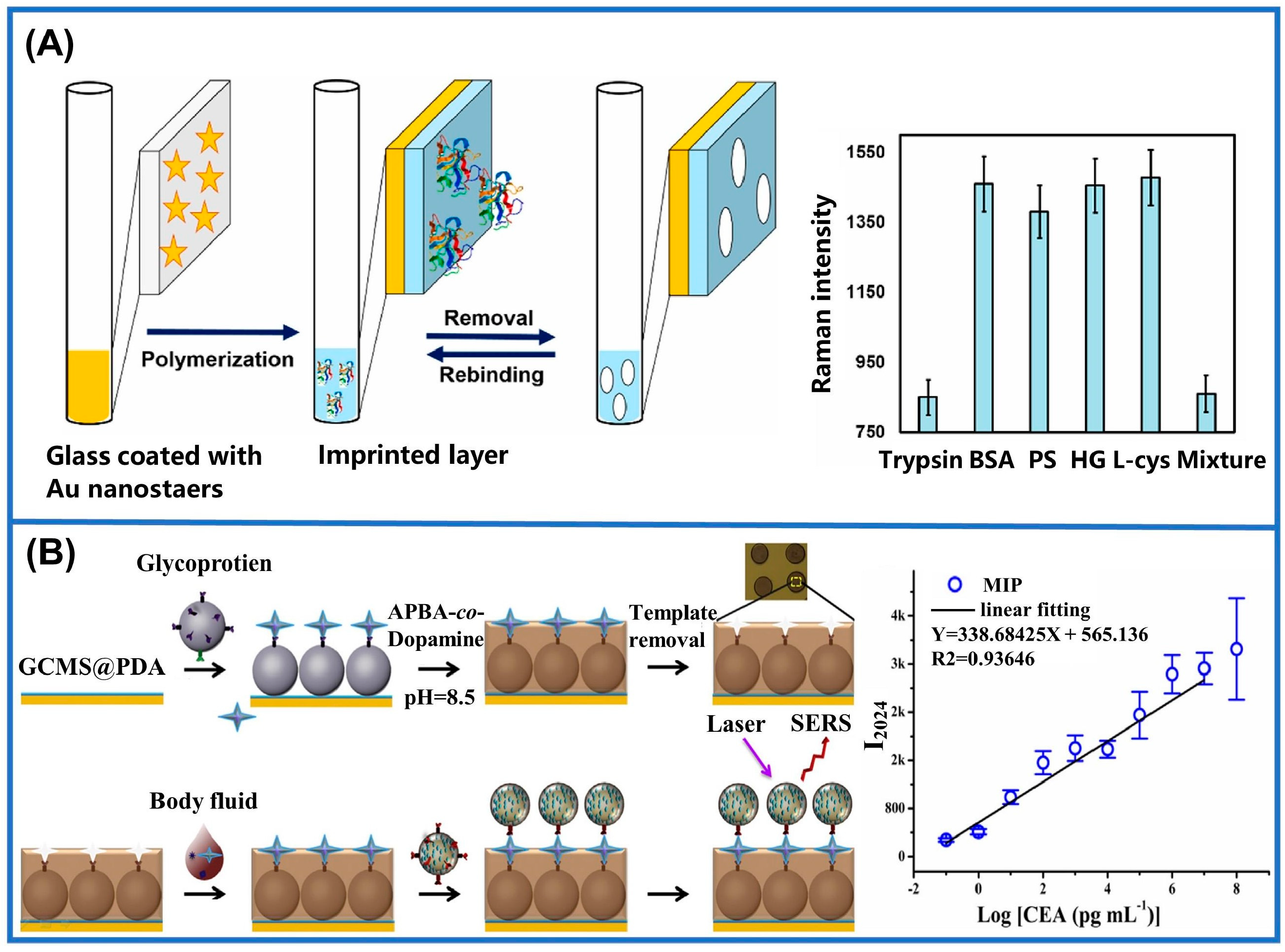
3.2. Surface-Enhanced Raman Scattering

SERS utilizes metal nanostructures to amplify weak Raman signals, enabling the detection of low concentrations or even single molecules [115,116,117,118,119]. SERS offers advantages such as a high sensitivity, spectral resolution, short response time, and molecular fingerprint information. However, interference from substances with similar chemical compositions or bonds may mask target signals, leading to false negatives or detection failure [120,121]. The combination of selective imprinting cavities from MIMs with the SERS method can effectively improve the anti-interference ability of SERS [122]. Furthermore, MIMs can serve as the adsorbent material to bring target molecules closer to the SERS substrate, thereby enhancing the sensitivity of SERS [123,124]. Consequently, the integration of MIMs with SERS technology has attracted significant interest in the fields of biomedicine, environmental monitoring, and food supervision [125].
MIM-SERS sensors are generally categorized into “one-step” and “two-step” methods based on their preparation process. In the one-step method, MIMs are directly attached to the SERS substrate, which enriches the target analyte and enhances SERS signals. This approach offers benefits such as simplicity, rapid detection, and minimal solvent consumption. Additionally, one-step MIM-SERS sensors can be further classified into planar and sandwich types. Planar sensors involve directly coating MIMs onto flat SERS substrates, which increases the surface area for a better interaction between target molecules and the substrate, thus enhancing scattering [126,127,128]. For example, multifunctional capillary SERS sensors were developed for protein detection with polydopamine imprinted layers (Figure 8A). The imprinted cavities can effectively capture target proteins while also serving as exclusive pathways for Raman indicators to access SERS substrates. When the imprinting cavities are blocked by target proteins, the reduction in SERS signals can reflect the quantity of captured molecules. Using bovine serum albumin (BSA), pepsin, and hemoglobin as target proteins, the sensor achieved versatility across different analytes [129]. Meanwhile, the detection of BSA can also be achieved with silver nanoparticle (AgNP)-modified graphene sensors. The incorporation of MIMs not only protected the AgNPs, but also enabled their specific binding with BSA. In comparison to conventional approaches, the sensor demonstrated a remarkable improvement in detection sensitivity by seven orders of magnitude. This method, however, still presents opportunities for enhancement, necessitating a further refinement of its materials and preparation techniques to improve its stability and longevity [130]. In addition to large proteins, MIM-SERS sensors are utilized in the identification of minute molecules as well. Through the polymerization of patulin (PAT) triggered by horseradish peroxidase, an Au-polydimethylsiloxane (PDMS)–concrete anodized aluminum oxide (AAO) substrate was constructed. This new approach exhibited an excellent selectivity towards PAT with a lower limit of detection at 8.5 × 10⁻¹¹ M [45]. As for sandwich-type MIM-SERS sensors, they utilize MIMs as the recognition layer to combine them with the target molecules and then generate SERS signals by binding to the upper Raman probes. Owing to the exceptional stability and sensitivity conferred by this sandwich structure, these sensors are highly suitable for detecting biological macromolecules [127]. In 2014, researchers pioneered the creation of a boron-affinity sandwich assay (BASA) for detecting AFP in serum. The target glycoproteins were first extracted by a boron-affinity MIM and then bound to boric-acid-modified SERS labels [131]. In addition, a novel dual-MIP-based immunoassay has been developed for the detection of proteins in complex biological samples. A carbon end epitope-imprinted layer is fabricated atop the assembled underlying material, functioning as a specialized SERS substrate specifically tailored for the intended protein. N-terminal epitope-imprinted Raman response Ag@SiO2 nanoparticles are employed as SERS labels in a sandwich structure, resulting in a significant enhancement in and the high specificity of the SERS signal [125]. Utilizing nucleoside diphosphate kinase A (NDKA) antibody as an SERS marker, an MIM-SERS sensor modified with 4-mercaptobenzoic acid (4-MBA) was prepared. The sensor employed polydopamine (PDA) as the substrate of the MIM to capture NDKA, achieving an exemplary test constraint of 0.25 pg/mL in human serum. But the intricate preparation process and the potential interference from substrate fluorescence or scattering present formidable challenges in regards to the sensor’s performance [132]. Except such enhanced Raman tags, the performance of MIM-SERS sensors can also be improved by using tags with specific sites to avoid interference. By employing probes with Raman signals located in the silent region (1800~2800 cm−1), researchers avoided optical noise from endogenous compounds within an area with a unique pattern (300~1800 cm−1). Due to the excellent anti-interference capability of the sensor, this method can be applied for the PoCT of CEA in real blood (Figure 8B) [133].
In contrast to the complexity of spectra obtained through the “one-step” method, the two-step SERS detection method was developed to separate and enrich the target using MIMs before the SERS detection of the eluted solution. This process effectively removes impurities and interferences, significantly improving the accuracy of results. In MIM-SERS sensors, MIMs function as molecular recognition cavities that fill a solid-phase extraction (SPE) column, specifically enriching the organic compounds from complex matrices [134]. For example, a sensor designed to detect the SERS signals of caffeine using theophylline as a virtual template was developed. The sensor, consisting of AgNPs@ molecularly imprinted SPE (MISPE), a syringe, and a removable microporous membrane, eliminated separate elution steps, thus reducing the overall analysis time. However, the preparation method of the sensor is intricate, necessitating AgNPs’ incorporation into the MIM through precipitation polymerization, thereby potentially compromising the stability and reproducibility of the fabrication process [135]. So MIM-SERS sensors also employ flexible polymer films as substrates, which are characterized by their transparency, flexibility, stability, and low cost. Conventionally, MIMs are coated or poured onto the surface of SERS substrates for detection. Polyvinyl chloride (PVC), as a type of polymer material, has wide applications in various aspects of daily life. As a brilliant example, an AgNP/PVC MIM-SERS sensor was fabricated with PVC as a carrier through the solvent casting technique. The sensor demonstrated undifferentiated detection on both sides, showcasing a robust linear correlation between the SERS intensity and solution concentration at 1595 cm−1 [43]. There are also other studies based on MIM-SERS PoCT sensors, such as those utilizing polyvinylidene fluoride (PVDF) for L-tyrosine analysis and an electrospun filament protein membrane (ESM) as a biocompatible scaffold for spiramycin detection [136,137]. Notably, these approaches have all exhibited exceptional nanomolar-level detection limits. However, MIM-SERS sensors still face challenges such as their limited anti-interference capability and complex regeneration processes. To address these issues, researchers are actively developing novel molecular imprinted materials with enhanced levels of specificity, sensitivity, and regeneration performance through precise design and preparation. Furthermore, strategies including portable Raman detectors for PoCT, nanotechnology for efficient SERS substrate fabrication, the integration of intelligent algorithms for data processing, and the optimization of template and monomer identification via boric acid affinity have been proposed. These approaches highlight significant advancements in overcoming the limitations associated with MIM-SERS sensors while offering new avenues to enhance their performance and broaden their application scope.

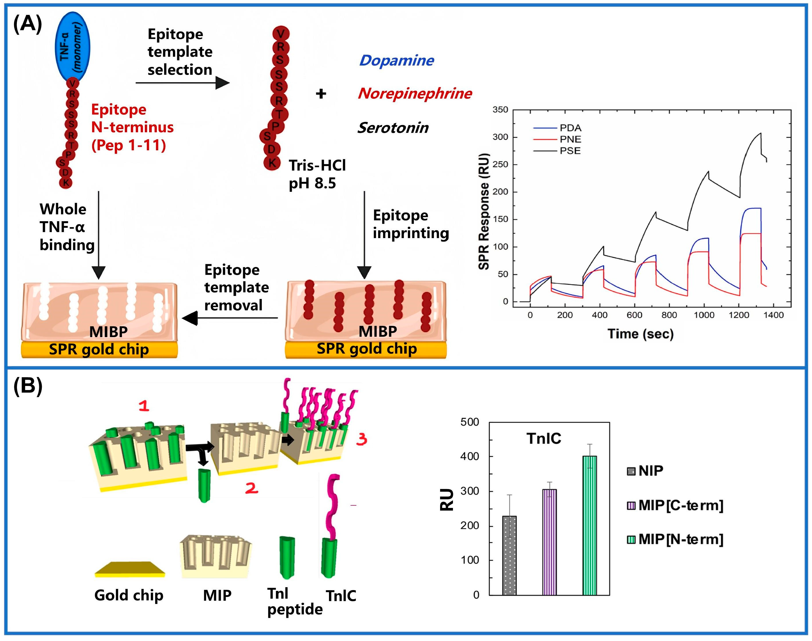
3.3. Surface Plasmon Resonance

Having explored the potential applications of SERS, we now turn our focus on another promising spectral analysis technique: SPR. In SPR-MIM sensors, MIMs selectively capture target analytes, thereby increasing their local concentration on the SPR sensor surface. This leads to a higher signal-to-noise ratio and improves their sensitivity in detection. SPR, originating in the 1990s, is widely used in biomedical fields for detecting ligand–analyte interactions on biosensing chips, facilitating real-time biomolecule analyses [26,138,139]. The principle of SPR involves collective electron oscillations on a metal surface when illuminated by light, generating plasma waves that interact with incident light to enable detection [140]. Its sensitivity to variations within the refractive index of the metal outer layer allows for the precise monitoring of biomolecular interactions via dynamic resonance angle adjustments [141,142]. With the application and development of SPR technology, it has been combined with various techniques to enhance its detection performance [143]. Among these techniques, the combination of MIT with SPR not only confers specificity to the SPR sensing chips but also enhances the sensitivity of MIMs [144,145,146]. This significantly broadens the application areas of SPR and MIT and has garnered significant interest from researchers [147].
For example, a carbohydrate Antigen-125 (CA-125)-imprinted SPR sensor was developed via electropolymerization utilizing pyrrole (Py) as a monomer. The grafting condition of CA-125 onto the SPR chips can be monitored by changes within the permittivity value of the chip surface and variations in the incident resonance angle of the sensor surface. According to the above principle, the sensor achieves a detection limit for CA-125 of 0.01 U/mL [148]. Researchers also utilized lysine (K) and alanine (A) residues as models to investigate diverse imprinting states. They determined that optimal imprinting occurred when three lysine residues were positioned at the N-terminus. Expanding on these findings, the efficacy of the 3K strategy was further assessed using a peptide template of soluble programmed cell death ligand 1 (PD-L1). Through systematic optimization, the strategy achieved an excellent level of reproducibility (AVCV% = 4.5). Ultimately, the SPR sensor constructed based on the N-3K strategy demonstrated an outstanding level of sensitivity (0.31 ± 0.04 ng/mL) in samples of serum from humans, thereby providing a crucial diagnostic tool for healthcare applications [149]. In another study, researchers constructed MIM-SPR sensors employing the naturally occurring neurotransmitter serotonin (SE) as a functional monomer to capture tumor necrosis factor-α (TNF-α) for the first time (Figure 9A). The detection limits of the polyserotonin (PSE) biosensor were 20 times and 13 times higher than those of dopamine (DA)- and norepinephrine (NE)-based MIM sensors, respectively. This indicates that the future use of imprinted PSE as a bionic receptor for PoCT is a promising approach. But from the perspective of clinical and diagnostic applications, further enhancement of its sensitivity is imperative to effectively compete with commercial immunoassay methods and encompass a wide range of clinically relevant concentrations across various conditions and diseases [150]. Similarly, sensors with polynorepinephrine (PNE) as MIMs have also attracted people’s attention. A sensor imprinted two short troponin I (TnI) peptides on PNE, overcoming the disadvantage of whole-protein imprinting in MIMs (Figure 9B). Figure 9B illustrates that the imprinting of N-terminal peptides resulted in a significantly enhanced SPR response, exhibiting an approximate 100 RU increase, when compared to that of TnI-printed C-terminal biosensors. Due to its minimization of non-specific binding, this method provides excellent detection capabilities in the low nanomolar range (7.1 ± 0.6 nM) [151]. MIM-SPR sensors typically exhibit an exceptionally low detection limit and a high sensitivity, rendering them well-suited for the quantification of bacterial factors (such as RoxP) at low concentrations. Compared with non-imprinted SPR chips, the selection of RoxP as the protein imprint resulted in a faster response and higher sensitivity. The sensor exhibited a high affinity constant of 3.3 × 10⁻⁹ M for the target protein, which can facilitate the early PoC identification and diagnosis of certain oxidative skin diseases. However, the performance of such biosensors can be compromised by signal fluctuations, leading to a substantial standard deviation and diminished robustness [152]. Furthermore, SPR sensors are sensitive to environmental variations, including fluctuations in temperature and humidity, which can potentially impact the sensors’ performance and accuracy. The utilization of MIM-SPR sensors is further hindered by the high costs associated with their preparation and maintenance, limited detection range, as well as their suitability for specific analytes only.

3.4. Quartz Crystal Microbalance

A QCM is an extremely precise instrument for measuring mass, capable of monitoring minute changes on the surface of sensing chips [153,154,155,156]. The principle of the QCM is based on the piezoelectric effect, which enables the detection of changes in resonance frequency caused by mass variations on the quartz crystal surface [157,158]. One of the current trends in QCM sensors is their joint application with MIT for sensing [159,160]. Most reported studies attach MIMs to the surface of QCM chips and specifically detect the QCM by introducing specific cavities on the MIMs. When the analyte enters the detection cavities of the MIM, the surface of the chip is compressed and the frequency changes [161,162]. Therefore, the mass signals of the analytes can be converted into frequency signals by the piezoelectric effect of the chips. Recently, numerous studies have focused on the clinical applications of MIM-QCM sensors.
An MIM-QCM assay array was constructed for the detection of low-density lipoprotein (LDL) and high-density lipoprotein (HDL) (Figure 10A). Two functional monomers, methacrylic acid and N-vinylpyrrolidone, were chosen to specifically recognize the target proteins, enhancing the sensor’s specificity. The proposed method does not require fasting, thus reducing the pain caused by conventional detection. However, in comparison to commercially available enzyme- and antibody-based methods, the measurement results obtained from this sensor exhibit a higher coefficient of variation, which may impact the accuracy of the findings [163]. A novel enhancement of MIPs, termed electric-field-assisted molecularly imprinted polymers (EFAMIPs), was developed to enhance MIP-modified QCM biosensors. In the existence of a vertical electric field, methacrylic acid is polymerized with immuno-globulin G (IgG) as a template, followed by the template’s removal to obtain EFAMIPs. Studies have demonstrated that EFAMIP-modified QCMs exhibited a significantly higher frequency shift of up to 113.5% in electric field detection when IgG was present, compared to typical MIPs. The ultimate alteration in frequency of EFAMIPs, which determined the IgG detection limit, was enhanced by 12.5% compared to conventional MIPs. Thus, EFAMIPs show promise for selectively detecting IgG in solutions that consist of different categories of immunoglobulins, presenting a novel detection method for clinical applications [164]. In another study, researchers functionalized the surface of QCM crystals with molecularly imprinted polydopamine (MIPDA) membranes for identifying the presence of hepatitis B core antigen (HBcAg) (Figure 10B). The frequency response of the MIPDA-QCM sensor to HBcAg was 7.8 times higher than that of human serum albumin (HSA) under the same conditions. Due to the sensitivity and simplicity of its operation, this sensor has practical application value in the PoCT of hepatitis B. Nevertheless, the stability of the sensor’s biometric elements (MIPDA films) may pose a potential concern as prolonged usage and storage could result in film degradation and failure, thereby impacting the sensor’s performance and reliability [165]. Hence, scholars developed computationally assisted simulation methods, aiming to facilitate the design of MIMs with enhanced levels of stability and sensitivity. As a brilliant example, hexestrol (DES) was detected through an MIM-QCM sensor, for which researchers optimized the ratio of functional monomers and cross-linkers by simulating the molecularly imprinted self-assembly system of the DES. Guided by their calculations, they found that the optimal molar ratio of DES to methacrylic acid was one to five. At this point, the sensor had a strong affinity for DES with a test constraint of 2.63 ng/mL. After numerous cycles, however, the sensor’s detection rate experienced a significant reduction. It is speculated that the internal pore structure of DES-MIMs on the surface underwent some damage during electrode cleaning, thus leading to an escalation in the error margin of the detection outcomes [166]. As another small molecule, L-tryptophan was monitored by in situ polymerization using methyl methacrylate as a monomer. After optimizing the quantity of the agent used for cross-linking, the MIM containing 10 mmol ethylene glycol dimethacrylate (EGDMA) possessed the best adsorption ability for L-tryptophan. The modified sensors exhibited an excellent level of selectivity and low response level to interferents such as D-tryptophan and ascorbic acid [161].
Additionally, MIM-QCM sensors can be utilized for the detection of other analytes, such as N-hexanoyl-L-homocysteine lactone, insulin, oxidized LDL, very-low-density lipoprotein (VLDL), and adenylate kinase 1 [167,168,169,170]. Analytical results from real samples demonstrate the great potential of MIM-QCM sensors in the detection field. However, the QCM is susceptible to environmental vibrations and is prone to surface contamination from grease and dust. Primarily employed for mass measurements in liquids, the QCM has a limited capability in measuring solid samples. Currently, molecular simulation computing, virtual template molecules, and computer-aided methods have been employed to design highly specific MIMs that cover the surface of QCM chips to alleviate the issues arising from the inherent shortcomings in QCM technology. It is expected that the advancement of MIM-QCM sensor technology will progressively overcome these limitations, thereby effectively harnessing the benefits offered by both technologies.

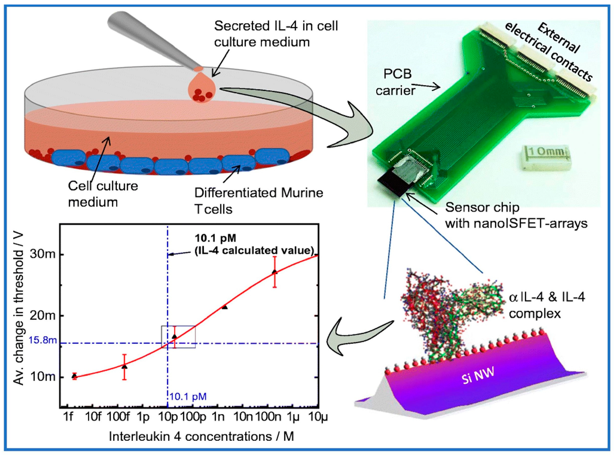
3.5. Ion-Sensitive Field-Effect Transistors

In recent years, FET-type biosensors leveraging various nanomaterials—such as silicon nanowires, graphene, and transition metal disulfides—have burgeoned. These biosensors have emerged as potent tools for detecting diverse biomolecular targets, both within clinical laboratory settings and for PoC diagnostics.
FETs are electronic devices designed for signal transmission and amplification, initially conceived in 1947 as a quantitative method for biomolecule detection. FET sensors comprise three distinct electrodes: the source, drain, and gate [171,172,173]. MIMs and FETs can be combined in two ways: firstly, by coating the MIMs on the gate, where the adsorption/removal of the template from the gate has an impact on the overall voltage applied to the gate, thereby influencing transistor conduction. MIMs can be combined with the two-dimensional materials in the transistor channel, allowing for the detection of electrical signal changes before and after the combination of materials, which negates the need for MIM conductive performance. FETs operate by applying a voltage to the gate, which induces electron flow from the source to the drain. Analytes affect charge transport in the channel, making the device responsive to the electric field generated by the gate electrode. This principle allows for the conversion of biological or biochemical signals into electrical signals, which is fundamental for biomolecule detection using transistors [174,175,176]. As sensors, transistors offer a straightforward, cost-effective, portable, and real-time detection mechanism, enabling interaction with a broad range of biomolecules. The sensitivity of these sensors is determined by the transistor’s signal conduction and amplification capabilities. Over time, FET technology has evolved, leading to the inclusion of various structures and signal generation/amplification methods, including ion-sensitive FETs (ISFETs) [177,178], organic electrochemical transistors (OECTs) [179,180,181], organic FETs (OFETs) [182,183,184], and so on. Integrating transistors with sensors and MIMs leverages both the signal transmission and amplification abilities of the transistors and the high selectivity of the MIMs. This combination addresses challenges such as interference from complex bodily fluids by using the specific affinity of target biomolecules and the imprinted cavities of the MIMs. The addition of surface nanostructuring can further enhance the affinity of MIM structures for gate electrodes [185], while the precise adjustment of their surface roughness to match the size of analyte molecules can improve their biosensing performance [186].
The integration of MIMs and FETs presents a powerful approach to chemical sensing and detection. Sensors created through this combination provide several benefits, including an enhanced specificity level, high sensitivity, rapid response times, and label-free detection. Firstly, the high selectivity of MIMs for specific target molecules reduces disturbance caused by additional substances present in the sample matrix, thereby improving the accuracy of FET detection. Secondly, FET sensors’ rapid response capability enables an immediate change in conductivity when target molecules bind to the MIM-functionalized FET surface, allowing for swift detection. Lastly, unlike conventional FET biosensors, MIM-functionalized FET sensors possess the potential for miniaturization and integration into compact, portable devices dedicated to PoC diagnostics, environmental monitoring, and field inspection applications. Recent studies have reported numerous applications of MIMs and FETs. For instance, one study integrated conductive MIMs with extended gate FET (EG-FET) sensors to identify specific antigenic regions of matrix metalloproteinase-1 (MMP-1) protein, a biomarker of idiopathic pulmonary fibrosis (IPF) [187]. This prepared sensor selectively detected MMP-1 protein within the concentration range of 50 to 500 nanomolar in serum (Figure 11A). Meanwhile, researchers developed a novel molecularly imprinted electrochemical sensor based on ISFET devices for urea detection [178]. MIMs were synthesized by the surface photopolymerization of ISFET devices using PMMA and urea as functional polymers and molecular templates, respectively. The LoD of the MIMs-modified ISFET sensor was determined to be 1.0 × 10⁻⁴ M. Figure 11B displays that a cyclic adenosine monophosphate (cAMP) mimetic ISFET sensor was fabricated using cyclic AMP as the molecularly imprinted material [188]. The ISFET electrode was coated with a polymer film that had been imprinted, leading to the development of sensors capable of effectively binding and selectively detecting cAMP in aqueous solutions. Furthermore, another study used an inorganic sol-gel polymerization mixture of titanium (IV) butanol–carboxylate complexes to create a thin layer on the surface of an ISFET gate [189]. The device provides repeatable measurement results at ambient temperatures with a voltage of ± 2 mV.
The proof-of-concept readiness of top-down fabricated nanoscale Si-based ISFET arrays (nanoISFETs) as a biosensor device for the label-free electrical detection of cell-secreted IL-4 and IL-2 was demonstrated (Figure 12) [190]. Increasingly, studies indicate that the combination of MIMs with FETs is not only widely applicable in trace detection but also addresses the stability issues associated with traditional antigen–antibody transistors. However, there is a paucity of literature on the combined application of these two technologies. This may be attributed to the organic reagents employed in the synthesis of MIMs and the cumbersome synthesis steps. Table 2 displays a compilation of diverse applications utilized in MIM biosensors.

4. Challenging and Limitations

MIMs present immense potential for enhancing the progression of all-solid-state biochemical sensors by virtue of their selective recognition and stability. Nonetheless, several noteworthy challenges and constraints persist in their practical utilization that necessitate careful attention and resolution. The foremost challenge lies in the imprinting efficiency of MIMs. Factors like the choice of functional monomers, the template-to-monomer ratio, and polymerization conditions profoundly influence the binding uniformity and specificity of MIMs. While significant strides have been made in polymerization techniques and functional monomer design, further enhancements in imprinting efficiency require extensive research endeavors.
Moreover, the long-term resilience of MIMs presents significant hurdles in real-world applications. Polymer degradation under varying environmental conditions such as pH, temperature, and humidity, along with shifts in binding affinity, also directly impact the reliability and longevity of sensors. Furthermore, ensuring reproducibility and standardization in MIM synthesis is paramount in current research endeavors. Consistency in MIM synthesis and sensor performance across different production batches and laboratories is pivotal for driving commercialization initiatives forward.
In summary, addressing these challenges necessitates collaborative efforts and innovative strategies from materials scientists, chemists, and sensor engineers. Overcoming these obstacles successfully will pave the way for broader advancements and applications of MIMs in diverse fields including environmental monitoring, food safety, and medical diagnostics.

5. Conclusions and Prospects

Through a comprehensive analysis of current research and methodologies, this paper unveils the unique benefits and potential applications of combining MIMs on 2D solid-supported substrates with state-of-the-art sensing technologies. This integration facilitates interdisciplinary research and has a significant impact on materials science and clinical testing applications. Supported by concrete examples and experimental data, these platforms enable the simultaneous detection of multiple disease markers and their seamless integration with various diagnostic technologies, including electrochemical and optical sensing, as well as mass spectroscopy and semiconducting detection. These cutting-edge sensing technologies facilitate the development of rapid on-site detection for precision diagnosis and personalized medicine.
The preceding discussion underscores the extensive utilization of MIMs across electricity, optics, and mechanics, yet PoC biosensors within the realm of FETs remain notably absent. Notably, sensors built upon FET technology offer compelling attributes such as their compactness, facile scalability, broad applicability, and cost-effectiveness. Therefore, we are confident that this research area will attract the interest of researchers and will be widely used in the future. A recent development in MITs was based on a molecular-imprinted self-assembled monolayer, for which a molecular-thick recognition layer was adsorbed on the nanostructured surface, providing a highly effective PoC diagnostic platform [191]. This technology can eventually be integrated with the processing of 2D solid-supported substrates to provide fast, accurate, and highly specific detection.
MIT’s increasing prominence due to its numerous advantages has been recognized. However, despite years of development, commercializing MIT innovations remains a slow and arduous process. Early proponents of using MIMs in separation science established several companies with some success. Nevertheless, the majority of commercial applications primarily focus on material separation rather than sensor technologies. While MIMs demonstrate a high separation performance, their utilization remains primarily confined to academic settings, with their integration into industrial applications still in its nascent stages. We posit that several factors impede their translation into industrial settings.
Firstly, in the development of MIM-based sensors, the occurrence of false-positive signals resulting from the non-specific binding of polymers to templated materials diminishes the sensors’ accuracy in practical applications. Due to the inherently adsorbent nature of polymers, some degree regarding the engagement with the molecule of interest is inevitable. Moreover, the presence of inhomogeneous binding sites can result in the rebinding of substances, limiting the relevant applications of polymers. Additionally, the employment of numerous environmentally unfriendly solvents and reagents in the manufacturing process poses challenges in adhering to stringent environmental regulations aimed at waste reduction and environmental protection. Clearly, the integrated application of MIMs is imperative to foster their widespread adoption and market penetration in sensing applications. As a result, there is growing interest among scientists and manufacturers in investigating production methods that are more eco-friendly and improving the effectiveness of separation processes to increase efficiency. We anticipate the imminent emergence of commercial applications leveraging MIM-based sensing technology.
Future research should prioritize the development of novel cross-linking agents and functional monomers to enhance the selectivity and specificity of MIMs. Additionally, there is a need to explore environmentally friendly materials and methods for MIMs to minimize environmental impact and promote sustainable sensing technologies. Research efforts should also focus on identifying 2D solid-supported substrates with enhanced stability and durability levels to further elevate sensor performance. The integration of MIMs and 2D solid-supported substrates can enable the creation of multifunctional sensing platforms capable of simultaneously detecting multiple target molecules. This advancement significantly broadens the application scope and practical utility of sensors, catering to diverse detection needs across various fields. In terms of applications, leveraging the advancements in the Internet and smart device technologies, MIMs can be integrated with smart sensing technologies to develop portable, real-time detection sensors. These sensors hold immense potential for widespread use in clinical diagnostics, environmental monitoring, and food safety, delivering timely and precise detection outcomes. Finally, it is investigated how to realize the large-scale preparation and commercial application of composite sensors with molecularly imprinted membranes and 2D solid-supported substrates. Optimizing production processes and reducing costs are crucial steps towards enhancing the competitiveness of these sensors in practical applications, thereby fostering the technology’s adoption and market penetration. By advancing research along the outlined directions, the application potential of MIMs on 2D solid-supported substrates will expand significantly, presenting new opportunities and challenges for the development of all-solid-supported biochemical sensors. Future innovative research holds the promise of accelerating progress in this field, ultimately benefiting various sectors of society.

Author Contributions

Conceptualization, L.W., X.Z. and W.F.; writing—original draft preparation, L.W. and N.L.; writing—review and editing, L.W., N.L., X.Z., I.B., I.G. and W.F. All authors have read and agreed to the published version of the manuscript.

Funding

This work is supported by National Natural Science Foundation of China (No. 52073160 and No. 52173007), the National Key Research and Development Program of China (No. 2020YFF01014706), and Beijing Municipal Science and Technology Commission (Z211100002421012), the European Union’s Horizon 2020 Research and Innovation Programme: the IPANEMA (No. 872662) and the ANTARES (SGA-CSA. No. 739570 under FPA No. 664387) (https://doi.org/10.3030/739570), and the Ministry of Science, Technological Development, and Innovation of the Republic of Serbia (grant 451-03-66/2024-03/200358).

Institutional Review Board Statement

Not applicable.

Informed Consent Statement

Not applicable.

Data Availability Statement

No new data were created or analyzed in this study. Data sharing is not applicable to this article.

Conflicts of Interest

The authors declare no conflicts of interest.

References

  1. DiMaio, J.T.M.; Doran, T.M.; Ryan, D.M.; Raymond, D.M.; Nilsson, B.L. Modulating supramolecular peptide hydrogel viscoelasticity using biomolecular recognition. Biomacromolecules 2017, 18, 3591–3599. [Google Scholar] [CrossRef] [PubMed]
  2. Chu, W.T.; Yan, Z.; Chu, X.; Zheng, X.; Liu, Z.; Xu, L.; Zhang, K.; Wang, J. Physics of biomolecular recognition and conformational dynamics. Rep. Prog. Phys. 2021, 84, 126601. [Google Scholar] [CrossRef]
  3. Lara, S.; Alnasser, F.; Polo, E.; Garry, D.; Lo Giudice, M.C.; Hristov, D.R.; Rocks, L.; Salvati, A.; Yan, Y.; Dawson, K.A. Identification of receptor binding to the biomolecular corona of nanoparticles. ACS Nano 2017, 11, 1884–1893. [Google Scholar] [CrossRef] [PubMed]
  4. Gianneli, M.; Polo, E.; Lopez, H.; Castagnola, V.; Aastrup, T.; Dawson, K.A. Label-free in-flow detection of receptor recognition motifs on the biomolecular corona of nanoparticles. Nanoscale 2018, 10, 5474–5481. [Google Scholar] [CrossRef] [PubMed]
  5. Liu, Y.; Wang, L.; Li, H.; Zhao, L.; Ma, Y.; Zhang, Y.; Liu, J.; Wei, Y. Rigorous recognition mode analysis of molecularly imprinted polymers—Rational design, challenges, and opportunities. Prog. Polym. Sci. 2024, 150, 101790. [Google Scholar] [CrossRef]
  6. Mirarefi, P.; Lee, C.T., Jr. Reversible control of enzyme-inhibitor interactions with light illumination using a photoresponsive surfactant. Proteins Struct. Funct. Bioinform. 2019, 87, 715–722. [Google Scholar] [CrossRef] [PubMed]
  7. Adamson, H.; Ajayi, M.O.; Campbell, E.; Brachi, E.; Tiede, C.; Tang, A.A.; Adams, T.L.; Ford, R.; Davidson, A.; Johnson, M.; et al. Affimer–enzyme–inhibitor switch sensor for rapid wash-free assays of multimeric proteins. ACS Sens. 2019, 4, 3014–3022. [Google Scholar] [CrossRef] [PubMed]
  8. Hang, Y.; He, X.P.; Yang, L.; Hua, J. Probing sugar–lectin recognitions in the near-infrared region using glyco-diketopyrrolopyrrole with aggregation-induced-emission. Biosens. Bioelectron. 2015, 65, 420–426. [Google Scholar] [CrossRef] [PubMed]
  9. Guan, P.; Guo, Z.; Xu, S.; Lu, H.; Wang, L.; Gu, Z.; Liu, Z. Molecularly imprinted nanobeacons redirect innate immune killing towards triple negative breast cancer. Angew. Chem. Int. Ed. 2023, 62, e202301202. [Google Scholar] [CrossRef]
  10. Bülbül, G.; Hayat, A.; Andreescu, S. Biomolecular recognition: ssDNA-functionalized nanoceria: A redox-active aptaswitch for biomolecular recognition. Adv. Healthc. Mater. 2016, 5, 864. [Google Scholar] [CrossRef]
  11. Karimi, A.; Hayat, A.; Andreescu, S. Biomolecular detection at ssDNA-conjugated nanoparticles by nano-impact electrochemistry. Biosens. Bioelectron. 2017, 87, 501–507. [Google Scholar] [CrossRef]
  12. Guest, J.D.; Vreven, T.; Zhou, J.; Moal, I.; Jeliazkov, J.R.; Gray, J.J.; Weng, Z.; Pierce, B.G. An expanded benchmark for antibody-antigen docking and affinity prediction reveals insights into antibody recognition determinants. Structure 2021, 29, 606–621.e5. [Google Scholar] [CrossRef]
  13. Mujahid, A.; Afzal, A.; Dickert, F.L. Transitioning from supramolecular chemistry to molecularly imprinted polymers in chemical sensing. Sensors 2023, 23, 7457. [Google Scholar] [CrossRef] [PubMed]
  14. Ostovan, A.; Arabi, M.; Wang, Y.; Li, J.; Li, B.; Wang, X.; Chen, L. Greenificated molecularly imprinted materials for advanced applications. Adv. Mater. 2022, 34, 2203154. [Google Scholar] [CrossRef]
  15. Haupt, K.; Medina Rangel, P.X.; Bui, B.T.S. Molecularly imprinted polymers: Antibody mimics for bioimaging and therapy. Chem. Rev. 2020, 120, 9554–9582. [Google Scholar] [CrossRef]
  16. Zhang, Z.; Liu, J. Molecular imprinting with functional DNA. Small 2019, 15, 1805246. [Google Scholar] [CrossRef] [PubMed]
  17. Dinc, M.; Esen, C.; Mizaikoff, B. Recent advances on core–shell magnetic molecularly imprinted polymers for biomacromolecules. TrAC Trends Anal. Chem. 2019, 114, 202–217. [Google Scholar] [CrossRef]
  18. Wulff, G.; Sarhan, A. The use of polymers with enzyme-analogous structures for the resolution of racemates. Angew. Chem. Int. Ed. 1972, 11, 341–346. [Google Scholar]
  19. Gong, H.; Hajizadeh, S.; Jiang, L.; Ma, H.; Ye, L. Dynamic assembly of molecularly imprinted polymer nanoparticles. J. Colloid Interface Sci. 2018, 509, 463–471. [Google Scholar] [CrossRef]
  20. Nsibande, S.A.; Forbes, P.B.C. Development of a quantum dot molecularly imprinted polymer sensor for fluorescence detection of atrazine. Luminescence 2019, 34, 480–488. [Google Scholar] [CrossRef]
  21. Lin, C.C.; Lin, H.Y.; Thomas, J.L.; Yu, J.X.; Lin, C.Y.; Chang, Y.H.; Lee, M.H.; Wang, T.L. Embedded upconversion nanoparticles in magnetic molecularly imprinted polymers for photodynamic therapy of hepatocellular carcinoma. Biomedicines 2021, 9, 1923. [Google Scholar] [CrossRef] [PubMed]
  22. Ratnaningsih, E.; Kadja, G.T.M.; Putri, R.M.; Alni, A.; Khoiruddin, K.; Djunaidi, M.C.; Ismadji, S.; Wenten, I.G. Molecularly imprinted affinity membrane: A review. ACS Omega 2022, 7, 23009–23026. [Google Scholar] [CrossRef]
  23. Yoshikawa, M.; Tharpa, K.; Dima, Ş.O. Molecularly imprinted membranes: Past, present, and future. Chem. Rev. 2016, 116, 11500–11528. [Google Scholar] [CrossRef] [PubMed]
  24. Villa, C.C.; Sánchez, L.T.; Valencia, G.A.; Ahmed, S.; Gutiérrez, T.J. Molecularly imprinted polymers for food applications: A review. Trends Food Sci. Technol. 2021, 111, 642–669. [Google Scholar] [CrossRef]
  25. Yang, W.; Ma, Y.; Sun, H.; Huang, C.; Shen, X. Molecularly imprinted polymers based optical fiber sensors: A review. TrAC Trends Anal. Chem. 2022, 152, 116608. [Google Scholar] [CrossRef]
  26. Ahmad, O.S.; Bedwell, T.S.; Esen, C.; Garcia Cruz, A.; Piletsky, S.A. Molecularly imprinted polymers in electrochemical and optical sensors. Trends Biotechnol. 2019, 37, 294–309. [Google Scholar] [CrossRef] [PubMed]
  27. Ying, X.; Zhu, X.; Kang, A.; Li, X. Molecular imprinted electrospun chromogenic membrane for l-tyrosine specific recognition and visualized detection. Talanta 2019, 204, 647–654. [Google Scholar] [CrossRef]
  28. Chen, J.; Wei, M.; Meng, M. Advanced development of molecularly imprinted membranes for selective separation. Molecules 2023, 28, 5764. [Google Scholar] [CrossRef] [PubMed]
  29. Li, T.; Fan, L.; Wang, Y.; Huang, X.; Xu, J.; Lu, J.; Zhang, M.; Xu, W. Molecularly imprinted membrane electrospray ionization for direct sample analyses. Anal. Chem. 2017, 89, 1453–1458. [Google Scholar] [CrossRef]
  30. Seraj, S.; Lotfollahi, M.N.; Nematollahzadeh, A. Synthesis and sorption properties of heparin imprinted zeolite beta/polydopamine composite nanoparticles. React. Funct. Polym. 2020, 147, 104462. [Google Scholar] [CrossRef]
  31. Tawfik, S.M.; Elmasry, M.R.; Sharipov, M.; Azizov, S.; Lee, C.H.; Lee, Y.-I. Dual emission nonionic molecular imprinting conjugated polythiophenes-based paper devices and their nanofibers for point-of-care biomarkers detection. Biosens. Bioelectron. 2020, 160, 112211. [Google Scholar] [CrossRef] [PubMed]
  32. Nontawong, N.; Ngaosri, P.; Chunta, S.; Jarujamrus, P.; Nacapricha, D.; Lieberzeit, P.A.; Amatatongchai, M. Smart sensor for assessment of oxidative/nitrative stress biomarkers using a dual-imprinted electrochemical paper-based analytical device. Anal. Chim. Acta 2022, 1191, 339363. [Google Scholar] [CrossRef] [PubMed]
  33. Piletsky, S.A.; Dubey, I.; Fedoryak, D.; Kukhar, V.P. Substrate-selective polymeric membranes. Selective transfer of nucleic acid components. Biopolym. Cell 1990, 6, 55–58. [Google Scholar] [CrossRef]
  34. Wang, H.Y.; Kobayashi, T.; Fujii, N. Molecular imprint membranes prepared by the phase inversion precipitation technique. Langmuir 1996, 12, 4850–4856. [Google Scholar] [CrossRef]
  35. Wang, H.Y.; Kobayashi, T.; Fukaya, T.; Fujii, N. Molecular imprint membranes prepared by the phase inversion precipitation technique. 2. Influence of coagulation temperature in the phase inversion process on the encoding in polymeric membranes. Langmuir 1997, 13, 5396–5400. [Google Scholar] [CrossRef]
  36. Yang, H.; Liu, H.B.; Tang, Z.S.; Qiu, Z.D.; Zhu, H.X.; Song, Z.X.; Jia, A.L. Synthesis, performance, and application of molecularly imprinted membranes: A review. J. Environ. Chem. Eng. 2021, 9, 106352. [Google Scholar] [CrossRef]
  37. Xing, R.; Ma, Y.; Wang, Y.; Wen, Y.; Liu, Z. Specific recognition of proteins and peptides via controllable oriented surface imprinting of boronate affinity-anchored epitopes. Chem. Sci. 2019, 10, 1831–1835. [Google Scholar] [CrossRef] [PubMed]
  38. Wang, H.; Qian, D.; Xiao, X.; Gao, S.; Cheng, J.; He, B.; Liao, L.; Deng, J. A highly sensitive and selective sensor based on a graphene-coated carbon paste electrode modified with a computationally designed boron-embedded duplex molecularly imprinted hybrid membrane for the sensing of lamotrigine. Biosens. Bioelectron. 2017, 94, 663–670. [Google Scholar] [CrossRef] [PubMed]
  39. El Hani, O.; García Guzmán, J.J.; Palacios Santander, J.M.; Digua, K.; Amine, A.; Cubillana Aguilera, L. Development of a molecularly imprinted membrane for selective, high-sensitive, and on-site detection of antibiotics in waters and drugs: Application for sulfamethoxazole. Chemosphere 2024, 350, 141039. [Google Scholar] [CrossRef]
  40. Yazdanian, N.; Akbari Adergani, B.; Kazemipour, M.; Ahmad Panahi, H.; Javanbakht, M. Improving the determination of celecoxib in body fluids and pharmaceuticals using a new selective and thermosensitive molecularly imprinted poly(vinylidene fluoride) membrane. Anal. Methods 2020, 12, 2185–2195. [Google Scholar] [CrossRef]
  41. Chen Legrand, D.; Mas, S.; Jugeau, B.; David, A.; Barus, C. Silicate marine electrochemical sensor. Sens. Actuators B Chem. 2021, 335, 129705. [Google Scholar] [CrossRef]
  42. del Campo, F.J. Self-powered electrochemical sensors. Curr. Opin. Electrochem. 2023, 41, 101356. [Google Scholar] [CrossRef]
  43. Li, Y.; Zhu, J.; Ma, Y.; Li, Y.; Shao, J.; Li, H. High transparent Ag NPs/PVC SERS membrane combined with molecular imprinting technology for selective detection of norfloxacin. J. Environ. Chem. Eng. 2022, 10, 108916. [Google Scholar] [CrossRef]
  44. Yang, Y.Y.; Li, Y.T.; Li, X.J.; Zhang, L.; Kouadio Fodjo, E.; Han, S. Controllable in situ fabrication of portable AuNP/mussel-inspired polydopamine molecularly imprinted SERS substrate for selective enrichment and recognition of phthalate plasticizers. Chem. Eng. J. 2020, 402, 125179. [Google Scholar] [CrossRef]
  45. Zhu, Y.; Wu, L.; Yan, H.; Lu, Z.; Yin, W.; Han, H. Enzyme induced molecularly imprinted polymer on SERS substrate for ultrasensitive detection of patulin. Anal. Chim. Acta 2020, 1101, 111–119. [Google Scholar] [CrossRef]
  46. Dabrowski, M.; Cieplak, M.; Noworyta, K.; Heim, M.; Adamkiewicz, W.; Kuhn, A.; Sharma, P.S.; Kutner, W. Surface enhancement of a molecularly imprinted polymer film using sacrificial silica beads for increasing l-arabitol chemosensor sensitivity and detectability. J. Mater. Chem. B 2017, 5, 6292–6299. [Google Scholar] [CrossRef]
  47. Chul Yang, J.; Won Hong, S.; Jeon, S.; Ik Park, W.; Byun, M.; Park, J. Molecular imprinting of hemispherical pore-structured thin films via colloidal lithography for gaseous formaldehyde Gravimetric sensing. Appl. Surf. Sci. 2021, 570, 151161. [Google Scholar] [CrossRef]
  48. Ikegami, T.; Mukawa, T.; Nariai, H.; Takeuchi, T. Bisphenol A-recognition polymers prepared by covalent molecular imprinting. Anal. Chim. Acta 2004, 504, 131–135. [Google Scholar] [CrossRef]
  49. Wulff, G. Selective binding to polymers via covalent bonds. The construction of chiral cavities as specific receptor sites. Angew. Chem. Int. Ed. 1982, 54, 2093–2102. [Google Scholar] [CrossRef]
  50. Awino, J.K.; Gunasekara, R.W.; Zhao, Y. Selective recognition of d-Aldohexoses in water by boronic acid-functionalized, molecularly imprinted cross-linked micelles. J. Am. Chem. Soc. 2016, 138, 9759–9762. [Google Scholar] [CrossRef]
  51. Mayes, A.G.; Whitcombe, M.J. Synthetic strategies for the generation of molecularly imprinted organic polymers. Adv. Drug Deliv. Rev. 2005, 57, 1742–1778. [Google Scholar] [CrossRef]
  52. Ye, L.; Mosbach, K. Molecular imprinting: Synthetic materials as substitutes for biological antibodies and receptors. Chem. Mater. 2008, 20, 859–868. [Google Scholar] [CrossRef]
  53. Arshady, R.; Mosbach, K. Synthesis of substrate-selective polymers by host-guest polymerization. Die Makromol. Chem. 1981, 182, 687–692. [Google Scholar] [CrossRef]
  54. Zhang, H.; Ye, L.; Mosbach, K. Non-covalent molecular imprinting with emphasis on its application in separation and drug development. J. Mol. Recognit. 2006, 19, 248–259. [Google Scholar] [CrossRef]
  55. Mosbach, K.; Haupt, K. Some new developments and challenges in non-covalent molecular imprinting technology. J. Mol. Recognit. 1998, 11, 62–68. [Google Scholar] [CrossRef]
  56. Dong, C.; Shi, H.; Han, Y.; Yang, Y.; Wang, R.; Men, J. Molecularly imprinted polymers by the surface imprinting technique. Eur. Polym. J. 2021, 145, 110231. [Google Scholar] [CrossRef]
  57. Shen, H.; Wang, S.; Xu, H.; Zhou, Y.; Gao, C. Preparation of polyamide thin film nanocomposite membranes containing silica nanoparticles via an in-situ polymerization of SiCl4 in organic solution. J. Membr. Sci. 2018, 565, 145–156. [Google Scholar] [CrossRef]
  58. Hijazi, H.Y.; Bottaro, C.S. Molecularly imprinted polymer thin-film as a micro-extraction adsorbent for selective determination of trace concentrations of polycyclic aromatic sulfur heterocycles in seawater. J. Chromatogr. A 2020, 1617, 460824. [Google Scholar] [CrossRef]
  59. Prabakaran, K.; Jandas, P.J.; Luo, J.; Fu, C. A highly sensitive surface acoustic wave sensor modified with molecularly imprinted hydrophilic PVDF for the selective amino acid detection. Sens. Actuators A Phys. 2022, 341, 113525. [Google Scholar] [CrossRef]
  60. Huang, Z.; Zhang, P.; Yun, Y. Preparing molecularly imprinted membranes by phase inversion to separate kaempferol. Polym. Adv. Technol. 2017, 28, 373–378. [Google Scholar] [CrossRef]
  61. Che Lah, N.F.; Ahmad, A.L.; Low, S.C. Molecular imprinted membrane biosensor for pesticide detection: Perspectives and challenges. Polym. Adv. Technol. 2021, 32, 17–30. [Google Scholar] [CrossRef]
  62. Bai, M.; Qiang, L.; Meng, M.; Li, B.; Wang, S.; Wu, Y.; Chen, L.; Dai, J.; Liu, Y.; Pan, J. Upper surface imprinted membrane prepared by magnetic guidance phase inversion method for highly efficient and selective separation of Artemisinin. Chem. Eng. J. 2021, 405, 126899. [Google Scholar] [CrossRef]
  63. Yang, Y.; Fang, G.; Liu, G.; Pan, M.; Wang, X.; Kong, L.; He, X.; Wang, S. Electrochemical sensor based on molecularly imprinted polymer film via sol–gel technology and multi-walled carbon nanotubes-chitosan functional layer for sensitive determination of quinoxaline-2-carboxylic acid. Biosens. Bioelectron. 2013, 47, 475–481. [Google Scholar] [CrossRef]
  64. Queirós, R.B.; Silva, S.O.; Noronha, J.P.; Frazão, O.; Jorge, P.; Aguilar, G.; Marques, P.V.S.; Sales, M.G.F. Microcystin-LR detection in water by the Fabry–Pérot interferometer using an optical fibre coated with a sol–gel imprinted sensing membrane. Biosens. Bioelectron. 2011, 26, 3932–3937. [Google Scholar] [CrossRef]
  65. Dunn, B.; Zink, J.I. Optical properties of sol–gel glasses doped with organic molecules. J. Mater. Chem. 1991, 1, 903–913. [Google Scholar] [CrossRef]
  66. Moein, M.M.; Javanbakht, M.; Karimi, M.; Akbari-adergani, B.; Abdel-Rehim, M. Three-phase molecularly imprinted sol–gel based hollow fiber liquid-phase microextraction combined with liquid chromatography–tandem mass spectrometry for enrichment and selective determination of a tentative lung cancer biomarker. J. Chromatogr. B 2015, 995–996, 38–45. [Google Scholar] [CrossRef]
  67. Giwa, A.; Dindi, A.; Kujawa, J. Membrane bioreactors and electrochemical processes for treatment of wastewaters containing heavy metal ions, organics, micropollutants and dyes: Recent developments. J. Hazard. Mater. 2019, 370, 172–195. [Google Scholar] [CrossRef]
  68. Gopalan, A.I.; Komathi, S.; Muthuchamy, N.; Lee, K.; Whitcombe, M.; Lakshmi, K.; Gopalan, S. Functionalized conjugated polymers for sensing and molecular imprinting applications. Prog. Polym. Sci. 2018, 88, 1–129. [Google Scholar]
  69. Fu, K.; Zhang, R.; He, J.; Bai, H.; Zhang, G. Sensitive detection of ketamine with an electrochemical sensor based on UV-induced polymerized molecularly imprinted membranes at graphene and MOFs modified electrode. Biosens. Bioelectron. 2019, 143, 111636. [Google Scholar] [CrossRef]
  70. Hu, J.; Zhou, R.; Lin, H.; Wei, Q.; Hu, F.; Yang, X. Novel plant flavonoid electrochemical sensor based on in-situ and controllable double-layered membranes modified electrode. PLoS ONE 2020, 15, e0237583. [Google Scholar] [CrossRef]
  71. Kim, S.; Fornasiero, F.; Park, H.G.; In, J.B.; Meshot, E.; Giraldo, G.; Stadermann, M.; Fireman, M.; Shan, J.; Grigoropoulos, C.; et al. Fabrication of flexible, aligned carbon nanotube/polymer composite membranes by in-situ polymerization. J. Membr. Sci. 2014, 460, 91–98. [Google Scholar] [CrossRef]
  72. Nasreen, S.A.A.N.; Sundarrajan, S.; Syed Nizar, S.A.; Balamurugan, R.; Ramakrishna, S. In situ polymerization of PVDF-HEMA polymers: Electrospun membranes with improved flux and antifouling properties for water filtration. Polym. J. 2014, 46, 167–174. [Google Scholar] [CrossRef]
  73. Erdem, Ö.; Eş, I.; Saylan, Y.; Atabay, M.; Gungen, M.A.; Ölmez, K.; Denizli, A.; Inci, F. In situ synthesis and dynamic simulation of molecularly imprinted polymeric nanoparticles on a micro-reactor system. Nat. Commun. 2023, 14, 4840. [Google Scholar] [CrossRef]
  74. Bai, H.; Wang, S.; Liu, P.; Xiong, C.; Zhang, K.; Cao, Q. Electrochemical sensor based on in situ polymerized ion-imprinted membranes at graphene modified electrode for palladium determination. J. Electroanal. Chem. 2016, 771, 29–36. [Google Scholar] [CrossRef]
  75. Bai, H.; Wang, C.; Chen, J.; Peng, J.; Cao, Q. A novel sensitive electrochemical sensor based on in-situ polymerized molecularly imprinted membranes at graphene modified electrode for artemisinin determination. Biosens. Bioelectron. 2015, 64, 352–358. [Google Scholar] [CrossRef]
  76. Yoshikawa, M.; Shimada, A.; Izumi, J.I. Novel polymeric membranes having chiral recognition sites converted from tripeptide derivatives. Analyst 2001, 126, 775–780. [Google Scholar] [CrossRef]
  77. Ramamoorthy, M.; Ulbricht, M. Molecular imprinting of cellulose acetate-sulfonated polysulfone blend membranes for Rhodamine B by phase inversion technique. J. Membr. Sci. 2003, 217, 207–214. [Google Scholar] [CrossRef]
  78. He, Z.; Meng, M.; Yan, L.; Zhu, W.; Sun, F.; Yan, Y.; Liu, Y.; Liu, S. Fabrication of new cellulose acetate blend imprinted membrane assisted with ionic liquid ([BMIM]Cl) for selective adsorption of salicylic acid from industrial wastewater. Sep. Purif. Technol. 2015, 145, 63–74. [Google Scholar]
  79. Zeng, J.; Lv, C.; Liu, G.; Zhang, Z.; Dong, Z.; Liu, J.; Wang, Y. A novel ion-imprinted membrane induced by amphiphilic block copolymer for selective separation of Pt(IV) from aqueous solutions. J. Membr. Sci. 2018, 572, 428–441. [Google Scholar] [CrossRef]
  80. Wu, J.; Gao, J.; Hou, Z.; Xing, W.; Dai, J.; Yan, Y.; Wu, Y. Accelerating the design of β-CD-PVDF-based molecularly imprinted nanocomposite membrane for selective separation: A surface functional monomer-directing strategy. Nano 2020, 15, 2050138. [Google Scholar] [CrossRef]
  81. Shaabani, N.; Chan, N.W.C.; Jemere, A.B. A molecularly imprinted sol-gel electrochemical sensor for naloxone determination. Nanomaterials 2021, 11, 631. [Google Scholar] [CrossRef]
  82. Zhang, K.; Li, Y.; Diao, Z.; Cui, H.; Ma, F.; Yan, M.; Wu, Y. Precise identification and transport of specific molecules through framework-functionalized membranes with multiple binding sites. J. Membr. Sci. 2023, 670, 121327. [Google Scholar] [CrossRef]
  83. Blanco López, M.C.; Lobo Castañón, M.J.; Miranda Ordieres, A.J.; Tuñón Blanco, P. Voltammetric sensor for vanillylmandelic acid based on molecularly imprinted polymer-modified electrodes. Biosens. Bioelectron. 2003, 18, 353–362. [Google Scholar] [CrossRef]
  84. You, Z.; Fu, Y.; Xiao, A.; Liu, L.; Huang, S. Magnetic molecularly imprinting polymers and reduced graphene oxide modified electrochemical sensor for the selective and sensitive determination of luteolin in natural extract. Arab. J. Chem. 2021, 14, 102990. [Google Scholar] [CrossRef]
  85. Li, S.; Wu, Y.; Ma, X.; Pang, C.; Wang, M.; Xu, Z.; Li, B. Monitoring levamisole in food and the environment with high selectivity using an electrochemical chiral sensor comprising an MOF and molecularly imprinted polymer. Food Chem. 2024, 430, 137105. [Google Scholar] [CrossRef]
  86. Yuan, L.; Zhang, J.; Zhou, P.; Chen, J.; Wang, R.; Wen, T.; Li, Y.; Zhou, X.; Jiang, H. Electrochemical sensor based on molecularly imprinted membranes at platinum nanoparticles-modified electrode for determination of 17β-estradiol. Biosens. Bioelectron. 2011, 29, 29–33. [Google Scholar] [CrossRef]
  87. Gui, R.; Jin, H.; Guo, H.; Wang, Z. Recent advances and future prospects in molecularly imprinted polymers-based electrochemical biosensors. Biosens. Bioelectron. 2018, 100, 56–70. [Google Scholar] [CrossRef]
  88. Zheng, C.; Ling, Y.; Chen, J.; Yuan, X.; Li, S.; Zhang, Z. Design of a versatile and selective electrochemical sensor based on dummy molecularly imprinted PEDOT/laser-induced graphene for nitroaromatic explosives detection. Environ. Res. 2023, 236, 116769. [Google Scholar] [CrossRef]
  89. Mei, X.; Yang, J.; Yu, X.; Peng, Z.; Zhang, G.; Li, Y. Wearable molecularly imprinted electrochemical sensor with integrated nanofiber-based microfluidic chip for in situ monitoring of cortisol in sweat. Sens. Actuators B Chem. 2023, 381, 133451. [Google Scholar] [CrossRef]
  90. Cesewski, E.; Johnson, B.N. Electrochemical biosensors for pathogen detection. Biosens. Bioelectron. 2020, 159, 112214. [Google Scholar] [CrossRef]
  91. Lu, Y.; Liang, X.; Niyungeko, C.; Zhou, J.; Xu, J.; Tian, G. A review of the identification and detection of heavy metal ions in the environment by voltammetry. Talanta 2018, 178, 324–338. [Google Scholar] [CrossRef]
  92. Karimi Maleh, H.; Orooji, Y.; Karimi, F.; Alizadeh, M.; Baghayeri, M.; Rouhi, J.; Tajik, S.; Beitollahi, H.; Agarwal, S.; Gupta, V.K.; et al. A critical review on the use of potentiometric based biosensors for biomarkers detection. Biosens. Bioelectron. 2021, 184, 113252. [Google Scholar] [CrossRef] [PubMed]
  93. Vu, O.T.; Nguyen, Q.H.; Nguy Phan, T.; Luong, T.T.; Eersels, K.; Wagner, P.; Truong, L.T.N. Highly sensitive molecularly imprinted polymer-based electrochemical sensors enhanced by gold nanoparticles for norfloxacin detection in aquaculture water. ACS Omega 2023, 8, 2887–2896. [Google Scholar] [CrossRef] [PubMed]
  94. Tran, T.T.T.; Do, M.N.; Dang, T.N.H.; Tran, Q.H.; Le, V.T.; Dao, A.Q.; Vasseghian, Y. A state-of-the-art review on graphene-based nanomaterials to determine antibiotics by electrochemical techniques. Environ. Res. 2022, 208, 112744. [Google Scholar] [CrossRef] [PubMed]
  95. Alam, M.W.; Najeeb, J.; Naeem, S.; Usman, S.M.; Nahvi, I.; Alismail, F.; Abuzir, A.; Farhan, M.; Nawaz, A. Electrochemical methodologies for investigating the antioxidant potential of plant and fruit extracts: A review. Antioxidants 2022, 11, 1205. [Google Scholar] [CrossRef]
  96. Zheng, C.; Liu, R.; Chen, J.; Li, S.; Ling, Y.; Zhang, Z. Development of a selective electrochemical microsensor based on molecularly imprinted polydopamine/ZIF-67/laser-induced graphene for point-of-care determination of 3-nitrotyrosine. Biosens. Bioelectron. 2024, 255, 116246. [Google Scholar] [CrossRef] [PubMed]
  97. Lai, Y.; Zhang, C.; Deng, Y.; Yang, G.; Li, S.; Tang, C.; He, N. A novel α-fetoprotein-MIP immunosensor based on AuNPs/PTh modified glass carbon electrode. Chin. Chem. Lett. 2019, 30, 160–162. [Google Scholar] [CrossRef]
  98. Meskher, H.; Belhaouari, S.B.; Deshmukh, K.; Hussain, C.M.; Sharifianjazi, F. A magnetite composite of molecularly imprinted polymer and reduced graphene oxide for sensitive and selective electrochemical detection of catechol in water and milk samples: An artificial neural network (ANN) application. J. Electrochem. Soc. 2023, 170, 047502. [Google Scholar] [CrossRef]
  99. Kuo, Y.C.; Lee, C.K.; Lin, C.T. Improving sensitivity of a miniaturized label-free electrochemical biosensor using zigzag electrodes. Biosens. Bioelectron. 2018, 103, 130–137. [Google Scholar] [CrossRef]
  100. Hassan Oghli, A.; Soleymanpour, A. Ultrasensitive electrochemical sensor for simultaneous determination of sumatriptan and paroxetine using molecular imprinted polymer/sol-gel/polyoxometalate/rGO modified pencil graphite electrode. Sens. Actuators B Chem. 2021, 344, 130215. [Google Scholar] [CrossRef]
  101. Zhang, X.; Yarman, A.; Kovács, N.; Bognár, Z.; Gyurcsányi, R.E.; Bier, F.F.; Scheller, F.W. Specific features of epitope-MIPs and whole-protein MIPs as illustrated for AFP and RBD of SARS-CoV-2. Microchim. Acta 2024, 191, 242. [Google Scholar] [CrossRef] [PubMed]
  102. Bognár, Z.; Kozma, J.; Kovács, N.; Gyurcsányi, R.E. Novel functional monomer for the electrochemical synthesis of highly affine epitope-imprinted polymers. Electroanalysis 2023, 35, e202300025. [Google Scholar] [CrossRef]
  103. Ratautaite, V.; Boguzaite, R.; Brazys, E.; Plausinaitis, D.; Ramanavicius, S.; Samukaite-Bubniene, U.; Bechelany, M.; Ramanavicius, A. Evaluation of the interaction between SARS-CoV-2 spike glycoproteins and the molecularly imprinted polypyrrole. Talanta 2023, 253, 123981. [Google Scholar] [CrossRef]
  104. Raziq, A.; Kidakova, A.; Boroznjak, R.; Reut, J.; Öpik, A.; Syritski, V. Development of a portable MIP-based electrochemical sensor for detection of SARS-CoV-2 antigen. Biosens. Bioelectron. 2021, 178, 113029. [Google Scholar] [CrossRef] [PubMed]
  105. Rawool, C.R.; Srivastava, A.K. A dual template imprinted polymer modified electrochemical sensor based on Cu metal organic framework/mesoporous carbon for highly sensitive and selective recognition of rifampicin and isoniazid. Sens. Actuators B Chem. 2019, 288, 493–506. [Google Scholar] [CrossRef]
  106. Li, Q.; Wu, J.T.; Liu, Y.; Qi, X.M.; Jin, H.G.; Yang, C.; Liu, J.; Li, G.L.; He, Q.G. Recent advances in black phosphorus-based electrochemical sensors: A review. Anal. Chim. Acta 2021, 1170, 338480. [Google Scholar] [CrossRef] [PubMed]
  107. Ma, T.; Huang, H.; Guo, W.; Zhang, C.; Chen, Z.; Li, S.; Ma, L.; Deng, Y. Recent progress in black phosphorus sensors. J. Biomed. Nanotechnol. 2020, 16, 1045–1064. [Google Scholar] [CrossRef] [PubMed]
  108. Yang, G.; Huang, H.; Xiao, Z.; Zhang, C.; Guo, W.; Ma, T.; Ma, L.; Chen, Z.; Deng, Y. A novel strategy for liquid exfoliation of ultrathin black phosphorus nanosheets. J. Biomed. Nanotechnol. 2020, 16, 548–552. [Google Scholar] [CrossRef]
  109. Li, G.; Qi, X.; Wu, J.; Xu, L.; Wan, X.; Liu, Y.; Chen, Y.; Li, Q. Ultrasensitive, label-free voltammetric determination of norfloxacin based on molecularly imprinted polymers and Au nanoparticle-functionalized black phosphorus nanosheet nanocomposite. J. Hazard. Mater. 2022, 436, 129107. [Google Scholar] [CrossRef]
  110. Olorunyomi, J.F.; Geh, S.T.; Caruso, R.A.; Doherty, C.M. Metal–organic frameworks for chemical sensing devices. Mater. Horiz. 2021, 8, 2387–2419. [Google Scholar] [CrossRef] [PubMed]
  111. Hatamluyi, B.; Rezayi, M.; Beheshti, H.R.; Boroushaki, M.T. Ultra-sensitive molecularly imprinted electrochemical sensor for patulin detection based on a novel assembling strategy using Au@Cu-MOF/N-GQDs. Sens. Actuators B Chem. 2020, 318, 128219. [Google Scholar] [CrossRef]
  112. Sun, Y.; He, J.; Waterhouse, G.I.N.; Xu, L.; Zhang, H.; Qiao, X.; Xu, Z. A selective molecularly imprinted electrochemical sensor with GO@COF signal amplification for the simultaneous determination of sulfadiazine and acetaminophen. Sens. Actuators B Chem. 2019, 300, 126993. [Google Scholar] [CrossRef]
  113. Liang, Y.; Wang, H.; Xu, Y.; Pan, H.; Guo, K.; Zhang, Y.; Chen, Y.; Liu, D.; Zhang, Y.; Yao, C.; et al. A novel molecularly imprinted polymer composite based on polyaniline nanoparticles as sensitive sensors for parathion detection in the field. Food Control 2022, 133, 108638. [Google Scholar] [CrossRef]
  114. Yang, B.; Fu, C.; Li, J.; Xu, G. Frontiers in highly sensitive molecularly imprinted electrochemical sensors: Challenges and strategies. TrAC Trends Anal. Chem. 2018, 105, 52–67. [Google Scholar] [CrossRef]
  115. Lee, H.K.; Lee, Y.H.; Koh, C.S.L.; Phan Quang, G.C.; Han, X.; Lay, C.L.; Sim, H.Y.F.; Kao, Y.C.; An, Q.; Ling, X.Y. Designing surface-enhanced Raman scattering (SERS) platforms beyond hotspot engineering: Emerging opportunities in analyte manipulations and hybrid materials. Chem. Soc. Rev. 2019, 48, 731–756. [Google Scholar] [CrossRef] [PubMed]
  116. Le Ru, E.C.; Auguié, B. Enhancement factors: A central concept during 50 years of surface-enhanced Raman spectroscopy. ACS Nano 2024, 18, 9773–9783. [Google Scholar] [CrossRef]
  117. Fang, S.; Wu, S.; Chen, Z.; He, C.; Lin, L.L.; Ye, J. Recent progress and applications of Raman spectrum denoising algorithms in chemical and biological analyses: A review. TrAC Trends Anal. Chem. 2024, 172, 117578. [Google Scholar] [CrossRef]
  118. Hang, Y.; Wang, A.; Wu, N. Plasmonic silver and gold nanoparticles: Shape- and structure-modulated plasmonic functionality for point-of-caring sensing, bio-imaging and medical therapy. Chem. Soc. Rev. 2024, 53, 2932–2971. [Google Scholar] [CrossRef] [PubMed]
  119. Vázquez Iglesias, L.; Stanfoca Casagrande, G.M.; García Lojo, D.; Ferro Leal, L.; Ngo, T.A.; Pérez Juste, J.; Reis, R.M.; Kant, K.; Pastoriza Santos, I. SERS sensing for cancer biomarker: Approaches and directions. Bioact. Mater. 2024, 34, 248–268. [Google Scholar] [CrossRef]
  120. Guo, X.; Li, J.; Arabi, M.; Wang, X.; Wang, Y.; Chen, L. Molecular-imprinting-based surface-enhanced Raman scattering sensors. ACS Sens. 2020, 5, 601–619. [Google Scholar] [CrossRef]
  121. Feng, S.; Hu, Y.; Ma, L.; Lu, X. Development of molecularly imprinted polymers-surface-enhanced Raman spectroscopy/colorimetric dual sensor for determination of chlorpyrifos in apple juice. Sens. Actuators B Chem. 2017, 241, 750–757. [Google Scholar] [CrossRef]
  122. Lv, Y.; Qin, Y.; Svec, F.; Tan, T. Molecularly imprinted plasmonic nanosensor for selective SERS detection of protein biomarkers. Biosens. Bioelectron. 2016, 80, 433–441. [Google Scholar] [CrossRef] [PubMed]
  123. Gao, F.; Grant, E.; Lu, X. Determination of histamine in canned tuna by molecularly imprinted polymers-surface enhanced Raman spectroscopy. Anal. Chim. Acta 2015, 901, 68–75. [Google Scholar] [CrossRef]
  124. Wang, Z.; Zhang, L.; Sun, L.; Bao, S.; Liu, D.; Li, H.; Liu, Y. Self-assembly flexible SERS imprinted membrane based on Ag nanocubes for selective detection of microcystin-LR. Microchim. Acta 2023, 191, 19. [Google Scholar] [CrossRef] [PubMed]
  125. Xing, R.; Wen, Y.; Dong, Y.; Wang, Y.; Zhang, Q.; Liu, Z. Dual molecularly imprinted polymer-based plasmonic immunosandwich assay for the specific and sensitive detection of protein biomarkers. Anal. Chem. 2019, 91, 9993–10000. [Google Scholar] [CrossRef]
  126. Ma, J.; Yan, M.; Feng, G.; Ying, Y.; Chen, G.; Shao, Y.; She, Y.; Wang, M.; Sun, J.; Zheng, L.; et al. An overview on molecular imprinted polymers combined with surface-enhanced Raman spectroscopy chemical sensors toward analytical applications. Talanta 2021, 225, 122031. [Google Scholar] [CrossRef]
  127. Tu, X.; Muhammad, P.; Liu, J.; Ma, Y.; Wang, S.; Yin, D.; Liu, Z. Molecularly imprinted polymer-based plasmonic immunosandwich assay for fast and ultrasensitive determination of trace glycoproteins in complex samples. Anal. Chem. 2016, 88, 12363–12370. [Google Scholar] [CrossRef]
  128. He, Q.; Wang, D.; Shao, J.; Li, Y.; Cheng, M.; Dong, L.; Li, Y.; Zhu, J.; Li, H. Multicomponent SERS imprinted bio-membrane based on eggshell membrane for selective detection of spiramycin in water. J. Mol. Struct. 2023, 1289, 135883. [Google Scholar] [CrossRef]
  129. Arabi, M.; Ostovan, A.; Zhang, Z.; Wang, Y.; Mei, R.; Fu, L.; Wang, X.; Ma, J.; Chen, L. Label-free SERS detection of Raman-inactive protein biomarkers by Raman reporter indicator: Toward ultrasensitivity and universality. Biosens. Bioelectron. 2021, 174, 112825. [Google Scholar] [CrossRef]
  130. Su, K.; Zhang, Y.; Chen, S.; Zuo, S.; Ha, Y.; Dan, J.; Chen, W.; Sun, C.; Dai, Z.; Shi, X. Selectively encapsulating Ag nanoparticles on the surface of two-dimensional graphene for surface-enhanced Raman scattering. Appl. Surf. Sci. 2019, 492, 108–115. [Google Scholar] [CrossRef]
  131. Ye, J.; Chen, Y.; Liu, Z. A boronate affinity sandwich assay: An appealing alternative to immunoassays for the determination of glycoproteins. Angew. Chem. Int. Ed. 2014, 53, 10386–10389. [Google Scholar] [CrossRef]
  132. Zhang, X.; Gan, T.; Xu, Z.; Zhang, H.; Wang, D.; Zhao, X.; Huang, Y.; Liu, Q.; Fu, B.; Dai, Z.; et al. Immune-like sandwich multiple hotspots SERS biosensor for ultrasensitive detection of NDKA biomarker in serum. Talanta 2024, 271, 125630. [Google Scholar] [CrossRef]
  133. Lin, X.; Wang, Y.; Wang, L.; Lu, Y.; Li, J.; Lu, D.; Zhou, T.; Huang, Z.; Huang, J.; Huang, H.; et al. Interference-free and high precision biosensor based on surface enhanced Raman spectroscopy integrated with surface molecularly imprinted polymer technology for tumor biomarker detection in human blood. Biosens. Bioelectron. 2019, 143, 111599. [Google Scholar] [CrossRef] [PubMed]
  134. Hu, Y.; Feng, S.; Gao, F.; Li Chan, E.C.Y.; Grant, E.; Lu, X. Detection of melamine in milk using molecularly imprinted polymers–surface enhanced Raman spectroscopy. Food Chem. 2015, 176, 123–129. [Google Scholar] [CrossRef] [PubMed]
  135. Hu, R.; Tang, R.; Xu, J.; Lu, F. Chemical nanosensors based on molecularly-imprinted polymers doped with silver nanoparticles for the rapid detection of caffeine in wastewater. Anal. Chim. Acta 2018, 1034, 176–183. [Google Scholar] [CrossRef]
  136. Li, H.; Wang, J.; Wang, X.; Yu, H.; Ji, L.; Zhou, T.; Liu, C.; Che, G.; Wang, D. A high-performance SERS imprinted membrane based on Ag/CNTs for selective detection of spiramycin. Spectrochim. Acta Part A Mol. Biomol. Spectrosc. 2022, 281, 121587. [Google Scholar] [CrossRef]
  137. Li, H.; Wang, J.; Fang, H.; Xu, H.; Yu, H.; Zhou, T.; Liu, C.; Che, G.; Wang, D. Hydrophilic modification of PVDF-based SERS imprinted membrane for the selective detection of L-tyrosine. J. Environ. Manag. 2022, 304, 114260. [Google Scholar] [CrossRef] [PubMed]
  138. Akgönüllü, S.; Yavuz, H.; Denizli, A. SPR nanosensor based on molecularly imprinted polymer film with gold nanoparticles for sensitive detection of aflatoxin B1. Talanta 2020, 219, 121219. [Google Scholar] [CrossRef] [PubMed]
  139. Rasheed, S.; Kanwal, T.; Ahmad, N.; Fatima, B.; Najam-ul-Haq, M.; Hussain, D. Advances and challenges in portable optical biosensors for onsite detection and point-of-care diagnostics. TrAC Trends Anal. Chem. 2024, 173, 117640. [Google Scholar] [CrossRef]
  140. D’Agata, R.; Bellassai, N.; Spoto, G. Exploiting the design of surface plasmon resonance interfaces for better diagnostics: A perspective review. Talanta 2024, 266, 125033. [Google Scholar] [CrossRef]
  141. Cennamo, N.; Arcadio, F.; Seggio, M.; Maniglio, D.; Zeni, L.; Bossi, A.M. Spoon-shaped polymer waveguides to excite multiple plasmonic phenomena: A multisensor based on antibody and molecularly imprinted nanoparticles to detect albumin concentrations over eight orders of magnitude. Biosens. Bioelectron. 2022, 217, 114707. [Google Scholar] [CrossRef] [PubMed]
  142. Gao, W.; Li, P.; Qin, S.; Huang, Z.; Cao, Y.; Liu, X. A highly sensitive tetracycline sensor based on a combination of magnetic molecularly imprinted polymer nanoparticles and surface plasmon resonance detection. Microchim. Acta 2019, 186, 637. [Google Scholar] [CrossRef] [PubMed]
  143. Uzun, L.; Turner, A.P.F. Molecularly-imprinted polymer sensors: Realising their potential. Biosens. Bioelectron. 2016, 76, 131–144. [Google Scholar] [CrossRef] [PubMed]
  144. Erdem, Ö.; Saylan, Y.; Cihangir, N.; Denizli, A. Molecularly imprinted nanoparticles based plasmonic sensors for real-time Enterococcus faecalis detection. Biosens. Bioelectron. 2019, 126, 608–614. [Google Scholar] [CrossRef] [PubMed]
  145. Torrini, F.; Palladino, P.; Baldoneschi, V.; Scarano, S.; Minunni, M. Sensitive ‘two-steps’ competitive assay for gonadotropin-releasing hormone detection via SPR biosensing and polynorepinephrine-based molecularly imprinted polymer. Anal. Chim. Acta 2021, 1161, 338481. [Google Scholar] [CrossRef] [PubMed]
  146. Torrini, F.; Battaglia, F.; Palladino, P.; Scarano, S.; Minunni, M. Imprinted biopolymers as green abiotic route in immunoglobulin affinity plasmonic sensing. Biosens. Bioelectron. 2022, 217, 114706. [Google Scholar] [CrossRef] [PubMed]
  147. Henderson, A.; Sullivan, M.V.; Hand, R.A.; Turner, N.W. Detection of selective androgen receptor modulators (SARMs) in serum using a molecularly imprinted nanoparticle surface plasmon resonance sensor. J. Mater. Chem. B 2022, 10, 6792–6799. [Google Scholar] [CrossRef] [PubMed]
  148. Rebelo, T.S.C.R.; Costa, R.; Brandão, A.T.S.C.; Silva, A.F.; Sales, M.G.F.; Pereira, C.M. Molecularly imprinted polymer SPE sensor for analysis of CA-125 on serum. Anal. Chim. Acta 2019, 1082, 126–135. [Google Scholar] [CrossRef]
  149. Torrini, F.; Goletta, G.; Palladino, P.; Scarano, S.; Minunni, M. A LysLysLys-tag as trigger in polynorepinephrine epitope imprinting: The case study of soluble PD-L1 detection in serum by optical-based sensing. Biosens. Bioelectron. 2023, 220, 114806. [Google Scholar] [CrossRef]
  150. Battaglia, F.; Torrini, F.; Palladino, P.; Scarano, S.; Minunni, M. Serotonin: A new super effective functional monomer for molecular imprinting. The case of TNF-α detection in real matrix by surface plasmon resonance. Biosens. Bioelectron. 2023, 242, 115713. [Google Scholar] [CrossRef]
  151. Baldoneschi, V.; Palladino, P.; Banchini, M.; Minunni, M.; Scarano, S. Norepinephrine as new functional monomer for molecular imprinting: An applicative study for the optical sensing of cardiac biomarkers. Biosens. Bioelectron. 2020, 157, 112161. [Google Scholar] [CrossRef] [PubMed]
  152. Ertürk Bergdahl, G.; Andersson, T.; Allhorn, M.; Yngman, S.; Timm, R.; Lood, R. In vivo detection and absolute quantification of a secreted bacterial factor from skin using molecularly imprinted polymers in a surface plasmon resonance biosensor for improved diagnostic abilities. ACS Sens. 2019, 4, 717–725. [Google Scholar] [CrossRef]
  153. Fauzi, F.; Rianjanu, A.; Santoso, I.; Triyana, K. Gas and humidity sensing with quartz crystal microbalance (QCM) coated with graphene-based materials—A mini review. Sens. Actuators A Phys. 2021, 330, 112837. [Google Scholar] [CrossRef]
  154. Wang, P.; Su, J.; Gong, L.; Shen, M.; Ruths, M.; Sun, H. Numerical simulation and experimental study of resonance characteristics of QCM-P devices operating in liquid and their application in biological detection. Sens. Actuators B Chem. 2015, 220, 1320–1327. [Google Scholar] [CrossRef]
  155. Wang, L.; Song, J.; Yu, C. The utilization and advancement of quartz crystal microbalance (QCM): A mini review. Microchem. J. 2024, 199, 109967. [Google Scholar] [CrossRef]
  156. Adel, M.; Allam, A.; Sayour, A.E.; Ragai, H.F.; Umezu, S.; Fath El-Bab, A.M.R. Design and development of a portable low-cost QCM-based system for liquid biosensing. Biomed. Microdevices 2024, 26, 11. [Google Scholar] [CrossRef] [PubMed]
  157. Migoń, D.; Wasilewski, T.; Suchy, D. Application of QCM in peptide and protein-based drug product development. Molecules 2020, 25, 3950. [Google Scholar] [CrossRef]
  158. Chen, Q.; Xu, S.; Liu, Q.; Masliyah, J.; Xu, Z. QCM-D study of nanoparticle interactions. Adv. Colloid Interface Sci. 2016, 233, 94–114. [Google Scholar] [CrossRef]
  159. Gu, Y.; Wang, Y.; Wu, X.; Pan, M.; Hu, N.; Wang, J.; Wang, S. Quartz crystal microbalance sensor based on covalent organic framework composite and molecularly imprinted polymer of poly(o-aminothiophenol) with gold nanoparticles for the determination of aflatoxin B1. Sens. Actuators B Chem. 2019, 291, 293–297. [Google Scholar] [CrossRef]
  160. Akgönüllü, S.; Battal, D.; Yalcin, M.S.; Yavuz, H.; Denizli, A. Rapid and sensitive detection of synthetic cannabinoids JWH-018, JWH-073 and their metabolites using molecularly imprinted polymer-coated QCM nanosensor in artificial saliva. Microchem. J. 2020, 153, 104454. [Google Scholar] [CrossRef]
  161. Prabakaran, K.; Jandas, P.J.; Luo, J.; Fu, C.; Wei, Q. Molecularly imprinted poly(methacrylic acid) based QCM biosensor for selective determination of L-tryptophan. Colloids Surf. A Physicochem. Eng. Asp. 2021, 611, 125859. [Google Scholar] [CrossRef]
  162. Ma, X.; He, X.W.; Li, W.Y.; Zhang, Y.K. Oriented surface epitope imprinted polymer-based quartz crystal microbalance sensor for cytochrome c. Talanta 2019, 191, 222–228. [Google Scholar] [CrossRef] [PubMed]
  163. Chunta, S.; Suedee, R.; Singsanan, S.; Lieberzeit, P.A. Sensing array based on molecularly imprinted polymers for simultaneous assessment of lipoproteins. Sens. Actuators B Chem. 2019, 298, 126828. [Google Scholar] [CrossRef]
  164. LariMojarad, I.; Mousavi, M.; Moeini Manesh, M.M.; Bouloorchi Tabalvandani, M.; Badieirostami, M. Electric field-assisted molecularly imprinted polymer-modified QCM sensor for enhanced detection of immunoglobulin. ACS Omega 2024, 9, 16026–16034. [Google Scholar] [CrossRef] [PubMed]
  165. Lim, H.J.; Saha, T.; Tey, B.T.; Tan, W.S.; Hassan, S.S.; Ooi, C.W. Quartz crystal microbalance-based biosensing of hepatitis B antigen using a molecularly imprinted polydopamine film. Talanta 2022, 249, 123659. [Google Scholar] [CrossRef] [PubMed]
  166. Cai, X.; Liu, J.; Liang, D.; Tang, S.; Xu, B. Construction of a QCM sensor for detecting diethylstilbestrol in water based on the computational design of molecularly imprinted polymers. Arab. J. Chem. 2023, 16, 104601. [Google Scholar] [CrossRef]
  167. Chunta, S.; Boonsriwong, W.; Wattanasin, P.; Naklua, W.; Lieberzeit, P.A. Direct assessment of very-low-density lipoprotein by mass sensitive sensor with molecularly imprinted polymers. Talanta 2021, 221, 121549. [Google Scholar] [CrossRef]
  168. Chunta, S.; Suedee, R.; Boonsriwong, W.; Lieberzeit, P.A. Biomimetic sensors targeting oxidized-low-density lipoprotein with molecularly imprinted polymers. Anal. Chim. Acta 2020, 1116, 27–35. [Google Scholar] [CrossRef]
  169. Kartal, F.; Çimen, D.; Bereli, N.; Denizli, A. Molecularly imprinted polymer based quartz crystal microbalance sensor for the clinical detection of insulin. Mater. Sci. Eng. C 2019, 97, 730–737. [Google Scholar] [CrossRef]
  170. Guha, A.; Ahmad, O.S.; Guerreiro, A.; Karim, K.; Sandström, N.; Ostanin, V.P.; van der Wijngaart, W.; Piletsky, S.A.; Ghosh, S.K. Direct detection of small molecules using a nano-molecular imprinted polymer receptor and a quartz crystal resonator driven at a fixed frequency and amplitude. Biosens. Bioelectron. 2020, 158, 112176. [Google Scholar] [CrossRef]
  171. Kang, H.; Wang, X.; Guo, M.; Dai, C.; Chen, R.; Yang, L.; Wu, Y.; Ying, T.; Zhu, Z.; Wei, D.; et al. Ultrasensitive detection of SARS-CoV-2 antibody by graphene field-effect transistors. Nano Lett. 2021, 21, 7897–7904. [Google Scholar] [CrossRef]
  172. De Simoni, G.; Paolucci, F.; Solinas, P.; Strambini, E.; Giazotto, F. Metallic supercurrent field-effect transistor. Nat. Nanotechnol. 2018, 13, 802–805. [Google Scholar] [CrossRef]
  173. Si, M.; Saha, A.K.; Gao, S.; Qiu, G.; Qin, J.; Duan, Y.; Jian, J.; Niu, C.; Wang, H.; Wu, W.; et al. A ferroelectric semiconductor field-effect transistor. Nat. Electron. 2019, 2, 580–586. [Google Scholar] [CrossRef]
  174. Fu, W.; Jiang, L.; van Geest, E.P.; Lima, L.M.C.; Schneider, G.F. Sensing at the surface of graphene field-effect transistors. Adv. Mater. 2017, 29, 1603610. [Google Scholar] [CrossRef] [PubMed]
  175. Béraud, A.; Sauvage, M.; Bazán, C.M.; Tie, M.; Bencherif, A.; Bouilly, D. Graphene field-effect transistors as bioanalytical sensors: Design, operation and performance. Analyst 2021, 146, 403–428. [Google Scholar] [CrossRef]
  176. Fan, H.; Sasaki, Y.; Zhou, Q.; Tang, W.; Nishina, Y.; Minami, T. Non-enzymatic detection of glucose levels in human blood plasma by a graphene oxide-modified organic transistor sensor. Chem. Commun. 2023, 59, 2425–2428. [Google Scholar] [CrossRef] [PubMed]
  177. Zhang, J.; Rupakula, M.; Bellando, F.; Garcia Cordero, E.; Longo, J.; Wildhaber, F.; Herment, G.; Guérin, H.; Ionescu, A.M. Sweat biomarker sensor incorporating picowatt, three-dimensional extended metal gate ion sensitive field effect transistors. ACS Sens. 2019, 4, 2039–2047. [Google Scholar] [CrossRef] [PubMed]
  178. Rayanasukha, Y.; Pratontep, S.; Porntheeraphat, S.; Bunjongpru, W.; Nukeaw, J. Non-enzymatic urea sensor using molecularly imprinted polymers surface modified based-on ion-sensitive field effect transistor (ISFET). Surf. Coat. Technol. 2016, 306, 147–150. [Google Scholar] [CrossRef]
  179. Nazeri, M.; Ghalamboran, M.; Grau, G. Laser-induced graphene electrodes for organic electrochemical transistors (OECTs). Adv. Mater. Technol. 2023, 8, 2300188. [Google Scholar] [CrossRef]
  180. Majak, D.; Fan, J.; Kang, S.; Gupta, M. Delta-9-tetrahydrocannabinol (Δ9-THC) sensing using an aerosol jet printed organic electrochemical transistor (OECT). J. Mater. Chem. B 2021, 9, 2107–2117. [Google Scholar] [CrossRef]
  181. Ait Yazza, A.; Blondeau, P.; Andrade, F.J. Simple approach for building high transconductance paper-based organic electrochemical transistor (OECT) for chemical sensing. ACS Appl. Electron. Mater. 2021, 3, 1886–1895. [Google Scholar] [CrossRef]
  182. Diemer, P.J.; Harper, A.F.; Niazi, M.R.; Petty Ii, A.J.; Anthony, J.E.; Amassian, A.; Jurchescu, O.D. Laser-printed organic thin-film transistors. Adv. Mater. Technol. 2017, 2, 1700167. [Google Scholar] [CrossRef]
  183. Wang, N.; Yang, A.; Fu, Y.; Li, Y.; Yan, F. Functionalized organic thin Film transistors for biosensing. Acc. Chem. Res. 2019, 52, 277–287. [Google Scholar] [CrossRef] [PubMed]
  184. Ren, H.; Cui, N.; Tang, Q.; Tong, Y.; Zhao, X.; Liu, Y. High-performance, ultrathin, ultraflexible organic thin-film transistor array via solution process. Small 2018, 14, 1801020. [Google Scholar] [CrossRef] [PubMed]
  185. Dabrowski, M.; Lach, P.; Cieplak, M.; Kutner, W. Nanostructured molecularly imprinted polymers for protein chemosensing. Biosens. Bioelectron. 2018, 102, 17–26. [Google Scholar] [CrossRef] [PubMed]
  186. Yu, Y.; Zhang, Q.; Chang, C.C.; Liu, Y.; Yang, Z.; Guo, Y.; Wang, Y.; Galanakis, D.K.; Levon, K.; Rafailovich, M. Design of a molecular imprinting biosensor with multi-scale roughness for detection across a broad spectrum of biomolecules. Analyst 2016, 141, 5607–5617. [Google Scholar] [CrossRef] [PubMed]
  187. Bartold, K.; Iskierko, Z.; Borowicz, P.; Noworyta, K.; Lin, C.Y.; Kalecki, J.; Sharma, P.S.; Lin, H.Y.; Kutner, W. Molecularly imprinted polymer-based extended-gate field-effect transistor (EG-FET) chemosensor for selective determination of matrix metalloproteinase-1 (MMP-1) protein. Biosens. Bioelectron. 2022, 208, 114203. [Google Scholar] [CrossRef] [PubMed]
  188. Kugimiya, A.; Kohara, K. Biomimetic sensor for cAMP using an ion-sensitive field-effect transistor. Mater. Sci. Eng. C 2009, 29, 959–962. [Google Scholar] [CrossRef]
  189. Zayats, M.; Lahav, M.; Kharitonov, A.B.; Willner, I. Imprinting of specific molecular recognition sites in inorganic and organic thin layer membranes associated with ion-sensitive field-effect transistors. Tetrahedron 2002, 58, 815–824. [Google Scholar] [CrossRef]
  190. Rani, D.; Singh, Y.; Salker, M.; Vu, X.T.; Ingebrandt, S.; Pachauri, V. Point-of-care-ready nanoscale ISFET arrays for sub-picomolar detection of cytokines in cell cultures. Anal. Bioanal. Chem. 2020, 412, 6777–6788. [Google Scholar] [CrossRef]
  191. Lee, W.I.; Subramanian, A.; Mueller, S.; Levon, K.; Nam, C.Y.; Rafailovich, M.H. Potentiometric biosensors based on molecular-imprinted self-assembled monolayer films for rapid detection of influenza a virus and SARS-CoV-2 spike protein. ACS Appl. Nano Mater. 2022, 5, 5045–5055. [Google Scholar] [CrossRef] [PubMed]
Figure 1. Overview of the principle and classification for molecularly imprinted membranes (MIMs) based on 2D solid-supported substrates for point-of-care testing via integration with electrochemical sensing, FET, QCM, SPR, and SERS technologies.
Sensors 24 05119 g001
Figure 2. The conceptual overview of the main synthetic methods covered in this review, which includes the merits of each method.
Sensors 24 05119 g002
Figure 3. The synthetic process of MIMs by in situ polymerization. (A) The MIM was synthesized using photo-radical polymerization on a thin solution layer composed of liquid pre-polymerization solution, which was sandwiched between a derivatized and a quartz cover glass slide [73]. (B) In situ synthesis of MIM in a microreactor [58].
Sensors 24 05119 g003
Figure 4. The technique of phase inversion employed in the fabrication of MIMs. (A) Schematic representation of a hybrid MIM made from cellulose acetate (CA) prepared by phase inversion for detecting salicylic acid (SA) [78]. (B) Manufacturing MIMs through the phase-inversion technique, facilitated by magnetic field forces [62].
Sensors 24 05119 g004
Figure 5. Preparation of MIMs by sol−gel polymerization. (A) The schematic illustrates the fabrication of an MIP sensor and the naloxone assay using the sol−gel method [81]. (B) Preparation process for preparing boron−affinity sol−gel MIM [82].
Sensors 24 05119 g005
Figure 6. The electrochemical polymerization method for the synthesis of MIMs. (A) The fabrication process of Fe3O4@MIP/rGO/GCE by electropolymerization [84]. (B) Preparation mechanism of electropolymerized chiral molecularly imprinted polymer sensor and schematic diagram of levamisole detection [85].
Sensors 24 05119 g006
Figure 8. Application of SERS with MIMs in real-world scenarios. (A) The schematic depicts the Raman scattering mechanism in a multifunctional capillary SERS sensor and illustrates the sensor’s response to various analytes [129]. (B) Construction of the boron−affinity molecularly imprinted sensor and the logarithmic concentration curve of CEA detection based on the average intensity observed at 2024 cm−1 [133].
Sensors 24 05119 g008
Figure 9. Utilization of SPR in MIMs for real-world applications. (A) Fabrication of a printed plasma sensor for detecting tumor necrosis factor-α (TNF-α) and response diagrams for three different analytes [150]. (B) Schematic depiction illustrating the procedure of assembling troponin I (TnI) peptide-imprinted sensor and the response of imprinted and non-imprinted sensors at a concentration of 1 mg/L troponin I-C complex (TnIC) [151].
Sensors 24 05119 g009
Figure 10. Application of QCM coupled with MIMs for practical use in scientific research. (A) Preparation of MIM sensor array for simultaneous assessment of lipoproteins and selectivity curves [163]. (B) Schematic illustration of the combination of hepatitis B core antigen (HBcAg) and recognition cavities, along with the corresponding frequency responses of the sensor to HBcAg and human serum albumin (HSA) [165].
Sensors 24 05119 g010
Figure 11. Practical FET applications in the field of MIMs. (A) Principle of preparation and detection of MIP-based extended-gate FET (EG−FET) [187]. (B) Preparation process of the molecular-imprinted ISFET and generation principle of imprinted cavies on the ISFET gate [188].
Sensors 24 05119 g011
Figure 12. Utilization of nanoISFET sensors in PoC applications. Development of nanoscale ISFET arrays with point-of-care capability for ultra-sensitive detection of cytokines in cell cultures [190].
Sensors 24 05119 g012
Table 1. Comparison of MIM synthetic methods.
MethodsMerits and DemeritsSynthetic ProcessImprinted TimeRemoval
Template
References
In situ polymerizationMerits: simple preparation process; low porosity of MIMs; high rigidity of MIMs
Demerits: poor membrane permeability; the templates are hard to clean
The membrane that is supported by glass or other substances is immersed in the solution, which is composed of template molecules, functional monomers, and cross-linking agents. Following the polymerization reaction, the template molecules are eluted.45 min–1 hThe mixture of methanol/acetic acid (90/10) is washed for 4 h[57,58,59]
Phase-inversion Merits: improves membrane flux; recognition cavities directly in polymeric materials
Demerits: complex synthesis process
A mixture of template molecules and functional monomers is dropped on the carrier and placed in a coagulation bath or an inert gas atmosphere.Almost 12 hMethanol/acetic acid mixture (9:1, v/v) is repeatedly washed until the UV detector cannot detect the template molecule (ARU).[60,61,62]
Sol-gelMerits: excellent selectivity; rapid adsorption rate
Demerits: longer preparation time; environmentally unfriendly reagents using
Functional monomers and cross-linking agents are initially dissolved in a solvent. Subsequently, a polymer network is formed by polymerization or cross-linking reactions. Finally, the template molecules are removed, resulting in the formation of MIMs. Almost 24 hThe mixture of methanol:acetic acid (80:20) is washed for 4 h[63,64,65,66]
Electrochemical Merits: fast preparation speed; controllable thickness; direct preparation on the electrode surface
Demerits: MIMs are brittle; template molecules cannot be aligned
The electrodes are cleaned and immersed in an electrolyte solution containing template molecules, which are used to electrochemically polymerize the imprinted film.A few minutesSoak in acetic acid/methanol mixture (v/v = 1:1) for 10–30 min[26,67,68,69,70]
Table 2. Compilation of diverse applications of MIM biosensors.
TransducerNanomaterialMIM Synthesis MethodAnalyteDetection RangeDetection LimitReferences
ElectrochemicalZIF-67/LIGElectropolymerization3-nitrotyrosine0.04 μM–100 μM6.71 nM[96]
PTh/AuNPs/GCEElectropolymerizationα-fetoprotein0.001 ng/mL–800 ng/mL0.8138 pg/mL[97]
rGO@Fe3O4/GCESol-gelCatechol1 μM–50 μM4.18 nM[98]
Polyoxometalate/rGO/PGESol-gelSumatriptan
Paroxetine
0.02 μM–3 μM
0.005 μM–2.2 μM
4.0 nM
0.7 nM
[100]
Gold wire electrodesElectropolymerizationSARS-CoV-2\\[101]
Gold SPRi chips Electropolymerization Receptor binding domain of SARS-CoV-2 apical protein2.5 μM–50 μM\[102]
Pt electrodeElectropolymerizationSARS-CoV-2 spike0 μg/mL–25 μg/mL\[103]
Disposable sensor chip/TFEElectropolymerizationSARS-CoV-2 antigen2.22 fM–111 fM15 fM[104]
BPNS-AuNP/GCEElectropolymerizationNorfloxacin0.1 nM–10 μM0.012 nM[109]
Au@Cu-MOF/N-GQDs/GCEElectropolymerizationPatulin0.001 ng/mL–70.0 ng/mL0.0007 ng/mL[111]
FUN-PANI/GCSol-gelParathion0.034 μM–0.0187 mM0.0113 μM[113]
SERSAuNPs/GlassIn situ polymerizationProtein biomarkers0.01 μg/L–1000 μg/L4.1 ng/L[129]
G/AgSol-gelBovine serum albumin\10 pM[130]
Au/PDMS/AAOIn situ polymerizationPatulin0.5 nM–1 μM0.085 nM[45]
AuNPs SAM/GlassIn situ polymerizationProtein biomarkers0.1 ng/mL–10 μg/mL\[125]
AuNPs/SiIn situ polymerizationNucleoside diphosphate kinase A100 pg/mL–10 ng/mL0.25 pg/mL[132]
GCMSIn situ polymerizationCarcinoembryonic antigen0.1 pg/mL–10 μg /mL0.064 pg/mL[133]
AgNPs@MISPEPrecipitate polymerizationCaffeine0.1 mmol/L–0.35 mmol/L100 ng/L[135]
AgNPs/PVCIn situ polymerizationNorfloxacin1 nM–0.01 mM1 nM[43]
Ag/CNTsSol-gelSpiramycin0.01 nM–1 μM0.01 nM[136]
WA/pDA/PVDFPrecipitate polymerizationL-tyrosine1 nM–1 mM1 nM[137]
SPRAu-SPEElectropolymerizationCarbohydrate antigen 1250.01 U/mL–500 U/mL0.01 U/mL[148]
Au chipPhase-inversionSoluble programmed cell death protein 1 ligand0.25 μg/mL–10 μg/mL4.2 ± 0.4 ng/mL[149]
Au chipIn situ polymerizationTumor necrosis factor-alpha\21 ± 4 pmol/L[150]
Au chipIn situ polymerizationNorepinephrine\193 ± 30 pM[151]
Au chipIn situ polymerizationRoxP\0.23 nM[152]
QCMAu-QCM electrodesSol-gelLDL-C
HDL-C
3 mg/dL–400 mg/dL
8 mg/dL–200 mg/dL
3 mg/dL
8 mg/dL
[163]
Au-QCM electrodesSol-gelImmunoglobulin G\\[164]
Au-QCM electrodesIn situ polymerizationHepatitis B core antigen0.88 μg/mL–25 μg/mL0.88 μg/mL[165]
QCM electrodesSol-gelDiethylstilbestrol50 ng/mL–350 ng/mL2.63 ng/mL[166]
Au-QCM electrodesSol-gelL-tryptophan0.0012 μM–0.204 μM0.73 ng/mL[161]
Au-QCM electrodesSol-gelVery-low-density lipoprotein2.5 mg/dL–100 mg/dL1.5 mg/dL[167]
Au-QCM electrodesSol-gelOxidized low-density lipoprotein86 μg/dL–5600 μg/dL86 μg/dL[168]
Au-QCM electrodesSol-gelInsulin0.008 ng/mL–1.0 ng/mL1.58 pg/mL[169]
QCM electrodesIn situ polymerizationN-hexanoyl-L-homocysteine lactone1 μM–50 μM1 μM[170]
FETAu/GlassElectropolymerizationMatrix metalloproteinase-150 nM–500 nM20 nM[187]
ISFET electrodeSol-gelUrea0.1 mM–0.1 M0.1 mM[178]
ISFET electrodeSol-gelcAMP0.1 mM–1.0 mM0.1 mM[188]
ISFET electrodeSol-gelChloroaromatic acids0.08 mM–1 mM0.15 mM[189]
Disclaimer/Publisher’s Note: The statements, opinions and data contained in all publications are solely those of the individual author(s) and contributor(s) and not of MDPI and/or the editor(s). MDPI and/or the editor(s) disclaim responsibility for any injury to people or property resulting from any ideas, methods, instructions or products referred to in the content.

