Review

A Survey on Blood Pressure Measurement Technologies: Addressing Potential Sources of Bias

by
Seyedeh Somayyeh Mousavi
1,
Matthew A. Reyna
1,
Gari D. Clifford
1,2 and
Reza Sameni
1,*
1
Department of Biomedical Informatics, Emory University, Atlanta, GA 30322, USA
2
Biomedical Engineering Department, Georgia Institute of Technology, Atlanta, GA 30332, USA
*
Author to whom correspondence should be addressed.
Sensors 2024, 24(6), 1730; https://doi.org/10.3390/s24061730
Submission received: 13 December 2023 / Revised: 14 February 2024 / Accepted: 2 March 2024 / Published: 7 March 2024
(This article belongs to the Special Issue Sensor Technologies for Human Health Monitoring: 2nd Edition)

Abstract

:
Regular blood pressure (BP) monitoring in clinical and ambulatory settings plays a crucial role in the prevention, diagnosis, treatment, and management of cardiovascular diseases. Recently, the widespread adoption of ambulatory BP measurement devices has been predominantly driven by the increased prevalence of hypertension and its associated risks and clinical conditions. Recent guidelines advocate for regular BP monitoring as part of regular clinical visits or even at home. This increased utilization of BP measurement technologies has raised significant concerns regarding the accuracy of reported BP values across settings. In this survey, which focuses mainly on cuff-based BP monitoring technologies, we highlight how BP measurements can demonstrate substantial biases and variances due to factors such as measurement and device errors, demographics, and body habitus. With these inherent biases, the development of a new generation of cuff-based BP devices that use artificial intelligence (AI) has significant potential. We present future avenues where AI-assisted technologies can leverage the extensive clinical literature on BP-related studies together with the large collections of BP records available in electronic health records. These resources can be combined with machine learning approaches, including deep learning and Bayesian inference, to remove BP measurement biases and provide individualized BP-related cardiovascular risk indexes.

1. Introduction

In 2021, the World Health Organization (WHO) reported that 32% of the world’s mortality is related to cardiovascular diseases (CVDs) [1]. In 2020, CVDs were the leading cause of death in the United States, surpassing cancer and COVID-19 [2]. Strokes and heart attacks are the leading causes of CVD-related mortalities [1,3,4], and hypertension is the most significant risk factor for CVDs [5,6,7]. Hypertension rarely shows early symptoms before causing severe damage to organs such as the heart, blood vessels, brain, eyes, and kidneys [8]. Therefore, it is known as the “silent killer” [9,10]. Monitoring blood pressure (BP) is one of the most effective and widely accessible methods for diagnosing and reducing CVD prevalence [11]. Abnormal BP is even more critical and life-endangering for vulnerable populations, including the elderly and pregnant women.
BP is measured manually and automatically in medical centers. Most commercial BP devices use a cuff—a non-elastic fabric commonly wrapped around the arm—to apply sufficient external pressure on the artery wall to temporarily block the blood flow and to measure the BP when the arterial BP and the monitored external pressure are balanced. Over the decades, cuff-based BP devices have evolved from manual mercury-based devices to the current ones based on pressure sensors and automatic electronic measurements [12].
Cuff-based BP devices have passed the test of time due to their simple operation, low cost, availability, and ease of interpretation [13]. Automatic and portable BP devices have also enabled out-of-clinic ambulatory BP monitoring by patients and their families. Although ambulatory BP is not always as accurate as in-clinic measurements, it can be acquired more frequently, thereby reducing clinic visits and costs, increasing patient satisfaction, and overcoming clinical environment stress, which can lead to the so-called white-coat hypertension [14,15]. Specifically, the latest guidelines advise patients with gestational and chronic hypertension to repeat BP measuring at home [14].
Hypertension diagnosis and the treatment of many other diseases are based on the accurate measurement of BP. It is, therefore, critical to assess the accuracy of BP values reported in ambulatory and in-clinic settings [16,17]. However, most users are unaware or neglectful of the standard BP measurement protocols that should be followed during BP acquisition to acquire accurate BP values. Therefore, BP measurements—even in clinical settings—can be significantly biased and variable due to the measurement circumstances (beyond the patient’s physiological factors). This results in misinterpretations of BP readings and hampers the reliability of this vital sign for clinical diagnosis.
In recent years, many studies have focused on the notion of bias and its significance in different areas of biomedical research, including bias in false beliefs about biological differences between various races [18], pain assessment and treatment recommendations [19], medical equipment [20], racial biases in algorithmic diagnosis [21], performance metrics in algorithmic diagnosis [22], and reducing bias in machine learning (ML) for medical applications [23].
In this survey, we focus on the potential sources of bias in BP measurement, which can influence BP-based diagnoses of hypertensive and hypotensive patients. We focus on the most popular commercial cuff-based devices, which are currently the most accurate and popular BP devices used for in- and out-patients. They are also used for the calibration of cuff-less BP devices. We have conducted a broad literature survey in terms of the various factors that can potentially impact BP measurements, including patient-related factors, BP acquisition session circumstances, and device-related factors.
It is important to note that the objective of this work is not to conduct a systematic review, as the notion of bias is very broad and beyond the scope of a single survey article. Instead, this survey is designed to investigate studies that address bias-related aspects, both directly and indirectly. To the best of our knowledge, although certain surveys have systematically reviewed specific sources of bias in BP measurement, there has not yet been a comprehensive survey broadly encompassing various potential sources of bias in BP measurement. Our goal is to bridge this gap in the literature. This paper is organized as follows. Section 2 reviews the biophysics of BP. Section 3 classifies BP measurement methods. Section 4 investigates different validation standards and reviews various commercial BP technologies and their operating principles. Section 5 presents potential sources of bias in BP technologies from different perspectives. Section 7 details future perspectives for using machine learning techniques for individualized BP assessment and bias correction. Section 8 concludes this study and discusses its impact and limitations.

2. A Review of Blood Pressure Physiology

Vital signs and physiological measurements serve as proxies for assessing fundamental body functions. Together with other vital signs, BP is one of the key clinical parameters obtained from the body [24]. A regulated BP guarantees a timely and adequate supply of blood [25], which is essential for the following blood functions: (1) transportation of nutrients, waste, hormones, oxygen, and carbon dioxide; (2) regulation of osmotic pressures, temperature, and pH; and (3) protection against infections via white blood cells, antibodies, and blood clotting to prevent excessive blood loss during injuries.

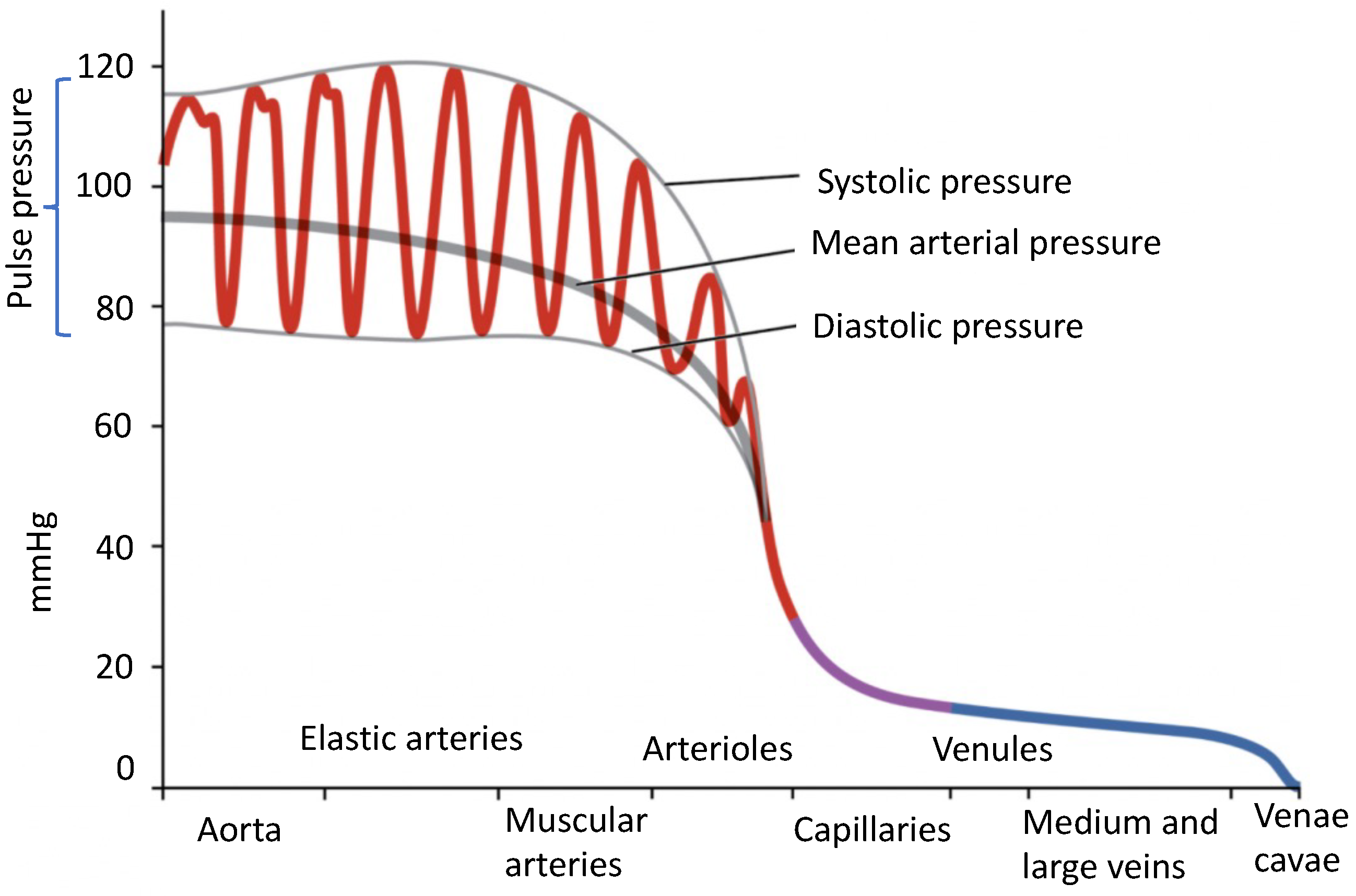
2.1. Blood Pressure Definition

Blood flows throughout the body due to the pressure difference in the arterial system [26]. BP assesses the mechanical function of the heart, acting as a pump. It is the force per unit area of the arterial system, commonly measured in millimeters of mercury (mmHg). In healthy subjects, the heart contracts between 60 and 100 times per minute, resulting in a pulsatile and almost periodic pressure wave in the arterial system. The pressure wave’s maximum occurs during systole when cardiac contraction exerts its maximum pressure on the blood and arterial walls. This is when blood is pumped from the heart into the arteries. The lowest pressure corresponds to diastole, when the myocardium relaxes, allowing the chambers to refill with blood [27]. Therefore, arterial BP fluctuates between the maximum and minimum levels and gradually decreases to zero as it reaches the end of the circulatory system, as shown in Figure 1 [28]. Throughout this work, we study the following BP parameters:
  • Systolic blood pressure (SBP), which represents the maximum pressure inside the arteries during cardiac contraction;
  • Diastolic blood pressure (DBP), which represents the minimum arterial pressure during cardiac rest;
  • Mean Arterial Pressure (MAP), an empirical weighted average of SBP and DBP used to approximate the average BP over the cardiac cycle using only its maximum and minimum values [29]:
    MAP = SBP + 2 × DBP 3
    where the higher weight of the DBP empirically accounts for the asymmetry of the continuous BP (see Figure 2).

2.2. Factors Impacting Blood Pressure

Arterial BP values are subjective and depend on many factors, including the physics and physiology of the body; body position; brain activities; digestive activities; muscle activities; nerval stimulations; environmental factors (air temperature and audio noise level); smoking; alcohol and coffee consumption; and medications [30,31]. There are also various biological factors that impact BP [32], as outlined below.
Figure 2. The auscultatory method measures blood pressure based on Korotkoff sounds. Note the difference between the actual and reported systolic/diastolic blood pressures; adapted from [32] (CC-BY-3.0).
Sensors 24 01730 g002

2.2.1. Cardiac Output

Cardiac output is the amount of blood pumped into the ventricles by the heart. BP and blood flow increase when factors such as sympathetic stimulation, epinephrine and norepinephrine, thyroid hormones, and increased calcium ion levels increase cardiac output (heart rate, stroke volume, or both). Conversely, factors that decrease heart rate, stroke volume, or both, such as parasympathetic stimulation, increased or decreased levels of potassium ions, decreased calcium levels, anoxia, and acidosis, decrease cardiac output [30,32].

2.2.2. Compliance

Compliance is the ratio of the change in volume to the change in pressure applied to a vessel [33]. Arterial compliance has a direct relationship with its efficiency. There is an inverse relationship between BP and the compliance of blood vessels. When vascular diseases cause artery stiffening, compliance decreases, and the heart works harder to push blood through the stiffened arteries, resulting in an increase in BP [32].

2.2.3. Blood Volume

The total amount of blood in the body directly affects blood flow and blood pressure. If blood volume decreases due to bleeding, dehydration, vomiting, severe burns, or certain medications, BP and blood flow also decrease. However, the body’s regulatory mechanisms are efficient in controlling BP, and symptoms may not appear until 10-20% of blood volume is lost. Hypovolemia can be treated with intravenous fluid replacement, but the underlying cause must also be addressed to restore homeostasis in these patients. Intravenous fluid replacement is typically part of the treatment [32,34].

2.2.4. Blood Viscosity

Blood viscosity refers to the fluid’s thickness (resistance to blood flow). Blood viscosity is inversely proportional to flow and directly proportional to resistance. As a result, any factor that increases blood viscosity raises the resistance and lowers the flow. In contrast, factors that decrease viscosity increase flow and lower the resistance. Blood viscosity typically does not vary over short time intervals. Plasma proteins and the formed elements are the two primary factors influencing blood viscosity. Any condition that affects the number of plasma constituents, such as red blood cells, can change viscosity [32]. Since the liver produces most plasma proteins, liver impairments or dysfunctions such as hepatitis, cirrhosis, alcohol damage, and drug toxicity can also alter viscosity and decrease blood flow [35].

2.2.5. Blood Vessel Length and Diameter

The resistance and length of vessels are directly related. Longer vessels have more resistance and lower flow. A higher surface area of the vessel makes it harder for blood to flow through. Similarly, shorter vessels have smaller resistance, resulting in higher flow. The length of blood vessels grows with age, but they tend to stabilize and remain constant in length during adulthood under normal physiological conditions [30,32]. The diameter of blood vessels differs depending on their type and can change throughout the day in response to chemical and neural signals. Unlike vessel length, vessel diameter is inversely related to resistance. Intuitively, a vessel with a larger diameter allows blood to flow with less friction and resistance (even with the same blood volume), because the blood has less contact with the vessel walls [32,36].

2.3. Blood Pressure Norms

In 2018, the Journal of the American Heart Association (AHA) published guidelines for the prevention, detection, evaluation, and management of BP [37]. Table 1 lists the BP ranges for adults in four categories. BP values for children and adolescents are generally lower than those for adults, and they gradually rise with age [38,39]. Hypertension (abnormally high BP) is characterized by a continuous elevation of BP values above the normal range. On the other hand, hypotension (abnormally low BP) occurs when BP values are below normal ranges. Hypotension can happen due to sudden blood loss or a decrease in blood volume, and hypertension is linked to an increased risk of various forms of CVDs [37].

3. Blood Pressure Measurement Methods

BP readings are also impacted by the measurement technology utilized, measurement setup, patient conditions, and time. We study these factors in the sequel to this paper.

3.1. Invasive Blood Pressure Measurement

In this type of measurement, a catheter—a thin tube—is inserted into a vessel to access blood for laboratory testing and to measure arterial blood pressure using a pressure transducer consisting of a delicate and sensitive diaphragm. The transducer’s resistance varies with the slightest pressure changes, allowing for the detection of BP fluctuations [16]. This technique is utilized to record and monitor changes in BP [40].
In modern healthcare facilities, disposable pressure transducers are commonly utilized for more precise and continuous measurement of BP in specialized settings, such as cardiac catheterization labs, intensive care units (ICUs), and operating rooms. Although pressure transducers can measure intracranial and intra-abdominal pressures, they are most frequently used for invasive monitoring of arterial and venous BP. Invasive methods of monitoring BP can be generally classified into two categories [41]: intravascular, where the pressure sensor is inserted into the vessel at the tip of the catheter, and extra-vascular, where the pressure sensor is located outside the vessel and along the catheter’s end.

3.2. Non-Invasive Blood Pressure Measurement

Non-invasive BP measurement techniques determine BP without any physical injury to the body. This technique is classified into two groups: cuff-based and cuff-less methods. Cuff-less methods are still in the developmental stage and are uncommon in clinical studies. Therefore, the scope of the current survey is on cuff-based methods.
All commercially available cuff-based BP measurement technologies consist of a manometer (digital or analog), a pressure pump (manual or automatic), and a cuff. The basic principle of these devices is to apply sufficient pressure to the extremities (arm, wrist, or leg) to temporarily block the blood flow through the artery. The cuff is then slowly deflated, and the pressure is reduced until the blood begins to flow through the artery. At this point, the pressure in the cuff is close to the SBP—the peak of BP (Figure 2). Since BP oscillates through the cardiac cycle, it repeatedly falls below the external pressure, which results in repeated obstruction of blood flow (which can be heard through a stethoscope or sensed via a measurement device). The cuff pressure is then further reduced until blood flows through the artery with no obstruction, where the pressure in the cuff drops below the DBP, and no further sounds are heard by the physician (or sensed by the automatic analysis hardware/software). In Figure 2, we can see how the actual and reported BP values can deviate, especially for the DBP.
There are various methods for identifying the moments when the cuff pressure is equal to the SBP and DBP. The main difference between these methods lies in their ability to accurately detect the external-internal pressure balance points [26]. These methods are outlined below.

3.2.1. Auscultatory

This method dates back to the late 18th century and remains the gold standard for validating novel BP measurement methods [42]. It is based on Korotkoff sounds, which are produced by the turbulent flow of blood through the compressed artery. As the cuff pressure is slowly released, the artery begins to open, and its pressure exceeds the cuff pressure. The pressure that the manometer displays when the first Korotkoff sound is heard corresponds to the systolic BP. As the cuff pressure reduces, the blood flows turbulently in the artery, and the sound continues until the pressure of the cuff reaches the lowest arterial pressure. At this moment, the Korotkoff sound disappears, and the corresponding manometer pressure is the diastolic BP, as shown in Figure 2 [42,43].
Initially, BP was measured using mercury sphygmomanometers, but the toxicity of mercury led to the adoption of aneroid sphygmomanometers, which use a mechanical pressure gauge that is calibrated to display the pressure readings. The precision of the aneroid sphygmomanometer depends on the operator’s proficiency in auscultation, use of a stethoscope, and visual acuity [42]. More recently, hybrid sphygmomanometers have been used to automate the detection of Korotkoff sounds and the display of the SBP, DSP, and pulse rate on digital monitors [44]. Notably, aneroid sphygmomanometers have varying accuracy across manufacturers. Over the past decade, surveys have been conducted to assess the accuracy of these devices. The results demonstrate that the BP readings from different manufacturers have significant deviations ranging from 1 to 44% [42,45,46,47]. Additionally, using a small gauge to read the pressure is another potential source of bias in these devices [42]. Therefore, mercury sphygmomanometers remain popular in clinical settings.

3.2.2. Oscillometric

This method is currently the most popular technology for automated BP measurement devices [13]. Using a pressure sensor, it measures the pulsatile BP in the artery during cuff inflation and deflation [11]. In this technology, the transducer detects the small variations in arterial pressure oscillations or the intra-cuff pressure, produced by the changes in pulse volume due to the heartbeats [13]. The oscillations begin when the cuff pressure exceeds the systolic BP and continue until it is lower than the diastolic BP. A microcontroller is used to control the inflating process, reading the analog output signal of the pressure sensor and its digitization. The micro-variations of the sensed pressure are filtered and pre-processed to obtain the mean arterial pressure, as defined in (1) [13]. Due to the indirect nature of this approach, the measured pressure value requires calibration to be mapped to the actual systolic and diastolic BPs. The microcontroller displays the measured BPs on a local screen [42].
Some of the advantages of the oscillometric method are as follows [13,42]: (1) ease of use for patients (placement and removal of the cuff); (2) ease of calibration (commonly via a button on the device); (3) portability; and (4) use with minimal training. The negative aspect of this method is that commercial oscillometric devices use different (and commonly proprietary) algorithms to estimate BP from the measurements [48]. This results in an intrinsic source of bias in BP measurements among devices from different manufacturers. It is important to note that although these algorithms have been validated in adult populations, there is a lack of validation in the youth population.

3.2.3. Ultrasound

This technique is based on the Doppler effect [13]. Similar to the previous methods, cuff inflation blocks the blood flow in the artery. Then, as the cuff deflates, the arterial wall starts to move at the systolic BP and produces a Doppler phase shift in the reflected ultrasound. As the arterial motion decreases and reaches its endpoint, the cuff pressure at that moment is considered the diastolic BP [42]. The detection of the onset and offset of arterial wall motions is performed by processing the Doppler reflection using a local (digital) processor.

4. Cuff-Based Blood Pressure Measurement Technologies

As with all medical equipment, BP measurement devices must meet regulatory requirements and standards. In this section, the essential BP measurement and validation standards of commercial cuff-based BP monitors are reviewed.

4.1. BP Measurement Device Standards

There are different standards for validating BP measurement devices. In 1987, the Association for the Advancement of Medical Instrumentation (AAMI) published the first standard for non-invasive BP medical devices [49]. In 1990, the British Hypertension Society (BHS) established another clinical protocol for validating these devices, which included many of the AAMI standards [50,51]. These parallel standards continued until 2018 when the AAMI, the European Society of Hypertension (ECH), and the International Organization for Standardization (IOS) published a universal protocol named the “single universal standard”, also referred to as ISO 81060-2:2018/Amd 1:2020 on “non-invasive sphygmomanometers”, [13,52]. This unified standard facilitated the validation and comparison of measurements made by BP devices manufactured globally.
As part of this standard, manufacturers are required to collect a database of measurements from at least 85 individuals, with each individual being measured at least three times. Therefore, at least 255 records are required to validate BP devices [26]. Moreover, the mean absolute error between the recorded BP values and the reference technology should be less than 5 mmHg, and the standard deviation of the multiple measurements should be less than 8 mmHg. The standard also categorizes the cumulative absolute error of BP measurement devices into three groups: less than 5 mmHg, 5 mmHg to less than 10 mmHg, and 10 mmHg to less than 15 mmHg. Devices can only pass the certification test if at least 85 percent of the reported results based on the noted criteria are less than 10 mmHg [13]. The reference BP measurement technology is the invasive BP measurement technique (Section 3.1), but it is also acceptable to compare the BP results with any non-invasive measurement method with a maximum error of 1 mmHg [13].

4.2. Commercial Cuff-Based BP Measurement Devices

Commercial BP devices can be categorized into three groups: ambulatory BP monitors (ABPMs), office BP (OBP) monitors, and home BP monitors (HBPMs).
The ABPM is a portable monitor that is carried by individuals with hypertension (or those who are at a higher risk of developing hypertension) for a period of 24 or 48 h while engaging in their regular daily activities and during sleep. Based on the physician’s required settings, the device measures the patient’s BP at specific time intervals, e.g., 15 or 30 min. After the required period, the patient returns to the clinical center, the device is taken off the patient, and the BP data are transferred to the computer or cloud and analyzed using software [26]. ABPMs are most commonly used for detecting non-dipping BP patterns, which refer to a phenomenon where an individual’s BP fails to exhibit the normal nocturnal decrease during sleep, potentially indicating an elevated risk of cardiovascular issues [53].
OBP monitors are the most common type of BP measurement device for clinical use. Their accuracy is critical, especially in emergency and surgical units, where physicians make essential real-time decisions from the values of the BP and other vital signs. In these situations, the physician may not have adequate time to repeat BP measurement (as advised by BP reading standards). These BP devices are either integrated into bedside monitors or used as discrete devices similar to HBPMs.

4.3. Standard Blood Pressure Measurement Conditions

Several guidelines have been published to improve the accuracy of BP measurement devices by standardizing BP acquisition procedures. Although there are different recommendations in different countries and organizations, they typically address the same fundamental issues [54]. Figure 3 illustrates the basic principles of BP measurement [55]. The following are some of the common items found in BP measurement guidelines:
  • To maintain a stable BP measurement environment, it is recommended to refrain from opening and closing windows and doors. [42].
  • The temperature and relative humidity of the BP measurement environment should be in the range of 15–25 °C and 20–85%, respectively [42].
  • BP should be measured in a quiet environment [55,56].
  • The patient should not smoke, eat, or drink for at least 30 min before measuring [55].
  • The patient should have adequate rest time before the measurement to stabilize BP.
  • The patient should not speak and should remain quiet during the measurement [55].
  • The patient should sit on a chair with back and arm supports and without crossing their legs [56].
  • The patient’s arm should be placed and remain at the same level as the heart throughout the BP measurement [42].
  • An appropriate cuff should be used for measuring according to AHA guidelines (Table 2) [42].
  • The antecubital fossa (the area between the anatomical arm and the forearm) should be 2-3 cm above the lower end of the cuff [57].
  • During the measurement, the patient’s feet should remain flat on the floor [55].
  • Measuring BP should be performed using direct contact of the cuff with the upper part of the arm (not over sleeves) [56].
  • It is recommended to take three BP measurements with one-minute intervals in between. The average of the results should be reported as the BP value [55,56].

5. Potential Sources of Bias in Blood Pressure Technologies

Patient positions or acquisition circumstances that do not meet the measurement guidelines may potentially result in BP measurement biases (over- or under-estimation) and misdiagnosis [48,54,58]. We study the potential sources of biases under three categories [48,59].

5.1. Biases Related to Blood Pressure Measurement Devices

The BP measurement device is the first source of measurement bias. BP devices comprise the main measurement unit and the consumable parts, as detailed below.

5.1.1. Main Blood Pressure Measurement Unit

Biases related to the main BP measurement unit are known as systematic errors. Manufacturers report the acceptable range of measurement errors in the user manuals of BP measurement devices. Measurements falling outside of this range are considered unacceptable, indicating that the device needs calibration. Therefore, they require maintenance and regular calibration to identify and reduce measurement uncertainties to an acceptable level. Medical instruments comprise numerous electro-mechanical elements that undergo natural aging and wear and are impacted by microscopic airborne contaminants that accumulate on their sensitive electronic elements and sensors. These effects change the electro-mechanical characteristics of the devices, even if they are not used, resulting in a gradual drift from their nominal operating point. The deviations of the device elements contribute to measurement biases at the system level. It should be highlighted that the effects of device aging are not the same as the failure of the elements or the device. Therefore, even though a BP device may appear new or be fully functional, it may deviate from the original calibration point. Hence, the regular calibration of medical devices is an essential requirement that should not be compromised. It is important to note that systematic errors, such as changes in the cuff deflation rate [60], are not mitigated by averaging, as they tend to introduce a constant bias into the reported values (unknown to the user) [61]. The identification and correction of systematic errors are performed using reference instruments that have been well maintained and calibrated by medical instrumentation experts.
Another source of systematic error arises from the software/algorithm used to calculate BP from the pressure sensor measurements. The lack of calibration for the electronic pressure sensors can result in systematic errors [61,62]. For example, in automatic BP measurement devices, the rate of cuff inflation and deflation is significant because the isometric exercise involved in inflating the cuff causes a temporary elevation of about 10 mmHg. Although this only takes around 20 s, if the cuff is deflated too quickly, the pressure may not have returned to baseline, resulting in a falsely high systolic pressure [48]. Other factors contributing to bias include sensor accuracy and the software/firmware logic [42]. Moreover, the cuff’s deflation rate, or ‘bleed rate’, is assumed to be consistent when the algorithm calculates the systolic and diastolic pressures [42], which may not hold in practice.
The WHO regularly publishes and updates guidelines for standard practices to ensure that healthcare providers are cognizant of calibration requirements and potential biases to provide quality healthcare [63]. The process of calibrating medical equipment is different for each device. Most devices have built-in mechanisms for calibration. Manufacturers also advise regular maintenance based on the rate of utilization. Automatically calibrated devices can be used within predefined tolerances, beyond which they should be returned to the manufacturer for technical inspection or disposed of.

5.1.2. Consumables of Blood Pressure Measurement Devices

All BP devices have consumable components that require regular replacement. This includes batteries, rubber tubing, hoses, fittings, and cuffs [13]. For example, since the BP device consumes variable power throughout the inflation-deflation cycle, weak batteries may result in erroneous BP values. Worn cuffs and rubber tubing with weak elasticity also impact the reported BP values. Consumable parts of BP devices should, therefore, be replaced according to the manufacturers’ recommended timelines.

5.2. Subject-Specific Biases

BP values can significantly fluctuate across subjects [64]. In the following section, we list some of the most important sources of subject-specific bias.

5.2.1. Demographic Features

Demographic features, such as sex, age, race, and genetic background, influence BP measurements due to variations in physiology and anatomy.
Sex is a prominent factor that influences BP [65,66]. Generally, males tend to have higher BP levels, which can be associated with differences in hormonal activities and anatomical differences between male and female bodies [66,67]. A notable study involving 32,833 individuals ranging from 5 to 98 years (54% women) was conducted over four decades [68]. The study showed that women exhibited a steeper increase in BP measurements compared to men, starting as early as the third decade of life and continuing throughout their lifespan [68]. Table 3 presents a compilation of studies reporting BP values based on sex.
Age is another significant demographic feature. Arterial stiffness is known to increase with age, leading to reduced arterial compliance and an increase in pulse pressure [57]. Elderly individuals may experience systolic hypertension, characterized by elevated SBP without a corresponding increase in DBP. This condition, commonly known as “pseudohypertension”, has been linked to a decline in arterial distensibility, which can lead to bias and inaccurately high BP readings, as the external cuff pressure on the artery is reduced [57]. Carrico et al. examined the relationship between age, sex, and BP in a group comprising 965 men and 1114 women [87]. Their findings indicated that BP values generally increase with age but decrease again after approximately 70 years of age. In most age groups, BP is higher in males than in females, but both sexes have similar BP until their teenage years. However, after about 70 years of age, females’ SBP values surpass those of males, whereas their DBP values remain approximately the same.
There has also been significant interest in examining health measures across racial groups. Numerous studies have examined the relationship between BP and race [88,89]. These studies have found that specific diseases are more common in certain racial groups, suggesting a potential link between race and hypertension prevalence [90,91]. For instance, Staessen et al. [92] showed that Asian populations have a higher rate of non-dipping compared to European populations, indicating possible variations in BP patterns among different races. Furthermore, individuals of African American descent have been observed to have higher rates of hypertension, cardiovascular, and cerebrovascular morbidity and mortality compared to those of European descent [93]. This suggests that race (or, more accurately, genetic factors) may play a significant role in hypertension susceptibility. Another possible explanation is that the experiences of some racial or ethnic groups, including but not limited to the medical care that these groups receive, affect their BP. Most likely, multiple factors contribute to the higher prevalence of hypertension in African Americans, including genetic, socioeconomic, systemic, and other factors. Table 4 summarizes the group of studies that have compared BP values based on race.

5.2.2. Subject-Specific Factors

Height and weight are two essential factors influencing BP values. Studies have shown that taller individuals tend to have lower SBP and higher DBP compared to shorter individuals [85]. Body mass index (BMI)—weight divided by the squared height (kg/m2)—can be used as a compound factor for BP assessment [94,95]. Table 5 presents the findings from various studies that have reported BP values based on BMI, showing an overall positive correlation between BP and BMI [96].

5.2.3. Subject-Specific Background Medical Conditions

Accurate interpretation of BP measurements should consider patient-specific conditions and comorbid factors, which can directly or indirectly influence BP readings. Often, the inherent biological rhythms in the disease process and their potential clinical implications are overlooked or inadequately considered when treating hypertensive patients [53]. In the following section, we discuss several situations where these considerations play a significant role.
Healthy individuals typically show BP variations throughout the day, rising during the day and dropping at night [115]. Most people have nighttime BP readings 10–20% lower than those in the daytime. However, some experience a non-dipping pattern, where nighttime BP drops by less than 10% [116]. About 25% of hypertensive individuals with unknown causes exhibit this non-dipping pattern [117].
The “white-coat effect”, caused by the medical environment and presence of a physician, can elevate a patient’s clinic BP [48]. This effect often results in higher SBP and DBP compared to baseline ambulatory BP [58]. Table 6 summarizes studies comparing BP measurements in settings like the home and office.
Pregnancy-related hemodynamic changes can affect BP readings. Although many automated BP device types exist, few are calibrated for pregnant women, including those with hypertensive disorders [14]. Nearly 10% of pregnant women face high BP, risking both the fetus(es) and the mother. Those with gestational diabetes or preeclampsia are especially at risk [54,120,121].
Obesity correlates with high BP in both children and adults. For each BMI unit increase, SBP and DBP rise by 0.56 and 0.54 mmHg, respectively, in obese children [94]. Table 7 presents studies examining the impact of obesity on children’s BP values.

5.2.4. Eating, Drinking, and Smoking

Eating, drinking, and smoking habits can significantly influence BP values [105,122,123]. Studies indicate that eating has both short-term and long-term effects on BP levels [124].
Postprandial (post-meal) BP initially rises due to increased activity but later drops, particularly due to decreased total peripheral resistance from visceral vasodilation [125]. This reduction is pronounced in elderly, hypertensive individuals and those with autonomic failure [126]. High-carbohydrate meals cause a larger decrease in BP compared to high-fat ones. Although the exact mechanism for post-meal hypotension remains unclear, impacting factors may include impaired baroreceptor reflexes, insulin-induced vasodilation, and the release of vasodilatory gastrointestinal polypeptides [127]. This BP decrease peaks around 1 hour post-meal and can last over 2 hours, influencing diurnal BP changes, especially in elderly subjects with hypertension [125]. Furthermore, BP effects from alcohol, caffeine, and nicotine vary by dose and individual [58].
For the long-term impacts of eating habits on BP, guidelines primarily recommend lifestyle and dietary changes to manage hypertension, with an emphasis on reducing salt intake. Adding potassium-rich foods like nuts, fruits, and vegetables to the diet can enhance BP control [128]. Table 8 summarizes studies on the impacts of eating, drinking, or smoking on BP.

5.2.5. Circadian Rhythm

The circadian clock significantly affects CVD risk factors like heart rate and BP [134]. It has been linked to two mechanisms: the central clock in the hypothalamic suprachiasmatic nucleus (SCN) and the peripheral clock in most body tissues and organs [135]. Environmental factors, like physical exercise, can also impact and align circadian rhythms, especially in skeletal muscle [136].
BP displays daily rhythmic fluctuations, peaking in the early morning and dipping around midnight. Boivin et al. [137] examined BP changes in the morning and evening among 52 controlled hypertensive patients. BP was measured six times in both the morning and evening, three times before and after resting. Each session was nine minutes with a minute between measurements and five minutes of rest. The study found significant BP differences between morning and evening of around 5 mmHg for DBP and 8 mmHg for SBP.
Psychological and physical activities also contribute to BP fluctuations, with higher values commonly observed during work hours and lower values at home. While several neurohormonal systems follow a circadian rhythm with a morning peak, the sympathetic nervous system appears to be the primary determinant of these BP circadian variations. However, this principle can be reversed for individuals with specific job roles, such as shift workers [53].
Table 9 provides a comparative analysis of studies exploring the effects of the circadian clock on BP values.

5.3. Biases Related to the Acquisition Session

There are specific guidelines for BP measurement, violations of which can lead to inaccurate readings. Here, we explore the often-overlooked factors during BP acquisition that can introduce bias and compromise measurement reliability.

5.3.1. Seasonal Variations and Ambient Temperature

Seasonal BP fluctuations, with peaks in winter and troughs in summer, have been reported in both home and clinic measurements [138,139]. Although the direct link between BP and ambient temperature has not been conclusively established, consistent seasonal patterns suggest a correlation [140]. Yang et al.’s research on 23,040 individuals in China found BP values to be highest in winter (December–February) and lowest in summer (July–August) [141]. Table 10 summarizes studies comparing BP values by environmental temperature. The short-term impact of temperature on BP underscores the importance of protective measures in colder weather to maintain stable BP and reduce BP-related disease risks [104].

5.3.2. Cuff Position

The brachial artery is commonly used for BP measurement [57,144]. While wrist and finger monitors have become popular, SBP and DBP values differ across the arterial tree [48]. Figure 4 depicts the influence of various body points on BP as blood flows through different arteries when upright. Table 11 summarizes studies comparing BP across different body points.

5.3.3. Body Position

Body position significantly affects BP readings. Guidelines suggest measuring BP in a seated position with back support [59]. Research indicates BP values are higher when seated compared to lying down [81,102,106]. Specifically, sitting upright can increase DBP by up to 6.5 mmHg compared to leaning back [48]. For accurate measurements, the BP cuff should be at the level of the patient’s right atrium, regardless of position [57]. Table 12 summarizes studies on BP values for various body positions.

5.3.4. Arm Position

Proper arm positioning during BP measurements is vital, with the arm ideally supported at heart level on a flat surface [48,102,152]. Shifting the arm from horizontal to vertical can raise the pressure by 5–6 mmHg due to hydrostatic changes [48]. Mariotti et al. explored the effects of arm positioning and postural hypotension during BP assessments [153], finding that incorrect arm positioning while standing resulted in overestimated BP readings. Table 13 summarizes studies on BP values considering different arm positions.

5.3.5. Leg Position

Proper leg positioning during BP measurements is crucial according to guidelines [155]. Studies show that crossed-leg positions yield higher BP readings compared to uncrossed legs or sitting with feet flat [58,156]. Table 14 provides a summary of studies on BP values with varying leg positions, emphasizing the importance of standardized leg positioning for accurate measurements.

5.3.6. Left vs. Right Arm

Clinical settings often show BP reading differences between the left and right arms [158]. Guidelines suggest measuring BP in both arms on the first visit, using the arm with higher readings thereafter [57]. Conditions like coarctation of the aorta or upper-extremity arterial obstruction can cause significant BP variations between arms [57]. Table 15 summarizes studies comparing BP measurements in both arms.

5.3.7. Cuff Size and Tightness

Using the correct cuff size is vital for accurate BP measurement [160]. Small cuffs can overestimate pressure, which is a frequent error [48,161]. Larger cuffs tend to report lower BP values [58]. For children, cuff size selection is challenging. The BHS suggests three sizes based on arm circumference [48]: 4 × 13 cm, 8 × 18 cm, and 12 × 35 cm (adult cuff).
In obese individuals, selecting the right cuff size is essential to ensure that the brachial artery is accurately compressed, yielding reliable BP readings [57]. Many obese individuals have tronco-conical arms, further complicating accurate BP measurement [161]. For these patients, conical cuffs can offer more accurate BP readings [57]. Table 16 presents studies exploring the influence of cuff size on BP readings.
When measuring BP, the cuff should be securely placed around the upper arm without any clothing interference, ensuring even snugness from top to bottom. The tightness can be gauged by fitting one finger easily and two fingers with comfort between the cuff’s top and bottom. Achieving the correct cuff tightness is vital for reliable and consistent BP readings [57].

5.3.8. Rest Period before Measuring BP

Studies have investigated the effects of rest durations before taking BP measurements. Their findings indicate that not resting sufficiently before a measurement can lead to elevated SBP and DBP readings. A rest period ranging from 10 to 16 min showed a modest decrease in SBP and a slight drop in DBP [163,164]. However, the exact duration of rest needed to compensate for the effects of prior physical activity remains uncertain. More research is needed to specify the ideal rest period for precise BP readings [58]. A summary of studies exploring the influence of the duration of rest on BP measurements can be found in Table 17.

5.3.9. Number of Measurements

For many individuals, the first BP reading taken in a clinical setting tends to be higher than the readings that follow. Research has explored this pattern by conducting three consecutive BP measurements [57]. Findings showed that when only the first measurement was considered, approximately 35% of adults exhibited an SBP range of 140–159 mmHg and a DBP range of 90–99 mmHg. However, when the average of all three measurements was taken into account, most participants registered SBP/DBP values below the 140/90 mmHg benchmark [57]. Therefore, relying exclusively on the initial reading can result in over-diagnosing hypertension, emphasizing the necessity of multiple readings to ensure an accurate diagnosis [165].

5.3.10. Clothing

Healthcare professionals are advised to measure BP by fully exposing the cuff on the upper arm. However, it is a common practice to measure BP by rolling up the sleeve or placing the cuff over the sleeves. Table 18 presents the findings from various studies that have examined the impact of clothing on BP values, demonstrating a bias in the BP readings due to clothing.

6. Cuff-Less Blood Pressure Technologies

Significant efforts have been made to develop novel and versatile methods for measuring BP. Traditional BP measurement techniques involving cuffs have several inherent limitations [43], such as (1) time-consuming procedures; (2) patient discomfort due to frequent cuff inflation, as seen in ABPM devices used for recording BP during the day and night [166]; (3) impracticality for continuous BP measurement in specific medical settings such as heart surgical units and acute burns cases; and (4) the necessity of allowing sufficient time between successive BP measurements to enable the blood vessels beneath the cuff to return to their baseline state and to prevent vessel collapse due to cuff pressure. To address these limitations, cuff-less BP measurement methods have been introduced, which can be categorized into three groups [11,26,167]: tonometry, volume clamps, and pulse-wave velocity. These innovative approaches offer the potential to overcome the drawbacks associated with traditional cuff-based BP measurement techniques, paving the way for more efficient and patient-friendly BP monitoring solutions.
The pulse-wave velocity (PWV) method of measuring BP was invented by Moens and Korteweg [40]. They defined a fundamental relationship between the vascular elasticity and pulse-wave velocity in the artery, known as the Moens–Korteweg equation [43,168]:
PWV = E · h ρ · D
where h represents the thickness of the vessel wall, ρ is the blood density, and D is the vessel’s inner diameter. The parameter E denotes Young’s modulus of elasticity, which indicates the vessel wall’s elasticity. Geddes empirically defined E as follows [43]:
E = E 0 e α · BP
where E 0 represents the modulus of elasticity at a pressure of 0 mmHg, α is a constant related to the vessel (typically ranging from 0.016 to 0.018 mmHg−1), e is the Euler number, and BP is the blood pressure. The Moens–Korteweg equation and Geddes’ formulation are fundamental in enabling the utilization of the PWV as a valuable and non-invasive method for assessing BP and vascular elasticity. Combining (2) and (3) leads to a compact relation between BP and PWV:
BP = 1 α ln ρ · D · PWV 2 h · E 0
Several methods are used to measure the PWV. The pulse transit time (PTT) is the most well-known indirect method of calculating the PWV [9]. The relationship between the PTT and PWV is defined as follows [43]:
PWV = d PTT
where d is the distance between the heart and a specific location where the blood flows and PTT is the time it takes for the blood pulse to propagate the distance d. PTT is calculated using different sensors and bio-signals, including [169] photoplethysmography (PPG) and the output signal of the Hall sensor [170]; the PPG signal and the modulated magnetic signature of the blood [171]; PPG and ballistocardiography [172]; PPG and impedance plethysmography [173]; PPG and electrocardiography [174]; or one or two PPG recordings [175].
The parameter d is challenging to accurately determine in practice. Therefore, PWV and subsequently BP in (5) are difficult to accurately determine. To address this challenge, cuff-less BP monitoring devices are increasingly integrating artificial intelligence (AI) algorithms to learn crucial and complex features of the cardiovascular system [11,16]. Ongoing research is focused on exploring how to extract the most relevant features from signals like the PTT to model the cardiovascular system and effectively predict BP values. However, it is essential to acknowledge that these studies are still in the prototype stage and must be validated in large cohorts to meet medical standards before being used in clinical settings [11]. Nonetheless, advances in this domain show promise for enhancing BP monitoring and improving patient care.
When developing cuff-less methods, cuff-based BP databases are typically used as references to demonstrate their “substantial equivalency” with existing technology, as required for regulatory clearance like FDA 510(k) premarket approval [176,177,178,179]. If standard requirements are not met during cuff-based BP acquisition, the calibration models for new technologies will be trained with biased recordings. This affects the long-term reliability of these technologies, potentially causing deviations between predicted and actual BP values. It is essential to address these biases for accurate measurements with cuff-less technologies, warranting further research to enhance their performance and reliability.

7. Future Perspectives: Using Machine Learning for Bias Removal and Individualized BP-Level Risk Assessment

In our study, we investigated BP and various measurement- and subject-related factors that impact the accurate collection and interpretation of BP. However, given the vast BP literature, it is unfeasible to comprehensively examine every potential source of bias. It is important to note that as of 4 February 2024, PubMed alone reports 687,705 instances of the keyword “blood pressure” in its databases (obtained using the Entrez Direct (EDirect) Linux tools from the NCBI [180]). In addition, there are billions of BP records in electronic health records (EHRs) worldwide. The one-by-one investigation of these rich resources is infeasible. We propose that future research can leverage machine learning and natural language processing techniques (especially the recent advances in large language models) to mine these massive data- and literature-driven resources in a more systematic manner. This could result in more conclusive findings regarding subject-specific BP-related risk factors across different demographic groups. Open-source codes for BP-related studies are another requirement. Schwenck et al. recently released a versatile open-source BP analysis and visualization toolkit in R [181]. Beyond visualization tools, rigorous statistical frameworks and models are required for building machine learning pipelines. In the sequel to this paper, we propose a stochastic framework that can be used in future research to use machine learning algorithms in BP-related studies.
The problem of BP measurement can be formulated as follows:
y k = BP k + e k
where y k represents the reported BP values in different trials/sessions, BP k is the ‘true’ BP value, and e k denotes the total measurement errors attributed to nonstandard devices or measurement errors. Both the BP and its error can be considered random variables, with presumed (time-variant) distributions. With this data model, the problem of accurate BP measurement and bias removal can be formulated as a classical estimation problem that could be addressed using standard techniques such as least squares, maximum likelihood, or Bayesian estimation, where the latter two benefit from prior distributions of the BP and measurement errors.
e k is device- and technology-dependent. Complying with standard BP measurement techniques and averaging over multiple trials ( k = 1 , , K ) can mitigate the impact of this term, resulting in a more accurate measurement of BP.
Even with accurate measurements ( e k 0 ), BP k is a random variable, which fluctuates over time and across trials. A comprehensive machine learning framework should benefit from the demographic-specific distributions of BP k , namely:
f ( BP k | p )
which is the conditional distribution of the BP (SBP, DBP, or both) given all the demographic and comorbid factors parameterized by the vector p (sex, race, age, background medical conditions, comorbidities, etc.).
We propose that our current survey of the BP literature—and more systematic future surveys that could benefit from massive EHR registries combined with NLP technologies for BP literature analysis—can be used to estimate f ( BP k | p ) across different demographic factors. As proof of concept, graphical representations of the BP data on sex and BMI from Table 3 and Table 5 are shown in Figure 5 and Figure 6, respectively. In Figure 5, each ellipse corresponds to an individual study from the studied literature, where each ellipse is centered at the mean SBP and DBP, with the horizontal and vertical radii of the ellipses representing the standard deviation of the reported SBP and DBP, respectively. Similar elliptical shapes can be constructed from massive EHR data while accounting for individualized and demographic factors. For multi-site studies, this information can be used to construct multi-modal distributions of f ( BP k | p ) . For instance, assuming that the reported BP values follow a normal distribution (which is an accurate assumption for population-wide studies), the multi-site data can be effectively modeled using Gaussian mixture models (GMMs) to capture underlying clusters or sub-populations [182]. The heatmaps in Figure 5a,b and Figure 6c,d correspond to GMM distributions fitted over the ensemble of the studied BP reports for sex and BMI.
An interesting observation from these figures is that while the literature on BP merely reports the mean and standard deviation of the SBP and DBP, as well as the pulse pressure, there is a trend of correlation between the SBP and DBP, which has not been studied in the BP literature. BP data from EHRs can additionally be used to identify the correlations between SBP and DBP. In recent research, we have calculated the correlation coefficient and other statistical properties of the SBP–DPB distribution on a large cohort of 75 million patient encounters [183].
Future research can integrate these distributions and the data model in (6) to provide Bayesian estimates of BP on a subject-specific basis. These distributions can also be integrated with EHR data and clinical outcomes to train machine learning technologies that quantify the risk of hypertension and BP-related complications of individuals across different demographics (see Figure 7), resulting in a more accurate assessment of BP-based diagnoses [184]. With such technology, we anticipate being able to express a patient’s BP-associated risk using terms such as “A 55-yr-old Hispanic female with a body mass index of 34.2, a consistent in-clinic cuff-based systolic BP > 129 mmHg, a diastolic BP > 85 mmHg while sitting, and a history of diabetes is 15% at risk of stroke in the next 12 years (p-value < 0.05)”.
We envision that this risk index will be integrated with well-established risk scores, such as the Framingham Risk Score (which estimates a person’s 10-year risk of developing cardiovascular disease, considering factors like sex, age, cholesterol levels, and BP) [185] or the HEART Score [186] (which is used in emergency settings to assess the risk of major adverse cardiac events in patients presenting with chest pain based on criteria such as medical history, ECG results, etc.).
Figure 7. A future perspective of blood pressure (BP) monitoring. AI-assisted and ML-based technologies are anticipated to integrate into future generations of BP monitoring technologies. Large electronic health record (EHR) data can be leveraged to extract population-wide norms of BP, accounting for variations due to biological sex, age, and race (from a genetic variability standpoint) [183]. Natural language processing and large language models can assist in analyzing clinical literature and physicians’ notes from EHR data [187]. These data resources can be combined using an ML framework to detect and correct BP measurement biases and provide individualized BP-related risk factors.
Sensors 24 01730 g007
BP measurement devices require maintenance and regular calibration to identify and reduce measurement uncertainties to an acceptable level. The identification and correction of systematic errors are performed using reference instruments that have been well maintained and calibrated by medical instrumentation experts. The process of calibrating medical equipment varies for each device. While integrating AI and ML techniques into current BP devices may not directly reduce systematic errors related to the primary BP measurement unit, envisioning the future of BP technologies suggests promising possibilities for minimizing such errors. In the future, BP devices could be equipped with integrated technologies designed to significantly enhance accuracy and address systematic errors. This could involve integrating online technologies that compare an individual’s BP with their historical records, clinical data, and broader population-wide information.
Integrating AI and ML techniques into the next generation of BP devices would require substantial computing capabilities. A possible technological advancement is to implement low-computation versions of machine and deep learning models in the software/firmware of BP devices or to link them with mobile phones to process and transmit the BP data to cloud platforms. The former would require more powerful processors and/or tensor flow units (TPUs) in BP devices, enabling the implementation of recent technologies such as TinyML and TensorFlow Lite on edge devices. Pre-trained models may also be integrated into the BP device’s software or released as mobile applications for efficient and accurate BP assessment without extensive computing power within the device itself.
Incorporating AI and ML into BP measurement technologies also promises enhanced accuracy and personalized monitoring. By utilizing large BP record databases and state-of-the-art machine and deep learning models, these algorithms aim to reduce biases, leading to precise BP monitoring and tailored cardiovascular risk assessments. These models can be provided with inputs like measurement setup (patient position, rest condition), environmental factors, and individual-specific details (sex, age, BMI, comorbidities, etc.), complemented by population-wide data. AI/ML models can be trained to utilize these inputs to improve precision in BP measurements and risk assessment.

8. Conclusions

BP is a crucial vital sign for monitoring health and making clinical decisions. In recent years, the adoption of cuff-based BP technologies has surged, primarily driven by the growing popularity of ambulatory and home-based BP monitoring, as well as its widespread use in medical centers and hospitals. Accurate BP values are essential, as discrepancies between reported and actual BP measurements can lead to misinterpretation and mistreatment. In this study, we highlighted the notion of “bias” and the factors that may result in bias in cuff-based BP monitoring across various social groups. This survey demonstrated that reported BP values can be diverse due to individualized and demographic factors or inaccurate due to a failure to adhere to BP measurement standards. Given these limitations, the development of a new generation of cuff-based BP devices supported by artificial intelligence (AI) and machine learning (ML) techniques is in high demand. Integrating AI and ML techniques into these devices is a promising approach to identifying and correcting bias, as well as customizing normal and abnormal BP ranges on an individualized level, thereby improving the accuracy of BP measurements and clinical decision making. In conclusion, addressing the issue of bias in cuff-based BP devices is essential for advancing patient care and ensuring reliable BP data for medical decision making. Developing ML-based devices that can detect and correct bias will undoubtedly be a valuable contribution to the field, enhancing the overall effectiveness and reliability of BP monitoring systems. By focusing on improving BP monitoring precision, we can significantly improve patient outcomes.

Author Contributions

Conceptualization, S.S.M. and R.S.; methodology, S.S.M. and R.S.; software, S.S.M.; validation, S.S.M., R.S. and M.A.R.; formal analysis, S.S.M. and R.S.; investigation, S.S.M.; resources, R.S. and G.D.C.; data curation, S.S.M.; writing—original draft preparation, S.S.M.; writing—review and editing, S.S.M., R.S., M.A.R. and G.D.C.; visualization, S.S.M. and R.S.; supervision, R.S., M.A.R. and G.D.C.; project administration, R.S.; funding acquisition, R.S. All authors have read and agreed to the published version of the manuscript.

Funding

This research received no external funding.

Institutional Review Board Statement

The Emory University IRB approved this study under the study protocol number STUDY00006568.

Informed Consent Statement

Not applicable.

Data Availability Statement

Not applicable.

Conflicts of Interest

The authors declare no conflicts of interest.

References

  1. World Health Organization. Cardiovascular Diseases (CVDs). 2021. Available online: https://www.who.int/news-room/factsheets/detail/cardiovascular-diseases-(cvds) (accessed on 21 June 2021).
  2. Ahmad, F.B.; Anderson, R.N. The leading causes of death in the US for 2020. JAMA 2021, 325, 1829–1830. [Google Scholar] [CrossRef]
  3. Leonardi-Bee, J.; Bath, P.M.; Phillips, S.J.; Sandercock, P.A. Blood Pressure and Clinical Outcomes in the International Stroke Trial. Stroke 2002, 33, 1315–1320. [Google Scholar] [CrossRef]
  4. Lawes, C.M.; Bennett, D.A.; Feigin, V.L.; Rodgers, A. Blood Pressure and Stroke. Stroke 2004, 35, 776–785. [Google Scholar] [CrossRef]
  5. Vasan, R.S.; Larson, M.G.; Leip, E.P.; Evans, J.C.; O’Donnell, C.J.; Kannel, W.B.; Levy, D. Impact of High-Normal Blood Pressure on the Risk of Cardiovascular Disease. N. Engl. J. Med. 2001, 345, 1291–1297. [Google Scholar] [CrossRef]
  6. Zhou, B.; Bentham, J.; Cesare, M.D.; Bixby, H.; Danaei, G.; Cowan, M.J.; Paciorek, C.J.; Singh, G.; Hajifathalian, K.; Bennett, J.E.; et al. Worldwide trends in blood pressure from 1975 to 2015: A pooled analysis of 1479 population-based measurement studies with 19·1 million participants. Lancet 2017, 389, 37–55. [Google Scholar] [CrossRef]
  7. Guyenet, P.G. The sympathetic control of blood pressure. Nat. Rev. Neurosci. 2006, 7, 335–346. [Google Scholar] [CrossRef]
  8. Brenner, B.M.; Garcia, D.L.; Anderson, S. Glomeruli and Blood Pressure: Less of One, More the Other? Am. J. Hypertens. 1988, 1, 335–347. [Google Scholar] [CrossRef]
  9. Mukkamala, R.; Hahn, J.O.; Inan, O.T.; Mestha, L.K.; Kim, C.S.; Töreyin, H.; Kyal, S. Toward ubiquitous blood pressure monitoring via pulse transit time: Theory and practice. IEEE Trans. Biomed. Eng. 2015, 62, 1879–1901. [Google Scholar] [CrossRef] [PubMed]
  10. Kalehoff, J.P.; Oparil, S. The Story of the Silent Killer. Curr. Hypertens. Rep. 2020, 22, 1–14. Available online: https://link.springer.com/article/10.1007/s11906-020-01077-7 (accessed on 27 August 2020). [CrossRef] [PubMed]
  11. Rastegar, S.; GholamHosseini, H.; Lowe, A. Non-invasive continuous blood pressure monitoring systems: Current and proposed technology issues and challenges. Phys. Eng. Sci. Med. 2020, 43, 11–28. [Google Scholar] [CrossRef] [PubMed]
  12. Tholl, U.; Forstner, K.; Anlauf, M. Measuring blood pressure: Pitfalls and recommendations. Nephrol. Dial. Transplant. 2004, 19, 766–770. [Google Scholar] [CrossRef]
  13. World Health Organization. WHO Technical Specifications for Automated Non-Invasive Blood Pressure Measuring Devices with Cuff. 2020; World Health Organization: Geneva, Switzerland, 2020; pp. 1–68. [Google Scholar]
  14. Van Den Heuvel, J.F.; Lely, A.T.; Franx, A.; Bekker, M.N. Validation of the iHealth Track and Omron HEM-9210T automated blood pressure devices for use in pregnancy. Pregnancy Hypertens. 2019, 15, 37–41. [Google Scholar] [CrossRef]
  15. Pickering, T.G.; Shimbo, D.; Haas, D. Ambulatory Blood-Pressure Monitoring. N. Engl. J. Med. 2006, 354, 2368–2374. [Google Scholar] [CrossRef]
  16. Ding, X.R.; Zhao, N.; Yang, G.Z.; Pettigrew, R.I.; Lo, B.; Miao, F.; Li, Y.; Liu, J.; Zhang, Y.T. Continuous blood pressure measurement from invasive to unobtrusive: Celebration of 200th birth anniversary of Carl Ludwig. IEEE J. Biomed. Health Inform. 2016, 20, 1455–1465. [Google Scholar] [CrossRef]
  17. Sewell, K.; Halanych, J.H.; Russell, L.B.; Andreae, S.J.; Cherrington, A.L.; Martin, M.Y.; Pisu, M.; Safford, M.M. Blood pressure measurement biases in clinical settings, Alabama, 2010–2011. Prev. Chronic Dis. 2016, 13, E01. [Google Scholar] [CrossRef]
  18. Hoffman, K.M.; Trawalter, S.; Axt, J.R.; Oliver, M.N. Racial bias in pain assessment and treatment recommendations, and false beliefs about biological differences between blacks and whites. Proc. Natl. Acad. Sci. USA 2016, 113, 4296–4301. [Google Scholar] [CrossRef] [PubMed]
  19. Lee, P.; Le Saux, M.; Siegel, R.; Goyal, M.; Chen, C.; Ma, Y.; Meltzer, A.C. Racial and ethnic disparities in the management of acute pain in US emergency departments: Meta-analysis and systematic review. Am. J. Emerg. Med. 2019, 37, 1770–1777. [Google Scholar] [CrossRef] [PubMed]
  20. Valbuena, V.S.M.; Merchant, R.M.; Hough, C.L. Racial and ethnic bias in pulse oximetry and clinical outcomes. JAMA Intern. Med. 2022, 182, 699–700. [Google Scholar] [CrossRef] [PubMed]
  21. Obermeyer, Z.; Powers, B.; Vogeli, C.; Mullainathan, S. Dissecting racial bias in an algorithm used to manage the health of populations. Science 2019, 366, 447–453. [Google Scholar] [CrossRef] [PubMed]
  22. Reyna, M.A.; Nsoesie, E.O.; Clifford, G.D. Rethinking algorithm performance metrics for artificial intelligence in diagnostic medicine. JAMA 2022, 328, 329–330. [Google Scholar] [CrossRef] [PubMed]
  23. Vokinger, K.N.; Feuerriegel, S.; Kesselheim, A.S. Mitigating bias in machine learning for medicine. Commun. Med. 2021, 1, 25. [Google Scholar] [CrossRef]
  24. Magder, S. The meaning of blood pressure. Crit. Care 2018, 22, 257. [Google Scholar] [CrossRef] [PubMed]
  25. Iaizzo, P.A. Handbook of Cardiac ANATOMY, Physiology, and Devices; Springer Science & Business Media: Berlin/Heidelberg, Germany, 2010. [Google Scholar]
  26. Mousavi, S.S.; Charmi, M.; Firouzmand, M.; Hemmati, M.; Moghadam, M.; Ghorbani, Y. Designing and Manufacturing a Mobile-Based Ambulatory Monitoring of Blood Pressure Using Electrocardiogram and Photoplethysmography Signals. Master’s Thesis, Zanjan University, Zanjan, Iran, 2018. [Google Scholar]
  27. Lowry, M.; Ashelford, S. Assessing the Pulse Rate in Adult Patients. Nurs. Times 2015, 111, 18–20. Available online: https://www.nursingtimes.net/clinical-archive/assessment-skills/assessing-the-pulse-rate-in-adult-patients-31-08-2015/ (accessed on 11 September 2015).
  28. Betts, J.; Desaix, P.; Johnson, E.; Johnson, J.; Korol, O.; Kruse, D.; Poe, B.; Wise, J.; Womble, M.; Young, K. Anatomy and Physiology; OpenStax: Houston, TX, USA, 2013. [Google Scholar]
  29. Vlachopoulos, C.; O’Rourke, M.; Nichols, W.W. McDonald’s Blood Flow in Arteries, 6th ed.; Hodder Arnold: London, UK, 2011. [Google Scholar] [CrossRef]
  30. Lapum, J.L.; Verkuyl, M.; Garcia, W.; St-Amant, O.; Tan, A. Vital Sign Measurement across the Lifespan—2nd Canadian Edition; Ryerson University: Toronto, ON, Canada, 2021; Available online: https://pressbooks.library.torontomu.ca/vitalsign/ (accessed on 21 January 2021).
  31. Sapra, A.; Malik, A.; Bhandari, P. Vital sign assessment. In StatPearls [Internet]; StatPearls Publishing: Treasure Island, FL, USA, 2021. [Google Scholar]
  32. DeSaix, P.; Betts, G.J.; Johnson, E.; Johnson, J.E.; Oksana, K.; Kruse, D.H.; Poe, B.; Wise, J.A.; Young, K.A. Anatomy & Physiology (OpenStax), 2013. Available online: https://assets.openstax.org/oscms-prodcms/media/documents/AnatomyandPhysiology-OP.pdf (accessed on 20 April 2022).
  33. Glasser, S. Vascular compliance and cardiovascular disease: A risk factor or a marker? Am. J. Hypertens. 1997, 10, 1175–1189. [Google Scholar] [CrossRef] [PubMed]
  34. Bazett, H. The time relations of the blood-pressure changes after excision of the adrenal glands, with some observations on blood volume changes. J. Physiol. 1920, 53, 320. [Google Scholar] [CrossRef] [PubMed]
  35. Letcher, R.L.; Chien, S.; Pickering, T.G.; Sealey, J.E.; Laragh, J.H. Direct relationship between blood pressure and blood viscosity in normal and hypertensive subjects. Role of fibrinogen and concentration. Am. J. Med. 1981, 70, 1195–1202. [Google Scholar] [CrossRef] [PubMed]
  36. Jeppesen, P.; Sanye-Hajari, J.; Bek, T. Increased blood pressure induces a diameter response of retinal arterioles that increases with decreasing arteriolar diameter. Invest. Ophthalmol. Vis. Sci. 2007, 48, 328–331. [Google Scholar] [CrossRef] [PubMed]
  37. Whelton, P.K.; Carey, R.M.; Aronow, W.S.; Casey, D.E.; Collins, K.J.; Himmelfarb, C.D.; DePalma, S.M.; Gidding, S.; Jamerson, K.A.; Jones, D.W.; et al. 2017 ACC/AHA/AAPA/ABC/ACPM/ AGS/APhA/ASH/ASPC/NMA/PCNA Guideline for the Prevention, Detection, Evaluation, and Management of High Blood Pressure in Adults. J. Am. Coll. Cardiol. 2018, 71, e127–e248. [Google Scholar] [CrossRef] [PubMed]
  38. Azegami, T.; Uchida, K.; Tokumura, M.; Mori, M. Blood Pressure Tracking From Childhood to Adulthood. Front. Pediatr. 2021, 9, 785356. [Google Scholar] [CrossRef]
  39. Falkner, B.; Gidding, S.S.; Baker-Smith, C.M.; Brady, T.M.; Flynn, J.T.; Malle, L.M.; South, A.M.; Tran, A.H.; Urbina, E.M. Pediatric Primary Hypertension: An Underrecognized Condition: A Scientific Statement From the American Heart Association. Hypertension 2023, 80, e101–e111. [Google Scholar] [CrossRef]
  40. Cole, E. Measuring central venous pressure. Nurs. Stand. 2007, 22, 40–42. [Google Scholar] [CrossRef]
  41. Webster, J. Medical instrumentation: Application and Design; John Wiley & Sons: Hoboken, NJ, USA, 2009. [Google Scholar]
  42. Kumar, R.; Dubey, P.K.; Zafer, A.; Kumar, A.; Yadav, S. Past, present and future of blood pressure measuring instruments and their calibration. Measurement 2021, 172, 108845. [Google Scholar] [CrossRef]
  43. Peter, L.; Noury, N.; Cerny, M. A review of methods for non-invasive and continuous blood pressure monitoring: Pulse transit time method is promising? Irbm 2014, 35, 271–282. [Google Scholar] [CrossRef]
  44. Lim, S.H.; Kim, S.H. Blood pressure measurements and hypertension in infants, children, and adolescents: From the postmercury to mobile devices. Clin. Exp. Pediatr. 2022, 65, 73–80. [Google Scholar] [CrossRef] [PubMed]
  45. Yarows, S.A.; Qian, K. Accuracy of aneroid sphygmomanometers in clinical usage: University of Michigan experience. Blood Press. Monit. 2001, 6, 101–106. [Google Scholar] [CrossRef]
  46. Canzanello, V.J.; Jensen, P.L.; Schwartz, G.L. Are Aneroid Sphygmomanometers Accurate in Hospital and Clinic Settings? Arch. Intern. Med. 2001, 161, 729. [Google Scholar] [CrossRef] [PubMed]
  47. Mion, D.; Pierin, A. How accurate are sphygmomanometers? J. Hum. Hypertens. 1998, 12, 245–248. [Google Scholar] [CrossRef] [PubMed]
  48. Ogedegbe, G.; Pickering, T. Principles and techniques of blood pressure measurement. Cardiol. Clin. 2010, 28, 571–586. [Google Scholar] [CrossRef] [PubMed]
  49. Association for the Advancement of Medical Instrumentation and others. ANSI/AAMI SP 10-1987; Association for the Advancement of Medical Instrumentation: Arlington, VA, USA, 1987. [Google Scholar]
  50. Geršak, G.; Žemva, A.; Drnovšek, J. A procedure for evaluation of non-invasive blood pressure simulators. Med. Biol. Eng. Comput. 2009, 47, 1221–1228. [Google Scholar] [CrossRef] [PubMed]
  51. Stergiou, G.S.; Alpert, B.; Mieke, S.; Asmar, R.; Atkins, N.; Eckert, S.; Frick, G.; Friedman, B.; Graßl, T.; Ichikawa, T.; et al. A universal standard for the validation of Blood Pressure measuring devices: Association for the Advancement of Medical Instrumentation/European Society of Hypertension/International Organization for Standardization (AAMI/ESH/ISO) collaboration statement. Hypertension 2018, 71, 368–374. [Google Scholar] [CrossRef]
  52. ISO 81060-2:2018/Amd.1:2020; Non-Invasive Sphygmomanometers—Part 2: Clinical Investigation of Intermittent Automated Measurement—Amendment 1. International Organization for Standardization: Geneva, Switzerland, 2020.
  53. Hassler, C.; Burnier, M. Circadian variations in blood pressure. Am. J. Cardiovasc. Drugs 2005, 5, 7–15. [Google Scholar] [CrossRef] [PubMed]
  54. Wagner, S.; Toftegaard, T.S.; Bertelsen, O.W. Challenges in blood pressure self-measurement. Int. J. Telemed. Appl. 2012, 2012, 437350. [Google Scholar] [CrossRef] [PubMed]
  55. Stergiou, G.S.; Palatini, P.; Parati, G.; O’Brien, E.; Januszewicz, A.; Lurbe, E.; Persu, A.; Mancia, G.; Kreutz, R.; European Society of Hypertension Council and the European Society of Hypertension Working Group on Blood Pressure Monitoring and Cardiovascular Variability. 2021 European Society of Hypertension practice guidelines for office and out-of-office blood pressure measurement. J. Hypertens. 2021, 39, 1293–1302. [Google Scholar] [CrossRef] [PubMed]
  56. Liu, J.; Li, Y.; Li, J.; Zheng, D.; Liu, C. Sources of automatic office blood pressure measurement error: A systematic review. Physiol. Meas. 2022, 43, 09TR02. [Google Scholar] [CrossRef]
  57. Muntner, P.; Shimbo, D.; Carey, R.M.; Charleston, J.B.; Gaillard, T.; Misra, S.; Myers, M.G.; Ogedegbe, G.; Schwartz, J.E.; Townsend, R.R.; et al. Measurement of blood pressure in humans: A scientific statement from the American Heart Association. Hypertension 2019, 73, e35–e66. [Google Scholar] [CrossRef]
  58. Kallioinen, N.; Hill, A.; Horswill, M.S.; Ward, H.E.; Watson, M.O. Sources of inaccuracy in the measurement of adult patients’ resting blood pressure in clinical settings: A systematic review. J. Hypertens. 2017, 35, 421. [Google Scholar] [CrossRef] [PubMed]
  59. ESH/ESC Task Force for the Management of Arterial Hypertension. 2013 Practice guidelines for the management of arterial hypertension of the European Society of Hypertension (ESH) and the European Society of Cardiology (ESC): ESH/ESC Task Force for the Management of Arterial Hypertension. J. Hypertens. 2013, 31, 1925–1938. [Google Scholar] [CrossRef] [PubMed]
  60. Yong, P.G.; Geddes, L.A. The effect of cuff pressure deflation rate on accuracy in indirect measurement of blood pressure with the auscultatory method. J. Clin. Monit. 1987, 3, 155–159. [Google Scholar] [CrossRef]
  61. Speechly, C.; Bignell, N.; Turner, M. Sphygmomanometer calibration: Why, how and how often? Aust. Fam. Physician 2007, 36, 834–837. [Google Scholar]
  62. Turner, M.J.; Baker, A.B.; Kam, P.C. Effects of systematic errors in blood pressure measurements on the diagnosis of hypertension. Blood Press. Monit. 2004, 9, 249–253. [Google Scholar] [CrossRef]
  63. Yayan, E.H.; Zengin, M. A Key Point in Medical Measurements: Device Calibration and Knowledge Level of Healthcare Professionals. Int. J. Caring Sci. 2020, 13, 1346–1354. [Google Scholar]
  64. Pickering, T.G.; Hall, J.E.; Appel, L.J.; Falkner, B.E.; Graves, J.; Hill, M.N.; Jones, D.W.; Kurtz, T.; Sheps, S.G.; Roccella, E.J. Recommendations for blood pressure measurement in humans and experimental animals: Part 1: Blood pressure measurement in humans: A statement for professionals from the Subcommittee of Professional and Public Education of the American Heart Association Council on High Blood Pressure Research. Hypertension 2005, 45, 142–161. [Google Scholar] [CrossRef]
  65. Reckelhoff, J.F. Sex Differences in Regulation of Blood Pressure. In Advances in Experimental Medicine and Biology; Springer International Publishing: Berlin/Heidelberg, Germany, 2018; pp. 139–151. [Google Scholar] [CrossRef]
  66. Sandberg, K.; Ji, H. Sex differences in primary hypertension. Biol. Sex Differ. 2012, 3, 7. [Google Scholar] [CrossRef]
  67. Reckelhoff, J.F. Gender differences in the regulation of blood pressure. Hypertension 2001, 37, 1199–1208. [Google Scholar] [CrossRef]
  68. Ji, H.; Kim, A.; Ebinger, J.E.; Niiranen, T.J.; Claggett, B.L.; Merz, C.N.B.; Cheng, S. Sex Differences in Blood Pressure Trajectories Over the Life Course. JAMA Cardiol. 2020, 5, 255. [Google Scholar] [CrossRef]
  69. Somani, Y.B.; Baross, A.W.; Brook, R.D.; Milne, K.J.; McGowan, C.L.; Swaine, I.L. Acute response to a 2-minute isometric exercise test predicts the blood pressure-lowering efficacy of isometric resistance training in young adults. Am. J. Hypertens. 2018, 31, 362–368. [Google Scholar] [CrossRef]
  70. Olatunji, L.; Aaron, A.; Michael, O.; Oyeyipo, I. Water ingestion affects orthostatic challenge-induced blood pressure and heart rate responses in young healthy subjects: Gender implications. Niger. J. Physiol. Sci. 2011, 26, 11–18. [Google Scholar]
  71. Kho, Y.L.; Yi, S.G.; Lee, E.H.; Chung, M.H. Acute Effects of Tobacco and Non-tobacco Cigarette Smoking on the Blood Pressure and Heart Rate. J. Environ. Health Sci. 2006, 32, 222–226. [Google Scholar]
  72. Papakonstantinou, E.; Kechribari, I.; Sotirakoglou, K.; Tarantilis, P.; Gourdomichali, T.; Michas, G.; Kravvariti, V.; Voumvourakis, K.; Zampelas, A. Acute effects of coffee consumption on self-reported gastrointestinal symptoms, blood pressure and stress indices in healthy individuals. Nutr. J. 2016, 15, 26. [Google Scholar] [CrossRef] [PubMed]
  73. Monnard, C.R.; Grasser, E.K. Water ingestion decreases cardiac workload time-dependent in healthy adults with no effect of gender. Sci. Rep. 2017, 7, 7939. [Google Scholar] [CrossRef] [PubMed]
  74. Helfer, S.G.; McCubbin, J.A. Does gender affect the relation between blood pressure and pain sensitivity? Int. J. Behav. Med. 2001, 8, 220–229. [Google Scholar] [CrossRef]
  75. Harshfield, G.A.; Alpert, B.S.; Willey, E.S.; Somes, G.W.; Murphy, J.K.; Dupaul, L.M. Race and gender influence ambulatory blood pressure patterns of adolescents. Hypertension 1989, 14, 598–603. [Google Scholar] [CrossRef]
  76. Costa-Hong, V.A.; Muela, H.C.S.; Macedo, T.A.; Sales, A.R.K.; Bortolotto, L.A. Gender differences of aortic wave reflection and influence of menopause on central blood pressure in patients with arterial hypertension. BMC Cardiovasc. Disord. 2018, 18, 123. [Google Scholar] [CrossRef]
  77. Ki, J.H.; Oh, M.K.; Lee, S.H. Differences in blood pressure measurements obtained using an automatic oscillometric sphygmomanometer depending on clothes-wearing status. Korean J. Fam. Med. 2013, 34, 145–151. [Google Scholar] [CrossRef] [PubMed]
  78. Wang, X.; Poole, J.C.; Treiber, F.A.; Harshfield, G.A.; Hanevold, C.D.; Snieder, H. Ethnic and gender differences in ambulatory blood pressure trajectories: Results from a 15-year longitudinal study in youth and young adults. Circulation 2006, 114, 2780–2787. [Google Scholar] [CrossRef] [PubMed]
  79. Song, B.M.; Kim, H.C.; Shim, J.S.; Lee, M.H.; Choi, D.P. Inter-arm difference in brachial blood pressure in the general population of Koreans. Korean Circ. J. 2016, 46, 374–383. [Google Scholar] [CrossRef]
  80. Lan, Y.L.; Chen, T.L. Prevalence of high blood pressure and its relationship with body weight factors among inpatients with schizophrenia in Taiwan. Asian Nurs. Res. 2012, 6, 13–18. [Google Scholar] [CrossRef]
  81. Privšek, E.; Hellgren, M.; Råstam, L.; Lindblad, U.; Daka, B. Epidemiological and clinical implications of blood pressure measured in seated versus supine position. Medicine 2018, 97, e11603. [Google Scholar] [CrossRef]
  82. Cui, J.; Hopper, J.L.; Harrap, S.B. Genes and family environment explain correlations between blood pressure and body mass index. Hypertension 2002, 40, 7–12. [Google Scholar] [CrossRef]
  83. Vallée, A.; Perrine, A.L.; Deschamps, V.; Blacher, J.; Olié, V. Relationship between dynamic changes in body weight and blood pressure: The ESTEBAN survey. Am. J. Hypertens. 2019, 32, 1003–1012. [Google Scholar] [CrossRef] [PubMed]
  84. Giggey, P.P.; Wendell, C.R.; Zonderman, A.B.; Waldstein, S.R. Greater coffee intake in men is associated with steeper age-related increases in blood pressure. Am. J. Hypertens. 2011, 24, 310–315. [Google Scholar] [CrossRef]
  85. Bourgeois, B.; Watts, K.; Thomas, D.M.; Carmichael, O.; Hu, F.B.; Heo, M.; Hall, J.E.; Heymsfield, S.B. Associations between height and blood pressure in the United States population. Medicine 2017, 96, e9233. [Google Scholar] [CrossRef] [PubMed]
  86. Pan, W.H.; Nanas, S.; Dyer, A.; Liu, K.; Mcdonald, A.; Schoenberger, J.A.; Shekelle, R.B.; Stamler, R.; Stamler, J. The role of weight in the positive association between age and blood pressure. Am. J. Epidemiol. 1986, 124, 612–623. [Google Scholar] [CrossRef]
  87. Carrico, R.J.; Sun, S.S.; Sima, A.P.; Rosner, B. The predictive value of childhood blood pressure values for adult elevated blood pressure. Open J. Pediatr. 2013, 3, 116. [Google Scholar] [CrossRef]
  88. Jones, D.W.; Hall, J.E. Racial and Ethnic Differences in Blood Pressure. Circulation 2006, 114, 2757–2759. [Google Scholar] [CrossRef] [PubMed]
  89. Hardy, S.T.; Chen, L.; Cherrington, A.L.; Moise, N.; Jaeger, B.C.; Foti, K.; Sakhuja, S.; Wozniak, G.; Abdalla, M.; Muntner, P. Racial and ethnic differences in blood pressure among US adults, 1999–2018. Hypertension 2021, 78, 1730–1741. [Google Scholar] [CrossRef] [PubMed]
  90. Cooper, R.S.; Kaufman, J.S. Race and Hypertension. Hypertension 1998, 32, 813–816. [Google Scholar] [CrossRef] [PubMed]
  91. Fryar, C.D.; Ostchega, Y.; Hales, C.M.; Zhang, G.; Kruszon-Moran, D. Hypertension Prevalence and Control among Adults: United States, 2015–2016; National Center for Health Statistics: Hyattsville, MA, USA, 2017. Available online: https://www.cdc.gov/nchs/products/databriefs/db289.htm (accessed on October 2017).
  92. Staessen, J.A.; Bieniaszewski, L.; O’Brien, E.; Gosse, P.; Hayashi, H.; Imai, Y.; Kawasaki, T.; Otsuka, K.; Palatini, P.; Thijs, L.; et al. Nocturnal blood pressure fall on ambulatory monitoring in a large international database. The “Ad Hoc’ Working Group. Hypertension 1997, 29, 30–39. [Google Scholar] [CrossRef]
  93. Mayet, J.; Chapman, N.; Li, C.K.C.; Shahi, M.; Poulter, N.R.; Sever, P.S.; Foale, R.A.; Thom, S.A.M. Ethnic differences in the hypertensive heart and 24-hour blood pressure profile. Hypertension 1998, 31, 1190–1194. [Google Scholar] [CrossRef]
  94. He, Q.; Ding, Z.Y.; Fong, D.Y.T.; Karlberg, J. Blood pressure is associated with body mass index in both normal and obese children. Hypertension 2000, 36, 165–170. [Google Scholar] [CrossRef]
  95. Jena, S.K.; Pattnaik, M. Relationship between body mass index and blood pressure in school students. CHRISMED J. Health Res. 2018, 5, 187. [Google Scholar]
  96. Neter, J.E.; Stam, B.E.; Kok, F.J.; Grobbee, D.E.; Geleijnse, J.M. Influence of weight reduction on blood pressure: A meta-analysis of randomized controlled trials. Hypertension 2003, 42, 878–884. [Google Scholar] [CrossRef]
  97. Tibana, R.A.; Pereira, G.B.; Navalta, J.W.; Bottaro, M.; Prestes, J. Acute effects of resistance exercise on 24-h blood pressure in middle aged overweight and obese women. Int. J. Sports Med. 2013, 34, 460–464. [Google Scholar] [CrossRef] [PubMed]
  98. Karatzi, K.; Rontoyanni, V.G.; Protogerou, A.D.; Georgoulia, A.; Xenos, K.; Chrysou, J.; Sfikakis, P.P.; Sidossis, L.S. Acute effects of beer on endothelial function and hemodynamics: A single-blind, crossover study in healthy volunteers. Nutrition 2013, 29, 1122–1126. [Google Scholar] [CrossRef]
  99. Fantin, F.; Bulpitt, C.J.; Zamboni, M.; Cheek, E.; Rajkumar, C. Arterial compliance may be reduced by ingestion of red wine. J. Hum. Hypertens. 2016, 30, 68–72. [Google Scholar] [CrossRef] [PubMed]
  100. Kayrak, M.; Ulgen, M.S.; Yazici, M.; Yilmaz, R.; Demir, K.; Dogan, Y.; Ozhan, H.; Alihanoglu, Y.; Koc, F.; Bodur, S. A comparison of blood pressure and pulse pressure values obtained by oscillometric and central measurements in hypertensive patients. Blood Press. 2010, 19, 98–103. [Google Scholar] [CrossRef] [PubMed]
  101. Cunha, R.M.; Vilaça-Alves, J.; Noleto, M.V.; Silva, J.S.; Costa, A.M.; Silva, C.N.F.; Póvoa, T.I.R.; Lehnen, A.M. Acute blood pressure response in hypertensive elderly women immediately after water aerobics exercise: A crossover study. Clin. Exp. Hypertens. 2017, 39, 17–22. [Google Scholar] [CrossRef] [PubMed]
  102. Netea, R.T.; Lenders, J.W.M.; Smits, P.; Thien, T. Both body and arm position significantly influence blood pressure measurement. J. Hum. Hypertens. 2003, 17, 459–462. [Google Scholar] [CrossRef]
  103. Talukder, M.R.R.; Rutherford, S.; Phung, D.; Islam, M.Z.; Chu, C. The effect of drinking water salinity on blood pressure in young adults of coastal Bangladesh. Environ. Pollut. 2016, 214, 248–254. [Google Scholar] [CrossRef]
  104. Xu, D.; Zhang, Y.; Wang, B.; Yang, H.; Ban, J.; Liu, F.; Li, T. Acute effects of temperature exposure on blood pressure: An hourly level panel study. Environ. Int. 2019, 124, 493–500. [Google Scholar] [CrossRef]
  105. Azar, R.R.; Frangieh, A.H.; Mroué, J.; Bassila, L.; Kasty, M.; Hage, G.; Kadri, Z. Acute effects of waterpipe smoking on blood pressure and heart rate: A real-life trial. Inhal. Toxicol. 2016, 28, 339–342. [Google Scholar] [CrossRef]
  106. Krzesiński, P.; Stańczyk, A.; Gielerak, G.; Piotrowicz, K.; Banak, M.; Wójcik, A. The diagnostic value of supine blood pressure in hypertension. Arch. Med. Sci. 2016, 12, 310–318. [Google Scholar] [CrossRef]
  107. Li, Y.; Thijs, L.; Zhang, Z.Y.; Asayama, K.; Hansen, T.W.; Boggia, J.; Björklund-Bodegård, K.; Yang, W.Y.; Niiranen, T.J.; Ntineri, A.; et al. Opposing age-related trends in absolute and relative risk of adverse health outcomes associated with out-of-office blood pressure. Hypertension 2019, 74, 1333–1342. [Google Scholar] [CrossRef]
  108. Walker, K.A.; Sharrett, A.R.; Wu, A.; Schneider, A.L.C.; Albert, M.; Lutsey, P.L.; Bandeen-Roche, K.; Coresh, J.; Gross, A.L.; Windham, B.G.; et al. Association of midlife to late-life blood pressure patterns with incident dementia. JAMA 2019, 322, 535–545. [Google Scholar] [CrossRef] [PubMed]
  109. Widlansky, M.E.; Vita, J.A.; Keyes, M.J.; Larson, M.G.; Hamburg, N.M.; Levy, D.; Mitchell, G.F.; Osypiuk, E.W.; Vasan, R.S.; Benjamin, E.J. Relation of season and temperature to endothelium-dependent flow-mediated vasodilation in subjects without clinical evidence of cardiovascular disease (from the Framingham Heart Study). Am. J. Cardiol. 2007, 100, 518–523. [Google Scholar] [CrossRef] [PubMed]
  110. Barba, G.; Troiano, E.; Russo, P.; Strazzullo, P.; Siani, A. Body mass, fat distribution and blood pressure in Southern Italian children: Results of the ARCA project. Nutr. Metab. Cardiovasc. Dis. 2006, 16, 239–248. [Google Scholar] [CrossRef] [PubMed]
  111. Sano, H.; Hara, A.; Asayama, K.; Miyazaki, S.; Kikuya, M.; Imai, Y.; Ohkubo, T. Antihypertensive drug effects according to the pretreatment self-measured home blood pressure: The HOMED-BP study. BMJ Open 2020, 10, e040524. [Google Scholar] [CrossRef]
  112. Zheng, S.; Wang, M.Z.; Cheng, Z.Y.; Kang, F.; Nie, Y.H.; Mi, X.Y.; Li, H.Y.; Jin, L.; Zhang, Y.W.; Bai, Y.N. Effects of outdoor temperature on blood pressure in a prospective cohort of northwest China. Biomed. Environ. Sci. 2021, 34, 89–100. [Google Scholar] [CrossRef]
  113. Kang, Y.; Han, Y.; Guan, T.; Wang, X.; Xue, T.; Chen, Z.; Jiang, L.; Zhang, L.; Zheng, C.; Wang, Z.; et al. Clinical blood pressure responses to daily ambient temperature exposure in China: An analysis based on a representative nationwide population. Sci. Total Environ. 2020, 705, 135762. [Google Scholar] [CrossRef]
  114. Lewington, S.; Li, L.; Sherliker, P.; Guo, Y.; Millwood, I.; Bian, Z.; Whitlock, G.; Yang, L.; Collins, R.; Chen, J.; et al. Seasonal variation in blood pressure and its relationship with outdoor temperature in 10 diverse regions of China. J. Hypertens. 2012, 30, 1383–1391. [Google Scholar] [CrossRef]
  115. Ohkubo, T.; Hozawa, A.; Yamaguchi, J.; Kikuya, M.; Ohmori, K.; Michimata, M.; Matsubara, M.; Hashimoto, J.; Hoshi, H.; Araki, T.; et al. Prognostic significance of the nocturnal decline in blood pressure in individuals with and without high 24-h blood pressure. J. Hypertens. 2002, 20, 2183–2189. [Google Scholar] [CrossRef] [PubMed]
  116. Routledge, F.; Mc Fetridge-Durdle, J. Nondipping blood pressure patterns among individuals with essential hypertension: A review of the literature. Eur. J. Cardiovasc. Nurs. 2007, 6, 9–26. [Google Scholar] [CrossRef] [PubMed]
  117. Pickering, T.G.; Kario, K. Nocturnal non-dipping: What does it augur? Curr. Opin. Nephrol. Hypertens. 2001, 10, 611–616. [Google Scholar] [CrossRef] [PubMed]
  118. Ragot, S.; Genes, N.; Vaur, L.; Herpin, D. Comparison of three blood pressure measurement methods for the evaluation of two antihypertensive drugs: Feasibility, agreement, and reproducibility of blood pressure response. Am. J. Hypertens. 2000, 13, 632–639. [Google Scholar] [CrossRef] [PubMed]
  119. Asayama, K.; Ohkubo, T.; Rakugi, H.; Miyakawa, M.; Mori, H.; Katsuya, T.; Ikehara, Y.; Ueda, S.; Ohya, Y.; Tsuchihashi, T.; et al. Direct comparison of the reproducibility of in-office and self-measured home blood pressures. J. Hypertens. 2022, 40, 398–407. [Google Scholar] [CrossRef] [PubMed]
  120. Katebi, N.; Clifford, G.D. Deep sequence learning for assessing hypertension in pregnancy from Doppler signals. medRxiv 2022. [CrossRef]
  121. Katebi, N.; Bremer, W.; Nguyen, T.; Phan, D.; Jeff, J.; Armstrong, K.; Phabian-Millbrook, P.; Platner, M.; Carroll, K.; Shoai, B.; et al. Automated image transcription for perinatal blood pressure monitoring using mobile health technology. medRxiv 2023. [CrossRef]
  122. Forman, J.P. Diet and Lifestyle Risk Factors Associated With Incident Hypertension in Women. JAMA 2009, 302, 401. [Google Scholar] [CrossRef]
  123. Buckman, J.F.; Eddie, D.; Vaschillo, E.G.; Vaschillo, B.; Garcia, A.; Bates, M.E. Immediate and complex cardiovascular adaptation to an acute alcohol dose. Alcohol. Clin. Exp. Res. 2015, 39, 2334–2344. [Google Scholar] [CrossRef]
  124. Appel, L.J.; Moore, T.J.; Obarzanek, E.; Vollmer, W.M.; Svetkey, L.P.; Sacks, F.M.; Bray, G.A.; Vogt, T.M.; Cutler, J.A.; Windhauser, M.M.; et al. A Clinical Trial of the Effects of Dietary Patterns on Blood Pressure. N. Engl. J. Med. 1997, 336, 1117–1124. [Google Scholar] [CrossRef]
  125. Kawano, Y. Diurnal blood pressure variation and related behavioral factors. Hypertens. Res. 2010, 34, 281–285. [Google Scholar] [CrossRef]
  126. Jansen, R.W.; Penterman, B.J.; Lier, H.J.V.; Hoefnagels, W.H. Blood pressure reduction after oral glucose loading and its relation to age, blood pressure and insulin. Am. J. Cardiol. 1987, 60, 1087–1091. [Google Scholar] [CrossRef]
  127. Sidery, M.B.; Cowley, A.J.; MacDonald, I.A. Cardiovascular responses to a high-fat and a high-carbohydrate meal in healthy elderly subjects. Clin. Sci. 1993, 84, 263–270. [Google Scholar] [CrossRef]
  128. Burnier, M. Should we eat more potassium to better control blood pressure in hypertension? Nephrol. Dial. Transplant. 2019, 34, 184–193. [Google Scholar] [CrossRef]
  129. Nishiwaki, M.; Kora, N.; Matsumoto, N. Ingesting a small amount of beer reduces arterial stiffness in healthy humans. Physiol. Rep. 2017, 5, e13381. [Google Scholar] [CrossRef] [PubMed]
  130. McMullen, M.K.; Whitehouse, J.M.; Shine, G.; Towell, A. Habitual coffee and tea drinkers experienced increases in blood pressure after consuming low to moderate doses of caffeine; these increases were larger upright than in the supine posture. Food Funct. 2011, 2, 197–203. [Google Scholar] [CrossRef] [PubMed]
  131. Carter, J.R.; Stream, S.F.; Durocher, J.J.; Larson, R.A. Influence of acute alcohol ingestion on sympathetic neural responses to orthostatic stress in humans. Am. J. Physiol. Endocrinol. Metab. 2011, 300, E771–E778. [Google Scholar] [CrossRef] [PubMed]
  132. Nowak, D.; Gośliński, M.; Wesołowska, A.; Berenda, K.; Popławski, C. Effects of acute consumption of Noni and chokeberry juices vs. Energy drinks on blood pressure, heart rate, and blood glucose in young adults. Evid. Based Complement. Alternat. Med. 2019, 2019, 6076751. [Google Scholar] [CrossRef] [PubMed]
  133. Luqman, A.; Khan, M. An Experimental Study of Short-term Physiological Effects of a Single Dose of Energy Drink in Healthy Male Medical Students. Pak. J. Med. Health Sci. 2019, 13, 685–689. [Google Scholar]
  134. Morris, C.J.; Yang, J.N.; Scheer, F.A. The impact of the circadian timing system on cardiovascular and metabolic function. In Progress in Brain Research; Elsevier: Amsterdam, The Netherlands, 2012; pp. 337–358. [Google Scholar] [CrossRef]
  135. Richards, J.; Gumz, M.L. Advances in understanding the peripheral circadian clocks. FASEB J. 2012, 26, 3602–3613. [Google Scholar] [CrossRef]
  136. Hower, I.M.; Harper, S.A.; Buford, T.W. Circadian rhythms, exercise, and cardiovascular health. J. Circadian Rhythm. 2018, 16, 7. [Google Scholar] [CrossRef]
  137. Boivin, J.M.; Boutte, E.; Fay, R.; Rossignol, P.; Zannad, F. Home blood pressure monitoring: A few minutes of rest before measurement may not be appropriate. Am. J. Hypertens. 2014, 27, 932–938. [Google Scholar] [CrossRef] [PubMed]
  138. Park, S.; Kario, K.; Chia, Y.C.; Turana, Y.; Chen, C.H.; Buranakitjaroen, P.; Nailes, J.; Hoshide, S.; Siddique, S.; Sison, J.; et al. The influence of the ambient temperature on blood pressure and how it will affect the epidemiology of hypertension in Asia. J. Clin. Hypertens. 2019, 22, 438–444. [Google Scholar] [CrossRef] [PubMed]
  139. Sega, R.; Cesana, G.; Bombelli, M.; Grassi, G.; Stella, M.L.; Zanchetti, A.; Mancia, G. Seasonal variations in home and ambulatory blood pressure in the PAMELA population. J. Hypertens. 1998, 16, 1585–1592. [Google Scholar] [CrossRef] [PubMed]
  140. Jansen, P.M.; Leineweber, M.J.; Thien, T. The effect of a change in ambient temperature on blood pressure in normotensives. J. Hum. Hypertens. 2001, 15, 113–117. [Google Scholar] [CrossRef] [PubMed]
  141. Yang, L.; Li, L.; Lewington, S.; Guo, Y.; Sherliker, P.; Bian, Z.; Collins, R.; Peto, R.; Liu, Y.; Yang, R.; et al. Outdoor temperature, blood pressure, and cardiovascular disease mortality among 23000 individuals with diagnosed cardiovascular diseases from China. Eur. Heart J. 2015, 36, 1178–1185. [Google Scholar] [CrossRef] [PubMed]
  142. Wu, S.; Deng, F.; Huang, J.; Wang, X.; Qin, Y.; Zheng, C.; Wei, H.; Shima, M.; Guo, X. Does ambient temperature interact with air pollution to alter blood pressure? A repeated-measure study in healthy adults. J. Hypertens. 2015, 33, 2414–2421. [Google Scholar] [CrossRef] [PubMed]
  143. Kim, Y.M.; Kim, S.; Cheong, H.K.; Ahn, B.; Choi, K. Effects of heat wave on body temperature and blood pressure in the poor and elderly. Environ. Health Toxicol. 2012, 27, e2012013. [Google Scholar] [CrossRef] [PubMed]
  144. Bilo, G.; Sala, O.; Perego, C.; Faini, A.; Gao, L.; Głuszewska, A.; Ochoa, J.E.; Pellegrini, D.; Lonati, L.M.; Parati, G. Impact of cuff positioning on blood pressure measurement accuracy: May a specially designed cuff make a difference? Hypertens. Res. 2017, 40, 573–580. [Google Scholar] [CrossRef]
  145. Hinghofer-Szalkay, H. Gravity, the hydrostatic indifference concept and the cardiovascular system. Eur. J. Appl. Physiol. 2011, 111, 163–174. [Google Scholar] [CrossRef]
  146. Al-Qatatsheh, A.; Morsi, Y.; Zavabeti, A.; Zolfagharian, A.; Salim, N.; Z Kouzani, A.; Mosadegh, B.; Gharaie, S. Blood pressure sensors: Materials, fabrication methods, performance evaluations and future perspectives. Sensors 2020, 20, 4484. [Google Scholar] [CrossRef] [PubMed]
  147. Sareen, P.; Saxena, K.; Sareen, B.; Taneja, B. Comparison of arm and calf blood pressure. Indian J. Anaesth. 2012, 56, 83. [Google Scholar] [CrossRef] [PubMed]
  148. Eşer, İ.; Khorshid, L.; Yapucu Güneş, Ü.; Demir, Y. The effect of different body positions on blood pressure. J. Clin. Nurs. 2007, 16, 137–140. [Google Scholar] [CrossRef] [PubMed]
  149. Chachula, K.; Lieb, F.; Hess, F.; Welter, J.; Graf, N.; Dullenkopf, A. Non-invasive continuous blood pressure monitoring (ClearSight™ system) during shoulder surgery in the beach chair position: A prospective self-controlled study. BMC Anesthesiol. 2020, 20, 271. [Google Scholar] [CrossRef]
  150. Netea, R.T.; Smits, P.; Lenders, J.W.; Thien, T. Does it matter whether blood pressure measurements are taken with subjects sitting or supine? J. Hypertens. 1998, 16, 263–268. [Google Scholar] [CrossRef]
  151. Cicolini, G.; Pizzi, C.; Palma, E.; Bucci, M.; Schioppa, F.; Mezzetti, A.; Manzoli, L. Differences in blood pressure by body position (supine, Fowler’s, and sitting) in hypertensive subjects. Am. J. Hypertens. 2011, 24, 1073–1079. [Google Scholar] [CrossRef] [PubMed]
  152. Adiyaman, A.; Verhoeff, R.; Lenders, J.W.; Deinum, J.; Thien, T. The position of the arm during blood pressure measurement in sitting position. Blood Press. Monit. 2006, 11, 309–313. [Google Scholar] [CrossRef]
  153. Mariotti, G.; Alli, C.; Avanzini, F.; Canciani, C.; Tullio, M.D.; Manzini, M.; Salmoirago, E.; Taioli, E.; Zussino, A.; Radice, M. Arm position as a source of error in blood pressure measurement. Clin. Cardiol. 1987, 10, 591–593. [Google Scholar] [CrossRef]
  154. Netea, R.; Lenders, J.; Smits, P.; Thien, T. Arm position is important for blood pressure measurement. J. Hum. Hypertens. 1999, 13, 105–109. [Google Scholar] [CrossRef]
  155. Foster-Fitzpatrick, L.; Ortiz, A.; Sibilano, H.; Marcantonio, R.; Braun, L.T. The Effects of Crossed Leg on Blood Pressure Measurement. Nurs. Res. 1999, 48, 105–108. [Google Scholar] [CrossRef]
  156. Adiyaman, A.; Tosun, N.; Elving, L.D.; Deinum, J.; Lenders, J.W.; Thien, T. The effect of crossing legs on blood pressure. Blood Press. Monit. 2007, 12, 189–193. [Google Scholar] [CrossRef]
  157. Pinar, R.; Sabuncu, N.; Oksay, A. Effects of crossed leg on blood pressure. Blood Press. 2004, 13, 252–254. [Google Scholar] [CrossRef] [PubMed]
  158. Fred, H.L. Accurate blood pressure measurements and the other arm: The doctor is ultimately responsible. Tex. Heart Inst. J. 2013, 40, 217. [Google Scholar] [PubMed]
  159. Lane, D.; Beevers, M.; Barnes, N.; Bourne, J.; John, A.; Malins, S.; Beevers, D.G. Inter-Arm Differences in Blood Pressure: When Are They Clinically Significant. J. Hypertens. 2002, 20, 1089–1095. Available online: https://journals.lww.com/jhypertension/Abstract/2002/06000/Inter_arm_differences_in_blood_pressure__when_are.19.aspx (accessed on 1 March 2024). [CrossRef] [PubMed]
  160. Bur, A.; Hirschl, M.M.; Herkner, H.; Oschatz, E.; Kofler, J.; Woisetschläger, C.; Laggner, A.N. Accuracy of oscillometric blood pressure measurement according to the relation between cuff size and upper-arm circumference in critically ill patients. Crit. Care Med. 2000, 28, 371–376. [Google Scholar] [CrossRef]
  161. Palatini, P.; Asmar, R. Cuff challenges in blood pressure measurement. J. Clin. Hypertens. 2018, 20, 1100–1103. [Google Scholar] [CrossRef]
  162. Bakx, C.; Oerlemans, G.; van den Hoogen, H.; van Weel, C.; Thien, T. The influence of cuff size on blood pressure measurement. J. Hum. Hypertens. 1997, 11, 439–445. [Google Scholar] [CrossRef]
  163. Nikolic, S.B.; Abhayaratna, W.P.; Leano, R.; Stowasser, M.; Sharman, J.E. Waiting a few extra minutes before measuring blood pressure has potentially important clinical and research ramifications. J. Hum. Hypertens. 2013, 28, 56–61. [Google Scholar] [CrossRef]
  164. Sala, C.; Santin, E.; Rescaldani, M.; Magrini, F. How Long Shall the Patient Rest Before Clinic Blood Pressure Measurement? Am. J. Hypertens. 2006, 19, 713–717. [Google Scholar] [CrossRef]
  165. Mancia, G. Effects of blood-pressure measurement by the doctor on patient’s blood pressure and heart rate. Lancet 1983, 322, 695–698. [Google Scholar] [CrossRef] [PubMed]
  166. Pickering, T.G.; White, W.B. When and how to use self (home) and ambulatory blood pressure monitoring. J. Am. Soc. Hypertens. 2008, 2, 119–124. [Google Scholar] [CrossRef]
  167. Sharma, M.; Barbosa, K.; Ho, V.; Griggs, D.; Ghirmai, T.; Krishnan, S.; Hsiai, T.; Chiao, J.C.; Cao, H. Cuff-less and continuous blood pressure monitoring: A methodological review. Technologies 2017, 5, 21. [Google Scholar] [CrossRef]
  168. Geddes, L.A. Handbook of Blood Pressure Measurement; Springer Science & Business Media: Berlin/Heidelberg, Germany, 2013. [Google Scholar]
  169. Mukkamala, R.; Hahn, J.O. Toward ubiquitous blood pressure monitoring via pulse transit time: Predictions on maximum calibration period and acceptable error limits. IEEE Trans. Biomed. Eng. 2018, 65, 1410–1420. [Google Scholar] [CrossRef]
  170. Nam, D.H.; Lee, W.B.; Hong, Y.S.; Lee, S.S. Measurement of spatial pulse wave velocity by using a clip-type pulsimeter equipped with a Hall sensor and photoplethysmography. Sensors 2013, 13, 4714–4723. [Google Scholar] [CrossRef]
  171. Zhang, Y.; Li, Y.; Chen, X.; Deng, N. Mechanism of magnetic pulse wave signal for blood pressure measurement. J. Biomed. Sci. Eng. 2016, 9, 29–36. [Google Scholar] [CrossRef]
  172. Chen, Z.; Yang, X.; Teo, J.T.; Ng, S.H. Noninvasive monitoring of blood pressure using optical Ballistocardiography and Photoplethysmograph approaches. In Proceedings of the 2013 35th Annual International Conference of the IEEE Engineering in Medicine and Biology Society (EMBC), Osaka, Japan, 3–7 July 2013; pp. 2425–2428. [Google Scholar] [CrossRef]
  173. Liu, S.H.; Cheng, D.C.; Su, C.H. A cuffless blood pressure measurement based on the impedance plethysmography technique. Sensors 2017, 17, 1176. [Google Scholar] [CrossRef]
  174. Chen, S.; Ji, Z.; Wu, H.; Xu, Y. A non-invasive continuous blood pressure estimation approach based on machine learning. Sensors 2019, 19, 2585. [Google Scholar] [CrossRef]
  175. Mousavi, S.S.; Firouzmand, M.; Charmi, M.; Hemmati, M.; Moghadam, M.; Ghorbani, Y. Blood pressure estimation from appropriate and inappropriate PPG signals using A whole-based method. Biomed. Signal Process. Control 2019, 47, 196–206. [Google Scholar] [CrossRef]
  176. Division of Small Manufacturers Assistance, Office of Training and Assistance. Premarket Notification, 510 (k): Regulatory Requirements for Medical Devices; U.S. Dept. of Health and Human Services, Public Health Service, Food and Drug Administration, Center for Devices and Radiological Health: Rockville, MD, USA; Washington, DC, USA, 1990.
  177. Alpert, B.S.; Dart, R.A.; Sica, D.A. Public-use blood pressure measurement: The kiosk quandary. J. Am. Soc. Hypertens. 2014, 8, 739–742. [Google Scholar] [CrossRef]
  178. Donawa, M.E. Continuing Evolution of the US FDA 510 (k) Process. Eur. Med. Device Technol. 2010, 1, 10–13. [Google Scholar]
  179. Alpert, B.S. Can ‘FDA-cleared’ blood pressure devices be trusted? A call to action. Blood Press. Monit. 2017, 22, 179–181. [Google Scholar] [CrossRef]
  180. Kans, J. Entrez Direct: E-utilities on the Unix Command Line; National Center for Biotechnology Information: Bethesda, MA, USA, 2013. [Google Scholar]
  181. Schwenck, J.; Punjabi, N.M.; Gaynanova, I. bp: Blood pressure analysis in R. PLoS ONE 2022, 17, e0268934. [Google Scholar] [CrossRef]
  182. Reynolds, D.A. Gaussian mixture models. Encycl. Biom. 2009, 741, 659–663. [Google Scholar]
  183. Mousavi, S.S.; Guo, Y.; Sarker, A.; Sameni, R. Learning from Two Decades of Blood Pressure Data: Demography-Specific Patterns Across 75 Million Patient Encounters. arXiv 2024, arXiv:2402.01598. [Google Scholar] [CrossRef]
  184. Praveen, D.; Peiris, D.; MacMahon, S.; Mogulluru, K.; Raghu, A.; Rodgers, A.; Chilappagari, S.; Prabhakaran, D.; Clifford, G.D.; Maulik, P.K.; et al. Cardiovascular disease risk and comparison of different strategies for blood pressure management in rural India. BMC Public Health 2018, 18, 1264. [Google Scholar] [CrossRef]
  185. Wilson, P.W.F.; D’Agostino, R.B.; Levy, D.; Belanger, A.M.; Silbershatz, H.; Kannel, W.B. Prediction of Coronary Heart Disease Using Risk Factor Categories. Circulation 1998, 97, 1837–1847. [Google Scholar] [CrossRef]
  186. Backus, B.; Six, A.; Kelder, J.; Bosschaert, M.; Mast, E.; Mosterd, A.; Veldkamp, R.; Wardeh, A.; Tio, R.; Braam, R.; et al. A prospective validation of the HEART score for chest pain patients at the emergency department. Int. J. Cardiol. 2013, 168, 2153–2158. [Google Scholar] [CrossRef]
  187. Guo, Y.; Mousavi, S.S.; Sameni, R.; Sarker, A. Leveraging Large Language Models for Analyzing Blood Pressure Variations Across Biological Sex from Scientific Literature. arXiv 2024, arXiv:2402.01826. [Google Scholar] [CrossRef]
Figure 1. The systolic, diastolic, mean arterial, pulse pressure, and overall blood pressure at different blood vessel points; adopted from [28] by OpenStax College (CC-BY-3.0). The color scheme acts as a visual guide, highlighting blood oxygenation levels and flow directions. The use of red symbolizes freshly oxygenated blood from the lungs, coursing through arteries to nourish body tissues. A gradual shift to blue indicates decreasing oxygen levels, signifying an increase in deoxygenated blood. Blue represents deoxygenated blood returning to the heart through veins after delivering oxygen to tissues.
Sensors 24 01730 g001
Figure 3. The basic principles and important standards of blood pressure measurement; adapted from [55]. See STRIDE-BP (https://stridebp.org/, accessed on 1 March 2024) for validated electronic cuff-based blood pressure devices.
Sensors 24 01730 g003
Figure 4. Schematic showing blood pressure variations across arterial sites (carotid, brachial, radial, femoral, posterior tibial) influenced by distance from the heart, artery type, and gravity in a standing position [145,146].
Sensors 24 01730 g004
Figure 5. Comparison of male (a) vs. female (b) blood pressure values from various studies. Each ellipse represents a study, centered on the mean SBP and DBP, with horizontal and vertical radii representing the corresponding standard deviations. Gaussian mixture model (GMM) distributions are fitted over these reports in (c) and (d). For further clarification, each ellipse is assigned a specific color.
Sensors 24 01730 g005
Figure 6. Comparison of blood pressure values by BMI from various studies. Each ellipse represents a study, centered on mean BMI and BP, with horizontal and vertical radii representing their respective standard deviations. For further clarification, each ellipse is assigned a specific color. Gaussian mixture model (GMM) distributions are fitted over these reports in (c) and (d).
Sensors 24 01730 g006
Table 1. Blood pressure norms based on the health status of an adult [37].
BP CategorySBP (mmHg) DBP (mmHg)
Normal BP<120AND<80
Elevated120–129AND<80
High BP (Hypertension) Stage1130–139OR80–89
High BP (Hypertension) Stage2≥140OR≥90
Table 2. AHA recommendations for the appropriate cuff sizes for patients [42].
CuffArm Circumference (cm)Bladder Width (cm)Bladder Length (cm)
Newborn<636
Infant6–15515
Child16–21821
Small Adult22–261024
Adult27–341330
Large Adult35–522042
Adult Thigh45–522042
Table 3. The results of studies reporting blood pressure values based on sex. N is the number of patients. Summaries of SBP and DBP include mean ± standard deviation.
Ref.Total NMaleFemale
NSBPDBPNSBPDBP
[69]2010126.0 ± 8.073.0 ± 5.010122.0 ± 5.073.0 ± 5.0
[69]2613117.0 ± 5.065.0 ± 7.013103.0 ± 6.062.0 ± 8.0
[70]3722121.2 ± 9.773.7 ± 8.515117.4 ± 13.974.8 ± 12.2
[71]3924122.9 ± 13.282.6 ± 10.115110.5 ± 8.874.5 ± 7.3
[72]4020128.2 ± 12.383.3 ± 5.820117.0 ± 14.475.5 ± 12.3
[73]4523119.0 ± 9.576.0 ± 4.722111.0 ± 4.672.0 ± 4.6
[74]5526129.1 ± 9.164.2 ± 8.329108.0 ± 9.861.7 ± 6.7
[75]9255105.6 ± 10.358.5 ± 9.337103.4 ± 11.856.9 ± 8.9
[75]10742114.3 ± 12.262.5 ± 13.365100.3 ± 10.063.6 ± 10.9
[76]12252137.0 ± 20.086.0 ± 12.070145.0 ± 26.087.0 ± 15.0
[77]141117128.8 ± 10.481.3 ± 5.324126.0 ± 11.877.6 ± 7.4
[78]312142116.3 ± 9.966.4 ± 7.1170112.3 ± 8.366.5 ± 6.8
[78]351184113.7 ± 9.064.5 ± 6.6167109.8 ± 7.564.1 ± 5.9
[79]806237120.6 ± 12.977.9 ± 8.7569112.7 ± 12.371.7 ± 8.4
[80]1030614123.3 ± 12.377.3 ± 8.2416117.1 ± 10.673.9 ± 7.1
[81]1298638127.4 ± 14.077.7 ± 10.5660124.4 ± 15.774.5 ± 9.7
[82]1378664122.0 ± 10.572.0 ± 9.4714113.0 ± 9.968.2 ± 8.6
[82]1534767132.0 ± 16.483.1 ± 9.3767126.0 ± 16.178.9 ± 9.2
[83]2105945132.0 ± 18.079.0 ± 11.01160122.0 ± 18.075.0 ± 10.0
[84]24421577129.7 ± 19.280.9 ± 10.6865123.2 ± 20.976.5 ± 10.3
[85]28491505124.6 ± 15.573.8 ± 15.51344120.0 ± 18.371.2 ± 14.6
[85]36541915120.0 ± 21.870.5 ± 17.51739115.0 ± 20.868.2 ± 16.6
[85]64853379121.7 ± 23.272.9 ± 17.43106117.8 ± 22.270.4 ± 16.7
[86]33,59919,704138.7 ± 18.481.3 ± 11.513895132.1 ± 19.377.4 ± 11.6
Table 4. Summary of results of studies reporting blood pressure values across races. N is the number of patients. Summaries of SBP and DBP include mean ± standard deviation.
Ref.NRaceSBPDBP
[75]199Black105.7 ± 10.963.2 ± 11.9
White104.7 ± 10.957.9 ± 9.1
[93]245White hypertensive145.0 ± 18.392.0 ± 10.7
Black hypertensive142.0 ± 14.993.0 ± 10.8
[78]663European American111.8 ± 8.364.3 ± 6.2
African American114.1 ± 9.066.4 ± 6.9
[85]6503Non-Hispanic Black122.4 ± 16.972.5 ± 21.3
Mexican American117.6 ± 30.269.4 ± 24.1
[85]9334Non-Hispanic White119.8 ± 32.271.7 ± 24.1
Non-Hispanic Black122.4 ± 16.972.5 ± 21.3
[85]10,139Non-Hispanic White119.8 ± 32.271.7 ± 24.1
Mexican American117.6 ± 30.269.4 ± 24.1
Table 5. The results of studies reporting blood pressure values based on BMI.
Ref.NBMISBPDBP
[97]1330.7 ± 4.2124.7 ± 13.082.4 ± 10.1
[98]1724.3 ± 2.4115.4 ± 6.268.5 ± 5.4
[99]2123.9 ± 3.3115.4 ± 13.571.2 ± 9.4
[72]4023.6 ± 3.5122.6 ± 14.479.4 ± 10.3
[100]4529.8 ± 4.7174.0 ± 14.195.8 ± 11.5
[73]4522.6 ± 2.6115.0 ± 6.774.0 ± 6.7
[101]5028.6 ± 3.9133.9 ± 12.366.4 ± 9.7
[102]5725.7 ± 4.4135.7 ± 24.879.5 ± 9.7
[100]7030.6 ± 5.6168.3 ± 18.483.4 ± 9.4
[103]8822.0 ± 4.4108.0 ± 10.065.0 ± 9.0
[104]10023.7 ± 2.9132.9 ± 16.580.0 ± 10.4
[105]16521.3 ± 4.1112.0 ± 10.067.0 ± 9.0
[103]19426.0 ± 5.0120.3 ± 15.876.4 ± 11.3
[106]28028.7 ± 4.2143.8 ± 14.392.4 ± 9.5
[107]28725.0 ± 3.9139.2 ± 16.974.6 ± 12.0
[78]31224.0 ± 7.0114.1 ± 9.066.4 ± 6.9
[78]35122.0 ± 5.0111.8 ± 8.364.3 ± 6.2
[108]38929.4 ± 5.7121.1 ± 16.353.8 ± 4.8
[109]50027.9 ± 5.3123.0 ± 17.070.0 ± 11.0
[109]59928.1 ± 5.1128.0 ± 18.072.0 ± 12.0
[81]63827.5 ± 3.5127.4 ± 14.077.7 ± 10.5
[81]66027.3 ± 5.2124.4 ± 15.774.5 ± 9.7
[109]73328.0 ± 5.2122.0 ± 17.069.0 ± 11.0
[109]73528.2 ± 5.5124.0 ± 18.071.0 ± 11.0
[79]80623.7 ± 3.0115.0 ± 13.073.5 ± 8.9
[108]83327.5 ± 4.7124.3 ± 9.568.5 ± 6.1
[108]92727.3 ± 5.6117.1 ± 14.353.9 ± 4.6
[83]94526.1 ± 4.4132.0 ± 18.079.0 ± 11.0
[108]103030.8 ± 6.3138.1 ± 18.671.9 ± 8.6
[83]116025.7 ± 5.2122.0 ± 18.075.0 ± 10.0
[81]129827.4 ± 4.5125.9 ± 14.976.1 ± 10.2
[85]134430.0 ± 7.3120.0 ± 18.371.2 ± 14.6
[82]137823.4 ± 3.5112.5 ± 10.170.0 ± 8.9
[85]150527.1 ± 7.7124.6 ± 15.573.8 ± 15.5
[82]153426.5 ± 3.9129.0 ± 16.281.0 ± 9.2
[108]155928.8 ± 5.2137.2 ± 16.471.8 ± 8.3
[85]173928.6 ± 8.3115.0 ± 20.868.2 ± 16.6
[85]191527.7 ± 8.7120.0 ± 21.870.5 ± 17.5
[110]193719.2 ± 3.896.5 ± 13.360.6 ± 9.4
[110]196819.5 ± 3.997.5 ± 13.261.3 ± 9.0
[83]2105 25.9 ± 5.1 127.0 ± 19.077.0 ± 11.0
[111]242324.3 ± 3.3154.7 ± 16.290.1 ± 11.9
[84]244224.9 ± 3.6127.4 ± 20.179.4 ± 10.7
[85]310626.9 ± 11.1117.8 ± 22.270.4 ± 16.7
[85]337927.5 ± 5.8121.7 ± 23.272.9 ± 17.4
[107]688725.7 ± 4.4134.3 ± 20.279.6 ± 11.6
[107]12,62425.5 ± 4.4131.9 ± 23.179.7 ± 11.9
[107]17,92125.6 ± 4.4133.1 ± 22.479.9 ± 11.8
[112]32,71023.6 ± 3.3123.6 ± 19.878.9 ± 12.4
[113]417,90723.8 ± 3.6128.1 ± 19.076.1 ± 10.4
[114]506,67323.7 ± 3.4131.0 ± 21.078.0 ± 11.0
Table 6. The results of studies reporting blood pressure values based on the measuring environment.
Ref.NHomeClinic
NSBPDBPNSBPDBP
[118]454199144.0 ± 18.088.6 ± 10.0255160.0 ± 13.099.7 ± 4.0
[119]574287125.7 ± 8.472.9 ± 8.6287139.2 ± 16.974.6 ± 12.0
[111]48462423152.4 ± 3.189.7 ± 9.32423154.7 ± 16.290.1 ± 11.9
[107]13,7746887127.3 ± 18.176.2 ± 9.96887134.3 ± 20.279.6 ± 11.6
[107]35,84217,921129.1 ± 18.676.9 ± 9.817921133.1 ± 22.479.9 ± 11.8
Table 7. The results of studies investigating the effect of obesity on blood pressure values in the child population.
Ref.BPSexObese GroupNon-Obese GroupAge (Years)
NMean ± SDNMean ± SD
[94]SBPBoys33096.0 ± 13.333190.0 ± 10.60.1–6.9
SBPGirls25395.0 ± 13.225190.0 ± 11.5
[94]DBPBoys33060.0 ± 10.733160.0 ± 9.50.1–6.9
DBPGirls25360.0 ± 11.025160.0 ± 10.0
[110]SBPBoys420103.3 ± 14.8103494.2 ± 11.86–11
SBPGirls401100.7 ± 14.1105093.5 ± 11.8
[110]DBPBoys42064.4 ± 9.8103459.6 ± 8.76–11
DBPGirls40163.0 ± 9.3105058.8 ± 9.2
[95]SBPBoys80103.0 ± 13.014398.0 ± 11.06–12
SBPGirls2899.0 ± 14.014494.0 ± 11.0
[95]DBPBoys8057.0 ± 9.014355.0 ± 6.06–12
DBPGirls2855.0 ± 11.014450.0 ± 6.0
Table 8. Summarized results of studies investigating the impact of eating, drinking, or smoking on blood pressure values. “B.” denotes before and “A.” denotes after each activity.
Ref.NConditionsSBPDBP
[129]11B. drinking AF200 a120.0 ± 9.969.0 ± 3.3
90 min. A. drinking AF200123 ± 6.674.0 ± 3.3
[129]11B. drinking B350 b123.0 ± 6.671.0 ± 6.6
90 min. A. drinking B350123.0 ± 9.976.0 ± 13.2
[130]12B. drinking placebo133.5 ± 14.186.4 ± 8.7
A. drinking placebo131.5 ± 11.882.9 ± 8.4
[130]12B. drinking C67 d127.6 ± 9.181.9 ± 6.7
A. drinking C67135.6 ± 10.184.7 ± 6.0
[130]12B. drinking C133 e126.9 ± 11.181.4 ± 7.7
A. drinking C133137.6 ± 14.186.5 ± 8.2
[130]12B. drinking C200 f127.5 ± 10.281.1 ± 5.5
A. drinking C200132.7 ± 10.783.5 ± 8.2
[131]15Pre-treatment of alcohol120.0 ± 11.664.0 ± 7.7
Post-treatment of alcohol124.0 ± 15.569.0 ± 7.7
[131]15Pre-treatment of placebo117.0 ± 7.764.0 ± 11.6
Post-treatment of placebo123.0 ± 7.771.0 ± 7.7
[99]18B. drinking alcohol c110.3 ± 12.080.0 ± 8.0
A. drinking alcohol109.5 ± 11.476.2 ± 7.1
[132]22B. drinking Noni juice119.6 ± 8.377.0 ± 6.6
A. drinking Noni juice113.6 ± 8.572.0 ± 4.8
[132]22B. drinking chokeberry juice125.6 ± 1484.0 ± 9.8
A. drinking chokeberry juice124.3 ± 16.181.0 ± 9.9
[132]22B. consuming energy drink119.2 ± 14.873.9 ± 8.4
A. consuming energy drink124.8 ± 14.184.8 ± 9.9
[132]22B. drinking water124.3 ± 13.577.7 ± 9.2
A. drinking water124.0 ± 11.475.8 ± 8.0
[133]35B. drinking STING h123.0 ± 14.978.7 ± 10.5
A. drinking STING123.7 ± 14.578.2 ± 9.8
[70]37B. drinking 50 mL water119.6 ± 11.574.1 ± 10.1
A. drinking 50 mL water122.5 ± 11.677.3 ± 7.7
[70]37B. drinking 500 mL water116.9 ± 8.673.8 ± 10.0
A. drinking 500 mL water125.8 ± 8.876.8 ± 10.7
[71]39B. non-tobacco smoking120.0 ± 13.578.9 ± 10.1
65 min. A. non-tobacco smoking125.8 ± 8.876.6 ± 6.9
[71]39B. tobacco smoking118.6 ± 12.879.7 ± 9.2
65 min. A. tobacco smoking116.9 ± 12.480.0 ± 8.9
[72]40B. drinking 200 mL cold espresso116.7 ± 9.775.3 ± 7.1
A. drinking 200 mL cold espresso120.0 ± 11.179.5 ± 9.1
[72]40B. drinking 200 mL filter coffee118.2 ± 12.377.1 ± 8.5
A. drinking 200 mL filter coffee121.2 ± 10.679.1 ± 6.7
[72]40B. drinking 200 mL cold inst. coffee116.7 ± 12.377.3 ± 8.5
A. drinking 200 mL cold inst. coffee121.3 ± 11.479.6 ± 7.3
[72]40B. drinking 200 mL hot inst. coffee118.5 ± 10.578.2 ± 9.3
A. drinking 200 mL hot inst. coffee122.6 ± 11.880.2 ± 8.7
[133]60B. drinking STING g121.2 ± 14.377.4 ± 9.6
A. drinking STING126.5 ± 14.181.0 ± 9.0
[123]72Pre-treatment beverage of juice117.0 ± 13.179.8 ± 10.1
Post-treatment beverage of juice125.9 ± 13.285.4 ± 9.6
[123]72Pre-treatment beverage of placebo126.2 ± 19.283.5 ± 13.9
Post-treatment beverage of placebo130.7 ± 18.285.7 ± 12.9
[123]72Pre-treatment beverage of alcohol116.9 ± 13.580.1 ± 8.7
Post-treatment beverage of alcohol113.2 ± 12.679.9 ± 9.7
[105]194B. water-pipe smoking120.3 ± 15.876.4 ± 11.3
15 min. A. water-pipe smoking121.1 ± 16.177.1 ± 10.8
a 200mL of alcohol-free beer; b 350mL of beer; c 2 glasses (2 × 125 mL) of red wine (12% ethanol); d 67mg of caffeine; e 133mg of caffeine; f 200mg of caffeine; g a single dose (500 mL) of energy drink; h 2 glasses (500 mL) of plain water.
Table 9. Results of studies investigating the impact of the circadian clock on blood pressure values.
Ref.NMean Daytime BPMean Nighttime BP
SBP/DBPSBP/DBP
[93] 46(149.0 ± 18.3)/(95.0 ± 10.7)(132.0 ± 21.7)/(81.0 ± 13.5)
[93] 46(145.0 ± 14.9)/(95.0 ± 11.5)(136.0 ± 17.6)/(86.0 ± 11.5)
[106] 280(144.7 ± 11.9)/(91.0 ± 8.6)(128.2 ± 12.9)/(77.8 ± 9.0)
[78] 312(119.5 ± 8.8)/(72.5 ± 6.6)(108.7 ± 9.3)/(60.4 ± 7.2)
[78] 351(117.7 ± 8.1)/(70.9 ± 6.4)(105.9 ± 8.4)/(57.7 ± 6.1)
[107] 17,921(129.3 ± 15.1)/(78.8 ± 9.3)(112.9 ± 15.6)/(65.1 ± 9.6)
Table 10. Results of studies investigating the impact of ambient temperature on blood pressure values.
Ref.NTemp. (°C)SBPDBP
[140]197.5 ± 0.7118.0 ± 7.865.0 ± 6.1
[140]2014.8 ± 1.3116.0 ± 6.364.0 ± 5.4
[140]202.0 ± 0.4121.0 ± 7.665.0 ± 6.3
[140]20−3.4 ± 3.0125.0 ± 18.067.0 ± 5.8
[142]3925.0117.065.0
[142]3917.6117.066.0
[142]3922.7122.065.0
[142]3921.6119.065.0
[104]10015.7 ± 8.7132.9 ± 16.580.0 ± 10.4
[143]32731.5 ± 1.0133.7 ± 24.581.7 ± 15.4
[109]50025 ± 1123.0 ± 17.070.0 ± 11.0
[109]59924 ± 1128.0 ± 18.072.0 ± 12.0
[109]73326 ± 1122.0 ± 17.069.0 ± 11.0
[109]75525 ± 1124.0 ± 18.071.0 ± 11.0
Table 11. Summary of studies investigating the impact of body points on recorded blood pressure values.
Ref.NMeasuring PlaceSBPDBP
[100]45Upper Arm174.0 ± 14.195.8 ± 11.5
Wrist163.8 ± 25.494.4 ± 11.5
[100]70Upper Arm168.3 ± 18.483.4 ± 9.4
Wrist159.2 ± 18.583.2 ± 10.5
[147]250Arm127.7 ± 15.780.7 ± 11.2
Leg143.0 ± 22.275.7 ± 11.9
Table 12. Results of studies investigating blood pressure values for different positions of the subject during measurement.
Ref.NBody PositionSBPDBP
[130]12Supine116.2 ± 11.768.1 ± 6.6
Upright133.5 ± 14.186.4 ± 8.7
[102]57Sitting135.7 ± 24.879.5 ± 9.7
Supine141.3 ± 25.584.6 ± 10.5
[148]157Sitting102.8 ± 11.465.7 ± 8.2
Standing99.9 ± 10.266.0 ± 8.7
Supine107.9 ± 10.766.9 ± 9.6
Supine; legs crossed107.0 ± 8.666.7 ± 7.3
[149]229Supine129.8 ± 27.572.5 ± 14.5
Beach chair114.6 ± 24.864.6 ± 11.2
[150]245Sitting136.7 ± 21.986.0 ± 14
Supine135.5 ± 20.383.5 ± 12.5
[151]250Supine139.3 ± 14.080.1 ± 9.1
Fowler’s138.1 ± 13.881.9 ± 9.4
Sitting137.2 ± 13.783.0 ± 9.6
[81]1298Sitting125.9 ± 14.976.1 ± 10.2
Supine124.7 ± 14.171.7 ± 9.0
Table 13. Results of studies investigating the impact of arm position on blood pressure values.
Ref.NArm PositionSBPDBP
[102]57Arm high (at heart level)137.4 ± 29.078.2 ± 14.4
Arm low (on the bed)142.1 ± 28.082.1 ± 13.4
[154]69Arm high (at heart level)133.3 ± 20.777.7 ± 9.9
Arm low (on chair arm-rest)143.0 ± 19.988.6 ± 9.1
Table 14. Results of studies investigating the impact of leg position on blood pressure values.
Ref.NLeg PositionSBPDBP
[155]100Uncrossed146.5 ± 18.680.9 ± 11.2
Crossed155.6 ± 19.384.9 ± 11.6
[157]238Uncrossed145.3 ± 20.386.4 ± 10.8
Crossed153.6 ± 20.292.1 ± 11.2
Table 15. Results of studies investigating the differences in blood pressure values between the right and left arms.
Ref.NRight ArmLeft Arm
[102]57SBP: 138.3 ± 29.2SBP: 137.4 ± 29.0
DBP: 77.8 ± 13.7DBP:78.2 ± 14.4
[154]69SBP: 133.3 ± 20.7SBP: 131.8 ± 19.1
DBP: 77.7 ± 9.9DBP: 78.0 ± 9.9
[159]400SBP: 131.2 ± 21.0SBP: 129.4 ± 21.2
DBP: 76.8 ± 11.9DBP: 77.1 ± 12.6
Table 16. Results of studies investigating the effect of cuff size on blood pressure values.
Ref.NCuff Size (cm)SBPDBP
[162]13013 × 36125.1 ± 19.275.4 ± 12.4
16 × 23123.7 ± 19.774.4 ± 13.2
13 × 23127.2 ± 19.277.0 ± 12.8
Table 17. Results of studies investigating the effect of resting before measuring BP.
Ref.NBefore RestingAfter RestingResting Time
[137]52SBP: 127.9 ± 12.0SBP: 121.5 ± 10.95 min
DBP: 78.0 ± 8.7DBP: 76.0 ± 9.0
Table 18. Results of studies investigating the impact of wearing clothing on the arm during blood pressure measurements on reported values.
Ref.NMeasuring PlaceSBPDBP
[77]141Sleeved128.5 ± 10.680.7 ± 6.3
Rolled sleeves128.3 ± 11.180.9 ± 6.3
Bare arm128.4 ± 10.880.8 ± 6.0
Disclaimer/Publisher’s Note: The statements, opinions and data contained in all publications are solely those of the individual author(s) and contributor(s) and not of MDPI and/or the editor(s). MDPI and/or the editor(s) disclaim responsibility for any injury to people or property resulting from any ideas, methods, instructions or products referred to in the content.

