
High-Density Glass Scintillators for Proton Radiography—Relative Luminosity, Proton Response, and Spatial Resolution

 ,
,  ,
,  , and
, and
Abstract
:1. Introduction
1.1. Developments in Proton Radiography
1.2. High-Density Scintillating Glass
2. Materials and Methods
2.1. Physical Measurements
2.1.1. Ionization Quenching Measurement
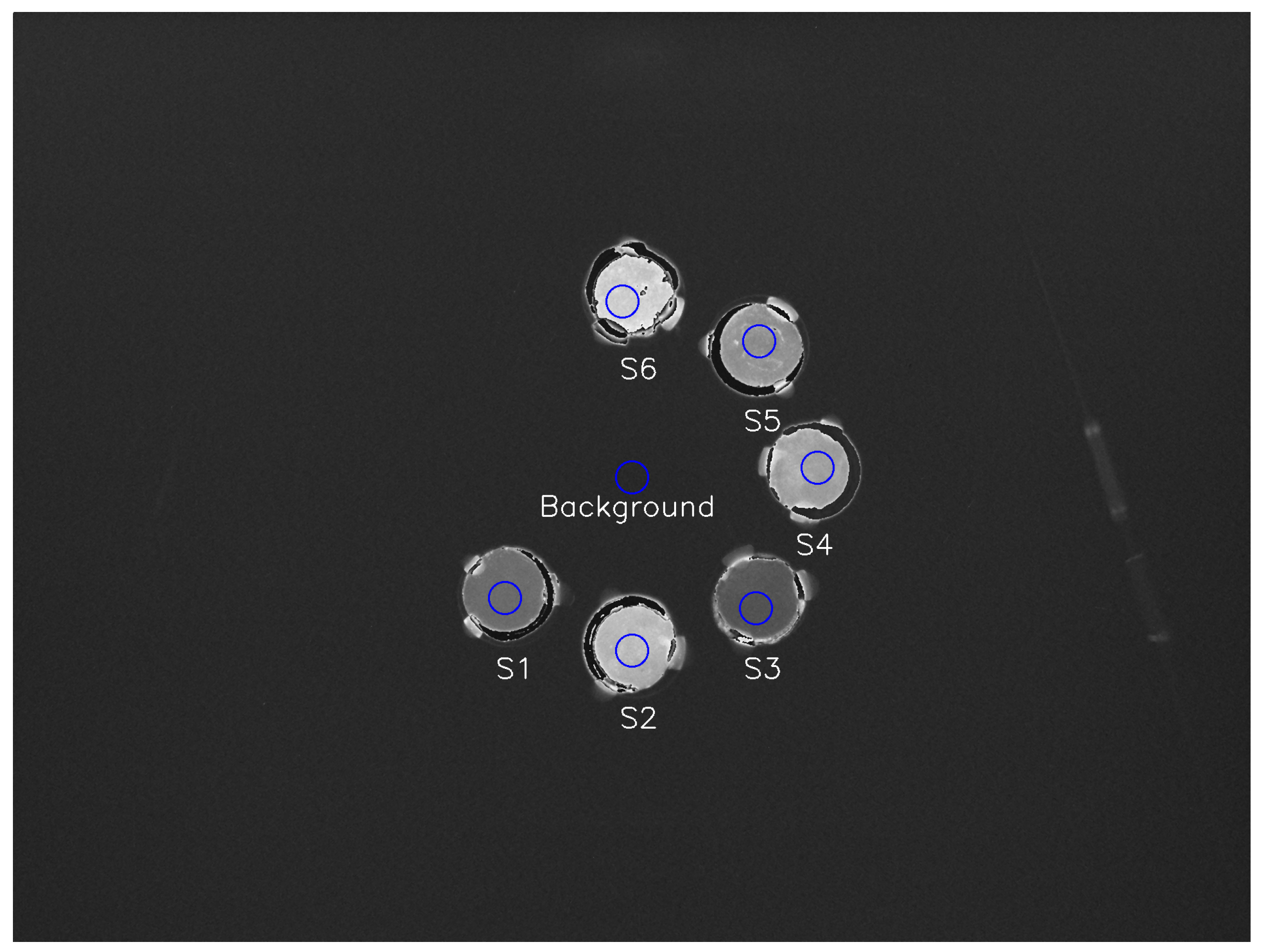
2.1.2. Relative Luminosity Measurement
2.2. Monte Carlo Simulations
2.2.1. Ionization Quenching
2.2.2. Beam Range and Spatial Resolution
3. Results
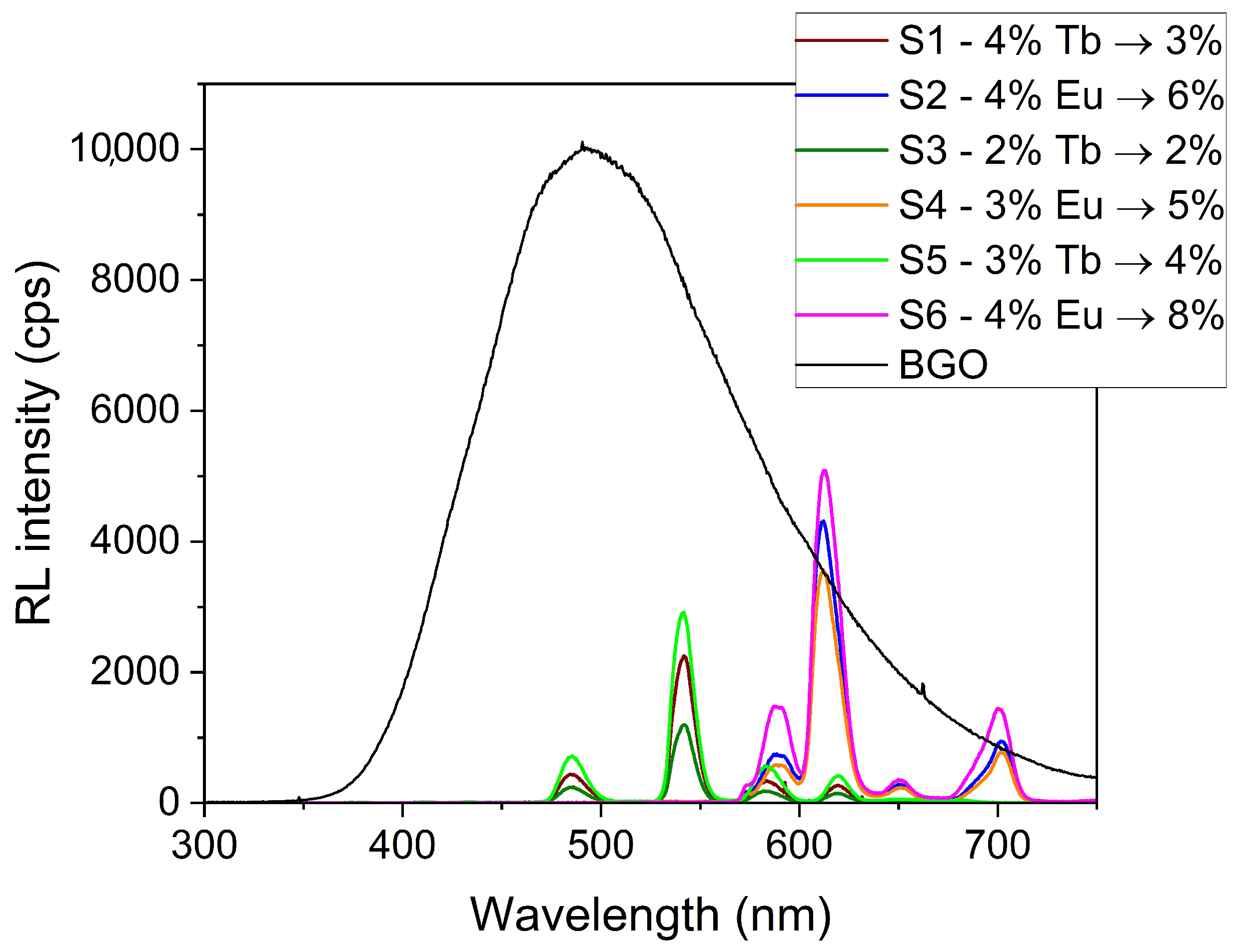
3.1. Relative Luminosity
3.2. Ionization Quenching
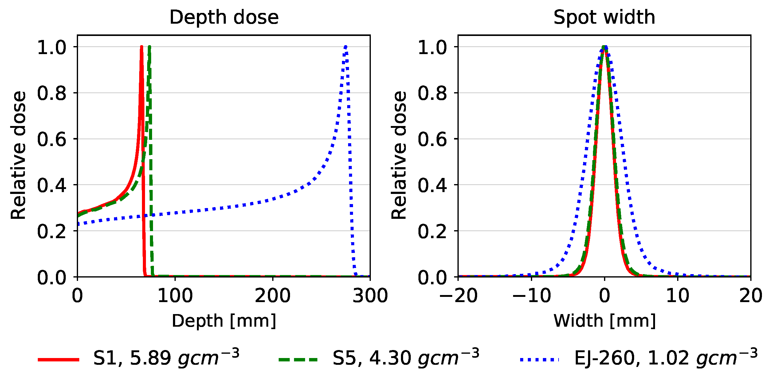
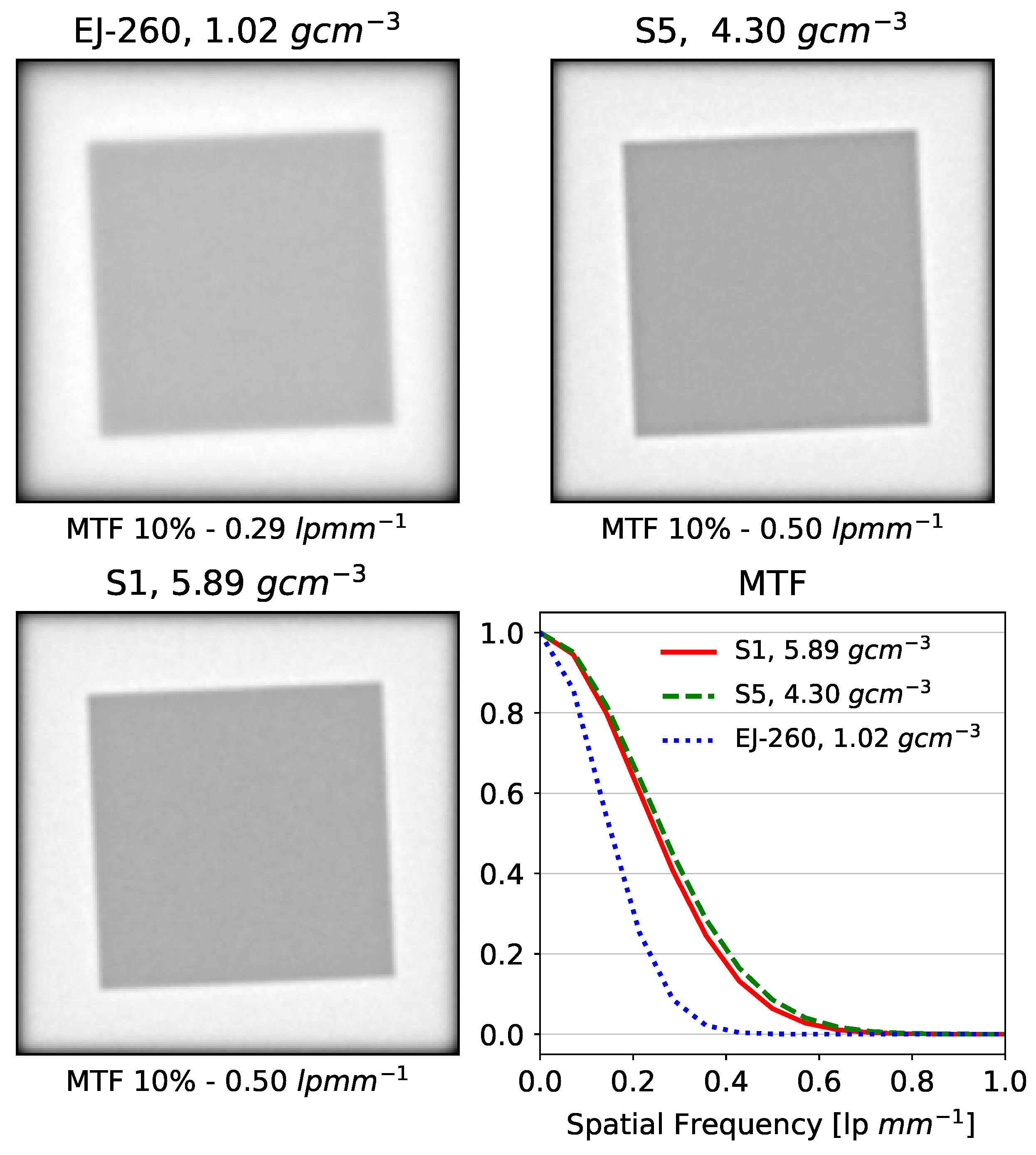
3.3. Beam Range and Spatial Resolution
4. Discussion
5. Conclusions
Author Contributions
Funding
Institutional Review Board Statement
Informed Consent Statement
Data Availability Statement
Acknowledgments
Conflicts of Interest
References
- Wilson, R.R. Radiological use of fast protons. Radiology 1946, 47, 487–491. [Google Scholar] [CrossRef]
- Ahmed, K.A.; Demetriou, S.K.; Mcdonald, M.; Johnstone, P.A.S. Clinical benefits of proton beam therapy for tumors of the skull base. Cancer Control 2016, 23, 213–219. [Google Scholar] [CrossRef]
- Fitzek, M.M.; Thornton, A.F.; Varvares, M.; Ancukiewicz, M.; Mcintyre, J.; Adams, J.; Rosenthal, S.; Joseph, M.; Amrein, P. Neuroendocrine tumors of the sinonasal tract: Results of a prospective study incorporating chemotherapy, surgery, and combined proton-photon radiotherapy. Cancer 2002, 94, 2623–2634. [Google Scholar] [CrossRef]
- Lee, C.T.; Bilton, S.D.; Famiglietti, R.M.; Riley, B.A.; Mahajan, A.; Chang, E.L.; Maor, M.H.; Woo, S.Y.; Cox, J.D.; Smith, A.R. Treatment planning with protons for pediatric retinoblastoma, medulloblastoma, and pelvic sarcoma: How do protons compare with other conformal techniques? Int. J. Radiat. Oncol. Biol. Phys. 2005, 63, 362–372. [Google Scholar] [CrossRef]
- Slater, J.D.; Yonemoto, L.T.; Mantik, D.W.; Bush, D.A.; Preston, W.; Grove, R.I.; Miller, D.W.; Slater, J.M. Proton radiation for treatment of cancer of the oropharynx: Early experience at Loma Linda University Medical Center using a concomitant boost technique. Int. J. Radiat. Oncol. Biol. Phys. 2005, 62, 494–500. [Google Scholar] [CrossRef]
- Tian, X.; Liu, K.; Hou, Y.; Cheng, J.; Zhang, J. The evolution of proton beam therapy: Current and future status (Review). Mol. Clin. Oncol. 2017. [Google Scholar] [CrossRef]
- Truong, M.T.; Kamat, U.R.; Liebsch, N.J.; Curry, W.T.; Lin, D.T.; Barker, F.G.; Loeffler, J.S.; Chan, A.W. Proton radiation therapy for primary sphenoid sinus malignancies: Treatment outcome and prognostic factors. Head Neck 2009, 31, 1297–1308. [Google Scholar] [CrossRef]
- Van De Water, T.A.; Lomax, A.J.; Bijl, H.P.; De Jong, M.E.; Schilstra, C.; Hug, E.B.; Langendijk, J.A. Potential benefits of scanned intensity-modulated proton therapy versus advanced photon therapy with regard to sparing of the salivary glands in oropharyngeal cancer. Int. J. Radiat. Oncol. Biol. Phys. 2011, 79, 1216–1224. [Google Scholar] [CrossRef]
- Yock, T.; Schneider, R.; Friedmann, A.; Adams, J.; Fullerton, B.; Tarbell, N. Proton radiotherapy for orbital rhabdomyosarcoma: Clinical outcome and a dosimetric comparison with photons. Int. J. Radiat. Oncol. Biol. Phys. 2005, 63, 1161–1168. [Google Scholar] [CrossRef]
- Hojo, H.; Dohmae, T.; Hotta, K.; Kohno, R.; Motegi, A.; Yagishita, A.; Makinoshima, H.; Tsuchihara, K.; Akimoto, T. Difference in the relative biological effectiveness and DNA damage repair processes in response to proton beam therapy according to the positions of the spread out Bragg peak. Radiat. Oncol. 2017, 12, 111. [Google Scholar] [CrossRef]
- Lievens, Y.; Pijls-Johannesma, M. Health economic controversy and cost-effectiveness of proton therapy. Semin. Radiat. Oncol. 2013, 23, 134–141. [Google Scholar] [CrossRef]
- Mohan, R.; Grosshans, D. Proton therapy––Present and future. Adv. Drug Deliv. Rev. 2017, 109, 26–44. [Google Scholar] [CrossRef]
- Schneider, U.; Pedroni, E.; Lomax, A. The calibration of CT Hounsfield units for radiotherapy treatment planning. Phys. Med. Biol. 1996, 41, 111–124. [Google Scholar] [CrossRef]
- Yang, M.; Zhu, X.R.; Park, P.C.; Titt, U.; Mohan, R.; Virshup, G.; Clayton, J.E.; Dong, L. Comprehensive analysis of proton range uncertainties related to patient stopping-power-ratio estimation using the stoichiometric calibration. Phys. Med. Biol. 2012, 57, 4095–4115. [Google Scholar] [CrossRef]
- Poludniowski, G.; Allinson, N.M.; Evans, P.M. Proton radiography and tomography with application to proton therapy. Br. J. Radiol. 2015, 88, 20150134. [Google Scholar] [CrossRef]
- Pemler, P.; Besserer, J.; De Boer, J.; Dellert, M.; Gahn, C.; Moosburger, M.; Schneider, U.; Pedroni, E.; Stäuble, H. A detector system for proton radiography on the gantry of the Paul-Scherrer-Institute. Nucl. Instrum. Methods Phys. Res. Sect. A 1999, 432, 483–495. [Google Scholar] [CrossRef]
- Schneider, U.; Besserer, J.; Pemler, P.; Dellert, M.; Moosburger, M.; Pedroni, E.; Kaser-Hotz, B. First proton radiography of an animal patient. Med. Phys. 2004, 31, 1046–1051. [Google Scholar] [CrossRef]
- Sarosiek, C.; DeJongh, E.A.; Coutrakon, G.; DeJongh, D.F.; Duffin, K.L.; Karonis, N.T.; Ordoñez, C.E.; Pankuch, M.; Rykalin, V.; Winans, J.R.; et al. Analysis of characteristics of images acquired with a prototype clinical proton radiography system. Med. Phys. 2021, 48, 2271–2278. [Google Scholar] [CrossRef]
- Bashkirov, V.A.; Schulte, R.W.; Hurley, R.F.; Johnson, R.P.; Sadrozinski, H.F.; Zatserklyaniy, A.; Plautz, T.; Giacometti, V. Novel scintillation detector design and performance for proton radiography and computed tomography. Med. Phys. 2016, 43, 664–674. [Google Scholar] [CrossRef]
- Johnson, R.P. Review of medical radiography and tomography with proton beams. Rep. Prog. Phys. 2018, 81, 016701. [Google Scholar] [CrossRef]
- Ulrich-Pur, F.; Adler, L.; Bergauer, T.; Burker, A.; De Franco, A.; Guidoboni, G.; Hirtl, A.; Irmler, C.; Kaser, S.; Nowak, S.; et al. Commissioning of low particle flux for proton beams at MedAustron. Nucl. Instrum. Methods Phys. Res. Sect. A 2021, 1010, 165570. [Google Scholar] [CrossRef]
- Curry, J.; Steward, V.W. Establishment of a beam line at the Fermi National Accelerator Laboratory for proton radiography. Med. Phys. 1978, 5, 188–194. [Google Scholar] [CrossRef]
- Whitlow, H.J.; Guibert, E.; Jeanneret, P.; Homsy, A.; Roth, J.; Krause, S.; Roux, A.; Eggermann, E.; Stoppini, L. Post-focus expansion of ion beams for low fluence and large area MeV ion irradiation: Application to human brain tissue and electronics devices. Nucl. Instrum. Methods Phys. Res. Sect. B 2017, 404, 87–91. [Google Scholar] [CrossRef]
- Darne, C.D.; Robertson, D.G.; Alsanea, F.; Collins-Fekete, C.A.; Beddar, S. A novel proton-integrating radiography system design using a monolithic scintillator detector: Experimental studies. Nucl. Instrum. Methods Phys. Res. Sect. A 2022, 1027, 166077. [Google Scholar] [CrossRef]
- Alaka, B.G.; Bentefour, E.H.; Chirvase, C.; Samuel, D.; Teo, B.K.K. Feasibility of energy-resolved dose imaging technique in pencil beam scanning mode. Biomed. Phys. Eng. Express 2020, 6, 065009. [Google Scholar] [CrossRef]
- Bentefour, E.H.; Schnuerer, R.; Lu, H.M. Concept of proton radiography using energy resolved dose measurement. Phys. Med. Biol. 2016, 61, N386–N393. [Google Scholar] [CrossRef]
- Doolan, P.J.; Royle, G.; Gibson, A.; Lu, H.; Prieels, D.; Bentefour, E.H. Dose ratio proton radiography using the proximal side of the Bragg peak. Med. Phys. 2015, 42, 1871–1883. [Google Scholar] [CrossRef]
- Gottschalk, B.; Tang, S.; Bentefour, E.H.; Cascio, E.W.; Prieels, D.; Lu, H. Water equivalent path length measurement in proton radiotherapy using time resolved diode dosimetry. Med. Phys. 2011, 38, 2282–2288. [Google Scholar] [CrossRef]
- Ryu, H.; Song, E.; Lee, J.; Kim, J. Density and spatial resolutions of proton radiography using a range modulation technique. Phys. Med. Biol. 2008, 53, 5461–5468. [Google Scholar] [CrossRef]
- Testa, M.; Schümann, J.; Lu, H.; Shin, J.; Faddegon, B.; Perl, J.; Paganetti, H. Experimental validation of the TOPAS Monte Carlo system for passive scattering proton therapy. Med. Phys. 2013, 40, 121719. [Google Scholar] [CrossRef]
- Tanaka, S.; Nishio, T.; Matsushita, K.; Tsuneda, M.; Kabuki, S.; Uesaka, M. Development of proton CT imaging system using plastic scintillator and CCD camera. Phys. Med. Biol. 2016, 61, 4156–4167. [Google Scholar] [CrossRef]
- Archambault, L.; Poenisch, F.; Sahoo, N.; Robertson, D.; Lee, A.; Gillin, M.T.; Mohan, R.; Beddar, S. Verification of proton range, position, and intensity in IMPT with a 3D liquid scintillator detector system: IMPT verification with 3D liquid scintillator. Med. Phys. 2012, 39, 1239–1246. [Google Scholar] [CrossRef]
- Beddar, S.; Archambault, L.; Sahoo, N.; Poenisch, F.; Chen, G.T.; Gillin, M.T.; Mohan, R. Exploration of the potential of liquid scintillators for real-time 3D dosimetry of intensity modulated proton beams. Med. Phys. 2009, 36, 1736–1743. [Google Scholar] [CrossRef]
- Darne, C.D.; Alsanea, F.; Robertson, D.G.; Sahoo, N.; Beddar, S. Performance characterization of a 3D liquid scintillation detector for discrete spot scanning proton beam systems. Phys. Med. Biol. 2017, 62, 5652–5667. [Google Scholar] [CrossRef]
- Hui, C.; Robertson, D.; Beddar, S. 3D reconstruction of scintillation light emission from proton pencil beams using limited viewing angles—A simulation study. Phys. Med. Biol. 2014, 59, 4477–4492. [Google Scholar] [CrossRef]
- Darne, C.D.; Alsanea, F.; Robertson, D.G.; Guan, F.; Pan, T.; Grosshans, D.; Beddar, S. A proton imaging system using a volumetric liquid scintillator: A preliminary study. Biomed. Phys. Eng. Express 2019, 5, 045032. [Google Scholar] [CrossRef]
- Knoll, G.F. Radiation Detection and Measurement, 4th ed.; John Wiley: Hoboken, NJ, USA, 2010. [Google Scholar]
- Ginther, R.J. New cerium activated scintillating glasses. IRE Trans. Nucl. Sci. 1960, 7, 28–31. [Google Scholar] [CrossRef]
- Bross, A.D. Properties of new scintillator glasses and scintillating fibers. Nucl. Instrum. Methods Phys. Res. Sect. A 1986, 247, 319–326. [Google Scholar] [CrossRef]
- Fu, J.; Parker, J.M.; Brown, R.M.; Flower, P.S. Compositional dependence of scintillation yield of glasses with high Gd2O3 concentrations. J. Non-Cryst. Solids 2003, 326-327, 335–338. [Google Scholar] [CrossRef]
- Wang, Q.; Yang, B.; Zhang, Y.; Xia, H.; Zhao, T.; Jiang, H. High light yield Ce3+-doped dense scintillating glasses. J. Alloys Compd. 2013, 581, 801–804. [Google Scholar] [CrossRef]
- Fu, J.; Kobayashi, M.; Sugimoto, S.; Parker, J.M. Eu3+-activated heavy scintillating glasses. Mater. Res. Bull. 2008, 43, 1502–1508. [Google Scholar] [CrossRef]
- Ginther, R.J.; Schulmian, J.H. Glass scintillators. IRE Trans. Nucl. Sci. 1958, 5, 92–95. [Google Scholar] [CrossRef]
- Jiang, J.; Zhang, G.; Poulain, M. Cerium-containing glasses for fast scintillators. J. Alloys Compd. 1998, 275–277, 733–737. [Google Scholar] [CrossRef]
- Tillman, I.J.; Dettmann, M.A.; Herrig, V.; Thune, Z.L.; Zieser, A.J.; Michalek, S.; Been, M.; Martinez-Szewczyk, M.; Koster, H.; Wilkinson, C.; et al. High-density scintillating glasses for a proton imaging detector. Opt. Mater. 2017, 68, 58–62. [Google Scholar] [CrossRef]
- Wilkinson, C.J.; Ruane, L.; Miller, W.; Gunsch, A.; Zieser, A.; Tillman, I.J.; Thune, Z.; Wang, D.; Akgun, U. CARNA—A compact glass proton imager. In Proceedings of the 2017 IEEE Nuclear Science Symposium and Medical Imaging Conference (NSS/MIC), Atlanta, GA, USA, 21–28 October 2017; pp. 1–5. [Google Scholar] [CrossRef]
- Tendler, I.; Robertson, D.; Darne, C.; Panthi, R.; Alsanea, F.; Collins-Fekete, C.A.; Beddar, S. Image quality evaluation of projection— and depth dose—based approaches to integrating proton radiography using a monolithic scintillator detector. Phys. Med. Biol. 2021, 66, 144001. [Google Scholar] [CrossRef]
- Jiang, H.; Kim, H.J.; Rooh, G.; Park, H.; Kim, S.; Fawad, U.; Cheon, J. Czochralski growth and scintillation properties of bismuth germanium silicon oxide (BGSO) single crystals. In Proceedings of the 2011 IEEE Nuclear Science Symposium Conference Record, Valencia, Spain, 23–29 October 2011; pp. 1580–1582. [Google Scholar] [CrossRef]
- Okazaki, K.; Fukushima, H.; Nakauchi, D.; Okada, G.; Onoda, D.; Kato, T.; Kawaguchi, N.; Yanagida, T. Investigation of Er:Bi4Ge3O12 single crystals emitting near-infrared luminescence for scintillation detectors. J. Alloys Compd. 2022, 903, 163834. [Google Scholar] [CrossRef]
- Matulewicz, T. Quenching of scintillation in BaF2 for light charged particles. Nucl. Instrum. Methods Phys. Res. Sect. A 1993, 325, 365–366. [Google Scholar] [CrossRef]
- Birks, J.B. Scintillations from organic crystals: Specific fluorescence and relative response to different radiations. Proc. Phys. Soc. Sect. A 1951, 64, 874–877. [Google Scholar] [CrossRef]
- Pöschl, T.; Greenwald, D.; Losekamm, M.J.; Paul, S. Measurement of ionization quenching in plastic scintillators. Nucl. Instrum. Methods Phys. Res. Sect. A 2021, 988, 164865. [Google Scholar] [CrossRef]
- Perl, J.; Shin, J.; Schümann, J.; Faddegon, B.; Paganetti, H. TOPAS: An innovative proton Monte Carlo platform for research and clinical applications. Med. Phys. 2012, 39, 6818–6837. [Google Scholar] [CrossRef]
- Testa, M.; Verburg, J.M.; Rose, M.; Min, C.H.; Tang, S.; Bentefour, E.H.; Paganetti, H.; Lu, H.M. Proton radiography and proton computed tomography based on time-resolved dose measurements. Phys. Med. Biol. 2013, 58, 8215–8233. [Google Scholar] [CrossRef]
- Agostinelli, S.; Allison, J.; Amako, K.; Apostolakis, J.; Araujo, H.; Arce, P.; Asai, M.; Axen, D.; Banerjee, S.; Barrand, G.; et al. Geant4—A simulation toolkit. Nucl. Instrum. Methods Phys. Res. Sect. A 2003, 506, 250–303. [Google Scholar] [CrossRef]
- Lechner, A.; Ivanchenko, V.N.; Knobloch, J. Validation of recent Geant4 physics models for application in carbon ion therapy. Nucl. Instrum. Methods Phys. Res. Sect. B 2010, 268, 2343–2354. [Google Scholar] [CrossRef]
- Hall, D.C.; Makarova, A.; Paganetti, H.; Gottschalk, B. Validation of nuclear models in Geant4 using the dose distribution of a 177 MeV proton pencil beam. Phys. Med. Biol. 2016, 61, N1–N10. [Google Scholar] [CrossRef]
- Goudsmit, S.; Saunderson, J.L. Multiple scattering of electrons. Phys. Rev. 1940, 57, 24–29. [Google Scholar] [CrossRef]
- Ivanchenko, V.N.; Kadri, O.; Maire, M.; Urban, L. Geant4 models for simulation of multiple scattering. J. Phys. Conf. Ser. 2010, 219, 032045. [Google Scholar] [CrossRef]
- ISO 12233:2023; Photography—Electronic Still Picture Imaging—Resolution and Spatial Frequency Responses. International Organization for Standardization: Geneva, Switzerland, 2023. Available online: https://www.iso.org/standard/79169.html (accessed on 1 January 2024).
- Zhao, L.; Das, I.J.; Zhao, Q.; Thomas, A.; Adamovics, J.; Oldman, M. Determination of the depth dose distribution of proton beam using PRESAGETM dosimeter. J. Phys. Conf. Ser. 2010, 250, 012035. [Google Scholar] [CrossRef]
- Alsanea, F.; Therriault-Proulx, F.; Sawakuchi, G.; Beddar, S. A real-time method to simultaneously measure linear energy transfer and dose for proton therapy using organic scintillators. Med. Phys. 2018, 45, 1782–1789. [Google Scholar] [CrossRef]
- Wang, L.L.W.; Perles, L.A.; Archambault, L.; Sahoo, N.; Mirkovic, D.; Beddar, S. Determination of the quenching correction factors for plastic scintillation detectors in therapeutic high-energy proton beams. Phys. Med. Biol. 2012, 57, 7767–7781. [Google Scholar] [CrossRef]
- Fu, J.; Kobayashi, M.; Parker, J.M. Terbium-activated heavy scintillating glasses. J. Lumin. 2008, 128, 99–104. [Google Scholar] [CrossRef]
- Hobson, P.; Imrie, D.; Price, T.; Sheikh, S.; Bell, K.; Brown, R.; Cockerill, D.; Flower, P.; Grayer, G.; Kennedy, B.; et al. The development of dense scintillating hafnium fluoride glasses for the construction of homogeneous calorimeters in particle physics. J. Non-Cryst. Solids 1997, 213-214, 147–151. [Google Scholar] [CrossRef]
- Tanner, P.A.; Mak, C.S.K.; Edelstein, N.M.; Murdoch, K.M.; Liu, G.; Huang, J.; Seijo, L.; Barandiarán, Z. Absorption and emission spectra of Ce3+ in elpasolite lattices. J. Am. Chem. Soc. 2003, 125, 13225–13233. [Google Scholar] [CrossRef]


| Parameter | Description | References |
|---|---|---|
| Code release date | TOPAS Version 3.8.1 | [53] |
| Validation | Validated for proton transport applications | [54] |
| CPU description | High-performance cluster, 100 h computation time on 32 CPUs | |
| Source description | 10 cm square field of 144.8 MeV monoenergetic protons | |
| Cross-sections | TOPAS default parameters | [53] |
| Transport parameters | TOPAS default parameters | [53] |
| Scored quantities | Dose to material, proton LET | |
| Number of histories | 1 × 10⁹ initial protons |
| Parameter | Description | References |
|---|---|---|
| Code release date | Geant4.10.6.p01 | [55] |
| Validation | ICRU 73 stopping powers incorporated into Geant4 including media such as water. | [56] |
| CPU description | High-performance cluster, 40 parallel computations of 1 h on 36 cores | |
| Source description | Pencil beams modeled as Double Gaussian functions: (1) 75% intensity with a standard deviation 2 mm, (2) 25% intensity with a standard deviation of 4 mm for wide-scattered protons | |
| Cross-sections | G4HadronElasticPhysics and emstandard_opt4 | [56,57] |
| Transport parameters | Multiple Coulomb scattering based on Lewis theory using the Urban model | [58,59] |
| Scored quantities | Energy deposition in material | |
| Number of histories | (a) a single proton pencil beam, and (b) 75 × 75 proton pencil beams evenly spaced 2 mm apart in a square pattern, with 4 × 10⁵ initial protons per pencil beam |
| Sample | Density (g cm−3) | Base Glass Composition | Activation Element | Peak-Plateau Ratio | Relative Luminosity (% BGO) | Birks Correction Factor (mg MeV−1 cm−2) |
|---|---|---|---|---|---|---|
| S1 | 5.6 | 21% Gd2O3, 35% WO3, 40% 2H3BO3 | 4% Tb2O3 | 1.5 | 3% | 30 |
| S2 | 5.6 | 21% Gd2O3, 35% WO3, 40% 2H3BO3 | 4% Eu2O3 | 1.6 | 6% | 22 |
| S3 | 5.9 | 21% Gd2O3, 45% WO3, 32% 2H3BO3 | 2% Tb2O3 | 1.6 | 2% | 33 |
| S4 | 5.8 | 22% Gd2O3, 45% WO3, 30% 2H3BO3 | 3% Eu2O3 | 1.6 | 5% | 28 |
| S5 | 4.3 | 30% PbO, 66% 2H3BO3 | 3% Tb2O3 | 1.5 | 4% | 30 |
| S6 | 4.5 | 30% PbO, 67% 2H3BO3 | 4% Eu2O3 | 1.5 | 8% | 21 |
Disclaimer/Publisher’s Note: The statements, opinions and data contained in all publications are solely those of the individual author(s) and contributor(s) and not of MDPI and/or the editor(s). MDPI and/or the editor(s) disclaim responsibility for any injury to people or property resulting from any ideas, methods, instructions or products referred to in the content. |
© 2024 by the authors. Licensee MDPI, Basel, Switzerland. This article is an open access article distributed under the terms and conditions of the Creative Commons Attribution (CC BY) license (https://creativecommons.org/licenses/by/4.0/).
Share and Cite
Stolen, E.; Fullarton, R.; Hein, R.; Conner, R.L.; Jacobsohn, L.G.; Collins-Fekete, C.-A.; Beddar, S.; Akgun, U.; Robertson, D. High-Density Glass Scintillators for Proton Radiography—Relative Luminosity, Proton Response, and Spatial Resolution. Sensors 2024, 24, 2137. https://doi.org/10.3390/s24072137
Stolen E, Fullarton R, Hein R, Conner RL, Jacobsohn LG, Collins-Fekete C-A, Beddar S, Akgun U, Robertson D. High-Density Glass Scintillators for Proton Radiography—Relative Luminosity, Proton Response, and Spatial Resolution. Sensors. 2024; 24(7):2137. https://doi.org/10.3390/s24072137
Chicago/Turabian StyleStolen, Ethan, Ryan Fullarton, Rain Hein, Robin L. Conner, Luiz G. Jacobsohn, Charles-Antoine Collins-Fekete, Sam Beddar, Ugur Akgun, and Daniel Robertson. 2024. "High-Density Glass Scintillators for Proton Radiography—Relative Luminosity, Proton Response, and Spatial Resolution" Sensors 24, no. 7: 2137. https://doi.org/10.3390/s24072137
APA StyleStolen, E., Fullarton, R., Hein, R., Conner, R. L., Jacobsohn, L. G., Collins-Fekete, C.-A., Beddar, S., Akgun, U., & Robertson, D. (2024). High-Density Glass Scintillators for Proton Radiography—Relative Luminosity, Proton Response, and Spatial Resolution. Sensors, 24(7), 2137. https://doi.org/10.3390/s24072137