Article

Cembrane Derivatives from the Soft Corals, Sinularia gaweli and Sinularia flexibilis

1
Graduate Institute of Marine Biotechnology and Department of Life Science and Institute of Biotechnology, National Dong Hwa University, Pingtung 944, Taiwan
2
National Museum of Marine Biology and Aquarium, Pingtung 944, Taiwan
3
Department of Chemistry, National Sun Yat-sen University, Kaohsiung 804, Taiwan
4
Department of Marine Biotechnology and Resources and Asia-Pacific Ocean Research Center, National Sun Yat-sen University, Kaohsiung 804, Taiwan
5
Department of Neurosurgery, Kaohsiung Chang Gung Memorial Hospital and Chang Gung University, College of Medicine, Kaohsiung 833, Taiwan
6
Graduate Institute of Food Science and Technology, National Taiwan University, Taipei 106, Taiwan
7
Department of Food Science, National Taiwan Ocean University, Keelung 202, Taiwan
8
School of Pharmacy, College of Pharmacy, China Medical University, Taichung 404, Taiwan
9
Chinese Medicine Research and Development Center, China Medical University Hospital, Taichung 404, Taiwan
10
Center for Molecular Medicine, China Medical University Hospital, Taichung 404, Taiwan
11
Graduate Institute of Natural Products, Kaohsiung Medical University, Kaohsiung 807, Taiwan
*
Authors to whom correspondence should be addressed.
These authors contributed equally to this work.
Mar. Drugs 2013, 11(6), 2154-2167; https://doi.org/10.3390/md11062154
Submission received: 19 April 2013 / Revised: 20 May 2013 / Accepted: 5 June 2013 / Published: 17 June 2013

Abstract

:
A new norcembranoidal diterpene, 1-epi-sinulanorcembranolide A (1), and a new cembranoidal diterpene, flexibilin D (2), were isolated from the soft corals, Sinularia gaweli and Sinularia flexibilis, respectively. The structures of new metabolites 1 and 2 were elucidated by spectroscopic methods, and compound 2 was found to significantly inhibit the accumulation of the pro-inflammatory iNOS and COX-2 proteins of the lipopolysaccharide (LPS)-stimulated RAW264.7 macrophage cells. In addition, S. flexibilis yielded a known cembrane, 5-dehydrosinulariolide (3); the structure, including its absolute stereochemistry, was further confirmed by single-crystal X-ray diffraction analysis.

1. Introduction

Octocorals belonging to the genus Sinularia, which are distributed widely in the tropical and subtropical Indo-Pacific Ocean, have been proven to be rich sources of bioactive terpenoid analogues [1,2,3,4]. In ongoing studies on the chemical constituents from marine invertebrates collected off the waters of Taiwan, the intersection of the Kuroshio current and the South China Sea surface current, the octocorals Sinularia gaweli and Sinularia flexibilis (phylum Cnidaria, class Anthozoa, subclass Octocorallia, order Alcyonacea, family Alcyoniidae) were studied, as their organic extract was found to show meaning signals in NMR studies. In previous studies, a series of interesting secondary metabolites were isolated from the octocorals, S. gaweli [5,6,7] and S. flexibilis [8,9,10,11,12,13,14,15,16,17,18], collected in the waters of Taiwan. We further isolated a new norcembranoidal diterpene, 1-epi-sinulanorcembranolide A (1), from S. gaweli and a new cembranoidal diterpene, flexibilin D (2), along with a known cembrane, 5-dehydrosinulariolide (3) [10,13,19,20,21,22,23], from S. flexibilis (Figure 1). In this paper, we describe the isolation, structure determination and bioactivity of terpenoids 13.
Figure 1. The structures of 1-epi-sinulanorcembranolide A (1), flexibilin D (2), 5-dehydrosinulariolide (3) and sinulanorcembranolide A (4).
Marinedrugs 11 02154 g001

2. Results and Discussion

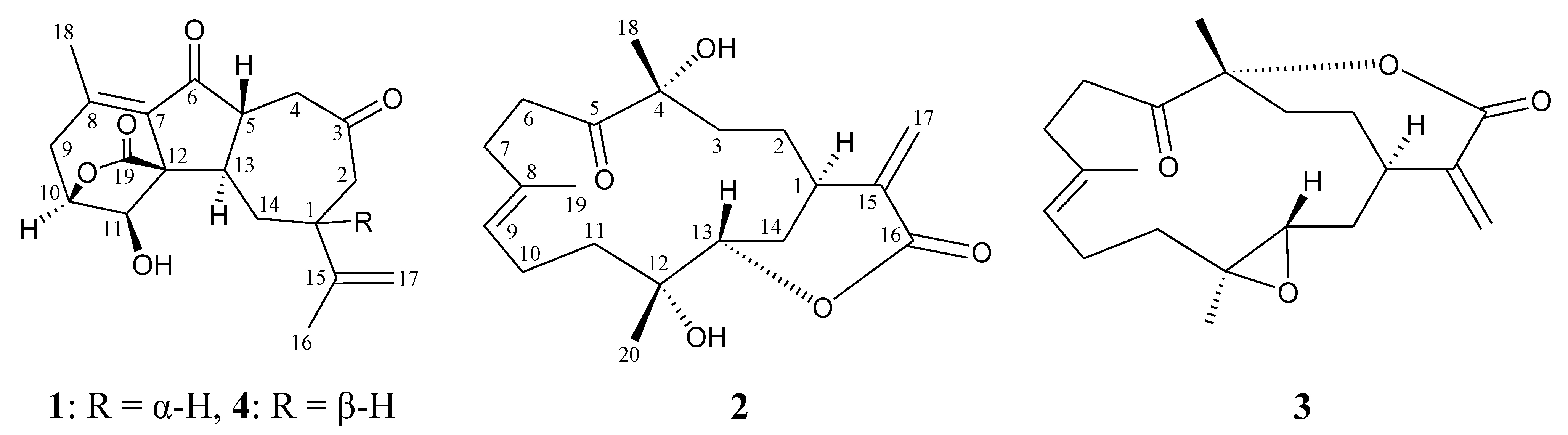
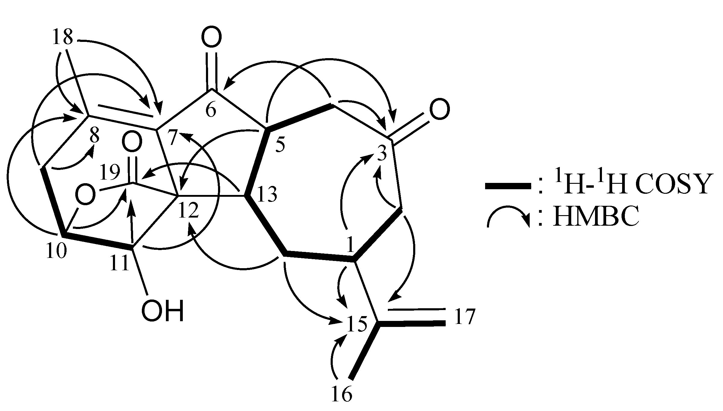
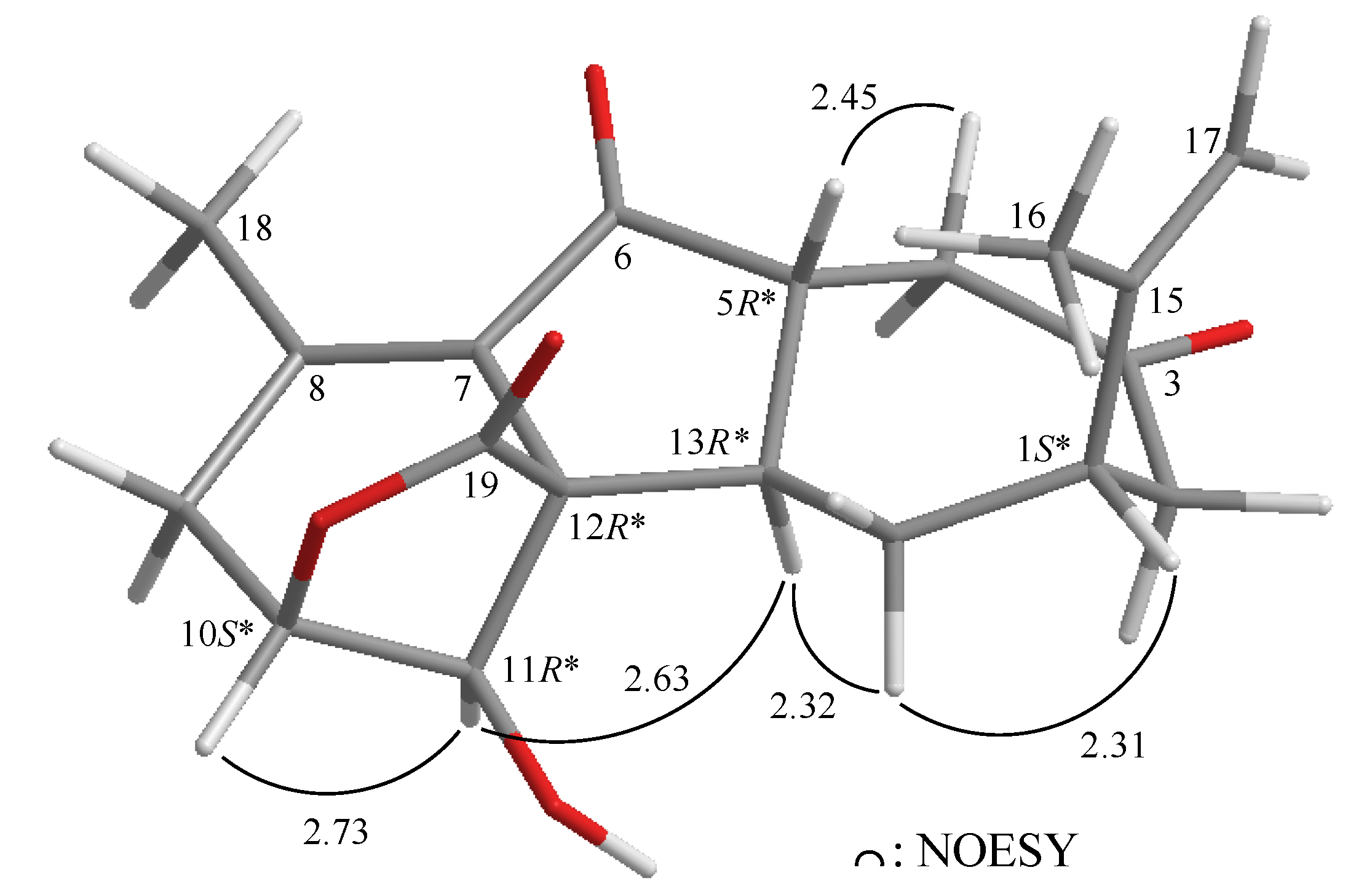
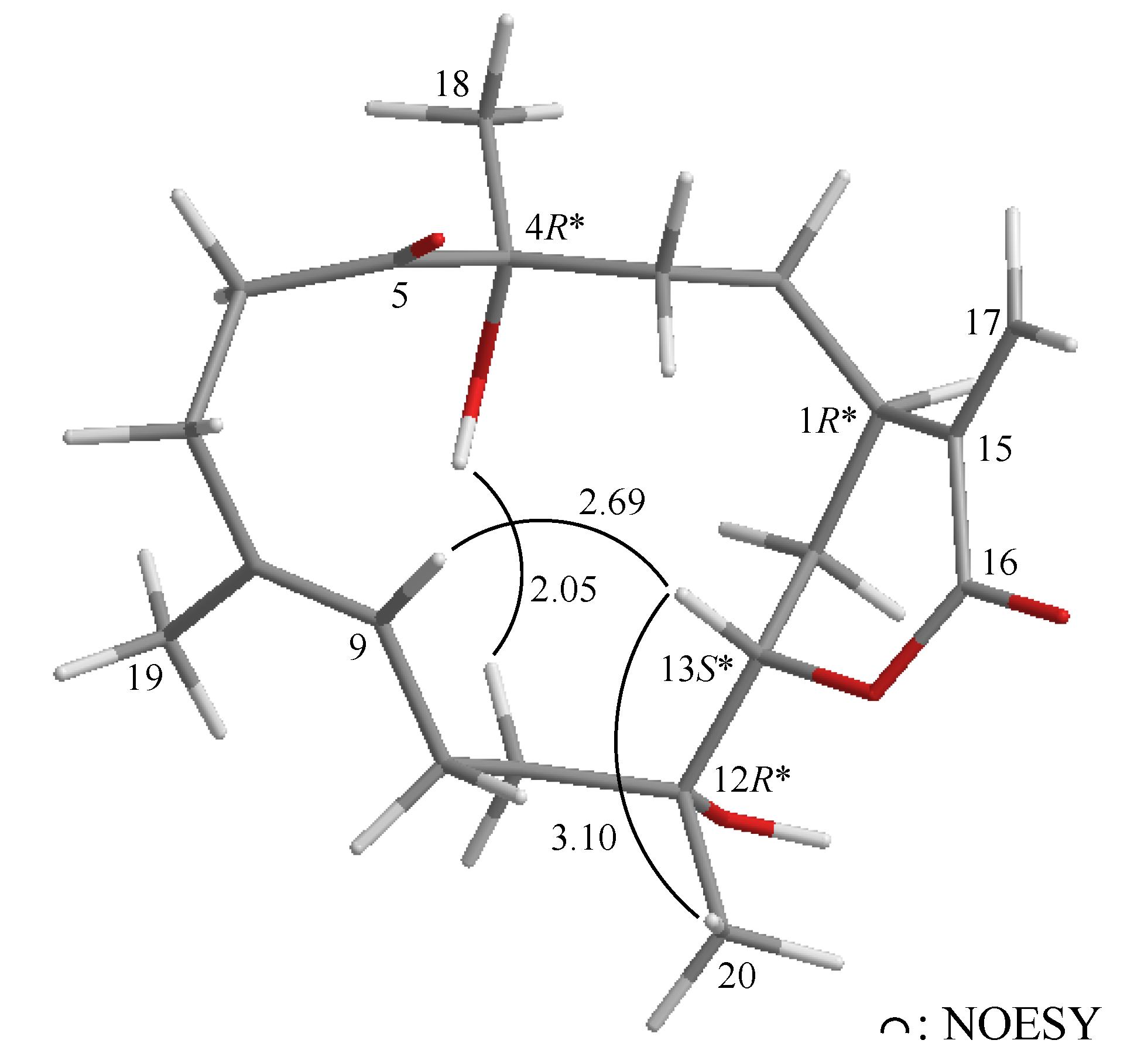
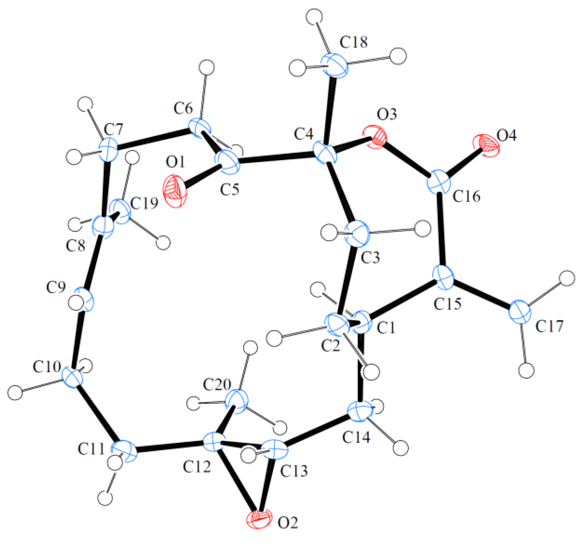
2.1. Isolation and Structure Determination of 1-epi-Sinulanorcembranolide A (1) from Sinularia gaweli

1-epi-Sinulanorcembranolide A (1) was isolated as an amorphous powder that gave a pseudomolecular ion [M + Na]+ at m/z 353.1368 in the HRESIMS, indicating the molecular formula, C19H22O5 (calcd. for C19H22O5Na, 353.1365), and implying nine degrees of unsaturation. The IR spectrum of 1 showed strong bands at 3448, 1769, 1701 and 1687 cm–1, consistent with the presence of hydroxy, ester, ketone and α,β-unsaturated ketone groups. The 13C NMR and DEPT spectra of 1 showed that this compound had 19 carbons (Table 1), including two methyls, four sp3 methylenes, five sp3 methines, an sp3 quaternary carbon, an sp2 methylene and six sp2 quaternary carbons. From the 13C NMR spectrum (Table 1), 1 was found to possess two keto carbonyls (δC 208.7, C-3; 198.0, C-6) and an ester group (δC 174.7, C-19). Two additional unsaturated functionalities were indicated by 13C resonances at δC 147.1 (C-15), 145.6 (C-8), 129.9 (C-7) and 112.3 (CH2-17), suggesting the presence of a tetrasubstituted olefin and an exocyclic carbon-carbon double bond. Thus, from the above data, five degrees of unsaturation were accounted for, and 1 was identified as a tetracyclic compound.
Table 1. 1H (500 MHz, CDCl3) and 13C (125 MHz, CDCl3) NMR data and 1H–1H COSY and HMBC correlations for norcembrane 1.
PositionδH (J in Hz)δC, Multiple1H–1H COSYHMBC
12.79 m39.4, CHH2-2, H2-14C-3, -13, -14, -15
2/2′2.71 d (16.0); 2.47 dd (16.0, 7.0)42.8, CH2H-1C-1, -3, -4, -14, -15
3 208.7, C
4/4′3.00 dd (15.5, 11.0); 2.56 (15.5, 7.0)38.9, CH2H-5C-3, -5, -6, -13
52.82 m46.6, CHH2-4, H-13C-3, -4, -12, -13, -14
6 198.0, C
7 129.9, C
8 145.6, C
92.68 br s39.1, CH2H-10C-7, -8
104.50 dd (2.5, 2.5)79.4, CHH2-9, H-11C-8, -11, -19
114.15 s77.0, CHH-10C-7, -10, -19
12 52.6, C
132.88 m32.9, CHH-5, H2-14C-1, -5, -19
14/14′2.71 br d (5.5); 2.11 m28.4, CH2H-1, H-13C-1, -5, -12, -15
15 147.1, C
161.76 s21.5, CH3H-17aC-1, -15, -17
17a/b4.86 s; 4.71 s112.3, CH2H3-16C-1, -16
182.02 s21.0, CH3 C-7, -8, -9
19 174.7, C
From the 1H–1H COSY spectrum of 1 (Table 1 and Figure 2), it was possible to differentiate among the separate spin systems of H2-4/H-5/H-13/H2-14/H-1/H2-2, H2-9/H-10/H-11 and H3-16/H-17a (by allylic coupling). These data, together with the key HMBC correlations between protons and quaternary carbons (Table 1 and Figure 2), such as H-1, H2-2, H-4, H-5/C-3; H2-4/C-6; H2-9, H-11, H3-18/C-7; H2-9, H-10, H3-18/C-8; H-5, H-14′/C-12; H-1, H2-2, H2-14, H3-16/C-15; and H-10, H-11, H-13/C-19, established the main carbon skeleton of 1. The vinyl methyls at C-8 and C-15 were confirmed by the HMBC correlations between H3-16/C-1, -15, -17 and H3-18/C-7, -8, -9. Thus, based on the above findings, 1 was revealed as a norcembranoidal diterpene possessing a γ-lactone moiety.
Figure 2. 1H–1H COSY and selected HMBC correlations (protons→quaternary carbons) for 1.
Marinedrugs 11 02154 g002
The relative configuration of 1 was elucidated mainly from a NOESY spectrum as being compatible with that of 1, ascertained using molecular mechanics calculations (MM2) [24]. In the NOESY experiment of 1 (Figure 3), one of the C-14 methylene protons (δH 2.11) exhibited correlations with H-1 and H-13, but not with H-5, indicated that these protons were situated on the same face and were assigned as α protons, since H-5 is a β-substituent at C-5. H-11 showed correlations with H-10 and H-13, as well as the lack of coupling detected between H-10 and H-11, indicating that the dihedral angle between H-10 and H-11 is approximately 90° and that the lactone moiety and 11-hydroxy group were β-oriented by molecular modeling analysis. From the above evidence, the relative configurations of the chiral carbons of 1 were assumed to be 1S*, 5R*, 10S*, 11R*, 12R* and 13R*. By comparison of the spectral data with those of a known norcembranoidal diterpene, sinulanorcembranolide A (4) (Figure 1), which was also isolated from S. gaweli [7], 1 was found to be the 1-epi-compound of 4 and should be named as 1-epi-sinulanorcembranolide A. To the best of our knowledge, compounds 1 and 4 are the only two norcembranoidal diterpenes with carbon-carbon linkages between C-5/13 and C-7/12. A plausible biosynthetic pathway for the compounds of this type was proposed in our previous study [7].
Figure 3. The computer-generated model of 1 using molecular mechanics calculations (MM2) force field calculations and the calculated distances (Å) between selected protons with key NOESY correlations.
Marinedrugs 11 02154 g003

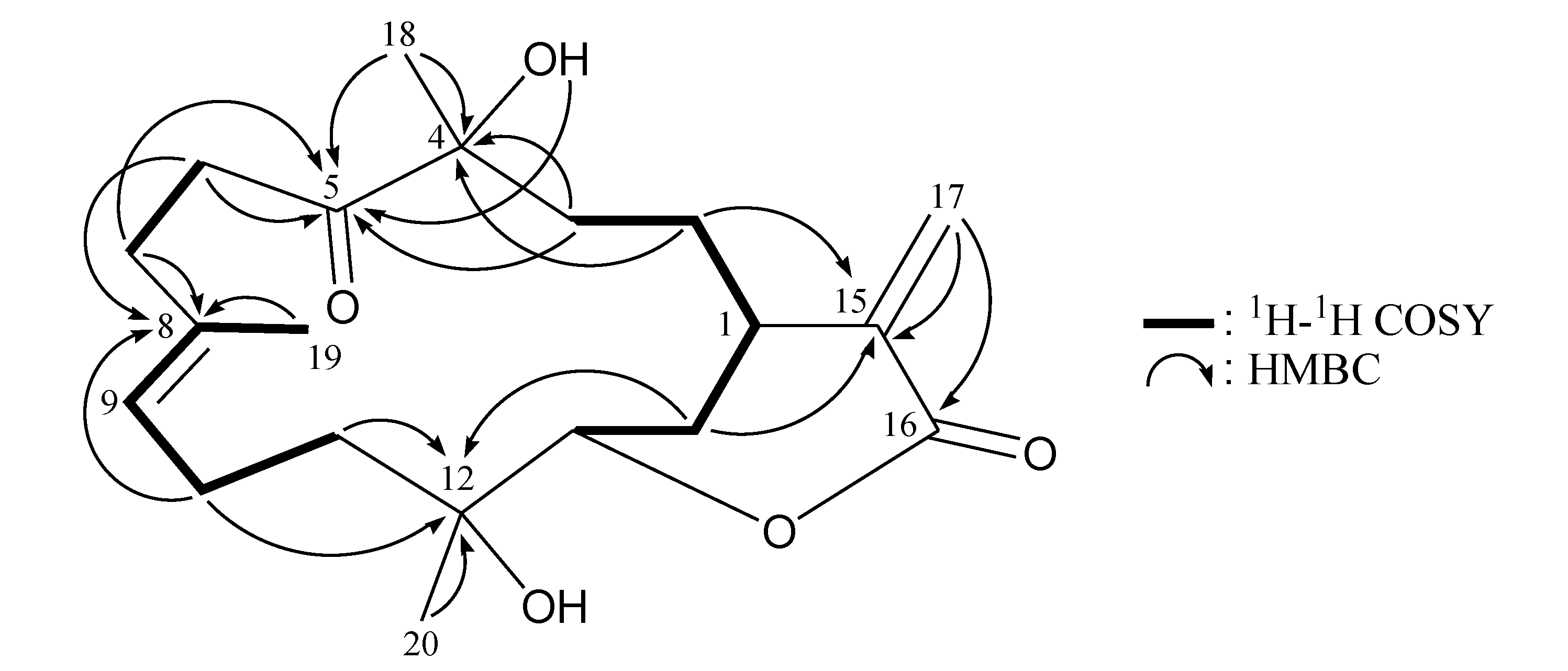
2.2. Isolation and Structure Determination of Flexibilin D (2) from Sinularia flexibilis

Flexibilin D (2) was obtained as a white powder. The molecular formula of 2 was established as C20H30O5 (6° of unsaturation) from a sodiated molecule at m/z 373 in the ESIMS spectrum and further supported by HRESIMS (m/z 373.1988, calcd. for C20H30O5Na, 373.1991). The IR spectrum of 2 exhibited the presence of hydroxy (νmax 3422 cm−1) and carbonyl (νmax 1712 cm−1) groups. From the 13C NMR data of 2 (Table 2), a suite of resonances at δC 165.5 (C-16), 138.0 (C-15), 128.4 (CH2-17), 79.1 (CH-13), 35.4 (CH-1) and 26.2 (CH2-14) could be assigned to the α-exomethylene-δ-lactone moiety. Two additional unsaturated functionalities were indicated by 13C resonances at δC 213.8 (C-5), 134.8 (C-8) and 126.1 (CH-9), suggesting the presence of a keto carbonyl and a trisubstituted olefin. On the basis of the overall unsaturation data, 2 was concluded to be a diterpenoid molecule possessing two rings. The 1H NMR spectrum of 2 showed the presence of three methyl groups: two singlets at δH 1.35 and 1.31, representing the methyl groups on oxygenated quaternary carbons, and a vinyl methyl at δH 1.66.
Table 2. 1H (400 MHz, CDCl3) and 13C (100 MHz, CDCl3) NMR data and 1H–1H COSY and HMBC correlations for cembrane 2.
PositionδH (J in Hz)δC, Multiple1H–1H COSYHMBC
12.85 m35.4, CHH2-2, H2-14n.o. a
2/2′1.49 m; 1.32 m28.2, CH2H-1, H2-3C-1, -3, -4, -14, -15
3/3′1.79 ddd (13.6, 11,2, 5.6); 1.64 m37.2, CH2H2-2C-1, -2, -4, -5, -18
4 78.9, C
5 213.8, C
6/6′2.76 ddd (18.0, 8.0, 3.2); 2.65 ddd (18.0, 10.2, 3.2)35.1, CH2H2-7C-5, -7, -8
7/7′2.48 m; 2.33 m31.8, CH2H2-6C-5, -6, -8, -9
8 134.8, C
95.15 dd (5.6, 5.6)126.1, CHH2-10, H3-19C-7, -10, -19
102.18 m23.1, CH2H-9, H2-11C-8, -9, -11, -12
11/11′1.87 dd (8.4, 2,8); 1.69 m36.7, CH2H2-10C-9, -12, -13
12 73.9, C
134.29 dd (9.2, 6.0)79.1, CHH2-14C-1, -14, -20
141.91 m26.2, CH2H-1, H-13C-12, -13, -15
15 138.0, C
16 165.5, C
17a/b6.43 d (1.2); 5.56 dd (1.2, 1.2)128.4, CH2 C-1, -15, -16
181.35 s25.2, CH3 C-3, -4, -5
191.66 s17.2, CH3H-9C-7, -8, -9
201.31 s24.1, CH3 C-11, -12, -13
OH-43.24 br s C-5
a n.o. = not observed.
The 1H NMR coupling information in the 1H–1H COSY spectrum of 2 enabled identification of the H-13/H2-14/H-1/H2-2/H2-3, H2-6/H2-7, H-9/H2-10/H2-11 and H-9/H3-19 (by allylic coupling) units, which were assembled with the assistance of an HMBC experiment (Table 2 and Figure 4), enabling establishment of the main carbon skeleton of 2. A vinyl methyl at C-8 was confirmed by the allylic coupling between H-9/H3-19 in the 1H–1H COSY spectrum and by the HMBC correlations between H3-19/C-7, -8, -9; and H-9/C-19. The tertiary methyls at C-4 and C-12 were confirmed by the HMBC correlations between H3-18/C-3, -4, -5 and H3-20/C-11, -12, -13.
Figure 4. 1H–1H COSY and selected HMBC correlations (protons→quaternary carbons) for cembrane 2.
Marinedrugs 11 02154 g004
The relative stereochemistry of 2 was elucidated by the analysis of NOE correlations, as shown in Figure 5. In the NOESY experiment of 2, H-13 was found to be correlated with H3-20, but not with H-1; this demonstrated that H-1, H-13 and Me-20 were α-, β- and β-oriented, respectively. Additionally, correlations between H-9 and H-13, and the absence of correlation between H-9/H3-19, reflected the E geometry of the double bond at C-8/9. The 4-hydroxy proton was found to be correlated with one of the C-11 methylene protons (δH 1.69), indicating that the 4-hydroxy group was α-oriented. Based on the above findings, the structure of 2 was elucidated, and the chiral carbons of 2 were assigned as 1R*, 4R*, 12R* and 13S*.
Figure 5. The computer-generated model of 2 using MM2 force field calculations and the calculated distances (Å) between selected protons with key NOESY correlations.
Marinedrugs 11 02154 g005
The known cembrane, 5-dehydrosinulariolide (=11-dehydrosinulariolide) (3) [10,13,19,20,21,22,23], was first isolated from an Indonesian soft coral, Sinularia flexibilis [19], and its structure was elucidated by spectroscopic and chemical methods and confirmed by single-crystal X-ray diffraction analysis [13,20]. In this study, the absolute stereochemistry of 3 was established by single-crystal X-ray diffraction analysis for the first time (Figure 6). The configurations for all chiral carbons of 3 were assigned as 1R, 4R, 12S and 13S. The proton and carbon chemical shifts for 3 were reassigned by detailed analyses of 1D and 2D NMR spectra (Table 3).
Figure 6. Molecular plot of 3 with confirmed absolute configuration.
Marinedrugs 11 02154 g006
Table 3. 1H (400 MHz, CDCl3) and 13C (100 MHz, CDCl3) NMR data and 1H–1H COSY and HMBC correlations for cembrane 3.
PositionδH (J in Hz)δC, Multiple1H–1H COSYHMBC
11.81 m34.7, CHH2-2, H2-14C-14, -15, -16, -17
2/2′2.24 ddd (18.0, 12.4, 6.0); 1.17 m30.8, CH2H-1, H2-3C-1, -3, -4, -14, -15
3/3′2.41 dd (15.6, 6.0); 1.87 m33.1, CH2H2-2C-1, -2, -4, -18
4 90.4, C
5 209.1, C
6/6′3.11 ddd (20.4, 10.8, 1.6); 2.63 ddd (20.4, 8.4, 1.6)33.5, CH2H2-7C-5, -7, -8
7/7′2.68 m; 1.94 m29.9, CH2H2-6C-5, -6, -8, -9
8 134.9, C
95.02 ddq (7.2, 7.2, 1.2)122.6, CHH2-10, H3-19C-7, -10, -19
102.13 m24.3, CH2H-9, H2-11C-8, -9, -11, -12
11/11′2.02 ddd (13.6, 4.4, 4.0); 1.11 m37.4, CH2H2-10C-9, -10, -12, -13
12 60.5, C
132.65 br s62.1, CHH2-14C-14
14/14′1.84 br s; 1.44 dd (22.8, 10.8)32.4, CH2H-1, H-13C-1, -2, -12, -13, -15
15 143.5, C
16 167.5, C
17a/b6.26 s; 5.45 s125.7, CH2 C-1, -15, -16
181.43 s29.5, CH3 C-3, -4, -5
191.59 d (1.2)17.1, CH3H-9C-7, -8, -9
201.13 s16.0, CH3 C-11, -12, -13
The in vitro anti-inflammatory effects of compounds 13 were tested. In this assay, the upregulation of the pro-inflammatory iNOS (inducible nitric oxide synthase) and COX-2 (cyclooxygenase-2) proteins expression of LPS (lipopolysaccharide)-stimulated RAW264.7 macrophage cells was evaluated using immunoblot analysis. At a concentration of 20 μM, compound 2 was found to significantly reduce the levels of iNOS and COX-2 to 19.27 ± 2.72 and 30.08% ± 9.07%, respectively, relative to the control cells stimulated with LPS only (Figure 7). Levels of β-actin protein (internal control) demonstrated no significant difference among concentrations of 10, 20 and 50 μM of compound 2 or compared with LPS only. Thus, compound 2 might be promising as an anti-inflammatory agent, as it does not exhibit cytotoxicity to RAW264.7 macrophage cells. Metabolites 1 and 3 did not attenuate the iNOS and COX-2 expression in LPS-stimulated macrophage cells at concentrations of 10 and 20 μM.
Figure 7. Effects of compound 2 on iNOS and COX-2 protein expression of RAW264.7 macrophage cells by immunoblot analysis. The values are the mean ± SEM (n = 5). Relative intensity of the lipopolysaccharide (LPS)-alone stimulated group was taken as 100%. Under the same experimental condition, CAPE (caffeic acid phenylethyl ester, 10 μM), reduces the levels of the iNOS and COX-2 to 2.8 ± 4.6 and 66.7% ± 9.6%, respectively. * Significantly different from LPS-alone stimulated group (p < 0.05).
Marinedrugs 11 02154 g007

3. Experimental Section

3.1. General Experimental Procedures

Optical rotations were measured on a Jasco P-1010 digital polarimeter (Japan Spectroscopic Corporation, Tokyo, Japan). Infrared spectra were recorded on a Varian Diglab FTS 1000 FT-IR spectrometer (Varian Inc., Palo Alto, CA, USA); peaks are reported in cm–1. NMR spectra were recorded on a Varian Inova 500 spectrometer (Varian Inc., Palo Alto, CA, USA)or a Varian Mercury Plus 400 NMR spectrometer (Varian Inc., Palo Alto, CA, USA) using the residual CHCl3 signal (δH 7.26 ppm) as the internal standard for 1H NMR and CDCl3 (δC 77.1 ppm) for 13C NMR. Coupling constants (J) are given in Hz. ESIMS and HRESIMS were recorded using a Bruker APEX II mass spectrometer (Bruker, Bremen, Germany). Column chromatography was performed on silica gel (230–400 mesh, Merck, Darmstadt, Germany). TLC was carried out on precoated Kieselgel 60 F254 (0.25 mm, Merck, Darmstadt, Germany); spots were visualized by spraying with 10% H2SO4 solution, followed by heating. The normal phase HPLC (NP-HPLC) was performed using a system comprised of a Hitachi L-7100 pump, a Hitachi L-7455 photodiode array detector (Hitachi Ltd. Tokyo, Japan), a Rheodyne 7725 injection port (Rheodyne LLC., Rohnert Park, CA, USA) and a normal phase column (Hibar 250 × 10 mm, silica gel 60, 5 μm, Merck, Darmstadt, Germany). The reverse phase HPLC (RP-HPLC) was performed using a system comprised of a Hitachi L-7100 pump, a Hitachi L-2455 photodiode array detector (Hitachi Ltd. Tokyo, Japan), a Rheodyne 7725 injection port (Rheodyne LLC. Rohnert Park, CA, USA) and a reverse phase column (Varian Polaris C18-A, 250 × 10 mm, 5 μm, Varian Inc., Palo Alto, CA, USA).

3.2. Animal Material

3.2.1. Sinularia gaweli

Specimens of the octocoral, S. gaweli, were collected by hand using scuba equipment off the coast of Sansiantai, Taitung County, Taiwan in October, 2011, and stored in a freezer (−20 °C) until extraction. A voucher specimen (NMMBA-TWSC-11007) was deposited in the National Museum of Marine Biology and Aquarium, Taiwan.

3.2.2. Sinularia flexibilis

Specimens of the octocoral, S. flexibilis, were collected by hand using scuba equipment off the coast of Southern Taiwan in July, 2011, and stored in a freezer (−20 °C) until extraction. A voucher specimen (NMMBA-TWSC-11005) was deposited in the National Museum of Marine Biology and Aquarium, Taiwan.

3.3. Extraction and Isolation

3.3.1. Sinularia gaweli

Specimens of the soft coral, S. gaweli (wet weight 1.30 kg, dry weight 328 g), were extracted with ethyl acetate (EtOAc). The EtOAc extract left after removal of the solvent (11.4 g) was separated by silica gel and eluted using n-hexane/EtOAc in a stepwise fashion from 100:1–pure EtOAc to yield 13 fractions, A–M. Fraction I was separated by NP-HPLC, using a mixture of n-hexane and acetone (5:1) to yield 8 subfractions, 1–8. Fraction I6 was purified by RP-HPLC, using a mixture of methanol and H2O (1:1, flow rate: 1.0 mL/min) as the mobile phase to afford 1 (2.0 mg, tR = 33 m).
1-epi-Sinulanorcembranolide A (1): white powder; mp 120–122 °C; [α]25D −46 (c 0.1, CHCl3); IR (neat) νmax 3448, 1769, 1701, 1687 cm–1; 1H (500 MHz, CDCl3) and 13C (125 MHz, CDCl3) NMR data, see Table 2; ESIMS: m/z 353 [M + Na]+; HRESIMS: m/z 353.1368 (calcd. for C19H22O5Na, 353.1365).

3.3.2. Sinularia flexibilis

Sliced bodies of the soft coral, S. flexibilis (wet weight 3.0 kg, dry weight 950 g), were extracted with ethyl acetate (EtOAc). The EtOAc extract was separated by silica gel and eluted using n-hexane/EtOAc in a stepwise fashion from pure n-hexane–100:1–pure EtOAc to yield 11 fractions, A–K. Fraction H was repurified by NP-HPLC using a mixture of n-hexane and acetone (7:1, flow rate: 2.0 mL/min) as the mobile phase to afford 3 (58.7 mg, tR = 240 m). Fraction J was chromatographed on silica gel and eluted using n-hexane/EtOAc (stepwise, 2:1–1:1) to afford subfractions 1–10. Fraction J7 was separated by NP-HPLC using a mixture of n-hexane and acetone (2:1, flow rate: 2.0 mL/min) as the mobile phase to afford 2 (3.1 mg, tR = 85 m).
Flexibilin D (2): white powder; mp 107–109 °C; [α]25D −16 (c 0.2, CHCl3); IR (neat) νmax 3422, 1712 cm–1; 1H (400 MHz, CDCl3) and 13C (100 MHz, CDCl3) NMR data, see Table 2; ESIMS: m/z 373 [M + Na]+; HRESIMS: m/z 373.1988 (calcd. for C20H30O5Na, 373.1991).
5-Dehydrosinulariolide (3): white powder; mp 109–111 °C ( 117–119 °C [10]; 120 °C [20]; 112–115 °C [21]); [α]25D +93 (c 0.79, CHCl3) ( [α]24D +95.5 (c 0.16, CHCl3) [10]; [α]D +87 (EtOH) [20]; [α]25D +45.1 (c 5.5, CHCl3) [21]); IR (neat) νmax 3411, 1713 cm–1; 1H (400 MHz, CDCl3) and 13C (100 MHz, CDCl3) NMR data, see Table 3; ESIMS: m/z 355 [M + Na]+.

3.4. Single-Crystal X-ray Crystallography of 5-Dehydrosinulariolide (3) [25]

Suitable colorless prisms of 3 were obtained from a solution of ethyl acetate. Crystal data and experimental details: C20H28O4, Mr = 332.42, crystal size 0.12 × 0.10 × 0.05 mm, crystal system monoclinic, space group P21 (#4), with a = 9.3510(5) Å, b = 10.8986(5) Å, c = 17.6895(8) Å, β = 101.873(4)°, V = 1764.22(15) Å3, , Z = 4, Dcalcd = 1.252 g/cm3, λ (Cu, kα) = 1.54178 Å. Intensity data were measured on a Bruker APEX-II CCD diffractometer equipped with a micro-focus Cu radiation source and a Montel mirror up to θmax of 26° at 100 K. All 4234 reflections were collected. The structure was solved by direct methods and refined by a full-matrix least-squares procedure. The refined structural model converged to a final R1 = 0.0810, wR2 = 0.2220 for 4023 observed reflection [I > 2σ(I)] and 433 variable parameters. The absolute configuration was determined by Flack’s method with Flack’s parameter determined as 0.00(45) [26].

3.5. Molecular Mechanics Calculations

Implementation of the MM2 force field [24] in CHEM3D PRO software from CambridgeSoft Corporation (Cambridge, MA, USA; ver. 9.0, 2005) was used to calculate molecular models.

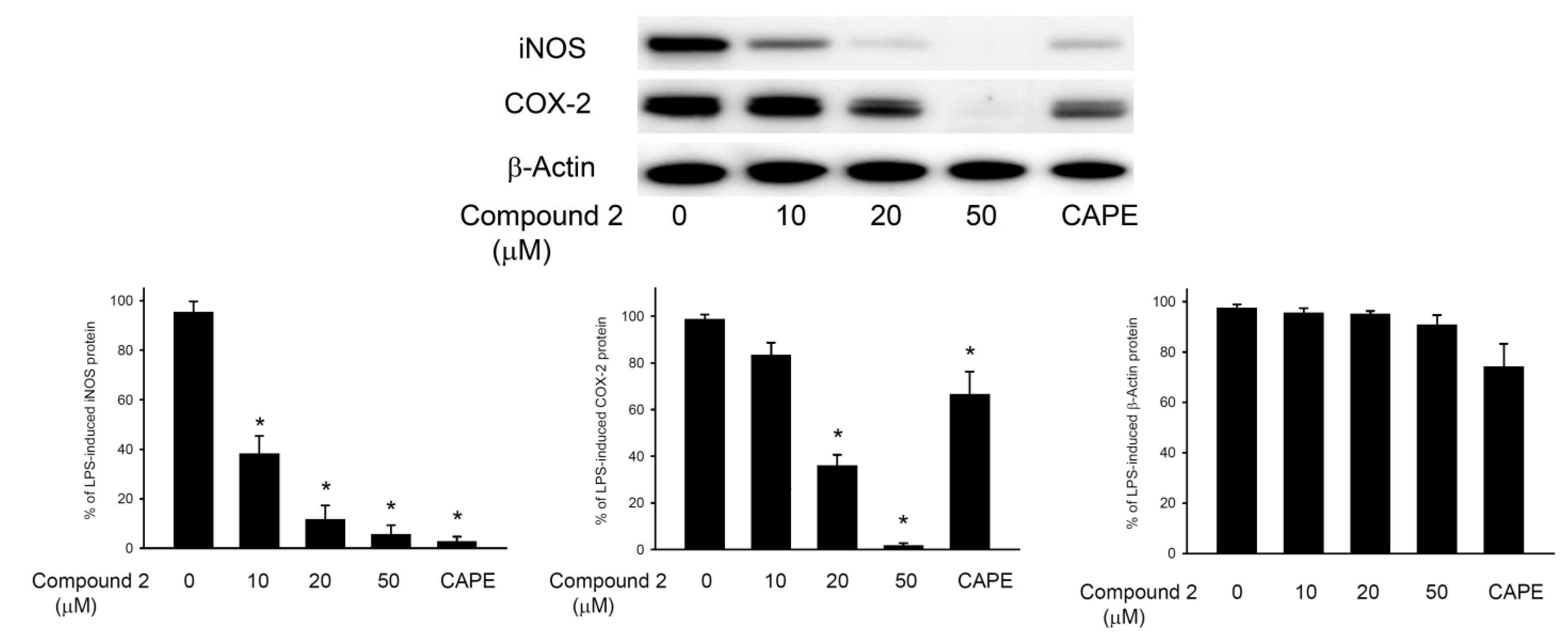
3.6. In Vitro Anti-Inflammatory Assay

The macrophage (RAW264.7) cell line was purchased from ATCC. In vitro anti-inflammatory activity of compounds 1 and 2 was measured by examining the inhibition of lipopolysaccharide (LPS)-induced upregulation of iNOS (inducible nitric oxide synthase) and COX-2 (cyclooxygenase-2) proteins in macrophage cells using Western blotting analysis [27,28,29].

4. Conclusions

In the present study, a new norcembranoidal diterpene, 1-epi-sinulanorcembranolide A (1), and a new cembranoidal diterpene, flexibilin D (2), were isolated from the soft corals, Sinularia gaweli and Sinularia flexibilis, respectively. In addition, a known cembrane, 5-dehydrosinulariolide (=11-dehydrosinulariolide) (3), which was found to show antitumor activities toward CAL-27 (human oral adenosquamous carcinoma) [30] and A2058 (human Caucasian metastatic melanoma) [31] cells and displayed neuroprotection in an in vitro Parkinson’s model [32], was also obtained from S. flexibilis. The structure, including its absolute stereochemistry of 3 was further confirmed by single-crystal X-ray diffraction analysis for the first time in this study. Moreover, in the anti-inflammatory test, compound 2 was found to significantly inhibit the accumulation of the pro-inflammatory iNOS and COX-2 proteins of the LPS-stimulated RAW264.7 macrophage cells. Because octocorals are claimed to be endangered species and based on the potential medicinal use, the soft coral S. flexibilis has begun to be transplanted to culturing tanks located in the National Museum of Marine Biology and Aquarium, Taiwan, for exhibition and the extraction of additional natural products to establish a stable supply of bioactive material.

Acknowledgments

This research was supported by grants from the National Museum of Marine Biology and Aquarium; the National Dong Hwa University; the Division of Marine Biotechnology, Asia-Pacific Ocean Research Center, National Sun Yat-Sen University (Grant No. 00C-0302-05); the Department of Health, Executive Yuan, Taiwan (Grant No. DOH101-TD-C-111-002); and the National Science Council (Grant No. NSC 102-2325-B-291-001, 101-2325-B-291-001, 100-2325-B-291-001 and 101-2320-B-291-001-MY3), Taiwan, awarded to Y.-C.W. and P.-J.S. We thank Chang-Feng Dai, Institute of Oceanography, National Taiwan University, Taiwan, for identification of the coral specimens.

Conflict of Interest

The authors declare no conflict of interest.

References and Notes

  1. Blunt, J.W.; Copp, B.R.; Keyzers, R.A.; Munro, M.H.G.; Prinsep, M.R. Marine natural products. Nat. Prod. Rep. 2013, 30, 237–323. [Google Scholar] [CrossRef]
  2. Chen, W.-T.; Li, Y.; Guo, Y.-W. Terpenoids of Sinularia soft corals: Chemistry and bioactivity. Acta Pharm. Sin. B 2012, 2, 227–237. [Google Scholar]
  3. Li, Y.; Pattenden, G. Novel macrocyclic and polycyclic norcembranoid diterpenes from Sinularia species of soft coral: Structural relationships and biosynthetic speculations. Nat. Prod. Rep. 2011, 28, 429–440. [Google Scholar] [CrossRef]
  4. Yang, B.; Zhou, X.-F.; Lin, X.-P.; Liu, J.; Peng, Y.; Yang, X.-W.; Liu, Y. Cembrane diterpenes chemistry and biological properties. Curr. Org. Chem. 2012, 16, 1512–1539. [Google Scholar] [CrossRef]
  5. Yen, W.-H.; Hu, L.-C.; Su, J.-H.; Lu, M.-C.; Twan, W.-H.; Yang, S.-Y.; Kuo, Y.-C.; Weng, C.-F.; Lee, C.-H.; Kuo, Y.-H.; Sung, P.-J. Norcembranoidal diterpenes from a Formosan soft coral Sinularia sp. Molecules 2012, 17, 14058–14066. [Google Scholar] [CrossRef]
  6. Yen, W.-H.; Chen, W.-F.; Cheng, C.-H.; Dai, C.-F.; Lu, M.-C.; Su, J.-H.; Su, Y.-D.; Chen, Y.-H.; Chang, Y.-C.; Chen, Y.-H.; et al. A new 5α,8α-epidioxysterol from the soft coral Sinularia gaweli. Molecules 2013, 18, 2895–2903. [Google Scholar] [CrossRef]
  7. Yen, W.-H.; Su, Y.-D.; Chang, Y.-C.; Chen, Y.-H.; Chen, Y.-H.; Dai, C.-F.; Wen, Z.-H.; Su, J.-H.; Sung, P.-J. Sinulanorcembranolide A, a novel norcembranoidal diterpene from the octocoral Sinularia gaweli. Tetrahedron Lett. 2013, 54, 2267–2270. [Google Scholar] [CrossRef]
  8. Duh, C.-Y.; Wang, S.-K.; Tseng, H.-K.; Sheu, J.-H.; Chiang, M.Y. Novel cytotoxic cembranoids from the soft coral Sinularia flexibilis. J. Nat. Prod. 1998, 61, 844–847. [Google Scholar] [CrossRef]
  9. Duh, C.-Y.; Wang, S.-K.; Tseng, H.-K.; Sheu, J.-H. A novel cytotoxic biscembranoid from the Formosan soft coral Sinularia flexibilis. Tetrahedron Lett. 1998, 39, 7121–7122. [Google Scholar]
  10. Hsieh, P.-W.; Chang, F.-R.; McPhail, A.T.; Lee, K.-H.; Wu, Y.-C. New cembranolide analogues from the Formosan soft coral Sinularia flexibilis and their cytotoxicity. Nat. Prod. Res. 2003, 17, 409–418. [Google Scholar] [CrossRef]
  11. Lo, K.-L.; Khalil, A.T.; Kuo, Y.-H.; Shen, Y.-C. Sinuladiterpenes A–F, new cembrane diterpenes from Sinularia flexibilis. Chem. Biodivers. 2009, 6, 2227–2235. [Google Scholar] [CrossRef]
  12. Su, J.-H.; Lin, Y.-F.; Lu, Y.; Yeh, H.-C.; Wang, W.-H.; Fan, T.-Y.; Sheu, J.-H. Oxygenated cembranoids from the cultured and wild-type soft corals Sinularia flexibilis. Chem. Pharm. Bull. 2009, 57, 1189–1192. [Google Scholar]
  13. Lin, Y.-S.; Chen, C.-H.; Liaw, C.-C.; Chen, Y.-C.; Kuo, Y.-H.; Shen, Y.-C. Cembrane diterpenoids from the Taiwanese soft coral Sinularia flexibilis. Tetrahedron 2009, 65, 9157–9164. [Google Scholar]
  14. Lo, K.-L.; Khalil, A.T.; Chen, M.-H.; Shen, Y.-C. New cembrane diterpenes from Taiwanese soft coral Sinularia flexibilis. Helv. Chim. Acta 2010, 93, 1329–1335. [Google Scholar] [CrossRef]
  15. Chen, B.-W.; Chao, C.-H.; Su, J.-H.; Huang, C.-Y.; Dai, C.-F.; Wen, Z.-H.; Sheu, J.-H. A novel symmetric sulfur-containing biscembranoid from the Formosan soft coral Sinularia flexibilis. Tetrahedron Lett. 2010, 51, 5764–5766. [Google Scholar]
  16. Shih, H.-J.; Tseng, Y.-J.; Huang, C.-Y.; Wen, Z.-H.; Dai, C.-F.; Sheu, J.-H. Cytotoxic and anti-inflammatory diterpenoids from the Dongsha Atoll soft coral Sinularia flexibilis. Tetrahedron 2012, 68, 244–249. [Google Scholar] [CrossRef]
  17. Su, C.-C.; Wong, B.-S.; Chin, C.; Wu, Y.-J.; Su, J.-H. Oxygenated cembranoids from the soft coral Sinularia flexibilis. Int. J. Mol. Sci. 2013, 14, 4317–4325. [Google Scholar] [CrossRef]
  18. Hu, L.-C.; Su, J.-H.; Chiang, M.Y.-N.; Lu, M.-C.; Hwang, T.-L.; Chen, Y.-H.; Hu, W.-P.; Lin, N.-C.; Wang, W.-H.; Fang, L.-S.; et al. Flexibilins A–C, new cembrane-type diterpenoids from the Formosan soft coral Sinularia flexibilis. Mar. Drugs 2013, 11, 1999–2012. [Google Scholar] [CrossRef]
  19. Tursch, B.; Braekman, J.C.; Daloze, D.; Herin, M.; Karlsson, R.; Losman, D. Chemical studies of marine invertebrates—XI. Sinulariolide, a new cembranolide diterpene from the soft coral Sinularia flexibilis (Coelenterata, Octocorallia, Alcyonacea). Tetrahedron 1975, 31, 129–133. [Google Scholar]
  20. Herin, M.; Tursch, B. Chemical studies of marine invertebrates. XXIV. Minor cembrane diterpenes from the soft coral Sinularia flexibilis (Coelenterata, Octocorallia). Bull. Soc. Chim. Belg. 1976, 85, 707–719. [Google Scholar] [CrossRef]
  21. Lin, Y.-F. Study on the Natural Products of the Wild-Type and Cultured Soft Corals Sinularia flexibilis. Master Thesis, Department of Marine Biotechnology and Resources, National Sun Yat-sen Univeristy, Kaohsiung, Taiwan, July 2008. [Google Scholar]
  22. Wen, T.; Ding, Y.; Deng, Z.; van Ofwegen, L.; Proksch, P.; Lin, W. Sinulaflexiolides A–K, cembrane-type diterpenoids from the Chinese soft coral Sinularia flexibilis. J. Nat. Prod. 2008, 71, 1133–1140. [Google Scholar] [CrossRef]
  23. Cembrane 3 was named as 5-dehydrosinulariolide in [13,22], and this compound was named as 11-dehydrosinulariolide in [10,20,21]. Due to the structure, including its absolute stereochemistry, of 3 was further determined in this study. The authors suggested that this compound should be named as 5-dehydrosinulariolide following the systematic numbering system of cembrane-type compound.
  24. Allinger, N.L. Conformational analysis. 130. MM2. A hydrocarbon force field utilizing V1 and V2 torsional terms. J. Am. Chem. Soc. 1977; 99, 8127–8134. [Google Scholar]
  25. Crystallographic data for the structure of 5-dehydrosinulariolide (3) have been deposited with the Cambridge Crystallographic Data Center as supplementary publication number CCDC 934485. Copies of the data can be obtained, free of charge, on application to CCDC, 12 Union Road, Cambridge, CB2 1EZ, UK (Fax: +44-(0)-1223-336033 or E-Mail: [email protected]).
  26. Flack, H.D. On enantiomorph-polarity estimation. Acta Cryst. 1983, A39, 876–881. [Google Scholar]
  27. Huang, S.-Y.; Chen, N.-F.; Chen, W.-F.; Hung, H.-C.; Lee, H.-P.; Lin, Y.-Y.; Wang, H.-M.; Sung, P.-J.; Sheu, J.-H.; Wen, Z.-H. Sinularin from indigenous soft coral attenuates nociceptive responses and spinal neuroinflammation in carrageenan-induced inflammatory rat model. Mar. Drugs 2012, 10, 1899–1919. [Google Scholar] [CrossRef]
  28. Jean, Y.-H.; Chen, W.-F.; Sung, C.-S.; Duh, C.-Y.; Huang, S.-Y.; Lin, C.-S.; Tai, M.-H.; Tzeng, S.-F.; Wen, Z.-H. Capnellene, a natural marine compound derived from soft coral, attenuates chronic constriction injury-induced neuropathic pain in rats. Br. J. Pharmacol. 2009, 158, 713–725. [Google Scholar] [CrossRef]
  29. Jean, Y.-H.; Chen, W.-F.; Duh, C.-Y.; Huang, S.-Y.; Hsu, C.-H.; Lin, C.-S.; Sung, C.-S.; Chen, I.-M.; Wen, Z.-H. Inducible nitric oxide synthase and cyclooxygenase-2 participate in anti-inflammatory and analgesic effects of the natural marine compound lemnalol from Formosan soft coral Lemnalia cervicorni. Eur. J. Pharmacol. 2008, 578, 323–331. [Google Scholar] [CrossRef]
  30. Liu, C.-I.; Chen, C.-C.; Chen, J.-C.; Su, J.-H.; Huang, H.H.; Chen, J.Y.-F.; Wu, Y.-J. Proteomic analysis of anti-tumor effects of 11-dehydrosinulariolide on CAL-27 cells. Mar. Drugs 2011, 9, 1254–1272. [Google Scholar] [CrossRef]
  31. Su, T.-R.; Tsai, F.-J.; Lin, J.-J.; Huang, H.H.; Chiu, C.-C.; Su, J.-H.; Yang, Y.-T.; Chen, J.Y.-F.; Wong, B.-S.; Wu, Y.-J. Induction of apoptosis by 11-dehydrosinulariolide via mitochondrial dysregulation and ER stress pathways in human melanoma cells. Mar. Drugs 2012, 10, 1883–1898. [Google Scholar] [CrossRef]
  32. Chen, W.-F.; Chakraborty, C.; Sung, C.-S.; Feng, C.-W.; Jean, Y.-H.; Lin, Y.-Y.; Hung, H.-C.; Huang, T.-Y.; Huang, S.-Y.; Su, T.-M.; et al. Neuroprotection by marine-derived compound, 11-dehydrosinulariolide, in an in vitro Parkinson’s model: A promising candidate for the treatment of Parkinson’s disease. Naunyn Schmiedebergs Arch. Pharmacol. 2012, 385, 265–275. [Google Scholar] [CrossRef]
  • Samples Availability: Sample of the cembrane 3 is available from the authors.

