2.1. Structure Determination
Compound
1 was isolated as colorless oil. High-resolution mass spectrometry data gave a molecular formula of C
15H
22O (
m/
z 219.1720 [M + H]
+). The IR spectrum suggested the presence of α,β-unsaturated carbonyl group (1662 cm
−1) in the molecule. The
13C-NMR spectral data of
1 (
Table 1) revealed the presence of 15 carbon atoms, including four methyls, three methylenes, four methines and four quaternary carbons, based on DEPT-135 and HSQC spectra, suggesting a chemical skeleton of sesquiterpenoid. The NMR signals of
1 (
Table 1) showed the presence of a ketone group at δC 206.6 (C), two trisubstituted double bonds at δ
C 146.5 (CH), 137.1 (C), 135.1 (C) and 125.2 (CH) (δH 6.09 (1H, ddq,
J = 12.4, 3.4, 1.4 Hz) and 4.87 (1H, d,
J = 10.3 Hz)), a cyclopropyl moiety at δC 26.1 (CH), 29.6 (CH) and 20.2 (C) (δH 1.32 (1H, dd,
J = 10.3, 8.9 Hz) and 0.77 (1H, ddd,
J = 11.7, 8.9, 2.8 Hz)), and four tertiary methyls at δC 13.3 (CH
3), 16.7 (CH
3), 15.9 (CH
3) and 29.3 (CH
3) (δH 1.74, 1.49, 1.12 and 1.08 (each 3H, s)) (see
1H- and
13C-NMR data of
1 in
Supplementary Information). Five degrees of unsaturation calculated
via HR-MS could be attributed to three double bonds and a bicyclic sesquiterpenoid framework leading to the structure of compound
1.
Table 1.
1H- and 13C-NMR data (600 MHz and 150 MHz, in CDCl3) of 1 and 2 (δ in ppm, J in Hz).
Table 1.
1H- and 13C-NMR data (600 MHz and 150 MHz, in CDCl3) of 1 and 2 (δ in ppm, J in Hz).
| Position | 1 | 2 |
|---|
| δC | δH | δC | δH |
|---|
| 1β | 26.1 (CH) | 1.32 dd (10.3, 8.9) | 27.9 (CH) | 1.45 t (8.9) |
| 2 | 125.2 (CH) | 4.87 d (10.3) | 122.9 (CH) | 4.61 d (8.9) |
| 3 | 137.1 (C) | | 137.6 (C) | |
| 4α | 38.9 (CH2) | 2.34 m | 39.3 (CH2) | 1.58 t (11.7) |
| β | 2.15 dd (11.7, 8.3) |
| 5α | 25.1 (CH2) | 2.47 td (12.4, 8.3) | 23.7 (CH2) | 2.25 td (12.2, 8.3) |
| β | 2.31 m | 2.01 dt (12.2, 8.3) |
| 6 | 146.5 (CH) | 6.09 ddq (12.4, 3.4, 1.4) | 130.2 (CH) | 5.40 tq (8.3, 1.4) |
| 7 | 135.1 (C) | | 140.0 (C) | |
| 8 | 206.6 (C) | | 211.5 (C) | |
| 9α | 37.1 (CH2) | 2.77 t (11.7) | 36.3 (CH2) | 2.32 d (8.9) |
| β | 2.37 dd (11.7, 2.8) |
| 10β | 29.6 (CH) | 0.77 ddd (11.7, 8.9, 2.8) | 28.3 (CH) | 1.38 q (8.9) |
| 11 | 20.2 (C) | | 21.7 (C) | |
| 12α | 15.9 (CH3) | 1.12 s | 16.1 (CH3) | 1.11 s |
| 13β | 29.3 (CH3) | 1.08 s | 29.2 (CH3) | 1.11 s |
| 14 | 13.3 (CH3) | 1.74 s | 21.9 (CH3) | 1.88 s |
| 15 | 16.7 (CH3) | 1.49 s | 17.4 (CH3) | 1.55 s |
Assignment of
1H-
13C correlations of
1 was determined by HSQC analysis. Two separate consecutive spin systems of “a” and “b” of
1 were revealed by
1H-
1H COSY correlations. Correlations corresponding to H
2-4/H
2-5/H-6 are represented by the “a” spin system and H-1/H-2/H
2-9/H-10 by the “b” spin system, and are depicted by the bold lines in
Figure 2. Both the “a” and “b” structural units were assembled to establish a bicyclic system comprised of fused 10- and 3-membered rings, suggesting a bicyclogermacrene skeleton for
1. This was achieved using key HMBC correlations of H
3-12α to C-1, C-10, C-11 and C-13; H
3-13β to C-1, C-10, C-11 and C-12; H
3-14 to C-6, C-7 and C-8; and H
3-15 to C-2, C-3 and C-4 as depicted by arrows in
Figure 2. Thus,
1 was deduced to possess a 10-membered ring with a ketone group at C-8, and a cyclopropyl moiety located at the C-1/C-10. This cyclopropyl ring was confirmed by the “b” partial structure based on the presence of HMBC correlations between H
3-12 to C-1, C-10, C-11 and C-13; and H
3-13 to C-1, C-10, C-11 and C-12. In addition, the upfield chemical shifts of H-6β (δ 1.32) and H-7β (δ 0.77), further supported the presence of this cyclopropyl moiety [
9]. Based on these analyses, the planar structure of
1 was determined to be as shown in
Figure 2.
Figure 2.
Selected 1H-1H COSY (▬) and HMBC (→) correlations of 1 and 2.
Figure 2.
Selected 1H-1H COSY (▬) and HMBC (→) correlations of 1 and 2.
The relative stereochemistry of compound
1 was deduced from NOESY correlations as shown in
Figure 3. In addition, the double bonds geometries were deduced from the
13C-NMR chemical shifts at C-14 (δC 13.3) and C-15 (δC 16.7), which suggested
E-configurations [
10,
11]. Besides, the lack of NOE correlations between H-6 with H
3-14 and H-2 with H
3-15, further supported this configuration. Other NOE analyses, H-6 showed NOE correlations to H-2, H
2-5α and H
2-9α; and H-2 showed NOE correlations with H
2-4α, H-6, H
2-9α and H
3-12α. Therefore, H
2-4α, H
2-5α, H
2-9α, and H
3-12α are suggested to be of the similar orientation, reflecting an α-orientation, hence H
2-4β, H
2-5β and H
2-9β are suggested to be of β-orientation. Furthermore, H-1β, H-10β, and H
3-13β were confirmed to be positioned on the β-orientation, due to NOE correlations observed between H-1β with H-10β, H
3-13β, and H
3-15. Besides that, the scalar coupling value between H-1β and H-10β was 8.9 Hz and this coupling value suggested a
cis-cyclopropyl group with both methine protons as β-orientated [
12]. Based on these configurations, the relative structure of
1 is reported as (1
S,10
R,2
E,6
E)-3,7,11,11-tetramethylbicyclo(8.1.0)undeca-2,6-dien-8-one. The compounds
1 and
2 were
trans and
cis isomers, but downfield shifts were observed at H-1β in
1 and
2, and H-10β in
2. The downfield shifts of both cyclopropane methines for
2 (δH 1.45 and 1.38) were similar to those observed in cyclocolorenone (δH 1.54 and 1.29) and its derivative [
13,
14]. It is reported that this cyclocolorenone also has one double bond beside these cyclopropane methines similar to those of
1 and
2, where the double bond was located at C2/C3. Thus, it could be due to the presence of this double bond at C2/C3 and its proton deshielding ability towards cyclopropane methines in
1 and
2. However, the H-10β (δH 0.77) in
1 was relatively more shielded as compared to those of
2. This could be due to the presence of
trans geometry double bond at C6/C7 and the position of oxygen atom (carbonyl group was oriented upward and outside the 10-membered ring) of
1 as compared that of
2. The chemical shifts of H-6 and C-6 in
1 were significantly downfield compared to those observed for
2, possibly due to the mesomeric effect [
15].
Figure 3.
Selective correlations of NOESY for 1 and 2.
Figure 3.
Selective correlations of NOESY for 1 and 2.
Compound
2 was obtained as colorless crystals. Its molecular formula, C
15H
22O was established by HR-MS (
m/
z 219.1725 [M + H]
+), revealing five degrees of unsaturation. The IR spectrum indicated the presence of α,β-unsaturated carbonyl functionality (1683 cm
−1) in the molecule. Comparison of the NMR data (
Table 1) of
2 with those of
1 also revealed the structure
2 to be of bicyclogermacrene type. Based on 2D NMR spectra, two separate consecutive proton spin systems “a” and “b” of
2 were revealed in
1H-
1H COSY spectrum (
Figure 2); “a” spin system for H
2-4/H
2-5/H-6 and “b” for H-1/H-2/H
2-9/H-10. Planar structure of
2 was established based on HMBC correlations. The relative configurations of the two successive chiral centers at C-1 and C-10 in
2 were determined based on NOESY data, as shown in
Figure 3. It was revealed that H-6 had NOE correlation with H
3-14 and H-2 had no NOE correlation with H
3-15, implying the
Z- and
E-configurations for double bonds at C-6/C-7 and C-2/C-3, respectively. Therefore, by comparison of the
13C-NMR of C-14 of
2 with that of
1, it was suggested that
2 had a
cis and
1 had a
trans double bond at C-6/C-7 [
10,
11]. In addition, H-2 showed NOE interactions with H
2-4α, H
2-9α, and H
3-12α; and H
3-15 exhibited NOE correlation with H
2-5α. Therefore, H
2-4α, H
2-5α, H
2-9α, and H
3-12α reflected an α-orientation, hence indicating H
2-4β, H
2-5β and H
2-9β were β-orientated. Further analysis of other NOE correlations and scalar coupling between H-1β and H-10β, revealed
2 possessed the similar relative configurations at C-1, C-10, and C-13 as that of
1. On the basis of above findings, the relative structure of
2 is reported as (1
S,10
R,2
E,6
Z)-3,7,11,11-tetramethylbicyclo(8.1.0)undeca-2,6-dien-8-one. The
1H- and
13C-NMR chemical shift at C-6 of
2 were more shielded as compared to those of
1 due to the absence of mesomeric effect.
2.2. Anti-Inflammatory Properties
Anti-inflammatory activity bioassay of compounds
1–
4 against RAW 264.7 macrophages were evaluated based on the accumulation of NO production and cell viability induced by LPS (1 μg/mL) (
Figure 4 and
Figure 5). The results showed compound
1 displayed potent anti-inflammatory potential by significantly reducing the NO production of LPS-induced RAW macrophages to 28.0% and 14.2% at 10 and 20 μg/mL, respectively. Cell viability of LPS-induced RAW macrophages in the presence of compound
1 was 105.1% and 74.6% at 10 and 20 μg/mL, respectively. Therefore, compound
1 was selected for further anti-inflammatory investigation against accumulation of NO, PGE
2, and pro-inflammatory cytokines (TNF-α, IL-1β, and IL-6) production and the expression of iNOS and COX-2 proteins induced by LPS (1 μg/mL) in RAW 264.7 cells were evaluated. The effect of compound
1 on NO production in LPS-treated RAW 264.7 macrophages was repeated at lower concentrations, as shown in
Figure 6. The findings showed NO production of compound
1 was 69.1%, 60.0%, and 24.6% at the concentrations of 5, 10, and 20 μg/mL, respectively. Based on the cell viability screening, compound
1 revealed the potential to significantly inhibit LPS-induced NO production in a concentration dependent manner, also suggesting absence of cytotoxic effects in RAW 264.7 macrophage cells. Dexamethasone used as positive control inhibited NO production to 16.01% at 5.0 μg/mL. At the concentration of 12.51 ± 0.16 μg/mL (57.34 μM) of compound
1, it inhibited 50% of NO production in LPS-stimulated RAW 264.7 macrophages.
Figure 4.
The effect of compounds 1–4 on NO production in RAW 264.7 macrophages.
Figure 4.
The effect of compounds 1–4 on NO production in RAW 264.7 macrophages.
Figure 5.
The effect of compounds 1–4 on cell viability in RAW 264.7 macrophages.
Figure 5.
The effect of compounds 1–4 on cell viability in RAW 264.7 macrophages.
Figure 6.
Concentration dependent effects of compound 1 on NO production in RAW 264.7 macrophages.
Figure 6.
Concentration dependent effects of compound 1 on NO production in RAW 264.7 macrophages.
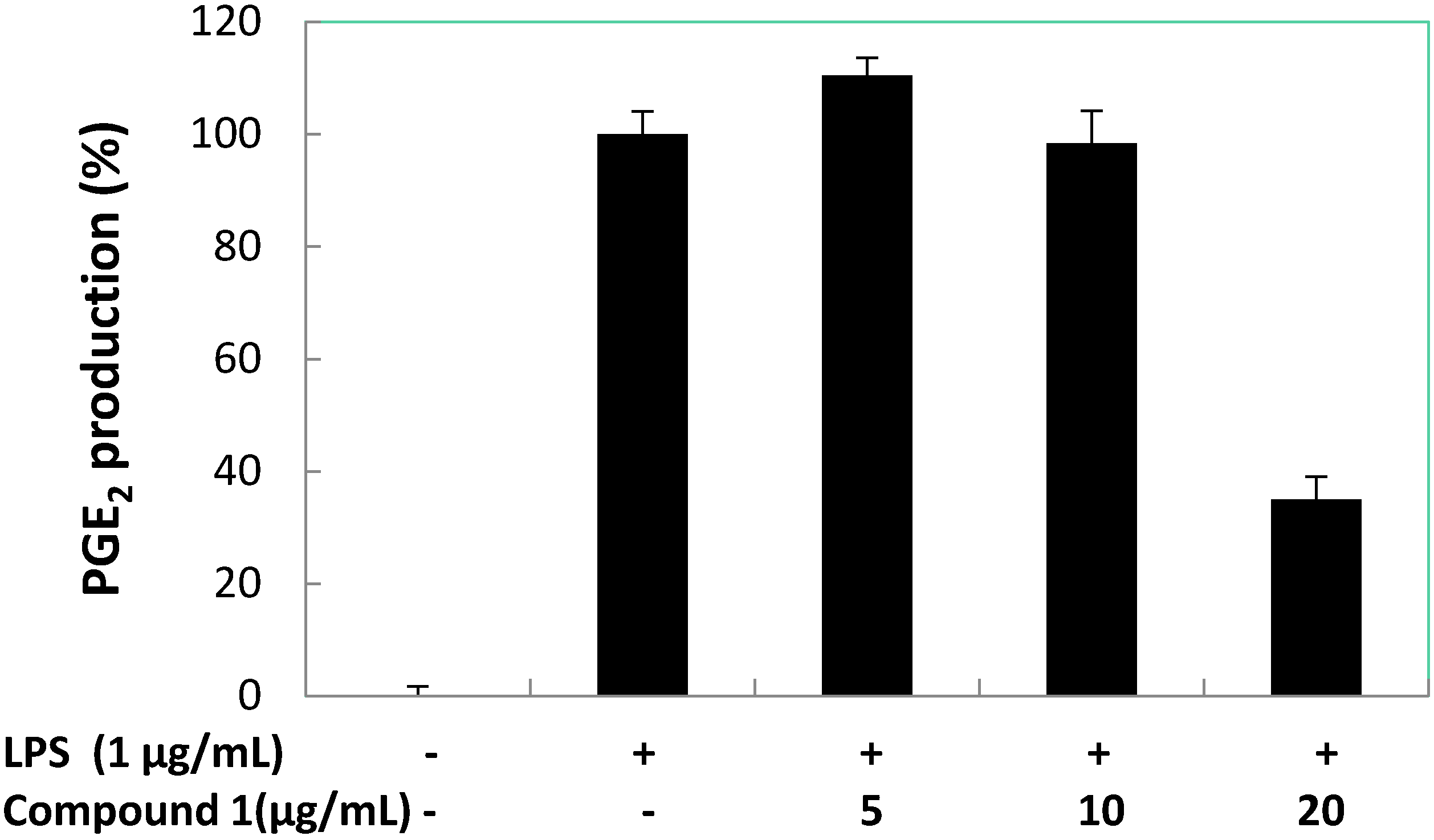
Further, we evaluated the effect of compound
1 on the LPS-induced production of PGE
2 in RAW 264.7 macrophage cells (
Figure 7). However, it was apparent that compound
1 was not a good inhibitor of PGE
2 production in LPS-treated RAW 264.7 macrophages at 5 and 10 μg/mL, but compound
1 reduced the PGE
2 production to below 40% at 20 μg/mL. Dexamethasone (positive control) inhibited PGE
2 production to 11.58% at 5.0 μg/mL. Compound
1 at 18.97 ± 0.63 μg/mL (86.93 μM) inhibited 50% of PGE
2 production in LPS-stimulated RAW 264.7 macrophages.
Figure 7.
Concentration dependent effects of compound 1 on LPS-induced PGE2 production in RAW 264.7 macrophages.
Figure 7.
Concentration dependent effects of compound 1 on LPS-induced PGE2 production in RAW 264.7 macrophages.
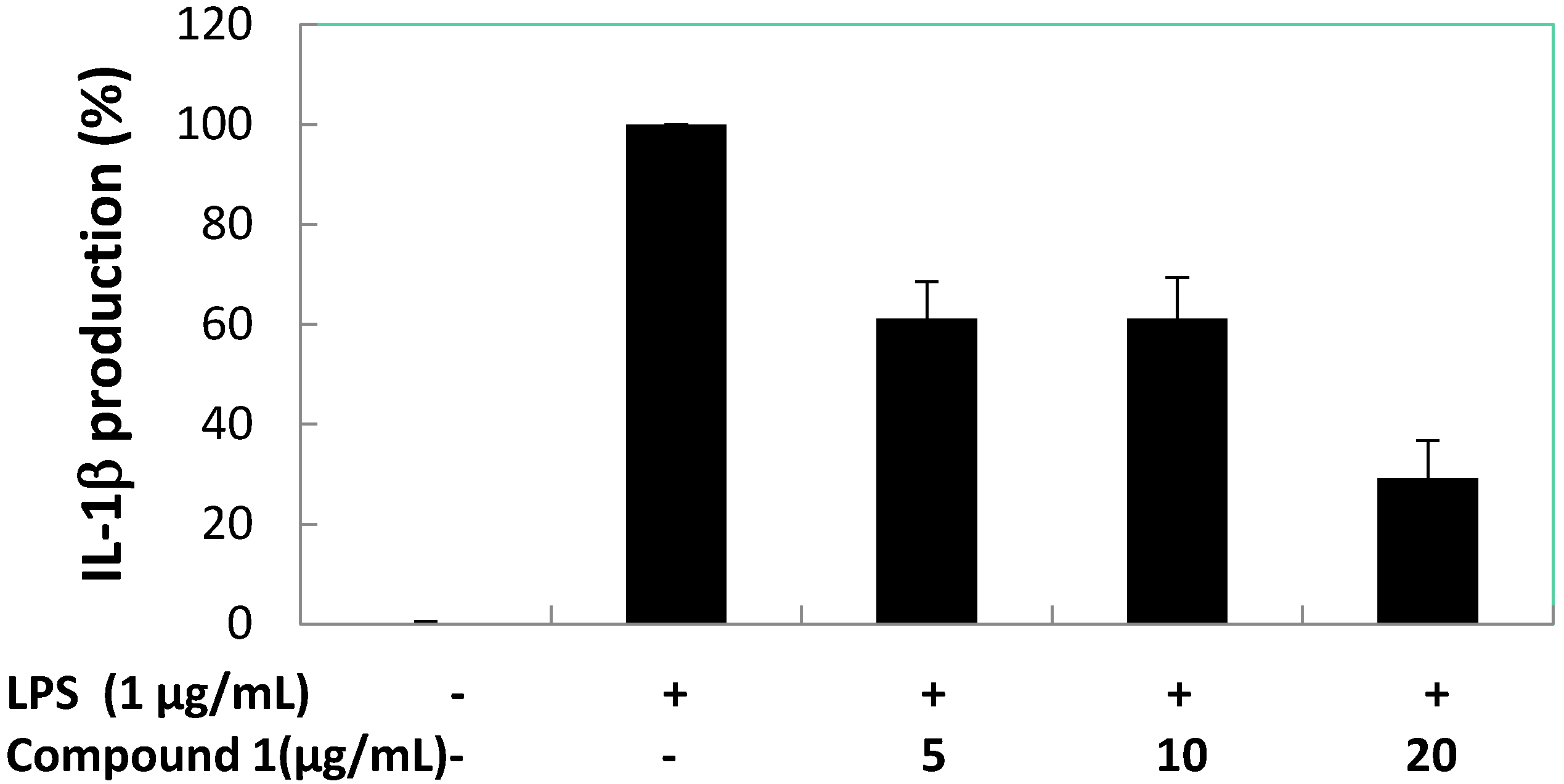
Subsequently, the effects of compound
1 on pro-inflammatory cytokines (TNF-α, IL-1β, and IL-6) production induced by LPS in RAW 264.7 macrophage cells were quantified (
Figure 8,
Figure 9 and
Figure 10). The result shows there was almost no inhibition of TNF-α and IL-6 production by compound
1 at 5, 10, and 20 μg/mL. However, compound
1 was able to inhibit the production of IL-1β with a reduction of 40% compared to the LPS-induced group at 5 and 10 μg/mL, and the IL-1β production value dropped to below 35% when 20 μg/mL of compound
1 was used. At 5.0 μg/mL of positive control (dexamethasone) inhibition of TNF-α, IL-1β and IL-6 reduced production to 15.31, 12.23 and 13.56%, respectively. At the concentration of 12.89 ± 1.38 μg/mL (59.06 μM) compound
1, inhibited 50% of IL-1β production in LPS-stimulated RAW 264.7 macrophages.
Figure 8.
Concentration dependent effects of compound 1 on LPS-stimulated TNF-α release in RAW 264.7 cells.
Figure 8.
Concentration dependent effects of compound 1 on LPS-stimulated TNF-α release in RAW 264.7 cells.
Figure 9.
Concentration dependent effects of compound 1 on LPS-treated IL-1β release in RAW 264.7 macrophage cells.
Figure 9.
Concentration dependent effects of compound 1 on LPS-treated IL-1β release in RAW 264.7 macrophage cells.
Figure 10.
Concentration dependent effects of compound 1 on LPS-induced IL-6 release in RAW 264.7 macrophage cells.
Figure 10.
Concentration dependent effects of compound 1 on LPS-induced IL-6 release in RAW 264.7 macrophage cells.
Further investigation pertaining to the mechanism of anti-inflammatory activity of compound
1 was evaluated by observing the expression of iNOS and COX-2 proteins via Western blot, as shown in
Figure 11. The result showed pretreatment with compound
1 significantly inhibited iNOS protein expression in a concentration dependent manner. However, compound
1 showed little inhibition of COX-2 protein expression at first concentration at 5 μg/mL, followed by 10 and 20 μg/mL by visual observation of bands densities, indicating little inhibition of PGE
2 production in LPS-treated RAW 264.7 macrophages. In this anti-inflammatory assay, we confirmed that LPS had significantly increased TNF-α, IL-1β, IL-6, PGE
2 and NO production, which resulted in the over expression of iNOS and COX-2. Pretreatment of LPS-induced RAW 264.7 macrophages with compound
1 exhibited the ability to inhibit NO and IL-1β production by down-regulating expression of iNOS. In addition, compound
1 also showed little inhibition of PGE
2 by little suppression of COX-2 expression.
Figure 11.
Concentration dependent effects of compound 1 on protein and mRNA expressions of iNOS and COX-2 in LPS-induced RAW 264.7 macrophages.
Figure 11.
Concentration dependent effects of compound 1 on protein and mRNA expressions of iNOS and COX-2 in LPS-induced RAW 264.7 macrophages.
Compound 1 and 2 were trans and cis isomers, but only compound 1 showed anti-inflammatory activity. It is our assumption that the differences in activity between these two compounds are due to their stereo differences. Compound 1 has a larger space of 10-membered ring as compared those of 2. The oxygen atom of 2 was positioned inside of the 10-membered ring, but oxygen atom of 1 was orientated outside of the 10-membered ring. Thus, the anti-inflammatory activity of compound 1 could be attributed to the larger space within the 10-membered ring and the position of oxygen atom. However, further SAR (Structure Activity Relation) analysis would be required to confirm this phenomenon.


