Article

Anti-Food Allergic Compounds from Penicillium griseofulvum MCCC 3A00225, a Deep-Sea-Derived Fungus

1
Key Laboratory of Marine Biogenetic Resources, Third Institute of Oceanography, Ministry of Natural Resources,184 Daxue Road, Xiamen 361005, China
2
Fujian Universities and Colleges Engineering Research Center of Marine Biopharmaceutical Resources, Xiamen Medical College, 1999 Guankouzhong Road, Xiamen 361023, China
3
College of Food and Biological Engineering, Jimei University, 43 Yindou Road, Xiamen 361021, China
*
Authors to whom correspondence should be addressed.
Mar. Drugs 2021, 19(4), 224; https://doi.org/10.3390/md19040224
Submission received: 25 March 2021 / Revised: 10 April 2021 / Accepted: 12 April 2021 / Published: 16 April 2021
(This article belongs to the Special Issue Bioactive Molecules from Extreme Environments II)

Abstract

:
Ten new (110) and 26 known (1136) compounds were isolated from Penicillium griseofulvum MCCC 3A00225, a deep sea-derived fungus. The structures of the new compounds were determined by detailed analysis of the NMR and HRESIMS spectroscopic data. The absolute configurations were established by X-ray crystallography, Marfey’s method, and the ICD method. All isolates were tested for in vitro anti-food allergic bioactivities in immunoglobulin (Ig) E-mediated rat basophilic leukemia (RBL)-2H3 cells. Compound 13 significantly decreased the degranulation release with an IC50 value of 60.3 μM, compared to that of 91.6 μM of the positive control, loratadine.

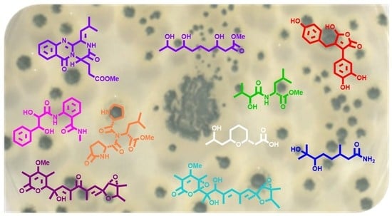
Graphical Abstract

1. Introduction

For the past decade, the trend to discover new compounds from marine microorganisms continues to rise [1], especially from marine fungi [2,3], which accounted for 68% of the reported new marine natural products in 2019 [4]. Of particular importance is the Penicillium species, which are recognized as the richest source for the discovery of biologically important and structurally unique secondary metabolites [5,6,7,8].
As our ongoing research for novel and bioactive secondary metabolites from the deep sea-derived microorganisms [8,9,10,11], the fungal strain Penicillium griseofulvum isolated from the Indian Ocean sediment was selected for a systematic chemical examination. As a result, five carotanes, four naphthalenes, and three viridicatol derivates were obtained [12,13]. A continuous study, however, led to the isolation of 10 new (Figure 1) and 26 known compounds. Herein, we report the isolation, structure elucidation, and biological activity of these compounds.

2. Results and Discussion

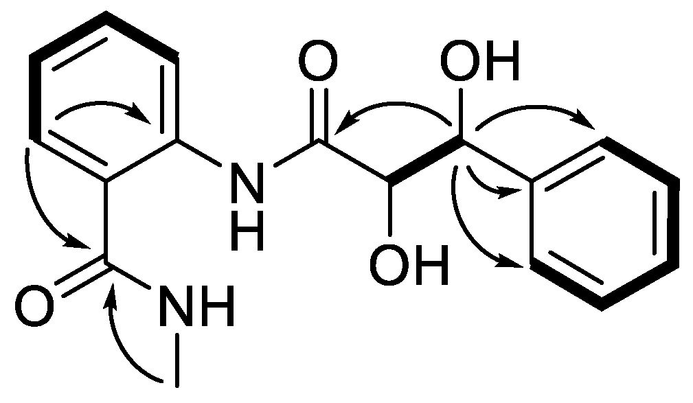
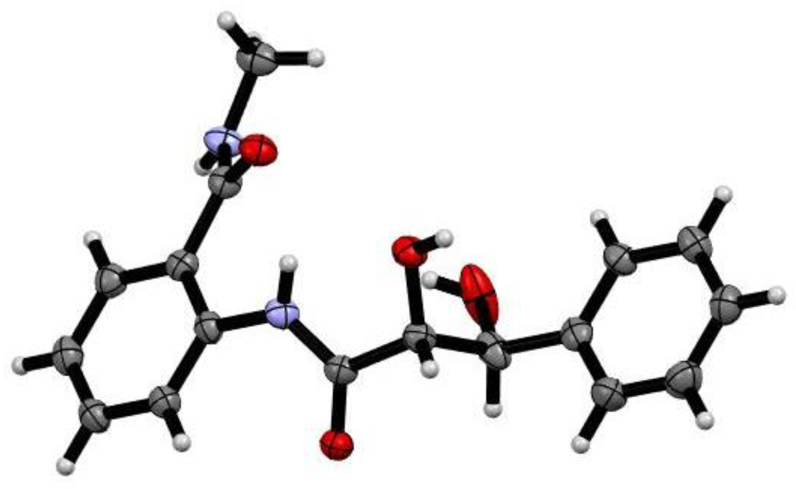
Compound 1 was isolated as a white powder. Its molecular formula was established as C17H18N2O4 according to the protonated molecule peak at m/z 337.1176 [M + Na]+ in its (+)−HRESIMS (High Resolution Electrospray Ionization Mass Spectroscopy) spectrum, requiring ten degrees of unsaturation. The 1H and 13C NMR spectroscopic data (Figures S1 and S2, Table 1) displayed 17 carbons, characteristics of one mono-substituted aromatic unit [δH 7.24 (1H, br t, J = 7.4 Hz, H-4), 7.33 (2H, dd, J = 7.8, 7.3 Hz, H-3, 5), 7.46 (2H, d, J = 7.8 Hz, H-2, 6); δC 127.5 (d × 2, C-2/C-6), 128.3 (d, C-4), 129.1 (d × 2, C-3/C-5), 143.2 (s, C-1)], one ortho-disubstituted benzene moiety [δH 7.16 (1H, td, J = 7.6, 1.0 Hz, H-5′), 7.47 (1H, td, J = 7.8, 1.5 Hz, H-4′), 7.60 (1H, dd, J = 7.8, 1.4 Hz, H-6′), 8.51 (1H, d, J = 8.1 Hz, H-3′); δC 122.4 (d, C-3′), 124.2 (s, C-1′), 124.7 (d, C-5′), 128.8 (d, C-6′), 132.7 (d, C-4′), 138.8 (s, C-2′)], one methyl [δH 2.89 (3H, s, 7′-NMe); δC 26.8 (q, 7′-NMe)], two oxygenated methines [δH 4.25 (1H, d, J = 2.3 Hz, H-8); 5.16 (1H, d, J = 2.0 Hz, H-7) δC 75.6 (d, C-7), 77.8 (d, C-8)], and two carbonyls [δC 171.3 (s, C-7′), 174.0 (s, C-9)]. In the 1H–1H COSY (Correlation Spectroscopy) spectrum, correlations of H-2 (H-6)/H-3 (H-5)/H-4, H-3′/H-4′/H-5′/H-6′, and H-7 (δH 5.16, d, J = 2.0 Hz)/H-8 (δH 4.25, d, J = 2.3 Hz) confirmed the two benzene units and deduced another fragment of C-7/C-8. By the HMBC (Heteronuclear Multiple-bond Correlation) correlations of H-7 (δH 5.16) to C-1/C-2/C-6/C-9 and H-6′ (δH 7.60)/7′-NMe (δH 2.89) to C-7′, 1 was then assigned a phenylpropionyl moiety and a benzamide groups (Figure 2). However, the limited HMBC correlations hindered the connection of these two fragments. Fortunately, crystals of 1 were obtained. By the single X-ray crystallography (Figure 3), the absolute configuration of 1 was then unambiguously assigned as 2-(2R,3S-dihydroxy-3-phenyl-propionylamino)-N-methyl-benzamide, and named penigrisamide.
Compound 2 was afforded as a colorless oil. The molecular formula C19H21N3O4 was deduced from (+)-HRESIMS data (m/z 378.1418 for [M + Na]+), indicative of eleven degrees of unsaturation. The 1H and 13C NMR spectroscopic data (Figures S7 and S8 from the Supplementary Materials, Table 2) exhibited 19 carbons, including three methyl singlets (one oxygenated), two methylenes, seven methines (five olefinic), and seven non-protonated carbons (one carbonyl and two ketone groups). These signals were closely similar to those of aurantiomide C (11) [14], except that the terminal amino group in 11 was replaced by the methoxy unit (δC 52.2) in 2. The assumption was confirmed by the HMBC correlation of 17-OMe (δH 3.46) to C-17 (δC 173.9). Accordingly, the structure of 2 was determined as 17-deamino-17-methoxylaurantiomide C, and named aurantiomoate C.
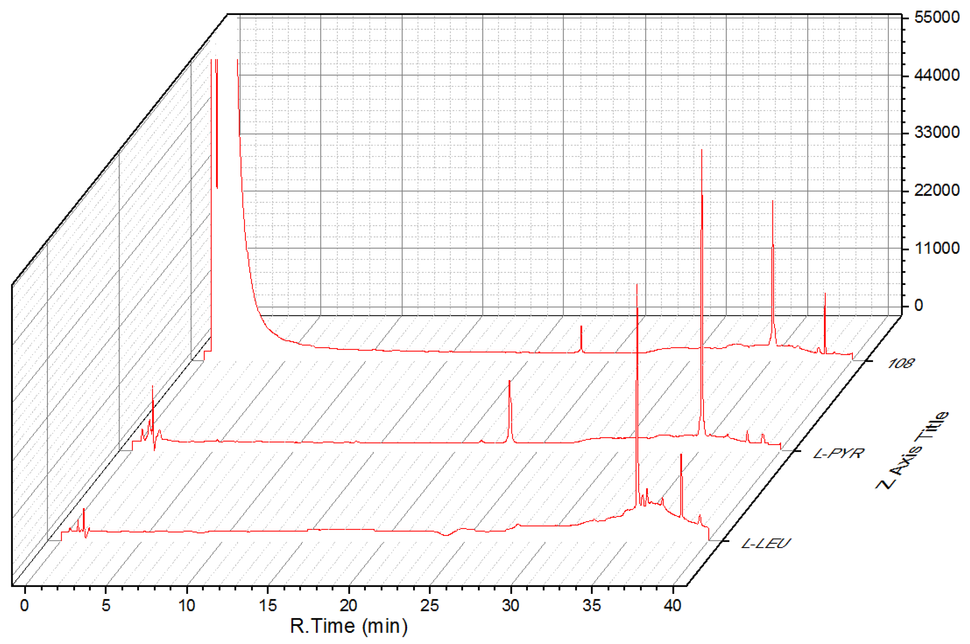
Compound 3 was obtained as a colorless oil. Its molecular formula was established as C17H27N3O5 on the basis of the protonated molecule peak at m/z 376.1841 [M + Na]+ in its (+)-HRESIMS spectrum, requiring six degrees of unsaturation. Diagnostic NMR data for 3 suggested the presence of a pyroglutamylleucinmethylester (20) [15]. Moreover, the 1H–1H COSY correlation of H2-4″ (δH 3.63 m)/H2-5″ (δH 2.02 m) and H2-6″ (δH 2.18 m, 2.00 m)/H-2″ (δH 4.47, dd, J = 8.4, 2.8 Hz), with HMBC correlations from H-2″ (δH 4.47, dd, J = 8.4, 2.8 Hz) to C-4″/C-5″, and H-6″ (δH 2.18 m, 2.00 m) to C-1″/C-4″, allowed for the presence of another pyroglutamyl moiety. The absolute configuration of 3 was determined by the hydrolysis and derivation using Marfey’s reagent, and Nα-(2,4-dinitro-5-fluorophenyl)-l-alaninamide (FDDA) derivatives were compared with the retention times of standard FDDA-amino acids (Figure 4). On the basis of the above evidences, 3 was then assigned as N,N-pyroglutamylleucinmethylester.
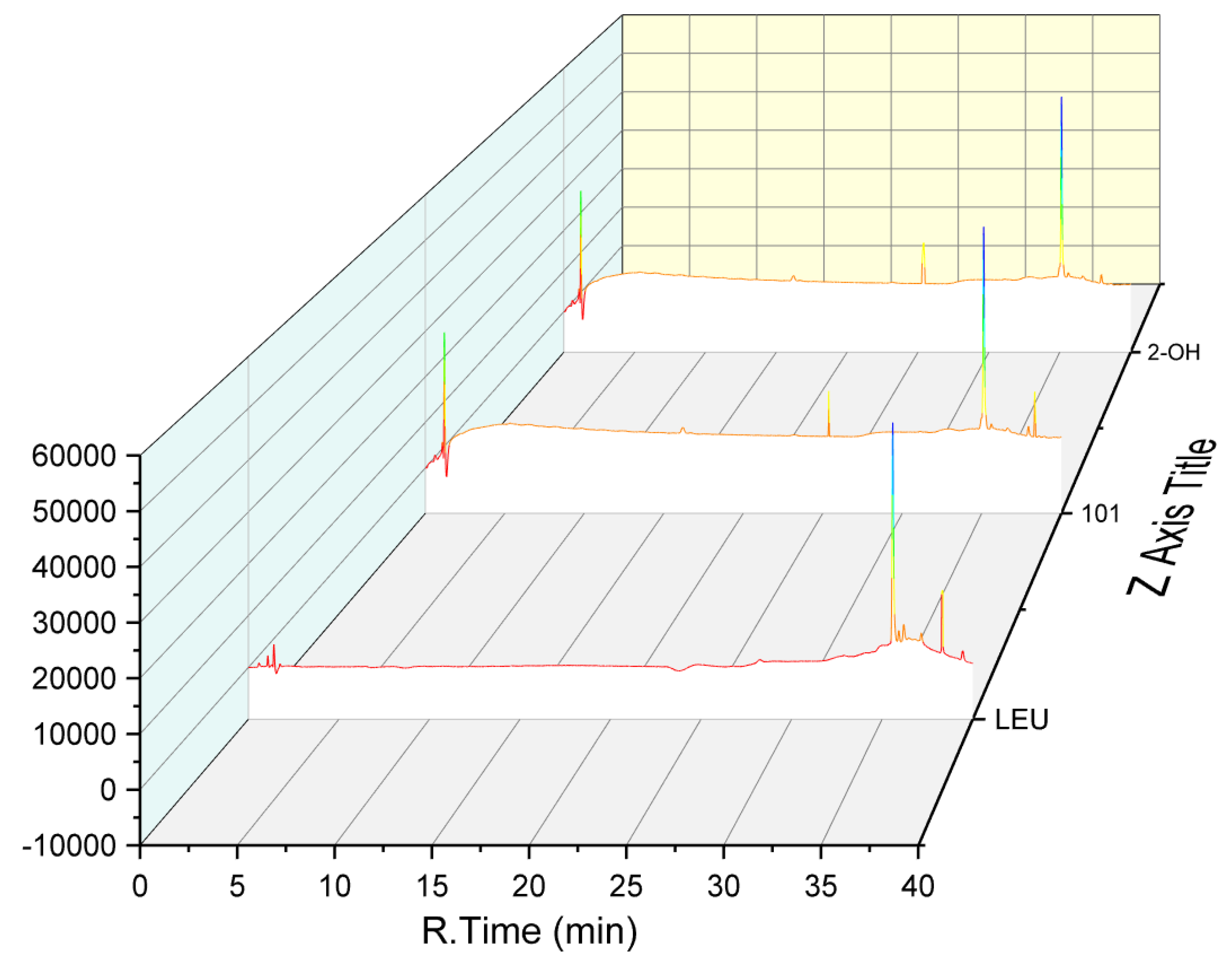
Compound 4 was obtained as a colorless oil. Its molecular formula was established as C12H23NO4 based on the sodium adduct ionic peak at m/z 268.1526 [M + Na]+ in its positive HRESIMS spectrum, requiring two degrees of unsaturation. Its 1H and 13C NMR spectra were very similar to those of pyroglutamylleucinmethylester (20) [15], except for a 2-hydroxy-3-methylbutanoyl unit instead of a pyroglutamyl moiety in 4. This was confirmed by the 1H–1H COSY correlations of H3-4′ (δH 1.00, d, J = 7.0 Hz) and H3-5′ (δH 0.84, d, J = 6.8 Hz) via H-3′ (δH 2.07 m) to H-2′ (δH 3.86, d, J = 3.7 Hz). Via detailed analysis of the HMBC spectroscopic data and using Marfey’s method (Figure 5), the absolute configuration of 4 was then assigned as methyl-2S-hydroxy-3-methylbutanoyl-l-leucinate.
The molecular formula of 5 was established as C24H32O7 by the ion peak at m/z 455.2040 [M + Na]+ in its positive HRESIMS. The 1H and 13C NMR spectra exhibited 24 carbons, including three doublets and five singlet methyls, one methoxyl, four methines (two oxygenated and two olefinic), and eleven quaternary carbons (six olefinic and two carbonyl carbons). These signals were closely similar to those of penicyrone A [16] except that the hydroxy (δC 82.6) at the C-9 position in penicyrone A was replaced by the carbonyl (δC 202.8) in 5. This was confirmed by the HMBC correlations from H-7 (δH 6.38, d, J = 1.4 Hz)/H-11(δH 6.28, d, J = 1.4 Hz)/H3-19 (1.67, s)/H3-20 (δH 2.02, d, J = 1.4 Hz) to δC 202.8. Accordingly, 5 was established to be 9-dehydroxy-9-oxopenicyrone A, and named verrucosidinol A.
Compound 6 presented a molecular formula of C24H34O8 by positive HRESIMS at m/z 473.2140 [M + Na]+. Comparison of the 1H and 13C NMR spectra of 6 with those of verrucosidinol (25) [17] showed they were very similar except that two olefinic carbons at C-4 and C-5 in 25 were replaced by an epoxy group in 6. This was evidenced by the HMBC correlations from H3-16 (δH 1.84) to C-1/C-2/C-3, H3-17 (δH 1.61) to C-3/C-4/C-5, and H3-18 (δH 1.28) to C-5/C-6/C-7. Therefore, 6 was established as 4,5-dihydro-4,5-epoxyverrucosidinol, and named verrucosidinol B.
Compound 7 had a molecular formula C17H14O6 as assigned by its positive HRESIMS at m/z 337.0690 [M + Na]+. Its 1H and 13C NMR spectroscopic data greatly resembled those of helvafuranone [18] except for an additional hydroxy substituent at the C-8 position. By detailed analysis of its 1D and 2D NMR spectroscopic data, 7 was then established as 8-hydroxyhelvafuranone.
Compound 8 gave a molecular formula C10H21NO3 as deduced by the protonated molecule peak at m/z 202.1504 [M − H] in its negative HRESIMS spectrum. The 1H NMR spectrum exhibited one methyl doublet at δH 0.96 (3H, d, J = 6.2 Hz, H-10), and two methyl singlets at δH 1.12 (3H, s, H-9) and δH 1.16 (3H, s, H-8). The 13C and DEPT spectra revealed the presence of 10 carbons, including three methyls, two methylenes, three methines, and one oxygenated and one carbonyl non-protonated carbon. In the 1H–1H COSY spectrum, correlations were found of H-6 via H-5 to H-4/H-3 and of H-3 to H3-10/H-2. By the HMBC correlations of H2-2 (δH 2.24, dd, J = 12.8, 5.2 Hz; 1.98, m) to C-1/C-4/C-10, H-6 (δH 3.21, d, J = 9.5 Hz) to C-4/C-7, and H3-8 (δH 1.16, s)/H3-9 (δH 1.12, s) to C-6/C-7, the planar structure of 8 was then established. To determine the absolute configuration of C-6, a dimolybdenum tetraacetate [Mo2(OAc)4]-induced circular dichroism (ICD) experiment was employed. The ICD spectrum exhibited a positive Cotton effect at 310 nm (Figure 6). The sign of the diagnostic band at about 310 nm was correlated to the absolute configuration of the chiral centers in the 1,2-diol moiety. According to the rule proposed by Snatzke, the positive sign suggested a positive torsional angle for the O-C-C-O moiety. It was ascertained that the 6R-form could maintain the favored conformation in which the bulkyl moiety and O-C-C-O center stayed away from each other. Based on the above evidence, the structure of 8 was then designated as 6R,7-dihydroxy-3,7-dimethyloctanamide.
Compound 9 was obtained as a white powder. The molecular formula C11H22O5 was deduced from (+)-HRESIMS data at m/z 257.1237 ([M + Na]+), indicative of one degree of unsaturation. The 1H NMR spectrum showed a methyl at δH 1.14 (d, J = 6.2 Hz, H-10) and a methoxyl at δH 3.65 (s, H-11). The 13C NMR and DEPT (Distortionless Enhancement by Polarization Transfer) data displayed 11 carbons, including one methyl, one methoxyl, five methylenes, three methines, and one carbonyl. In the 1H–1H COSY spectrum, two isolated spin systems were observed as H2-2 (δH 2.49, 2.42)/H-3 (δH 3.79)/H2-4 (δH 1.22)/H2-5 (δH 1.82) and H2-6 (δH 1.21)/H-7 (δH 3.54)/H2-8 (δH 1.61, 1.48)/H-9 (δH 3.94)/H3-10 (δH 1.14). These two fragments could be connected by the HMBC correlations of H2-2 (δH 2.49, 2.42) and H-11(δH 3.65) to C-1 (δC 173.7). Therefore, 9 was established as methyl-3,7,9-trihydroxydecanate.
Compound 10 was obtained as a colorless oil. Its molecular formula was established as C10H18O4 on the basis of the protonated molecule peak at m/z 225.1109 [M + Na]+ in its positive HRESIMS spectrum, requiring two degrees of unsaturation. The 13C NMR spectrum in association with the DEPT spectrum indicated 10 carbon signals ascribed to one methyl doublet (δc 23.9, C-10), five sp3 methylenes (δc 42.8, C-2; 32.2, C-4; 24.6, C-5; 32.8, C-6; 46.5, C-8), three sp3 methines (δc 75.8, C-3; 76.2, C-7; 65.3, C-9), and one carbonyl (δc 175.9, C-1). In the 1H–1H COSY spectrum, a long chain of C-2/C-3/C-4/C-5/C-6/C-7/C-8/C-9/C-10 could be deduced by correlations of H2-2 (δH 2.41)/H-3 (δH 3.76)/H2-4 (δH 1.64, 1.21)/H2-5 (δH 1.84, 1.59)/H2-6 (δH 1.52, 1.21)/H-7 (δH 3.57)/H2-8 (δH 1.48)/H-9 (δH 3.93)/H3-10 (δH 1.13). In the HMBC spectrum, H-3 (δH 3.76) was correlated to C-7 and C-1, which constructed a hexacyclic ring via an ether bond between C-1 and C-7. Accordingly, 10 was established as 9-hydroxy-3,7-epoxydecanoic acid.
By comparison of the NMR and MS data with those published in the literatures, 26 known compounds were determined to be aurantiomide C (11) [14], cyclopenin (12) [19], (−)-cyclopenol (13) [20], (3S)-1,4-benzodiazepine-2,5-diones (14) [21], 3-benzylidene-3,4-dihydro-4-methyl-lH-l,4-benzodiazepine-2,5-dione (15) [22], 3-methyl-3,4-dihydroquinazoline-4-one (16) [23], 1,2-dihydro-2,3-dimethyl4(3H)quinazolinone (17) [24], N,N’-1,2-phenylenebis-acetamide (18) [25], aconicarpyrazine B (19) [26], pyroglutamylleucinmethylester (20) [15], cyclo-(l-Trp-l-Phe) (21) [27], fructigenine A (22) [28], fructigenine B (23) [28], brevicompanine B (24) [29], verrucosidinol (25) [17], (S)-penipratynolene (26) [30], (S)-4-(2-hydroxybutynoxy)benzoic acid (27) [31], (S)-4-(2-hydroxybutoxy)benzoic acid (28) (CAS:1357392-03-0), (S)-2,4-dihydroxy-1-butyl(4- hydroxy)benzoate (29) [32], methyl p-hydroxybenzeneacetate (30) [33], 2-hydroxy phenyl acetic acid (31) [34], methyl homogentisate (32) [35], 5-hydroxymethyl-furaldehyde (33) [36], leptosphaerone A (34) [37], 3-methyl-2-penten-5-olide (35) [38], and (R)-mevalonolactone (36) [39].
All isolated compounds (136) were evaluated for their antifood allergic activities in RBL-2H3 cells. Compound 13 showed potent degranulation-inhibitory activity with an IC50 value of 60.3 μM, which was stronger than the commercially available antifood allergy medicine, loratadine (IC50 = 91.6 μM), while 14 and 29 showed weak effects with IC50 values of 167.0 and 134.0 μM, respectively (Table 3).

3. Materials and Methods

3.1. General Experimental Procedures and Fungal Fermentation

Penicillium griseofulvum, isolated from a sediment sample of the Indian Ocean at a depth of 1420 m, was deposited at the Marine Culture Collection of China (MCCC) with the accession number MCCC 3A00225. It was cultivated on corn medium in 100 × 1 L Erlenmeyer flasks for 62 days. The detailed general experimental procedures, fungal fermentation, and extraction were reported previously [12].

3.2. Isolation and Purification

The defatted extract (55.4 g) was separated by column chromatography (CC) over silica gel (500 g) using a CH2Cl2-MeOH gradient (0→100%, 49 mm × 460 mm) to give six fractions (Fr.1−Fr.6). Fr.2 (1.9 g) was subjected to ODS (octadecylsilyl) (H2O-MeOH, 5→100%, 15 × 460 mm, 0.5 L for each fraction) to attain five subfractions (sfrs) (sfrs.2.1–sfrs.2.5). Sfr.2.3 (155.0 mg) was purified by column chromatography on Sephadex LH-20 (100 g) (MeOH, 2.0 × 120 cm, 300 mL) to afford 26 (12.7 mg). Fr.3 (2.1 g) was subjected to column chromatography (CC) on ODS (70 g) (H2O-MeOH, 5→100%, 15 × 460 mm, 0.5 L for each fraction) to attain eleven subfractions (sfrs) (sfrs.3.1–sfrs.3.11). Sfr.3.3 (111.6 mg) was subjected to CC over Sephadex LH-20 (70 g) (MeOH, 2.0 × 120 cm, 300 mL) and silica gel (PE-EtOAc, 2:1, 17 × 305 mm) to yield 35 (25.7 mg). Sfr.3.5 (131.2 mg) was chromatographed on Sephadex LH-20 (100 g) (MeOH, 2.0 cm × 180 cm, 500 mL) resulting in two sub-subfractions (ssfrs) (ssfrs.3.5.1− ssfrs.3.5.2). Ssfr.3.5.1 (3.8 mg) was further purified by HPLC using gradient MeOH-H2O (20→70%, 10 × 250 mm, 4 mL/min) to provide 17 (2.4 mg). Ssfr.3.5.2 (41.0 mg) was purified using preparative TLC (CH2Cl2-Me2CO, 20:1) to give 16 (16.6 mg). Compound 15 (30.7 mg) was isolated from Sfr.3.6 (66.9 mg) by CC over Sephadex LH-20 (70 g) (MeOH, 2.0 × 120 cm, 300 mL). Sfr.3.8 (52.4 mg) was chromatographed on a Sephadex LH-20 (70 g) (MeOH, 2.0 × 120 cm, 300 mL) to give two sub-subfractions (ssfrs) (ssfrs.3.8.1− ssfrs.3.8.2), ssfrs.3.8.1 and ssfrs.3.8.2 were purified by preparative TLC on silica gel (CH2Cl2-MeOH, 20:1) to provide 24 (4.9 mg) and 9 (1.7 mg), respectively. Fr.4 (4.9 g) was subjected to ODS (130 g) (H2O-MeOH, 10→100%, 26 × 310 mm, 1.5 L for each fraction) to obtain twelve subfractions (sfrs) (sfrs.4.1− sfrs.4.12). Compound 12 (216.4 mg) was isolated from sfr.4.1 (304.0 mg) by CC over Sephadex LH-20 (100 g) (MeOH, 2.0 × 180 cm, 500 mL). Sfr.4.2 (906.0 mg) was chromatographed on a Sephadex LH-20 (225 g) column (MeOH, 3.5 × 180 cm, 800 mL) and silica gel (PE-EtOAc, 2:1, 46 × 457 mm) to yield 36 (152.9 mg). Sfr.4.3 (644.0 mg) was fractionated by CC over Sephadex LH-20 (225 g) (MeOH, 3.5 × 180 cm, 800 mL) to attain three sub-subfractions (ssfrs) (ssfrs.4.3.1–ssfrs.4.3.3), ssfr.4.3.3 (78.2 mg) was purified by Sephadex LH-20 (70 g) (MeOH, 2.0 × 120 cm, 200 mL), followed by preparative TLC (CH2Cl2- Me2CO, 10:1) to provide 33 (10.0 mg) and 34 (10.5 mg). Sfr.4.4 (270.9 mg) was subjected to CC over Sephadex LH-20 (100 g) (MeOH, 2.0 × 180 cm, 500 mL), further purified using preparative TLC (PE-EtOAc, 1:2) to obtain 14 (48.6 mg). Sfr.4.5 (33.3 mg) was purified by Sephadex LH-20 (70 g) (MeOH, 2.0 cm × 120 cm, 300 mL) to yield 18 (8.6 mg). Sfr.4.6 (270.9 mg) and sfr.4.7 (342.5 mg) were subjected to CC over Sephadex LH-20 (225 g) (MeOH, 3.5 × 180 cm, 800 mL) to attain 32 (4.0 mg) and 31 (2.9 mg), respectively. Sfr.4.9 and sfr.4.10 (376.6 mg) were fractionated by CC on Sephadex LH-20 (225 g) (MeOH, 3.5 × 180 cm, 800 mL) to obtain four sub-subfractions (ssfrs) (ssfrs.4.10.1–ssfrs.4.10.4). Ssfr.4.10.1 (191.0 mg) was subjected to Sephadex LH-20 (100 g) (MeOH, 2.0 × 180 cm, 500 mL) to attain 22 (117.2 mg), while 28 (3.5 mg) was isolated from ssfr.4.10.3 (10.3 mg) by preparative TLC (CH2Cl2-MeOH, 5:1). Sfr.4.12 (239.5 mg) was chromatographed on Sephadex LH-20 (100 g) (MeOH, 2.0 cm × 180 cm, 500 mL), further purified using preparative TLC (PE-EtOAc, 2:1) to yield 23 (46.2 mg). Fr.5 (40.0 g) separated by column chromatography (CC) over ODS (650 g) (H2O-MeOH, 5→80%, 49 × 460 mm, 3 L for each fraction) to obtain fifteen subfractions (sfrs.5.1–sfrs.5.15). Sfr.5.2 (1.7 g) was separated by CC over Sephadex LH-20 (225 g) (CH2Cl2-MeOH, 1:1, 3.5 × 180 cm, 1000 mL) to give three sub-subfractions (ssfrs) (ssfrs.5.2.1− ssfrs.5.2.3), ssfr.5.2.2 (126.0 mg) was subjected to Sephadex LH-20 (100 g) (MeOH, 2.0 × 180 cm, 500 mL), followed by preparative TLC (CH2Cl2-MeOH, 20:1) to provide 19 (2.9 mg). Ssfr.5.2.3 (103.0 mg) was purified by preparative TLC (CH2Cl2-MeOH, 10:1) to attain 29 (8.6 mg). Sfr.5.3 (625.0 mg) was subjected to CC over Sephadex LH-20 (225 g) (MeOH, 3.5 × 180 cm, 800 mL) to furnish five sub-subfractions (ssfrs) (ssfrs.5.3.1–ssfrs.5.3.5), ssfr.5.3.1(228.0 mg) was separated by silica gel (CH2Cl2-MeOH 50:1→10:1, 46 mm × 305 mm), then subjected to HPLC (MeOH-H2O, 55→65%, 10 × 250 mm, 5 mL/min) to yield 20 (22.8 mg). Compounds 27 (9.3 mg) and 30 (5.1 mg) were isolated from ssfr.5.3.3 (54.0 mg) and ssfr.5.3.5 (29.9 mg) by preparative TLC (CH2Cl2-MeOH, 20:1), respectively, while 10 (3.7 mg) was isolated from ssfr.5.3.4 (29.5 mg) by preparative TLC (EtOAc -MeOH, 50:1), and further purified by preparative TLC (CH2Cl2-MeOH, 20:1). Sfr.5.4 (3.3 g) was fractionated by CC over Sephadex LH-20 (225 g) (3.5 × 180 cm, CH2Cl2-MeOH 1:1, 1200 mL) to attain five sub-subfractions (ssfrs) (ssfrs.5.4.1−ssfrs.5.4.5), ssfr.5.4.2 (73.0 mg) was purified by preparative TLC (CH2Cl2-MeOH, 20:1) to provide 3 (11.0 mg). Ssfr.5.4.3 (1.6 g) was subjected to CC over Sephadex LH-20 (225 g) (3.5 × 180 cm, MeOH, 1200 mL) and preparative TLC (CH2Cl2-MeOH, 20:1) to yield 11 (29.4 mg). Sfr.5.5 (484.0 mg) was subjected to HPLC (MeOH-H2O, 20→40%, 10 × 250 mm, 5 mL/min), followed by preparative TLC on silica gel (CH2Cl2-MeOH, 10:1) to attain 7 (4.5 mg), 13 (34.1 mg), and 8 (4.2 mg). Sfr.5.7 (180 mg) was chromatographed on a Sephadex LH-20 (100 g) (MeOH, 2.0 × 180 cm, 500 mL), and then subjected to preparative TLC (CH2Cl2-MeOH, 20:1) to obtain 1 (1.5 mg). Sfr.5.11 (753.0 mg) was purified by CC over repeated Sephadex LH-20 (225 g) (MeOH, 3.5 × 180 cm, 800 mL) to obtain four sub-subfractions (ssfrs) (ssfrs.5.11.1- ssfrs.5.11.4), 21 (30.9 mg) was isolated from ssfr.5.11.2 (127.5 mg) by preparative TLC on silica gel using CH2Cl2-MeOH (10:1), while 5 (6.1 mg) was isolated from ssfr.5.11.3 (235.7 mg) by preparative TLC on silica gel (PE-EtOAc, 1:1). Sfr.5.12 (3.5 g) was separated by CC over SephadexLH-20 (CH2Cl2-MeOH, 1:1, 3.5 × 180 cm, 1200 mL) to attain three sub-subfractions (ssfrs) (ssfrs.5.12.1–ssfrs.5.12.3). Ssfr.5.12.1 (489.0 mg) was purified by Sephadex LH-20 (225 g) (MeOH, 3.5 × 180 cm, 800 mL) and silica gel (PE-EtOAc, 5:1→1:1, 46 × 305 mm), finally, by preparative TLC (CH2Cl2-MeOH, 10:1) to provide 6 (6.9 mg) and 25 (22.9 mg). Ssfr.5.12.2 (1.6 g) was purified by CC over repeated Sephadex LH-20 (225 g) (MeOH, 3.5 × 180 cm, 1000 mL) and preparative TLC (CH2Cl2-MeOH, 20:1) to yield 4 (3.1 mg) and 2 (24.6 mg).
Penigrisamide (1): Colorless needles; [α ] D 25 +34.5 (c 0.20, MeOH); UV (MeOH) λmax (log ε) 212 (3.03), 252 (2.77) nm; ECD (ACN) Δε195 +3.67, Δε203 +1.78, Δε203 +1.78, Δε213 +4.40, Δε225 −0.62, Δε250 +1.98; 1H and 13C NMR data, see Table 1; (+)-HRESIMS m/z 337.1176 [M + Na]+ (calculated for C17H18N2O4Na, 337.1164).
Aurantiomoate C (2): Colorless oil; [α ] D 25 −20.8 (c 1.20, MeOH), [α ] D 25 +19.4 (c 1.20, CHCl3); UV (MeOH) λmax (log ε) 211 (4.40), 305 (3.94) nm; ECD (ACN) Δε191 −20.6, Δε228 +10.7, Δε249 −7.66, Δε272 −1.60, Δε294 −2.81, Δε330 +2.17; 1H and 13C NMR data, see Table 2; (+)-HRESIMS m/z 378.1418 [M + Na]+ (calculated for C19H21N3O4Na, 378.1430).
5-Deoxypyroglutamyl-pyroglutamylleucinmethylester (3): colorless oil; [α ] D 25 −85.6 (c 0.27, MeOH); UV (MeOH) λmax (log ε) 205 (3.77) nm; ECD (ACN) Δε217 +1.96, Δε235 −0.39, Δε249 +0.16; 1H and 13C NMR data, see Table 1; (+)-HRESIMS m/z 376.1841 [M + Na]+ (calculated for C17H27N3O5Na, 376.1848).
Methyl-2-hydroxy-3-methylbutanoyl-L-leucinate (4): colorless oil; [α ] D 25 −42.9 (c 0.27, MeOH); UV (MeOH) λmax (log ε) 203 (3.31) nm; ECD (ACN) Δε210 +0.98, Δε234 −0.11; 1H and 13C NMR data, see Table 1; (+)-HRESIMS m/z 268.1526 [M + Na]+ (calculated for C12H23NO4Na, 268.1525).
Verrucosidinol A (5): Colorless oil; [α ] D 20 +86.8 (c 0.22, MeOH), [α ] D 25 +82.7 (c 0.22, MeOH); UV (MeOH) λmax (log ε) 205 (4.13), 231 (4.00), 298 (3.67) nm; ECD (ACN) Δε187 +1.57, Δε205 −7.27, Δε296 +7.91; 1H and 13C NMR data, see Table 2; (+)-HRESIMS m/z 455.2040 [M + Na]+ (calculated for C24H32O7Na, 455.2046).
Verrucosidinol B (6): Colorless oil; [α ] D 20 + 32.3 (c 0.35, MeOH), [α ] D 25 +34.6 (c 0.35, MeOH); UV (MeOH) λmax (log ε) 240 (3.98) nm; ECD (ACN) Δε195 +0.66, Δε214 −0.89, Δε254 +4.30; 1H and 13C NMR data, see Table 2; (+)-HRESIMS m/z 473.2140 [M + Na]+ (calculated for C24H34O8Na, 473.2151).
8-Hydroxyhelvafuranone (7): Colorless oil; [α ] D 25 − 16.7 (c 0.03, MeOH); UV (MeOH) λmax (log ε) 204 (4.28) nm; ECD (MeOH) Δε193 +2.23; 1H and 13C NMR data, see Table 2; (+)-HRESIMS m/z 337.0690 [M + Na]+ (calculated for C17H14O6Na, 337.0688).
6,7-Dihydroxy-3,7-dimethyloctanamide (8): Colorless oil; [α ] D 25 −7.3 (c 0.15, MeOH); UV (MeOH) λmax (log ε) 203 (3.09) nm; ECD (MeOH) Δε225 +0.02; 1H and 13C NMR data, see Table 1; (−)-HRESIMS m/z 202.1504 [M − H] (calculated for C10H20NO3, 202.1443).
Methyl-3,7,9-trihydroxydecanate (9): White powder; [α ] D 20 −6.8 (c 0.19, MeOH), [α ] D 20 −8.9 (c 0.19, CHCl3); UV (MeOH) λmax (log ε) 205 (2.21) nm; ECD (MeOH) Δε210 +0.11; 1H and 13C NMR data, see Table 1; (+)-HRESIMSm/z 257.1237 [M + Na]+.
9-Hydroxy-3,7-epoxydecanoic acid (10): Colorless oil; [α ] D 25 +15.7 (c 0.21, MeOH); UV (MeOH) λmax (log ε) 205 (3.10) nm; ECD (MeOH) Δε211 +0.18; 1H and 13C NMR data, see Table 2; (+)-HRESIMS m/z 225.1109[M + Na]+ (calculated for C10H18O4Na, 225.1103).

3.3. X-ray Crystallography of 1

Compound 1 was obtained as colorless needles from MeOH. Its crystallographic data were measured by an Xcalibur and Gemini single-crystal diffractometer with Cu Kα radiation (λ = 1.54184 Å). Space group P212121, a = 4.7555(2) Å, b = 14.7379(7) Å, c = 22.971(1) Å, α = β = γ = 90°, V = 1609.95(12) Å3, Z = 4, Dcalcd = 1.371 mg/cm3; µ = 0.847 mm−1, F (000) = 704. The final R indicates R = 0.0484 (2682), wR2 = 0.1337 (3174). Crystallographic data of 1 have been deposited in the Cambridge Crystallographic Data Center, with deposition number 2072655. Copies of the data can be obtained, free of charge, on application to CCDC, 12 Union Road, Cambridge CB21EZ, U.K. (fax +44(0)-1233-336033; email: [email protected]).

3.4. Maryer’s Method

As reported [40], compounds 3 and 4 (each for 1 mg) were separately dissolved in HCl (1 mL) and incubated for 24 h. The hydrolysate was dried and dissolved in acetone. Then NaHCO3 and FDAA were added to incubate for 1 h. After being cooled, the mixture was dissolved in 50% aqueous CH3CN to yield FDDA derivatives. The corresponding standard amino acids were treated with the same procedures. The FDAA derivates were analyzed by HPLC at 254 and 340 nm by comparing the retention times with those of standards.

3.5. Induced CD (ICD) Experiment

Compound 8 and dimolybdenum tetracetate [Mo2(OAc)4] were resolved in dried DMSO. Their CD spectra were recorded immediately. Then the ICD spectra were measured every 3 min until they were stationary. The inherent CD data of compound 8 was subtracted to provide its induced CD spectrum as described previously [41,42].

3.6. Anti-Food Allergic Experiment

The in vitro anti-food allergic experiment was conducted according to the reported method [43]. Briefly, IgE-sensitized RBL-2H3 cells were treated with tested compounds for 1 h. Then cells were stimulated with dinitrophenyl-bovine serum albumin. The bioactivities were quantified by measuring the fluorescence intensity of the hydrolyzed substrate in an Infinite M200PRO fluorometer (Tecan, Zurich, Switzerland). Phosphate-buffered saline (PBS) buffer and loratadine were used as negative and positive controls, respectively.

4. Conclusions

From the deep sea-derived fungus Penicillium griseofulvum MCCC 3A00225, 10 new and 26 known compounds were obtained. The structures of the new compounds were determined by extensive analysis of their NMR and HRESIMS spectra, the absolute configurations were confirmed by different methods including the single X-ray crystallography, Marfey’s method, and ICD experiment etc. (−)-Cyclopenol (13) showed the strongest in vitro anti-food allergic activity with an IC50 value of 60.3 μM in IgE-mediated RBL-2H3 cells.

Supplementary Materials

The following are available online at https://www.mdpi.com/article/10.3390/md19040224/s1, Figures S1–S60: 1D and 2D NMR spectra of 110.

Author Contributions

X.-W.Y. designed the project; C.-P.X. isolated all compounds. Q.L. and G.L. performed the bioactive experiments. Z.S. provided the fungus. C.-L.X. conducted fermentation. D.C. and L.-Z.L. performed the ICD and Marfey’s methods. T.-H.Z. obtained NMR data. C.-P.X., L.-Z.L., and X.-W.Y. wrote the paper, while critical revision of the publication was performed by all authors. All authors have read and agreed to the published version of the manuscript.

Funding

The work was supported by grants from the National Natural Science Foundation of China (21877022), the COMRA program (DY135-B2-08), and the Xiamen Southern Oceanographic Center (17GYY002NF02).

Conflicts of Interest

The authors declare no conflict of interest.

References

  1. Carroll, A.R.; Copp, B.R.; Davis, R.A.; Keyzers, R.A.; Prinsep, M.R. Marine natural products. Nat. Prod. Rep. 2020, 37, 175–223. [Google Scholar] [CrossRef]
  2. Tang, Y.; Liu, Y.; Ruan, Q.; Zhao, M.; Zhao, Z.; Cui, H. Aspermeroterpenes A–C: Three meroterpenoids from the marine-derived fungus Aspergillus terreus GZU-31-1. Org. Lett. 2020, 22, 1336–1339. [Google Scholar] [CrossRef] [PubMed]
  3. Jiao, W.H.; Xu, Q.H.; Ge, G.B.; Shang, R.Y.; Zhu, H.R.; Liu, H.Y.; Cui, J.; Sun, F.; Lin, H.W. Flavipesides A–C, PKS-NRPS hybrids as pancreatic lipase inhibitors from a marine sponge symbiotic fungus Aspergillus flavipes 164013. Org. Lett. 2020, 22, 1825–1829. [Google Scholar] [CrossRef]
  4. Carroll, A.R.; Copp, B.R.; Davis, R.A.; Keyzers, R.A.; Prinsep, M.R. Marine natural products. Nat. Prod. Rep. 2021, 38, 362–413. [Google Scholar] [CrossRef]
  5. Xie, C.L.; Zhang, D.; Lin, T.; He, Z.H.; Yan, Q.X.; Cai, Q.; Zhang, X.K.; Yang, X.W.; Chen, H.F. Antiproliferative sorbicillinoids from the deep-sea-derived Penicillium allii-sativi. Front. Microbiol. 2021, 11, 636948. [Google Scholar] [CrossRef] [PubMed]
  6. Kong, F.D.; Fan, P.; Zhou, L.M.; Ma, Q.Y.; Xie, Q.Y.; Zheng, H.Z.; Zheng, Z.H.; Zhang, R.S.; Yuan, J.Z.; Dai, H.F.; et al. Penerpenes A–D, four indole terpenoids with potent protein tyrosine phosphatase inhibitory activity from the marine-derived fungus Penicillium sp. KFD28. Org. Lett. 2019, 21, 4864–4867. [Google Scholar] [CrossRef]
  7. Frank, M.; Hartmann, R.; Plenker, M.; Mándi, A.; Kurtán, T.; Özkaya, F.C.; Müller, W.E.G.; Kassack, M.U.; Hamacher, A.; Lin, W.; et al. Brominated azaphilones from the sponge-associated fungus Penicillium canescens strain 4.14.6a. J. Nat. Prod. 2019, 82, 2159–2166. [Google Scholar] [CrossRef] [PubMed]
  8. Niu, S.; Fan, Z.; Xie, C.L.; Liu, Q.; Luo, Z.H.; Liu, G.; Yang, X.W. Spirograterpene A, a tetracyclic spiro-diterpene with a fused 5/5/5/5 ring system from the deep-sea-derived fungus Penicillium granulatum MCCC 3A00475. J. Nat. Prod. 2017, 80, 2174–2177. [Google Scholar] [CrossRef]
  9. Zou, Z.B.; Zhang, G.; Li, S.M.; He, Z.H.; Yan, Q.X.; Lin, Y.K.; Xie, C.L.; Xia, J.M.; Luo, Z.H.; Luo, L.Z.; et al. Asperochratides A–J, Ten new polyketides from the deep-sea-derived Aspergillus ochraceus. Bioorg. Chem. 2020, 105, 104349. [Google Scholar] [CrossRef]
  10. Niu, S.; Xie, C.L.; Xia, J.M.; Liu, Q.M.; Peng, G.; Liu, G.M.; Yang, X.W. Botryotins A–H, Tetracyclic diterpenoids representing three carbon skeletons from a deep-sea-derived Botryotinia fuckeliana. Org. Lett. 2019, 22, 580–583. [Google Scholar] [CrossRef]
  11. Xie, C.L.; Liu, Q.; He, Z.H.; Gai, Y.B.; Zou, Z.B.; Shao, Z.Z.; Liu, G.M.; Chen, H.F.; Yang, X.W. Discovery of andrastones from the deep-sea-derived Penicillium allii-sativi MCCC 3A00580 by OSMAC strategy. Bioorg. Chem. 2021, 108, 104671. [Google Scholar] [CrossRef]
  12. Xing, C.P.; Xie, C.L.; Xia, J.M.; Liu, Q.M.; Lin, W.X.; Ye, D.Z.; Liu, G.M.; Yang, X.W. Penigrisacids A–D, four new sesquiterpenes from the deep-sea-derived Penicillium griseofulvum. Mar. Drugs 2019, 17, 507. [Google Scholar] [CrossRef] [PubMed] [Green Version]
  13. Shu, Z.; Liu, Q.; Xing, C.; Zhang, Y.; Zhou, Y.; Zhang, J.; Liu, H.; Cao, M.; Yang, X.; Liu, G. Viridicatol isolated from deep-sea Penicillium griseofulvum alleviates anaphylaxis and repairs the intestinal barrier in mice by suppressing mast cell activation. Mar. Drugs 2020, 18, 517. [Google Scholar] [CrossRef] [PubMed]
  14. Xin, Z.H.; Fang, Y.C.; Du, L.; Zhu, T.J.; Duan, L.; Chen, J.; Gu, Q.Q.; Zhu, W.M. Aurantiomides A-C, quinazoline alkaloids from the sponge-derived fungus Penicillium aurantiogriseum SP0-19. J. Nat. Prod. 2007, 70, 853–855. [Google Scholar] [CrossRef] [PubMed]
  15. Orlowska, A.; Witkowska, E.; Izdebski, J. Sequence dependence in the formation of pyroglutamyl peptides in solid phase peptide synthesis. Int. J. Pept. Protein Res. 2009, 30, 141–144. [Google Scholar] [CrossRef]
  16. Bu, Y.Y.; Yamazaki, H.; Takahashi, O.; Kirikoshi, R.; Ukai, K.; Namikoshi, M. Penicyrones A and B, an epimeric pair of α-pyrone-type polyketides produced by the marine-derived Penicillium sp. J. Antibiot. 2015, 69, 57–61. [Google Scholar] [CrossRef]
  17. Yu, K.; Ren, B.; Wei, J.; Chen, C.; Sun, J.; Song, F.; Dai, H.; Zhang, L. Verrucisidinol and verrucosidinol acetate, two pyrone-type polyketides isolated from a marine derived fungus, Penicillium aurantiogriseum. Mar. Drugs 2010, 8, 2744–2754. [Google Scholar] [CrossRef] [Green Version]
  18. Furukawa, T.; Fukuda, T.; Nagai, K.; Uchida, R.; Tomoda, H. Helvafuranone produced by the fungus Aspergillus nidulans BF0142 isolated from hot spring-derived soil. Nat. Prod. Commun. 2016, 11, 1001–1003. [Google Scholar] [CrossRef] [Green Version]
  19. Hodge, R.P.; Harris, C.M.; Harris, T.M. Verrucofortine, a major metabolite of Penicillium verrucosum var. cyclopium, the fungus that produces the mycotoxin verrucosidin. J. Nat. Prod. 1988, 51, 66–73. [Google Scholar] [CrossRef]
  20. Fremlin, L.J.; Piggott, A.M.; Lacey, E.; Capon, R.J. Cottoquinazoline A and cotteslosins A and B, metabolites from an Australian marine-derived strain of Aspergillus versicolor. J. Nat. Prod. 2009, 72, 666–670. [Google Scholar] [CrossRef]
  21. Sugimori, T.; Okawa, T.; Eguchi, S.; Kakehi, A.; Yashima, E.; Okamoto, Y. The first total synthesis of (-)-benzomalvin A and benzomalvin B via the intramolecular aza-wittig reactions. Tetrahedron 1998, 54, 7997–8008. [Google Scholar] [CrossRef]
  22. Sun, W.N.; Chen, X.T.; Tong, Q.Y.; Zhu, H.C.; He, Y.; Lei, L.; Xue, Y.B.; Yao, G.M.; Luo, Z.W.; Wang, J.P.; et al. Novel small molecule 11 β-HSD1 inhibitor from the endophytic fungus Penicillium commune. Sci. Rep. 2016, 6, 1–10. [Google Scholar] [CrossRef] [Green Version]
  23. Spulak, M.; Pourova, J.; Voprsalova, M.; Mikusek, J.; Kunes, J.; Vacek, J.; Ghavre, M.; Gathergood, N.; Pour, M. Novel bronchodilatory quinazolines and quinoxalines: Synthesis and biological evaluation. Eur. J. Med. Chem. 2014, 74, 65–72. [Google Scholar] [CrossRef]
  24. Moehrle, H.; Seidel, C.M. ChemInform Abstract: Dehydrogenation of N-secondary cyclic aminals and acylaminals. Chem. Inf. 1976, 7, 471–479. [Google Scholar] [CrossRef]
  25. Park, J.; Lee, J.; Chang, S. Iterative C−H functionalization leading to multiple amidations of anilides. Angew. Chem. Int. Ed. 2017, 56, 4256–4260. [Google Scholar] [CrossRef] [PubMed]
  26. Guo, L.; Peng, C.; Dai, O.; Geng, Z.; Guo, Y.P.; Xie, X.F.; He, C.J.; Li, X.H. Two new pyrazines from the parent roots of Aconitum carmichaelii. Biochem. Syst. Ecol. 2013, 48, 92–95. [Google Scholar] [CrossRef]
  27. Kimura, Y.; Tani, K.; Kojima, A.; Sotoma, G.; Okada, K.; Shimada, A. Cyclo-(l-tryptophyl-l-phenylalanyl), a plant growth regulator produced by the fungus Penicillium sp. Phytochemistry 1996, 41, 665–669. [Google Scholar] [CrossRef]
  28. Arai, K.; Kimura, K.; Mushiroda, T.; Yamamoto, Y. Structures of fructigenines A and B, new alkaloids isolated from Penicillium fructigenum takeuchi. Chem. Pharm. Bull. 1989, 37, 2937–2939. [Google Scholar] [CrossRef] [Green Version]
  29. Kusano, M.; Sotoma, G.; Koshino, H.; Uzawa, J.; Chijimatsu, M.; Fujioka, S.; Kawano, T.; Kimura, Y. Brevicompanines A and B: New plant growth regulators produced by the fungus, Penicillium brevicompactum. J. Chem. Soc. Perkin Trans. 1 1998, 1, 2823–2826. [Google Scholar] [CrossRef]
  30. Jian, Y.J.; Wu, Y. On the structure of penipratynolene and WA. Tetrahedron 2010, 66, 637–640. [Google Scholar] [CrossRef]
  31. Oh, H.; Swenson, D.C.; Gloer, J.B.; Shearer, C.A. New bioactive rosigenin analogues and aromatic polyketide metabolites from the freshwater aquatic fungus Massarina tunicata. J. Nat. Prod. 2003, 66, 73–79. [Google Scholar] [CrossRef]
  32. Xin, Z.H.; Zhu, W.M.; Gu, Q.Q.; Fang, Y.C.; Duan, L.; Cui, C.B. A new cytotoxic compound from Penicillium auratiogriseum, symbiotic or epiphytic fungus of sponge Mycale plumose. Chin. Chem. Lett. 2005, 16, 1227–1229. [Google Scholar]
  33. Mandava, N.; Finegold, H. 1H and 13C Nuclear magnetic resonance spectra of phenylacetic acid derivatives. Spectrosc. Lett. 1980, 13, 59–68. [Google Scholar] [CrossRef]
  34. Gutierrez-Lugo, M.T.; Woldemichael, G.M.; Singh, M.P.; Suarez, P.A.; Maiese, W.M.; Montenegro, G.; Timmermann, B.N. Isolation of three new naturally occurring compounds from the culture of Micromonospora sp. P1068. Nat. Prod. Res. 2005, 19, 645–652. [Google Scholar] [CrossRef] [PubMed]
  35. Dai, J.; Kardono, L.B.; Tsauri, S.; Padmawinata, K.; Pezzuto, J.M.; Kinghorn, A.D. Phenylacetic acid derivatives and a thioamide glycoside from Entada phaseoloides. Phytochemistry 1991, 30, 3749–3752. [Google Scholar] [CrossRef]
  36. Zhang, Z.; Wang, N.; Zhao, Y.; Gao, H.; Hu, Y.H.; Hu, J.F. Fructose-derived carbohydrates from Alisma orientalis. Nat. Prod. Res. 2009, 23, 1013–1020. [Google Scholar] [CrossRef] [PubMed]
  37. Davison, J.; Al Fahad, A.; Cai, M.; Song, Z.; Yehia, S.Y.; Lazarus, C.M.; Bailey, A.M.; Simpson, T.J.; Cox, R.J. Genetic, molecular, and biochemical basis of fungal tropolone biosynthesis. Proc. Natl. Acad. Sci. USA 2012, 109, 7642–7647. [Google Scholar] [CrossRef] [Green Version]
  38. Shimomura, H.; Sashida, Y.; Mimaki, Y.; Adachi, T.; Yoshinari, K. A new mevalonolactone glucoside derivative from the bark of Prunus buergeriana. Chem. Pharm. Bull. 1989, 37, 829–830. [Google Scholar] [CrossRef] [Green Version]
  39. Kishida, M.; Yamauchi, N.; Sawada, K.; Ohashi, Y.; Eguchi, T.; Kakinuma, K. Diacetone-glucose architecture as a chirality template. Part 9.1 Enantioselective synthesis of (R)-mevalonolactone and (R)-[2H9]mevalonolactone on carbohydrate template. J. Chem. Soc. Perkin Trans. 1 1997, 1, 891–896. [Google Scholar] [CrossRef]
  40. Krasnoff, S.B.; Keresztes, I.; Gillilan, R.E.; Szebenyi, D.M.E.; Donzelli, B.G.G.; Churchill, A.C.L.; Gibson, N.M. Serinocyclins A and B, cyclic heptapeptides from Metarhizium anisopliae. J. Nat. Prod. 2007, 70, 1919–1924. [Google Scholar] [CrossRef]
  41. Xia, M.W.; Cui, C.B.; Li, C.W.; Wu, C.J. Three new and eleven known unusual C25 steroids: Activated production of silent metabolites in a marine-derived fungus by chemical mutagenesis strategy using diethyl sulphate. Mar. Drugs 2014, 12, 1545–1568. [Google Scholar] [CrossRef] [PubMed] [Green Version]
  42. Niu, S.; Peng, G.; Xia, J.M.; Xie, C.L.; Li, Z.; Yang, X.W. A new pimarane diterpenoid from the Botryotinia fuckeliana fungus isolated from deep-sea water. Chem. Biodivers. 2019, 16, e1900519. [Google Scholar] [CrossRef] [PubMed]
  43. Xie, C.L.; Liu, Q.; Xia, J.M.; Gao, Y.; Yang, Q.; Shao, Z.Z.; Liu, G.; Yang, X.W. Anti-allergic compounds from the deep-sea-derived actinomycete Nesterenkonia flava MCCC 1K00610. Mar. Drugs 2017, 15, 71. [Google Scholar] [CrossRef] [PubMed] [Green Version]
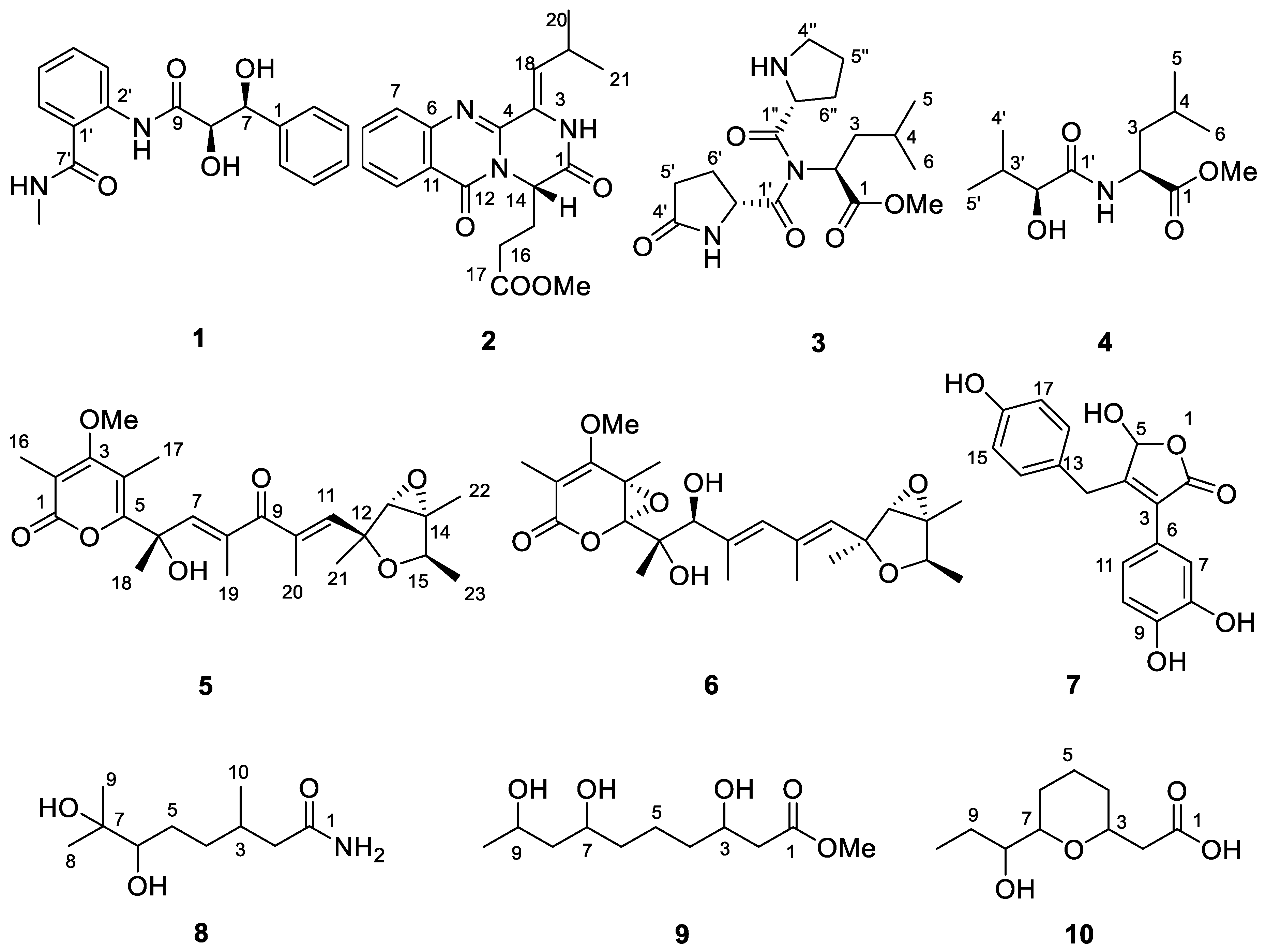
Figure 1. Compounds 110 from Penicillium griseofulvum MCCC 3A00225.
Marinedrugs 19 00224 g001
Figure 2. The key 1H–1H COSY (bold) and HMBC (arrow) correlations of 1.
Marinedrugs 19 00224 g002
Figure 3. The X-ray crystallography of 1.
Marinedrugs 19 00224 g003
Figure 4. FDDA derivatives of 3 compared with the retention times of standard FDDA-amino acids.
Marinedrugs 19 00224 g004
Figure 5. FDDA derivatives of 4 compared with the retention times of standard FDDA-amino acids.
Marinedrugs 19 00224 g005
Figure 6. The induced CD spectrum of 8 in DMSO solution of Mo2(OAc)4.
Marinedrugs 19 00224 g006
Table 1. 1H (400 MHz) and 13C (100 MHz) NMR spectroscopic data of 1, 3, 4, 8, and 9 in CD3OD.
No.13489
δCδHδCδHδCδHδCδHδCδH
1143.2 C 174.7 C 174.5 C 178.8 C 173.7 C
2127.5 CH7.46 (d, 7.8)52.3 CH4.41 (dd, 8.9, 6.2)51.6 CH4.52 (dd, 9.8, 4.8)44.0 CH22.24 (dd, 12.8, 5.2)1.98 m42.2 CH22.49 (dd, 15.1, 4.3)2.42 (dd, 15.1, 8.9)
3129.1 CH7.33 (dd, 7.8, 7.3)41.4 CH21.60 m41.6 CH21.67 m32.1 CH1.94 m75.9 CH3.79 (tdd, 8.9, 4.4, 2.0)
4128.3 CH7.24 (br t, 7.4)25.9 CH1.74 m26.0 CH1.68 m35.4 CH21.64 m; 1.22 m32.1 CH21.62 m; 1.22 m
5129.1 CH7.33 (dd, 7.8, 7.3)23.3 CH30.95 (d, 6.6)23.3 CH30.95 (d, 6.2)29.6 CH21.66 m; 1.23 m24.3 CH21.82 m; 1.58 m
6127.5 CH7.46 (d, 7.8)21.9 CH30.91 (d, 6.6)21.7 CH30.92 (d, 6.2)79.8 CH3.21 (d, 9.5)32.5 CH21.57 m; 1.21 m
775.6 CH5.16 (d, 2.0) 73.8 C 78.5 CH3.54 (tdd, 10.2, 3.7, 1.7)
877.8 CH4.25 (d, 2.3) 25.8 CH31.16 s45.9 CH21.61 m; 1.48 (dt, 14.0, 4.4)
9174.0 C 24.8 CH31.12 s67.4 CH3.94 m
10 20.2 CH30.96 (d, 6.2)23.1 CH31.14 (d, 6.2)
1′124.2 C 172.9 C 176.7 C
2′138.8 C 56.2 CH4.55 (dd, 8.8, 4.0)77.0 CH3.86 (d, 3.7)
3′122.4 CH8.51 (d, 8.1) 33.0 CH2.07 m
4′132.7 CH7.47 (td, 7.8, 1.5)181.6 C 19.5 CH31.00 (d, 7.0)
5′124.7 CH7.16 (td, 7.6, 1.0)30.3 CH22.36 m; 2.30 m16.3 CH30.84 (d, 6.8)
6′128.8 CH7.60 (dd, 7.8, 1.4)25.5 CH22.47 m; 2.16 m
7′ (1″)171.3 C 174.4 C
2″ 61.3 CH4.47 (dd, 8.4, 2.8)
4″ 48.1 CH23.63 m
5″ 25.9 CH22.02 m
6″ 30.3 CH22.18 m; 2.00 m
NMe/OMe26.8 CH32.89 s52.6 CH33.69 s52.7 CH33.70 s 52.1 CH33.65 s
Table 2. 1H (400 MHz) and 13C (100 MHz) NMR spectroscopic data of 2, 5, 6, 7, and 10.
No.2 a5 a6 a7 b10 a
δCδHδCδHδCδHδCδHδCδH
1167.7 C 167.1 C 167.9 C 175.9 C
2 111.1 C 109.1 C 170.6 C 42.8 CH22.41 (d, 6.5)
3126.3 C 171.2 C 168.3 C 120.3 C 75.8 CH3.76, m
4147.0 C 113.4 C 80.4 C 157.7 C 32.2 CH21.64, m; 1.21, m
5 160.8 C 109.1 C 96.5 CH5.77 (d, 6.8)24.6 CH21.84, m; 1.59, m
6121.1 C 75.2 C 82.1 C 127.8 C 32.8 CH21.52, m; 1.21, m
7128.4 CH7.64 (d, 8.1)147.7 CH6.38 (d, 1.4)90.5 CH4.09 s116.1 CH6.92 (d, 1.8)76.2 CH3.57, m
8136.0 CH7.77 (td, 8.4, 1.4)137.3 C 134.1 C 145.1 C 46.5 CH21.48, m
9128.0 CH7.46 (t, 7.6)202.8 C 133.3 CH5.84 s146.1 C 65.3 CH3.93, m
10127.6 CH8.14 (dd, 8, 1.1)139.3 C 136.6 C 115.6 CH6.79 (d, 8.2)23.9 CH31.13 (d, 6.3)
11148.7 C 143.7 CH6.28 (d, 1.4)133.2 CH5.53 s120.2 CH6.75 (dd, 8.2, 1.8)
12162.1 C 81.3 C 81.6 C 31.0 CH23.85 (d, 15.1); 3.57 (d, 15.1)
13 68.0 CH3.64 s68.7 CH3.55 s126.5 C
1456.2 CH5.34 (t, 6.6)68.7 C 68.7 C 129.6 CH6.99 (d, 8.4)
1528.7 CH22.65 m; 2.15 m78.5 CH4.08 (dt, 6.8, 6.8)78.3 CH4.05 (d, 6.8)115.5 CH6.69 (d, 8.4)
1630.6 CH22.44 m14.6 CH32.03 s10.2 CH31.84 s156.1 C
17173.9 C 11.1 CH32.09 s20.8 CH31.61 s115.5 CH6.69 (d, 8.4)
18129.6 CH6.33 (d, 10.4)27.0 CH31.68 (d, 0.8)19.7 CH31.28 s129.6 CH6.99 (d, 8.4)
1927.1 CH2.87 m13.4 CH31.67 s15.8 CH31.86 (d, 0.9)
2022.5 CH31.13 (d, 6.6)10.3 CH32.02 (d, 1.4)19.1 CH31.93 s
2122.6 CH31.16 (d, 6.6)21.1 CH31.38 s22.1 CH31.37 s
22 13.7 CH31.45 s13.8 CH31.45 s
23 19.3 CH31.17 (d, 6.8)19.2 CH31.20 (d, 6.8)
OMe52.2 CH33.46 s61.3 CH33.87 s61.1 CH33.92 s
a CD3OD. b DMSO-d6.
Table 3. Inhibition effects of compounds 136 on RBL-2H3 cell degranulation (n = 3).
CompoundIC50 (μM)
1360.3
14167.0
29134.0
Others a≥ 200
Loratadine b91.6
a Other compounds, including 112, 1528, and 3036. b Loratadine was a commercially available anti-food allergic medicine.
Publisher’s Note: MDPI stays neutral with regard to jurisdictional claims in published maps and institutional affiliations.